Diabetologia (2005) 48: 720–731 DOI 10.1007/s00125-005-1692-8
A RT I C L E
H. Wang . M. Iezzi . S. Theander . P. A. Antinozzi . B. R. Gauthier . P. A. Halban . C. B. Wollheim
Suppression of Pdx-1 perturbs proinsulin processing,
insulin secretion and GLP-1 signalling in INS-1 cells
Received: 30 June 2004 / Accepted: 6 December 2004 / Published online: 9 March 2005 # Springer-Verlag 2005
Abstract Aims/hypothesis: Mutations in genes encoding HNF-4α, HNF-1α and IPF-1/Pdx-1 are associated with, re-spectively, MODY subtypes-1, -3 and -4. Impaired glucose-stimulated insulin secretion is the common primary defect of these monogenic forms of diabetes. A regulatory circuit between these three transcription factors has also been sug-gested. We aimed to explore how Pdx-1 regulates beta cell function and gene expression patterns. Methods: We studied two previously established INS-1 stable cell lines permitting inducible expression of, respectively, Pdx-1 and its dominant-negative mutant. We used HPLC for insulin processing, adenovirally encoded aequorin for cytosolic [Ca2+], and tran-sient transfection of human growth hormone or patch-clamp capacitance recordings to monitor exocytosis. Results: In-duction of DN-Pdx-1 resulted in defective glucose-stimulated and K+-depolarisation-induced insulin secretion in INS-1 cells, while overexpression of Pdx-1 had no effect. We found that DN-Pdx-1 caused down-regulation of fibroblast growth factor receptor 1 (FGFR1), and consequently prohormone convertases (PC-1/3 and -2). As a result, DN-Pdx-1 severely impaired proinsulin processing. In addition, induction of Pdx-1 suppressed the expression of glucagon-like peptide 1 receptor (GLP-1R), which resulted in marked reduction of both basal and GLP-1 agonist exendin-4-stimulated cellular cAMP levels. Induction of DN-Pdx-1 did not affect glu-cokinase activity, glycolysis, mitochondrial metabolism or
ATP generation. The K+-induced cytosolic [Ca2+] rise and Ca2+-evoked exocytosis (membrane capacitance) were not abrogated. Conclusions/interpretation: The severely im-paired proinsulin processing combined with decreased GLP-1R expression and cellular cAMP content, rather than metabolic defects or altered exocytosis, may contribute to the beta cell dysfunction induced by Pdx-1 deficiency.
Keywords Beta cell . cAMP . FGFR1 . GLP-1 receptor . IPF-1 . MODY4 . Pdx-1 . Prohormone convertase-1 Abbreviations EMSA: electrophoretic mobility-shift assay . FGFR1: fibroblast growth factor receptor 1 . GLP-1R: glucagon-like peptide 1 receptor .
PC: prohormone convertase
Introduction
MODY is a monogenic form of type 2 diabetes characterised by early age of onset and autosomal dominant transmission. It is not usually associated with insulin resistance. With the exception of MODY-2 (glucokinase), MODY (-1, -3, -4, -5 and -6) has been linked to mutations in genes coding for the transcription factors, respectively, HNF-4α, HNF-1α, IPF-1/PDX-1/IDX-1, HNF-1β and NeuroD/BETA-2. Although mutations in these transcription factors display heteroge-neous clinical phenotypes, the primary cause of the various MODY subtypes has been attributed to beta cell dysfunc-tion. Only one MODY-4 family has been described in which a homozygous mutation carrier had pancreatic agenesis while heterozygous subjects developed early-onset diabetes [1,2]. The mutation in this family (IPF-1-P63fsdelC) directs expression of a dominant-negative protein [3]. Diabetic MODY-4 family members heterozygous for the mutation exhibit severe impairment of insulin secretion and enhanced insulin sensitivity, indicating that the primary defect is beta cell dysfunction [4]. Missense mutations in the IPF-1 gene have also been demonstrated to predispose to late-onset type 2 diabetes [5,6].
Electronic Supplementary Material Supplementary material is available for this article at http://dx.doi.org/10.1007/s00125-005-1692-8.
H. Wang (*) . M. Iezzi . S. Theander . P. A. Antinozzi . B. R. Gauthier . C. B. Wollheim
Department of Cell Physiology and Metabolism, University Medical Center,
1211 Geneva 4, Switzerland
e-mail: [email protected] Tel.: +41-22-3795570
Fax: +41-22-3795543 P. A. Halban
Department of Medical Genetics and Development, University Medical Center,
In the mouse, Pdx-1 has been reported to regulate early pancreatic development and to control the expression of insulin as well as other beta-cell-specific genes [7–10]. Tar-geted disruption of the PDX-1 gene results in pancreatic aplasia, while heterozygous mice exhibit impaired glucose tolerance [8,9,11,12]. Beta-cell-specific inactivation of the PDX-1 gene causes diabetes with increasing age, whereas mice with one deleted PDX-1 allele only develop glucose intolerance [13]. Inducible suppression of Pdx-1 function in mouse beta cells using an antisense ribozyme specific for Pdx-1 mRNA under control of the reverse tetracycline trans-activator also evokes age-dependent diabetes [14].
Pdx-1 plays an essential role in the regulation of beta cell neogenesis, differentiation and perhaps apoptosis [15–18]. Accordingly, transgenic overexpression of PDX-1 restores beta cell mass and function, thereby preventing the onset of diabetes in IRS2-null mice [16]. Adenovirus-mediated Pdx-1 overexpression in the pancreas of the mouse favours beta cell neogenesis, whereas ectopic expression of Pdx-1 in mouse liver or human fetal liver progenitor cells promotes differentiation into insulin-producing cells [18–20].
Glucose intolerance due to defective glucose-stimulated insulin secretion has been reported in the heterozygous PDX-1-mutant mouse [12]. Both reduced Glut-2 expression and decreased glucose-evoked NAD(P)H generation in islets are also observed, suggesting impaired glucose metab-olism [12]. However, the Pdx-1 target genes responsible for this secretory defect are yet to be identified.
Using INS-1 stable cell lines capable of expressing HNF-1α and HNF-4α or their dominant-negative mutants DN-HNF-1α and DN-HNF-4α in a doxycycline-dependent manner, we identified several target genes of HNF-1α and HNF-4α and suggested that the metabolism–secretion coupling in beta cells is affected in MODY-1 and MODY-3 [21–23]. It has been proposed that HNF-1α mediates transcription of the PDX-1 gene [24,25]. Furthermore, a MODY-1 family was reported in which the Pdx-1 binding site in the HNF-4α P2 promoter is mutated, suggesting a regulatory circuit between Pdx-1, HNF-4α and HNF-1α [26]. In a previous study on INS-1 cell lines permitting inducible overexpression or sup-pression of Pdx-1 we reported that this transcription factor is necessary for the beta-cell-like phenotype by inhibiting ex-pression of glucagon, the hormone secreted by the alpha cells [27]. To substantiate phenotype determination and to elu-cidate the mechanisms underlying the impaired insulin se-cretion caused by Pdx-1 deficiency, we examined insulin secretion, glucose metabolism, intracellular ATP and cAMP levels, beta cell gene expression profiles, cytosolic [Ca2+] and membrane capacitance in our established cellular models, which allow inducible expression of Pdx-1 or its dominant-negative mutant DN-Pdx-1 [27].
Materials and methods
Cell culture INS-1 stable cell lines, Pdx-1#6 and DN-Pdx-1#28, allowing inducible expression of, respectively, wild-type Pdx-1 and DN-Pdx-1 (lacking the N-terminal 79 amino acids), were cultured as previously described [27].
Immunoblotting and immunofluorescence Cells were cul-tured with or without 500 ng/ml doxycycline for 24 h. Nuclear proteins were extracted as previously described [28]. For total cellular protein extraction, cells were son-icated in lysis buffer containing (mmol/l): 20 Tris–HCl, pH 7.4, 2 EDTA, 2 EGTA, 1 PMSF and 1% Triton X-100. Nuclear extracts and total cellular proteins were fraction-ated by 7–11% SDS-PAGE. Immunoblotting was performed as described previously using enhanced chemiluminescence (Pierce, Rockford, IL, USA) for detection [21]. The dilu-tions for antibodies against Pdx-1 (a kind gift from Dr. H. Edlund), glucokinase (Santa Cruz, Basel, Switzerland), syntaxin A (Sigma, Buchs, Switzerland) and PC1/3 (a gen-erous gift from Dr D. F. Steiner) were 1:6,000, 1:2,000, 1:2,000, 1:1,000, 1:5,000 and 1:2,000, respectively.
For immunofluorescence, cells grown on polyornithine-coated glass coverslips were treated for 24 h with or without 500 ng/ml doxycycline. Cells were then washed, fixed in 4% paraformaldehyde, and permeabilised with 0.1% Triton X-100 in phosphate-buffered saline containing 1% BSA (PBS–BSA). The preparation was then blocked with PBS– BSA before incubating with the first antibody, anti-Pdx1 (1:1,000 dilution), followed by the second antibody labelling. Nuclear extract preparation and electrophoretic mobility-shift assay Nuclear extracts from Pdx-1#6 and DN-Pdx-1#28 cells grown in culture medium with or without 500 ng/ml doxycycline for 24 h were prepared according to Schreiber et al. [28]. The double-stranded oligonucleotides corre-sponding to the rat insulin I FLAT element [29], 5′gatcttg ttaataatctaattacc3′, was used as a probe. Electrophoretic mobility-shift assay (EMSA) procedures including condi-tions for probe labelling and binding reaccondi-tions were per-formed as in Wang et al. [30].
Measurements of insulin secretion and cellular insulin content Cells in 12-well plates were cultured in 11.2 mmol/l glucose medium with or without 500 ng/ml doxycycline for 4 days, followed by an additional 5 h equilibration in 2.5 mmol/l glucose medium. Insulin secretion was mea-sured over a period of 30 min in Krebs –Ringer–Bicarbon-ate–HEPES buffer (KRBH) (mmol/l: 140 NaCl, 3.6 KCl, 0.5 NaH2PO4, 0.5 MgSO4, 1.5 CaCl2, 2 NaHCO3, 10
HEPES and 0.1% BSA) containing indicated stimuli. In-sulin content was determined after extraction with acid ethanol following the procedures of Wang et al. [30]. Insulin was determined by radioimmunoassay using rat insulin as a standard [30] and a commercial antibody, not distinguishing between proinsulin and insulin (Linco, St. Charles, MO, USA).
Assay of glucokinase activity Cytosolic proteins were extracted, according to Wang and Iynedian [31], from cells cultured in 11.2 mmol/l glucose medium in the presence or absence of 500 ng/ml doxycycline for 4 days. Total hexo-kinase activity was measured at 30°C by a glucose-6-phos-phate dehydrogenase-coupled assay in a spectrophotometer (Lambda Bio20, Perkin Elmer, Switzerland), monitoring NADH production [31]. Glucokinase activity was calculated
as the differences in NADH produced at 100 and 0.5 mmol/l glucose and expressed in nmol/min (=mU) per mg of protein. Measurement of glucose utilisation Cells in 24-well dishes were cultured in standard medium (11.2 mmol/l glucose) with or without 500 ng/ml doxycycline for 72 h. The culture was continued in 2.5 mmol/l glucose medium with or without 500 ng/ml doxycycline for a further 24 h. The rate of glycolysis was estimated from the production of [3H] water from D-[5-3H]glucose according to Wang and Iynedjian
[31].
[14C]Pyruvate oxidation DN-Pdx-1#28 cells in 24-well plates were cultured in standard medium (11.2 mmol/l glucose) with or without 500 ng/ml doxycycline for 4 days. The production of14CO2from [2-14C] pyruvate was
mea-sured over 1 h in KRBH containing either 0.05 or 1.0 mmol/l pyruvate as reported previously [32].
Measurement of intracellular ATP and cAMP Cells in six-well dishes were cultured with (+Dox) or without (−Dox) 500 ng/ml doxycycline in standard medium (11.2 mmol/l glucose) for 3 days and then equilibrated in 2.5 mmol/l glucose medium for a further 24 h. Cellular ATP was measured after 8 min of stimulation with 20 mmol/l glu-cose as previously described [21]. Cellular cAMP was mea-sured after 30 min of stimulation with or without 10 nmol/l Exendin-4 in KRBH containing 2.5 or 20 mmol/l glucose according to the manufacturer’s protocol (cAMP Biotrak enzyme-immunoassay system; Amersham Biosciences, Freiburg, Germany).
Total RNA isolation and Northern blotting Pdx-1#6 cells in 10-cm diameter dishes were cultured in standard medium (11.2 mmol/l glucose) with or without the indicated con-centration of doxycycline for 96 h, unless otherwise specified. DN-Pdx-1#28 cells in 10-cm diameter dishes were cultured in standard medium (11.2 mmol/l glucose) with or without 500 ng/ml doxycycline for 72 h. The culture was continued in 2.5 mmol/l glucose medium with or without 500 ng/ml doxycycline for 16 h, followed by an additional 8 h in culture medium with 2.5, 6, 12 or 24 mmol/l glucose. Total RNA was extracted and blotted on nylon membranes as described previously [31]. The membrane was prehybridised and then hybridised to 32P-labelled random primer cDNA probes according to Wang and Iynedijian [31]. To ensure equal RNA loading and even transfer, all mem-branes were stripped and rehybridised with a‘housekeeping gene’ probe cyclophilin. cDNA fragments used as probes for glucokinase, Glut-2, L-pyruvate kinase, Rab3A, VAMP-2,
SNAP25A, Synaptotagmin-1 and Pdx-1 mRNA detection were digested from corresponding plasmids. cDNA probes for rat aldolase B, glyceraldehyde-3 phosphate dehydroge-nase (GAPDH), adenine nucleotide translocator-1 and 2 (ANT-1, ANT-2), mitochondrial uncoupling protein-2 (UCP-2), clathrin heavy chain, clathrin light chain, cyclin-depen-dent kinase-4 (CDK4), insulin receptor substrate-2 (IRS2), connexin-36, E-cadherin, N-cadherin, neural cell adhesion molecule (N-CAM), suppressor of cytokine signalling-3
(SOCS-3), signal transducer and activator of transcription (STAT)-1, -3, -5, Cav1.3 (α1D), prohormone convertases
(PC-1/3 and -2), adiponectin receptor (AdipoR), short-chain 3-hydroxyacyl-CoA dehydrogenase (SCHAD), fibroblast growth factor receptor 1 (FGFR1), and glucagon-like peptide-1 receptor (GLP-1R) were prepared by RT-PCR and confirmed by sequencing.
Measurement of cytosolic [Ca2+] in DN-Pdx1#28 cells Recombinant adenovirus encoding cytosolic aequorin under the chicken actin promoter (rAdCAGcAQ) was used [33]. DN-Pdx-1#28 cells were grown on polyornithine-coated plastic coverslips for 3 days in the absence or presence of doxycycline (500 ng/ml) and infected with rAdCAGcAQ for 90 min at approximately 50 PFU/ml. The [Ca2+]c was
measured 20 h later as previously described [33]. Briefly, cells were pre-incubated with 2.5 μmol/l coelenterezine (Calbiochem, San Diego, CA, USA) for 2–3 h and then perfused with KRBH containing 2.5 mmol/l glucose and 30 mmol/l KCl as stimulator. Emitted photons were col-lected with a photomultiplier apparatus (Thorn-Emi Electron tubes, UK).
Electrophysiological measurements Cells were seeded on glass coverslips and cultured without (−Dox) or with (+Dox) 500 ng/ml doxycycline for 4 days. The extracel-lular solution contained (mmol/l): 140 NaCl, 3.6 KCl, 2 NaHCO3, 2.6 CaCl2, 0.5 NaH2PO4, 0.5 MgSO4, 5 HEPES
and 2.5 glucose. The pH was set to 7.40 using NaOH. For membrane capacitance recordings, the pipette solu-tions contained (mmol/l): 125 potassium glutamate, 10 KCl, 10 NaCl, 1 MgCl2, 5 HEPES, 3 Mg-ATP, 10 EGTA
and 0 or 9.57 CaCl2. Using the Ca2+ chelator program
WEBMAXC (http://www.stanford.edu/~cpatton/webmaxc/
webmaxcS.htm) the free Ca2+ concentrations were
esti-mated to be lower than 0.1 nmol/l and 5 μmol/l, re-spectively. The pH was set to 7.15 using KOH. For patch clamp capacitance recordings a glass coverslip was trans-ferred to a temperature-controlled perfusion chamber. The chamber was perfused, using a gravity-driven perfusion system, with extracellular solution at a rate of 1.3 ml/min. Bath temperature was maintained at 32–33°C. Patch pipettes were pulled from borosilicate glass capillaries (GC150F-10; Clark Instruments, Reading, UK) on a Model P-97 puller from Sutter Instruments (Novato, CA, USA). Pipette re-sistance was between 4 and 6 MΩ. Patch-clamp recordings were performed with a List EPC 9 amplifier (HEKA, Darmstadt, Germany) in voltage-clamp mode. Capacitance was mea-sured after applying a 1-kHz, 28-mV peak-to-peak sinusoid stimulus from a dc holding potential of−70 mV. The ‘sine + dc’ mode of the software lock-in extension of the PULSE software was used to calculate the equivalent circuit param-eters Cm(membrane capacitance), membrane conductance
(Gm) and access resistance (Ra) from the current recordings.
Measurement of human growth hormone release DN-Pdx-1#28 cells in 24-well plates were transiently transfected with 2μg/plate of pcDNA3-hGH encoding human growth hormone using Effectene (Qiagen, Basel, Switzerland)
according to manufacturer’s protocol [34]. After 16 h of transfection, cells were cultured with or without 500 ng/ml doxycycline for 4 days in standard medium (11.2 mmol/l glucose) and continued for a further 5 h in 2.5 mmol/l glucose medium. Human growth hormone secretion stim-ulated by 24 mmol/l glucose, 100μmol/l tolbutamide and 20 mmol/l KCl was measured over a period of 30 min in KRBH containing 2.5 mmol/l glucose (basal). Human growth hormone was quantified by ELISA (Roche, Rotkreuz, Switzerland).
Measurement of proinsulin conversion DN-Pdx-1#28 cells in 3.5-cm diameter dishes were cultured in standard me-dium (11.2 mmol/l glucose) with or without 500 ng/ml doxycycline for 4 days. The cells were then washed and radiolabelled (2–4 mBq [3H]leucine) for 10 min at 37°C in KRBH, 16.7 mmol/l glucose. After washing in KRBH, 1.7 mmol/l glucose with 1 mmol/l unlabelled leucine, the pulse-labelled cells were incubated at 37°C for 60 or 120 min (chase) in this same buffer. At the end of the chase incubation, cells were extracted and analysed by reversed-phase HPLC as described in detail previously [35]. Statistics Results are expressed as means±SEM and sta-tistical analyses were performed by Student’s t-test for un-paired data.
Results
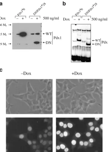
Pdx-1 and DN-Pdx-1 were expressed in a doxycycline-dependent manner We previously established Pdx-1#6 and DN-Pdx-1#28 cells using the parental INS-rβ (r9) cells [27]. Western blotting with an antibody against the C terminus of Pdx-1 demonstrated that both Pdx-1 and DN-Pdx-1 proteins were induced, respectively, in DN-Pdx-1#6 and DN-Pdx-1#28 cells treated with 500 ng/ml doxycycline for 24 h (Fig. 1a). EMSA with the rat insulin I promoter element showed that Pdx-1 protein binding was increased by 10-fold and diminished by 90%, respectively, in Pdx-1#6 and DN-Pdx-1#28 cells under the same conditions (Fig. 1b). The Pdx-1 and DN-Pdx-1 binding complexes were supershifted by anti-Pdx-1 antibody (data not shown). It is noteworthy that DN-Pdx-1 protein was undetectable under non-induced conditions (Fig. 1a and b). Immuno-fluorescence staining of DN-Pdx-1#28 cells with the same antibody illustrated that all cells uniformly expressed the nuclear localised DN-Pdx-1 protein (Fig. 1c). Similar re-sults were obtained in Pdx-1#6 cells (data not shown). Dominant-negative suppression of Pdx-1 impaired insulin secretion As demonstrated in Fig. 2a, overexpression of Pdx-1 in INS-1 cells did not alter glucose-stimulated insulin secretion. In contrast, similar induction of DN-Pdx-1 blunted glucose-, leucine-, and K+-evoked insulin release (Fig.2b). After 4 days of treatment with 500 ng/ml doxycycline, insulin content was increased by 37% (−Dox: 1.497±0.199; +Dox: 2.033±0.281 μg/mg protein) in Pdx-1#6 cells and decreased by 57% (−Dox: 1.957±0.303; +Dox: 0.857±
0.22 μg/mg protein) in DN-Pdx-1#28 cells. Therefore, in-sulin secretion in these cells was expressed as percentage of cellular insulin content.
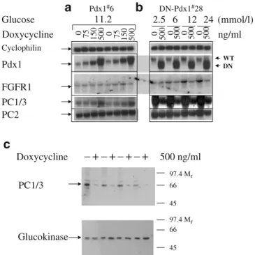
Induction of DN-Pdx1 down-regulated the expression of fibroblast growth factor receptor 1 (FGFR1) and prohor-mone-convertase (PC)-1/3 and -2 Since Pdx-1 has been proposed to regulate PC-1/3 expression through FGFR1 signalling [36], we examined the mRNA levels of FGFR1 and PC-1/3 in Pdx-1#6 and DN-Pdx-1#28 cells cultured with or without 500 ng/ml doxycycline for 4 days. As demonstrated by quantitative Northern blotting in Fig. 3, induction of DN-Pdx-1 for 4 days suppressed the mRNA levels of FGFR1 by 50%. Graded overexpression of Pdx-1 for 4 days resulted in a modest increase in the mRNA levels of PC1/3 and PC2 (Fig. 3a). In contrast, 4 days of induction of DN-Pdx1 suppressed the expression of PC1/3 and PC2 by 90 and 40%, respectively (Fig. 3b). The protein levels of PC-1/3 were also reduced by 80%, whereas the expression of glucokinase protein was unaltered
Pdx1 DN Dox Pd+ + 500 ng/ml x1#6 DN Pdx1 #28
a
WT 66 Mr 45 Mr 29 Mrb
Dox – + – – – Pd + 500 ng/ml x1 #6 DN Pdx1 #28 Pdx1 DN WTc
+Dox –DoxFig. 1 Nuclear localised proteins encoded by Pdx-1 and DN-Pdx-1 were induced in a doxycycline-dependent manner. Cells were cultured with or without 500 ng/ml doxycycline for 24 h. a Im-munoblotting of nuclear extracts from Pdx-1#6 and DN-Pdx-1#28 cells with antibody against Pdx-1 C terminus. b Gel-shift assay of nuclear extracts from Pdx1#6 and DN-Pdx-1#28 cells using the rat insulin I promoter fragment. c Immunofluorescence staining of DN-Pdx-1#28 cells with antibody against Pdx-1 C terminus
(Fig. 3c). Both PC1/3 and PC2, which are expressed in pancreatic beta cells and INS-1 cells, are implicated in the processing of proinsulin to insulin [37–39]. PC1/3 cleaves preferentially at the B-chain/C-peptide junction and thereby facilitates the second maturation cleavage by PC2 at the C-peptide/A-chain junction [35,37–40].
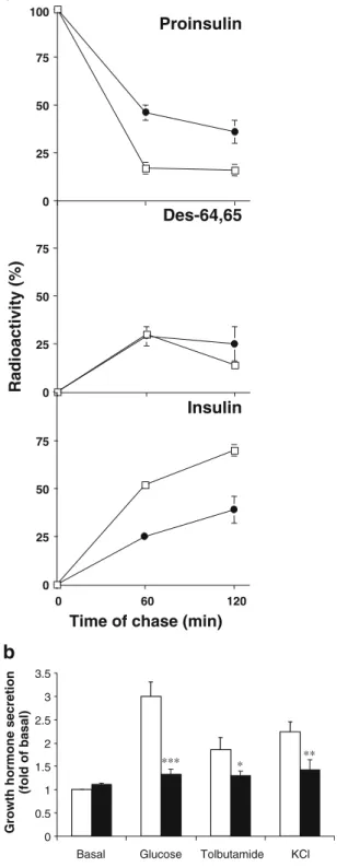
Induction of DN-Pdx-1 impaired proinsulin conversion in INS-1 cells To examine the consequences of reduced expression of PC1/3 on proinsulin processing, we analysed the pattern of radiolabelled proinsulin-related products dur-ing a pulse-chase protocol [35]. As depicted in Fig. 4a, dominant-negative suppression of Pdx-1 led to severely de-fective proinsulin processing. Thus at 120 min, proinsulin conversion was inhibited by more than 60%.
Induction of DN-Pdx-1 inhibited release of human growth hormone co-secreted with insulin To investigate hormone independently of regulation of insulin gene expression and processing, we transfected DN-Pdx-1#28 cells with a cDNA encoding human growth hormone. This polypeptide hor-mone is stored and co-secreted with insulin but is not processed by prohormone-convertase 1/3 (PC1/3) [34,39, 41]. As illustrated in Fig.4b, induction of DN-Pdx-1 caused significant decrease in human growth hormone secretion stimulated by glucose, tolbutamide and K+. Tolbutamide blocks ATP-dependent K+-( KATP) channels, whereas K+
directly depolarises the plasma membrane potential. Induction of DN-Pdx1 reduced the expression of GLP-1R, intracellular cAMP levels and GLP-1R signalling DN-Pdx-1*28 cells were cultured with or without 500 ng/ml doxycycline for 3 days in standard glucose medium. The culture was continued for a further 24 h in 2.5 mmol/l glucose medium. Overexpression of Pdx-1 had minor effects on GLP-1R expression, whereas induction of DN-Pdx-1 for 0 0.2 0.4 0.6 0.8 1 1.2 1.4 1.6 1.8 2.5 6 12 24 Glucose (mmol/l) Insulin secretion
(% of cellular insulin content)
a
Pdx1#6 0 1 2 3 4 5 6Basal Glucose Leucine KCl
Insulin secretion
(% of cellular insulin content)
b
DN-Pdx1#28***
***
***
Fig. 2 Dominant-negative suppression of Pdx-1-function-impaired insulin secretion. Cells were cultured with (+Dox, closed bar) or without (−Dox, open bar) 500 ng/ml doxycycline in standard medium (11.2 mmol/l glucose) for 4 days and then equilibrated in 2.5 mmol/l glucose medium for a further 5 h. a Induction of Pdx1 for 4 days did not alter glucose-stimulated insulin secretion. Insulin release from Pdx-1#6 cells in KRBH buffer containing indicated
concentrations of glucose was determined by radioimmunoassay and expressed as percentage of cellular insulin content. Cellular insulin content was increased by 36.7±4.3% (−Dox: 1.497±0.199; +Dox:
2.033±0.281μg/mg protein, n=6, p<0.005) after Pdx-1 induction. b Similar induction of DN-Pdx1 resulted in defective insulin release induced by (mmol/l): 24 glucose, 20 leucine and 20 KCl. Insulin secretion from DN-Pdx-1#28 cells stimulated by (mmol/l): 24 glu-cose, 20 leucine and 20 KCl was measured in KRBH containing 2.5 mmol/l glucose (basal). Cellular insulin content was reduced by 56.9±5.2% (−Dox: 1.957±0.303; +Dox: 0.857±0.22 μg/mg protein, n=6, p<0.001). Data represent means±SEM of six independent experiments. ***p<0.001 Cyclophilin WT DN PC1/3 PC2 0 75 150 500 0 75 150 500 0 500 0 500 0 500 0 500 ng/ml
a
Pdx1#6b
DN-Pdx1#28 11.2 Glucose 2.5 6 12 24 (mmol/l) Doxycycline Pdx1 FGFR1 Doxycycline –+–+–+–+ 500 ng/ml 97.4 Mr 66 45 97.4 Mr 66 45 PC1/3 Glucokinasec
Fig. 3 Pdx-1 regulated the expression of FGFR1 and PC-1/3. a dx-1#6 cells were cultured in standard medium (11.2 mmol/l glucose) with or without indicated concentration of doxycycline for 4 days. b DN-Pdx-1#28 cells were cultured in standard medium (11.2 mmol/l glucose) with or without 500 ng/ml doxycycline for 72 h. The culture was continued in 2.5 mmol/l glucose medium with or without 500 ng/ml doxycycline for 16 h, followed by an additional 8 h in culture medium with 2.5, 6, 12 and 24 mmol/l glucose. The gene expression pattern in these cells was quantitatively evaluated by Northern blotting. Total RNA samples (20μg) were analysed by hybridising with indicated cDNA probes. The experiments were repeated two to four times with similar results. c Induction of DN-Pdx-1 decreased the protein levels of PC-1/3. DN-Pdx-1#28 cells in 10-cm diameter dishes were cultured in standard medium (11.2 mmol/l glucose) without (−Dox) or with (+Dox) 500 ng/ml doxycycline for 4 days. Western blotting of total cell extracts (100μg protein) with antibodies against PC1/3 and glucoki-nase. Data represent four independent experiments
4 days suppressed the mRNA levels of GLP-1R by 90% (Fig.5).
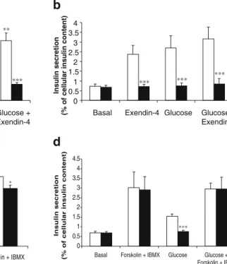
We therefore measured the intracellular cAMP levels and GLP-1 agonist-stimulated insulin secretion under the same conditions. DN-Pdx-1 reduced the intracellular cAMP content by 40% at basal (2.5 mmol/l) glucose conditions (Fig.6a). In non-induced cells, the GLP-1R agonist exendin-4 raised the cAMP concentrations by 2-fold at both 2.5 and 20 mmol/l glucose (Fig. 6a). In contrast, the exendin-4-induced cAMP increase was completely inhibited in INS-1
cells expressing DN-Pdx-1 (Fig. 6a). Consequently, ex-endin-4-induced insulin secretion was also blocked by induc-tion of DN-Pdx-1 (Fig.6b). Forskolin stimulates adenylate cyclase and IBMX inhibits phosphodiesterases, leading to robust increases in cellular cAMP levels (Fig. 6c). The insulin secretion induced by these agents was unaffected by DN-Pdx-1 (Fig.6d). The marked increase in cellular cAMP levels induced by forskolin plus IBMX were, however, slightly reduced by DN-Pdx-1 (Fig.6c) (p<0.05).
To examine the possibility that suppression of Pdx-1 function impairs metabolism-secretion coupling [12], we measured glucose metabolism in DN-Pdx-1#28 cells. Induction of DN-Pdx-1 did not affect glycolysis, mitochon-drial oxidation or ATP generation Pdx-1 was previously reported to regulate glucokinase gene expression [42]. However, we found that glucokinase activity was un-altered in Pdx-1#6 (−Dox: 10.06±2.07; +Dox: 11.70± 1.48 mU mg protein−1h−1; n=7) and DN-Pdx-1#28 (−Dox: 10.80±1.33; +Dox: 12.41±1.33 mU mg protein−1h−1; n=7) Cyclophilin GLP-1R Dox (ng/ml) 0 75 150 500 0 57 1 5 0 500 0 50 0 0 5 0 0 0 5 0 0 0 500 Glucose 11.2 2.5 6 12 24 (mmol/l)
a
Pdx1#6b
DN-Pdx1#28 Pdx1 WT DNFig. 5 Induction of DN-Pdx-1 suppressed the expression of GLP-1R. a Pdx-1#6 cells were cultured in standard medium (11.2 mmol/l glucose) with or without indicated concentration of doxycycline for 4 days. b DN-Pdx-1#28 cells in 10-cm diameter dishes were cultured in standard medium with or without 500 ng/ml doxycycline for 72 h. The culture was continued in 2.5 mmol/l glucose medium with or without 500 ng/ml doxycycline for 16 h, followed by an additional 8 h in culture medium with 2.5, 6, 12 and 24 mmol/l glucose. The gene expression profile in these cells was quantified by northern blotting. Total RNA samples (20μg) were analysed by hybridising with indicated cDNA probes. The experiments were repeated two to three times with similar results 0 25 50 75 Des-64,65 0 25 50 75 100 0 25 50 75 0 60 120 Radioactivity (%)
Time of chase (min)
Insulin Proinsulin 0 0.5 1 1.5 2 2.5 3 3.5
Basal Glucose Tolbutamide KCl
Growth hormone secretion
(fold of basal)
*** * **
a
b
3Fig. 4 Induction of DN-Pdx-1 resulted in defective proinsulin processing and impaired release of human growth hormone that co-secreted with insulin. a DN-Pdx-1#28 cells in 3.5-cm diameter dishes were cultured in standard medium (11.2 mmol/l glucose) with (+Dox, closed circle) or without (−Dox, open square) 500 ng/ml doxycycline for 4 days. Cells were labelled 10 min with [3H]leucine and chased for up to 2 h. Cell extracts were analysed by HPLC to determine the percentage of radioactivity in each proinsulin-related product. Results are means±SEM for three independent experiments. b DN-Pdx1#28 cells were transiently transfected with a plasmid encoding human growth hormone as a reporter for secretion. Cells were then cultured with (+Dox, closed bar) or without (−Dox, open bar) 500 ng/ml doxycycline for 4 days in standard medium (11.2 mmol/l glucose) and continued for further 5 h in 2.5 mmol/l glucose medium. Human growth hormone secretion stimulated by 24 mmol/l glucose, 100μmol/l tolbutamide and 20 mmol/l KCl was measured over a period of 30 min in KRBH containing 2.5 mmol/l glucose (basal). Data represent means±SEM of six independent experiments. *p<0.01, **p<0.005, ***p<0.001
cells after treatment with 500 ng/ml doxycycline for 4 days. Under the same conditions, neither glucose utilisation (Fig.7) nor mitochondrial oxidation (Fig.7b) was altered in DN-Pdx-1#28 cells. In good agreement, cellular ATP con-tent was unchanged by induction of DN-Pdx-1 (Fig. 7c). These results suggest that the target genes of Pdx-1 are distinct from those of HNF-1α and HNF-4α, the latter controlling mitochondrial function [21–23]. To substantiate this, we analysed the gene expression patterns in Pdx-1#6 and DN-Pdx-1#28 cells under non-induced and induced conditions.
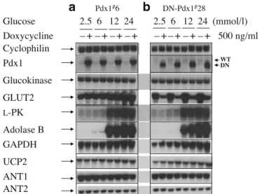
Pdx-1 did not regulate the expression of glucokinase and other genes involved in glucose metabolism As shown in Fig.8a, up-regulation of Pdx1 for 4 days did not alter the mRNA levels of glucokinase, L-pyruvate kinase (L-PK),
aldolase B, glyceraldehyde-3-phosphate dehydrogenase (GAPDH), uncoupling protein 2 (UCP2), or adenine nu-cleotide translocator 1 and 2 (ANT1 and ANT2). Similarly, dominant-negative suppression of Pdx-1 for 4 days had no effects on these transcripts (Fig.8b). However, there was a 90% decrease in Glut2 expression. As the capacity of this sugar transporter is in large excess of the activity of glucokinase and other glycolytic enzymes, even this de-crease would not have been expected to affect glucose
metabolism. Therefore in the present cellular model, Pdx-1 does not control glycolysis and mitochondrial metabolism. Consequently, we investigated further the effects of Pdx-1 on the expression of genes implicated in insulin processing and secretion, as well as granule maturation and exocytosis. Effects of DN-Pdx-1 on cytosolic [Ca2+] and exocytosis As shown in Fig. 9a and b, K+ elicited a pronounced increase in cytosolic [Ca2+]. This response was not atten-uated but actually increased by 15% following induction of DN-Pdx-1. This suggests that DN-Pdx-1-induced im-pairment of insulin secretion is not caused by reduced cytosolic [Ca2+].
In order to investigate whether Pdx-1 controls genes implicated in the exocytotic machinery we measured mem-brane capacitance to study the exocytotic response to infusion of Ca2+(5μmol/l) through the patch pipette. Mea-surement of capacitance began a few seconds after establish-ment of the whole cell configuration. Typical traces are shown in Fig.9c. It is seen that capacitance increases in a continuous manner following patch rupture. No change in capacitance was obtained in the absence of Ca2+ in the pipette solution (not shown). In order to compare the exocytotic response to the Ca2+infused through the patch pipette we measured the average increase in capacitance
0 2 4 6 8 10 12 14 16
Basal Exendin-4 Glucose Glucose + Basal Exendin-4 Glucose Exendin-4
Glucose + Exendin-4
Cellular cAMP concentrations
(pmol/mg protein) *** ** ** *** ** *** 0 0.5 1 1.5 2 2.5 3 3.5 4 Insulin secretion
(% of cellular insulin content)
*** *** *** 0 0.5 1 1.5 2 2.5 3 3.5 4 4.5
Basal Forskolin + IBMX Glucose Glucose + Forskolin + IBMX
Insulin secretion
(% of cellular insulin content)
*** 0 10 20 30 40 50 60 70 80 90
Basal Exendin-4 Forskolin + IBMX
Cellular cAMP concentrations
(pmol/mg protein) *** *** *
a
b
c
d
Fig. 6 DN-Pdx-1 decreased intracellular cAMP levels and GLP-1R signalling. DN-Pdx-1#28 cells in 24-well plates were cultured with (+Dox, closed bar) or without (open bar) 500 ng/ml doxycycline for 3 days in standard glucose medium. The culture was continued for a further 24 h in 2.5 mmol/l glucose medium. a Cellular cAMP con-tent was measured by stimulation with or without 10 nmol/l exen-din-4 for 30 min in KRBH buffer containing 2.5 (basal) or 20 mmol/ l glucose. Data represent five independent experiments. DN-Pdx-1 reduced both basal and exendin-4-stimulated cAMP concentrations. b Insulin secretion was measured under the same conditions. DN-Pdx-1 inhibited both exendin-4- and glucose-induced insulin secretion. Data
represent six independent experiments. c Cellular cAMP content was measured by stimulation with or without 10 nmol/l exendin-4 or 200 nmol/l forskolin plus 100μmol/l IBMX for 30 min in KRBH buffer containing 2.5 mmol/l glucose. Data represent five separate experiments performed in duplicate. d DN-Pdx-1 did not affect forskolin plus IBMX-induced insulin release. Insulin secretion was measured by stimulation with or without 200 nmol/l forskolin plus 100μmol IBMX for 30 min in KRBH containing 2.5 (basal) or 20 mmol/l glucose. Data represent six separate experiments. *p<0.05, **p<0.005, ***p<0.001
between 60 and 120 s after patch rupture. As shown in Fig.9d, there was no difference between cells cultured in the presence or absence of doxycycline. Likewise, the initial capacitance (6.33±0.32 and 6.39±0.32, respectively) was unaffected by doxycycline treatment. In conclusion, Ca2+ -induced exocytosis does not seem to be affected by dom-inant-negative suppression of Pdx-1.
To further investigate whether Pdx-1 controls genes participating in exocytosis, we examined the transcript levels of clathrin heavy and light chains, VAMP-2, Rab3A, SNAP25A, synaptotagmin-1, voltage-dependent Ca2+ chan-nel Cav1.3 (α1D) subunit [see Electronic Supplementary
Material (ESM), address on first page of article]. Clathrin seems to play an important role in the removal of proteases from maturing granules [43]. VAMP-2, Rab3A, SNAP25A and synaptotagmin I are implicated in insulin exocytosis [44–46]. Voltage-dependent Ca2+ channel Cav1.3 (α1D)
subunit is involved in mediating beta cell insulin secretion in response to rising concentrations of glucose [47]. All these proteins are required for normal glucose-stimulated insulin secretion. Pdx-1 did not regulate mRNA levels of these genes (see ESM).
We characterised further the effects of Pdx-1 on expres-sion of other genes potentially involved in beta cell function (see ESM), including cyclin-dependent kinase-4 (CDK4), insulin receptor substrate-2 (IRS2) [13, 36, 48–51], con-nexin-36, E-cadherin, N-cadherin, neural cell adhesion molecule (N-CAM), suppressor of cytokine signalling-3 (SOCS-3), signal transducer and activator of transcription (STAT)-1, -3, -5 [52–55], adiponectin receptor (AdipoR) [56] and short-chain 3-hydroxyacyl-CoA dehydrogenase (SCHAD) [57]. Neither overexpression nor dominant-negative suppression of Pdx-1 altered the mRNA levels of these genes, except that induction of DNPdx-1 inhibited STAT-5 but increased STAT-1 expression (see ESM).
0 1 2 3 4 5 2.5 6 12 24 Glucose (mmol/l) Glucose utilisation (nmol/mg DNA) 0 10 20 30 40 50 60 70 80 0.05 1 Pyruvate (mmol/l) Pyruvate oxidation (nmol CO 2 h -1mg protein -1) 0 0.2 0.4 0.6 0.8 1 1.2 1.4 1.6 1.8 2 Basal Glucose
Cellular ATP content
(fold of basal)
a
b
c
Fig. 7 Induction of DN-Pdx1 does not affect glucose metabolism. a Induction of DN-Pdx-1 for 4 days had no effect on glycolytic flux. DN-Pdx-1#28 cells were cultured with (+Dox, closed bar) or without (−Dox, open bar) 500 ng/ml doxycycline in standard medium (11.2 mmol/l glucose) for 3 days and then equilibrated in 2.5 mmol/l glucose medium for a further 24 h. Cells were then incubated with the indicated concentrations of glucose andD-[5-3H]glucose for 60 min.
Data are expressed perμg cellular DNA and represent means±SE from six separate experiments. b Induction of DN-Pdx1 did not alter mito-chondrial oxidation. [2-14C]Pyruvate oxidation was measured during 1 h of incubation in KRBH containing either 0.05 or 1.0 mmol/l pyruvate. Data represent means±SEM performed in triplicate from one of three similar experiments. Similar negative results were obtained using uniformly labelled [U-14C]glucose (data not shown). c DN-Pdx-1 did not affect cellular ATP content. DN-Pdx-1#28 cells were cultured with (+Dox) or without (−Dox) 500 ng/ml doxycycline in standard medium for 3 days and then equilibrated in 2.5 mmol/l glucose medium for a further 24 h. Cellular ATP content, measured at basal (2.5 mmol/l glucose) and after 8 min of incubation with 20 mmol/l glucose, is presented as fold of basal. Data are means±SEM from six separate experiments Cyclophilin ANT1 ANT2 UCP2 GAPDH Adolase B L-PK WT DN Glucokinase GLUT2 Doxycycline –+–+–+–+ –+–+–+–+ 500 ng/ml Glucose 2.5 6 12 24 2.5 6 12 24 (mmol/l)
a
Pdx1#6b
DN-Pdx1#28 Pdx1Fig. 8 Pdx-1 did not alter the mRNA levels of glucokinase and other genes involved in glucose metabolism. Pdx-1#6 (a) and DN-Pdx-1#28 (b) cells in 10-cm diameter dishes were cultured in standard medium (11.2 mmol/l glucose) with or without 500 ng/ml doxycycline for 72 h. The culture was continued in 2.5 mmol/l glucose medium with or without 500 ng/ml doxycycline for 16 h, followed by an additional 8 h in culture medium with 2.5, 6, 12 and 24 mmol/l glucose. The gene expression profile in these cells was quantified by northern blotting. 20μg total RNA samples were analysed by hybridising with indicated cDNA probes. The experiments were repeated two times with similar results
Discussion
Reduced expression of FGFR1 has been reported in islets of mice with beta-cell-specific deletion of PDX-1 [13]. Con-cordantly, we found that dominant-negative suppression of Pdx-1 decreased mRNA levels of FGFR1 in INS-1 cells. Our data also support previous findings that Pdx-1 acts upstream of FGFR1 signalling in beta cells to maintain insulin processing [36]. The mice with attenuated FGFR1c signalling develop diabetes with age and exhibit decreased beta cell mass, reduced Glut-2 levels and increased pro-insulin content in beta cells owing to defective expression of PC1/3 and 2 [36]. The present study demonstrated that induction of DNPdx-1 caused 80–90% reduction in PC1/3 expression at both mRNA and protein levels. In addition, PC1/3 deficiency correlated well with the severe impair-ment of proinsulin conversion. PC1/3 is the predominant hormone convertase in beta cells [37,39,58].
In agreement with previous studies in PDX-1 (+/−) mice [12, 15], dominant-negative suppression of Pdx-1 also caused defective insulin secretion stimulated by glucose, leucine and K+. This was not due to the decreased insulin content, since the data were expressed as percentage of cellular insulin. The reduced insulin content is an expected consequence of the lower insulin mRNA levels after sup-pression of Pdx-1 function [27]. The inhibition of insulin
mRNA levels was not seen in rat islets after adenovirus-mediated expression of DN-Pdx-1 [59]. Expression of DN-Pdx-1 in rat islets also caused impaired mitochondrial metabolism through suppression of the nuclear factor TFAM leading to decreased expression of mitochondrially encoded genes, including subunits of the respiratory chain complexes [59]. In the primary cells, the reduced ATP generation from glucose explains the inhibition of insulin secretion [59]. Induction of DN-Pdx-1 in INS-1 cells did not affect glucose metabolism and ATP generation but rather inhibited insulin secretion probably by affecting steps distal to the generation of mitochondrial coupling factors. It may be speculated that the apparent discrepancy between islets and INS-1 cells lies in different control of mitochondrial transcriptional activity in mature beta cells and proliferating insulinoma cells with higher energy requirement.
Although it has been previously reported that Pdx-1 regulates beta cell glucokinase gene expression [42], the present data and other in vivo studies do not support this notion [13,60]. In contrast, these studies suggest that Pdx-1 may act in coordination with some other factors rather than functioning independently on the glucokinase pro-moter. Therefore, manipulation of Pdx-1 function alone is not sufficient to regulate the beta glucokinase expression. The defective secretory response cannot be explained by an increase in impaired cytosolic [Ca2+], as the K+ -0.0 0.5 1.0 1.5 0 100 200 300 Time (s) Cm (pF) 0 1 2 3 4 5 fF/s –DOX +DOX –DOX +DOX 0 250 500 750 1000 1250 1500 1750 2000 Time (s) 30 mmol/l KCl + DOX 0 250 500 750 1000 1250 1500 1750 2000 a b c d 2 52 102 152 202 252 302 352 402 452 2 52 102 152 202 252 302 352 402 452 [Ca 2+ ] (nmol/l) [Ca 2+ ] (nmol/l) 30 mmol/l KCl – DOX 0 200 400 600 800 1000 1200 1400 1600 – DOX +DOX [Ca 2+ ] (nmol/l) *
Fig. 9 Measurement of cytosolic [Ca2+] and membrane capaci-tance in DN-Pdx-1#28 cells. a DN-Pdx1#28 cells were treated with doxycycline and infected with rAdCAGcAQ as described inMaterials and methods. In-fected cells were perifused with KRBH containing 2.5 mmol/l glucose and then stimulated with 30 mmol/l KCl. b Peak [Ca2+] response to KCl in the absence or presence of doxycycline. Values are means±SEM of six indepen-dent experiments, each performed in duplicate. *p<0.05 vs non-induced cells. c Representative traces from patch-clamp capaci-tance recordings of cells cultured in the presence or absence of doxycycline. The rate of exocy-tosis was evaluated during the time interval (gray shading) be-tween 60 and 120 s after the start of the recording by fitting the capacitance values to a linear function. d Average of the rates of exocytosis, as obtained in c doxycycline-treated and un-treated cells (n=16)
evoked response was not inhibited. Moreover, the process of exocytosis was unaffected as monitored by Ca2+ -in-duced changes in membrane capacitance or as judged from the expression profile of the principal genes implicated in the final steps of insulin secretion. Similarly, the attenuated insulin secretion is not due to inhibition of proinsulin pro-cessing, as co-secreted human growth hormone (transiently transfected), which is not processed by PC-1/3, was equally unaffected. The most likely explanation for the impaired insulin secretion in response to nutrients and K+ depolar-isation is the pronounced decrease in cAMP levels after suppression of Pdx-1 function.
This is a consequence of reduced GLP-1R expression. It has been demonstrated that the GLP-1R is capable of regulating basal cellular cAMP levels even in the absence of the ligand [61]. We found that both basal and GLP-1R agonist exendin-4-induced cellular cAMP levels were markedly reduced by dominant-negative suppression of Pdx-1. DN-Pdx-1 also inhibited exendin-4- but not forskolin+IBMX-stimulated insulin secretion, the latter combination resulting in a more marked rise in cellular cAMP levels. Increasing cAMP levels with forskolin and IBMX also completely restored glucose-stimulated insulin secretion in rat islets infected with adenoviral DN-Pdx-1 [59]. The preserved exocytotic response of DNPdx-1 cells to Ca2+ infusion through the patch-clamp pipette indicates that dialysis of the cells has eliminated the inhibition observed when K+ depolarisation was used to raise cytosolic [Ca2+]. We there-fore propose that Pdx-1 may affect insulin secretion by changing cellular cAMP levels through GLP-1R pathways. The preserved secretory response to forskolin/IBMX may be explained by direct activation of exocytosis through the low-affinity, protein kinase A-independent cAMP-GEF II mechanism operative in the beta cell [62]. In addition, as GLP-1 has been shown to regulate Pdx-1 activation [63– 65], there may exist a functional regulatory loop between GLP-1 and Pdx-1. Furthermore, an elegant study on mice lacking GLP-1 and GIP receptors has demonstrated that gluco-incretin hormones regulate insulin secretion by three different mechanisms [66]. First, they have additive in-sulinotropic effects on cells during oral glucose tolerance tests. Second, in the absence of the GLP-1 receptor, there is suppression of the first phase of insulin secretion measured in vivo, but not in the isolated islets, indicating a role for this receptor in the function of glucose sensors that are located outside the endocrine pancreas and that control first-phase secretion. Third, absence of both receptors causes a cell-autonomous insulin secretion defect at a site distal to plasma membrane depolarisation [66]. GLP-1R plays an important role in the regulation of beta cell neogenesis, development, differentiation and survival as well as insulin secretion [50, 51, 67–69]. GLP-1-stimulated insulin secretion was not reduced in Pdx-1+/− mice [12], but there was an attenuation in mice with beta-cell-specific deletion of Pdx-1 [70]. The results obtained in the latter mouse model resemble those of our INS-1 expressing DN-Pdx-1, which could be explained by the shift from a beta cell to an alpha cell phenotype seen in mice with beta-cell-specific deletion of Pdx-1 and our INS-1 cells expressing DN-Pdx-1 [13, 27] (as discussed
below). This phenotype shift is not obvious in Pdx-1+/− mice [12]. Since it is not known which mouse model most closely resembles the phenotype of MODY-4, data from different studies should be taken into account.
We have previously demonstrated that dominant-nega-tive suppression of Pdx-1 in INS-1 cells led to reduced beta-cell-specific gene expression and increased glucagon levels [27]. Concordantly, beta-cell-specific deletion of Pdx-1 also results in decreased beta cell mass and elevated alpha cell population [13]. The loss of beta-cell-like phenotypes may contribute to the impaired insulin secretion and defective expression of GLP-1R and PC1/3 in our INS-1 cells ex-pressing DN-Pdx-1. Likewise, the glucose transporter Glut-2, which is absent from alpha cells, was repressed in INS-1 cells expressing DN-Pdx-1, coinciding with a marked increase in glucagon transcript levels [27]. Both GLP-1R and PC1/3 are highly expressed in beta cells and are, re-spectively, not detected or barely detected in alpha cells [39, 58,71]. Although the shift from a beta cell to an alpha cell phenotype has also been demonstrated in mice with beta-cell-specific deletion of Pdx-1 [13], the pathogenesis implicated in beta cell dysfunction in MODY-4 patients re-mains to be defined.
In conclusion, the severely impaired proinsulin processing combined with decreased expression of GLP-1R and re-duced intracellular cAMP levels, rather than metabolic de-fects or altered exocytosis, may contribute to the beta cell dysfunction induced by Pdx-1 deficiency. Suppression of Pdx-1 favours loss of beta cell phenotype and beta-cell-specific gene expression. The molecular mechanisms under-lying beta cell pathology of MODY-4 are thus distinct from those of MODY-1 and MODY-3.
Acknowledgements We are grateful to S. Dupuis, D. Cornut-Harry, D. Nappey, Y. Dupré, S. Mouche and V. Calvo for expert technical assistance. We are indebted to H. Edlund (Pdx-1 cDNA and antibody), D. F. Steiner (PC1/3 antibody), H. Bujard (PUHD 10-3 vector) and N. Quintrell (pTKhygro plasmid). This work was supported by the Swiss National Science Foundation (grant no. 32-66907.01 to C. B. Wollheim and 3200-06177.00 to P. A. Halban) and the European Network Grant (GrowBeta) through the Swiss Office for Education and Science (grant no. 01.0260 to C. B. Wollheim), and a generous donation from Hubertus Spierings (Geneva, Switzerland) through the Foundation of Medical Research.
References
1. Stoffers DA, Zinkin NT, Stanojevic V, Clarke WL, Habener JF (1997) Pancreatic agenesis attributable to a single nucleotide dele-tion in the human IPF1 gene coding sequence. Nat Genet 15:106– 110
2. Stoffers DA, Ferrer J, Clarke WL, Habener JF (1997) Early-onset type-II diabetes mellitus (MODY4) linked to IPF1. Nat Genet 17:138–139
3. Stoffers DA, Stanojevic V, Habener JF (1998) Insulin promoter factor-1 gene mutation linked to early-onset type 2 diabetes mellitus directs expression of a dominant negative isoprotein. J Clin Invest 102:232–241
4. Clocquet AR, Egan JM, Stoffers DA et al (2000) Impaired insulin secretion and increased insulin sensitivity in familial maturity-onset diabetes of the young 4 (insulin promoter factor 1 gene). Diabetes 49:1856–1864
5. Macfarlane WM, Frayling TM, Ellard S et al (1999) Missense mutations in the insulin promoter factor-1 gene predispose to type 2 diabetes. J Clin Invest 104:33–39
6. Hani EH, Stoffers DA, Chèvre J-C et al (1999) Defective mutations in the insulin promoter factor-1 (IPF-1) gene in late-onset type 2 diabetes mellitus. J Clin Invest 104:41–48
7. Ohlsson H, Karlsson K, Edlund T (1993) IPF1, a homeodomain-containing transactivator of the insulin gene. EMBO J 12:4251– 4259
8. Johnson J, Carisson L, Edlund T, Edlund H (1994) Insulin-promoter-factor 1 is required for pancreas development in mice. Nature 371:606–609
9. Offield MF, Jetton TL, Labosky PA et al (1996) PDX-1 is required for pancreatic outgrowth and differentiation of the rostral duode-num. Development 122:983–995
10. Wang H, Maechler P, Ritz-Laser B et al (2001) Pdx1 level defines pancreatic gene expression pattern and cell lineage differentia-tion. J Biol Chem 276:25279–25286
11. Dutta S, Bonner-Weir S, Montminy M, Wright C (1998) Reg-ulatory factor linked to late-onset diabetes? Nature 392:560 12. Brissova M, Shiota M, Nicholson WE et al (2002) Reduction in
pancreatic transcription factor PDX-1 impairs glucose-stimu-lated insulin secretion. J Biol Chem 277:11225–11232 13. Ahlgren U, Johnson J, Johnson L, Simu K, Edlund H (1998)
Beta-cell-specific inactivation of the mouse Ipf1/Pdx1 gene results in loss of the beta-cell phenotype and maturity onset diabetes. Genes Dev 12:1763–1768
14. Thomas MK, Devon ON, Lee JH et al (2001) Development of diabetes mellitus in aging transgenic mice following suppres-sion of pancreatic homeoprotein IDX-1. J Clin Invest 108:319– 329
15. Johnson JD, Ahmed NT, Luciani DS et al (2003) Increased islet apoptosis in Pdx 1+/− mice. J Clin Invest 111:1147–1160 16. Kushner JA, Ye J, Schubert M et al (2002) Pdx1 restores beta
cell function in Irs2 knockout mice. J Clin Invest 109:1193– 1201
17. Kitamura T, Nakae J, Kitamura Y et al (2002) The forkhead transcription factor Foxo1 links insulin signaling to Pdx1 regula-tion of pancreatic beta cell growth. J Clin Invest 110: 1839–1847 18. Taniguchi H, Yamato E, Tashiro F, Ikegami H, Ogihara T, Miyazaki J (2003) Beta-cell neogenesis induced by adenovirus-mediated gene delivery of transcription factor pdx-1 into mouse pancreas. Gene Ther 10:15–23
19. Ferber S, Halkin A, Cohen H et al (2000) Pancreatic and duodenal homeobox gene 1 induces expression of insulin genes in liver and ameliorates streptozotocin-induced hyperglycemia. Nat Med 6:568–572
20. Zalzman M, Gupta S, Giri RK et al (2003) Reversal of hyper-glycemia in mice by using human expandable insulin-producing cells differentiated from fetal liver progenitor cells. Proc Natl Acad Sci U S A 100:7253–7258
21. Wang H, Maechler P, Hagenfeldt KA, Wollheim CB (1998) Dominant-negative suppression of HNF-1alpha function results in defective insulin gene transcription and impaired metabolism– secretion coupling in a pancreatic beta-cell line. EMBO J 17:6701– 6713
22. Wang H, Antinozzi PA, Hagenfeldt KA, Maechler P, Wollheim CB (2000) Molecular targets of a human HNF1alpha mutation responsible for pancreatic beta-cell dysfunction. EMBO J 19:1–8 23. Wang H, Maechler P, Antinozzi PA, Hagenfeldt KA, Wollheim CB (2000) Hepatocyte nuclear factor 4alpha regulates the expression of pancreatic beta-cell genes implicated in glucose metabolism and nutrient-induced insulin secretion. J Biol Chem 275:35953–35959
24. Gerrish K, Cissell MA, Stein R (2001) The role of hepatic nuclear factor 1 alpha and PDX-1 in transcriptional regulation of the pdx-1 gene. J Biol Chem 276:47775–47784
25. Wang H, Hagenfeldt-Johansson KA, Otten LA, Gauthier BR, Herrera PL, Wollheim CB (2002) Experimental models of tran-scription factor-associated maturity onset diabetes of the young. Diabetes 51(Suppl 3):S333–S342
26. Thomas H, Jaschkowitz K, Bulman M et al (2001) A distant up-stream promoter of the HNF-4alpha gene connects the transcrip-tion factors involved in maturity-onset diabetes of the young. Hum Mol Genet 10:2089–2097
27. Wang H, Maechler P, Ritz-Laser B et al (2001) Pdx1 level defines pancreatic gene expression pattern and cell lineage differentia-tion. J Biol Chem 276:25279–25286
28. Schreiber E, Matthias P, Muller MM, Schaffner W (1988) Identification of a novel lymphoid specific octamer binding protein (OTF-2B) by proteolytic clipping bandshift assay (PCBA). EMBO J 7:4221–4229
29. Emens LA, Landers DW, Moss LG (1992) Hepatocyte nuclear factor 1 alpha is expressed in a hamster insulinoma line and trans-activates the rat insulin I gene. Proc Natl Acad Sci U S A 89:7300– 7304
30. Wang H, Maechler P, Hagenfeldt KA, Wollheim CB (1998) Dominant-negative suppression of HNF-1alpha function results in defective insulin gene transcription and impaired metabo-lism-secretion coupling in a pancreatic beta-cell line. EMBO J 17:6701–6713
31. Wang H, Iynedjian PB (1997) Modulation of glucose responsive-ness of insulinoma beta-cells by graded overexpression of glu-cokinase. Proc Natl Acad Sci U S A 94:4372–4377
32. Antinozzi PA, Ishihara H, Newgard CB, Wollheim CB (2002) Mitochondrial metabolism sets the maximal limit of fuel-stimulated insulin secretion in a model pancreatic beta cell: a survey of four fuel secretagogues. J Biol Chem 277:11746– 11755
33. Ishihara H, Maechler P, Gjinovci A, Herrera PL, Wollheim CB (2003) Islet beta-cell secretion determines glucagon release from neighbouring alpha-cells. Nat Cell Biol 5:330–335
34. Iezzi M, Regazzi R, Wollheim CB (2000) The Rab3-interacting molecule RIM is expressed in pancreatic beta-cells and is implicated in insulin exocytosis. FEBS Lett 474:66–70 35. Neerman-Arbez M, Sizonenko SV, Halban PA (1993) Slow
cleav-age at the proinsulin B-chain/connecting peptide junction asso-ciated with low levels of endoprotease PC1/3 in transformed beta cells. J Biol Chem 268:16098–16100
36. Hart AW, Baeza N, Apelqvist A, Edlund H (2000) Attenuation of FGF signalling in mouse beta-cells leads to diabetes. Nature 408:864–868
37. Furuta M, Carroll R, Martin S et al (1998) Incomplete processing of proinsulin to insulin accompanied by elevation of Des-31,32 proinsulin intermediates in islets of mice lacking active PC2. J Biol Chem 273:3431–3437
38. Irminger JC, Meyer K, Halban P (1996) Proinsulin processing in the rat insulinoma cell line INS after overexpression of the endo-proteases PC2 or PC3 by recombinant adenovirus. Biochem J 320:11–15
39. Zhu X, Zhou A, Dey A et al (2002) Disruption of PC1/3 expression in mice causes dwarfism and multiple neuroendocrine peptide processing defects. Proc Natl Acad Sci U S A 99: 10293– 10298
40. Rhodes CJ, Lincoln B, Shoelson SE (1992) Preferential cleavage of des-31,32-proinsulin over intact proinsulin by the insulin se-cretory granule type II endopeptidase. Implication of a favored route for prohormone processing. J Biol Chem 267:22719–22727 41. Iezzi M, Escher G, Meda P et al (1999) Subcellular distribution and function of Rab3A, B, C, and D isoforms in insulin-secreting cells. Mol Endocrinol 13:202–212
42. Watada H, Kajimoto Y, Umayahara Y et al (1996) The human glucokinase gene beta-cell-type promoter. An essential role of insulin promoter factor 1/PDX-1 in its activation in HIT-T15 cells. Diabetes 45:1478–1488
43. Molinete M, Dupuis S, Brodsky FM, Halban PA (2001) Role of clathrin in the regulated secretory pathway of pancreatic beta-cells. J Cell Sci 114:3059–3066
44. Coppola T, Magnin-Luthi S, Perret-Menoud V, Gattesco S, Schiavo G, Regazzi R (2001) Direct interaction of the Rab3 effector RIM with Ca2+channels, SNAP-25, and synaptotag-min. J Biol Chem 276:32756–32762
45. Wollheim CB (2000) Beta-cell mitochondria in the regulation of insulin secretion: a new culprit in type II diabetes. Diabetologia 43:265–277
46. Lang J (1999) Molecular mechanisms and regulation of insulin exocytosis as a paradigm of endocrine secretion. Eur J Biochem 259:3–17
47. Ihara Y, Yamada Y, Fujii Y et al (1995) Molecular diversity and functional characterization of voltage-dependent calcium chan-nels (CACN4) expressed in pancreatic beta-cells. Mol Endocri-nol 9: 121–130
48. Rane SG, Dubus P, Mettus RV et al (1999) Loss of Cdk4 expression causes insulin-deficient diabetes and Cdk4 activa-tion results in beta-islet cell hyperplasia. Nat Genet 22:44–52 49. Withers DJ, Gutierrez JS, Towery H et al (1998) Disruption of
IRS-2 causes type 2 diabetes in mice. Nature 39:900–904 50. Li Y, Hansotia T, Yusta B, Ris F, Halban PA, Drucker DJ (2003)
Glucagon-like peptide-1 receptor signaling modulates beta cell apoptosis. J Biol Chem 278: 471–478
51. Drucker DJ (2003) Glucagon-like peptides: regulators of cell proliferation, differentiation, and apoptosis. Mol Endocrinol 17:161–171
52. Serre-Beinier V, Le Gurun S, Belluardo N et al (2000) Cx36 preferentially connects beta-cells within pancreatic islets. Dia-betes 49:727–734
53. Esni F, Taljedal IB, Perl AK, Cremer H, Christofori G, Semb H (1999) Neural cell adhesion molecule (N-CAM) is required for cell type segregation and normal ultrastructure in pancreatic islets. J Cell Biol 144:325–337
54. Esni F, Johansson BR, Radice GL, Semb H (2001) Dorsal pancreas agenesis in N-cadherin-deficient mice. Dev Biol 238:202–212 55. Ronn SG, Hansen JA, Lindberg K, Karlsen AE, Billestrup N
(2002) The effect of suppressor of cytokine signaling 3 on GH signaling in beta-cells. Mol Endocrinol 16:2124–2134 56. Yamauchi T, Kamon J, Ito Y et al (2003) Cloning of adiponectin
receptors that mediate antidiabetic metabolic effects. Nature 423:762–769
57. Clayton PT, Eaton S, Aynsley-Green A et al (2001) Hyperinsu-linism in short-chainL-3-hydroxyacyl-CoA dehydrogenase
de-ficiency reveals the importance of beta-oxidation in insulin secretion. J Clin Invest 108:457–465
58. Zhu X, Orci L, Carroll R, Norrbom C, Ravazzola M, Steiner DF (2002) Severe block in processing of proinsulin to insulin ac-companied by elevation of des-64,65 proinsulin intermediates in islets of mice lacking prohormone convertase 1/3. Proc Natl Acad Sci U S A 99:10299–10304
59. Gauthier BR, Brun T, Sarret EJ et al (2004) Oligonucleotide microarray analysis reveals PDX1 as an essential regulator of mitochondrial metabolism in rat islets. J Biol Chem 279:31121– 31130
60. Chakrabarti SK, James JC, Mirmira RG (2002) Quantitative assessment of gene targeting in vitro and in vivo by the pancreatic transcription factor, Pdx1. Importance of chromatin structure in directing promoter binding. J Biol Chem 277:13286– 13293
61. Serre V, Dolci W, Schaerer E et al (1998) Exendin-(9–39) is an inverse agonist of the murine glucagon-like peptide-1 receptor: implications for basal intracellular cyclic adenosine 3′,5′-mono-phosphate levels and beta-cell glucose competence. Endocri-nology 139:4448–4454
62. Ozaki N, Shibasaki T, Kashima Y et al (2000) cAMP-GEFII is a direct target of cAMP in regulated exocytosis. Nat Cell Biol 2:805–811
63. Wang X, Zhou J, Doyle ME, Egan JM (2001) Glucagon-like peptide-1 causes pancreatic duodenal homeobox-1 protein translocation from the cytoplasm to the nucleus of pancreatic beta-cells by a cyclic adenosine monophosphate/protein kinase A-dependent mechanism. Endocrinology 142:1820–1827 64. Wang X, Cahill CM, Pineyro MA, Zhou J, Doyle ME, Egan JM
(1999) Glucagon-like peptide-1 regulates the beta cell tran-scription factor, PDX-1, in insulinoma cells. Endocrinology 140:4904–4907
65. Buteau J, Roduit R, Susini S, Prentki M (1999) Glucagon-like peptide-1 promotes DNA synthesis, activates phosphatidylino-sitol 3-kinase and increases transcription factor pancreatic and duodenal homeobox gene 1 (PDX-1) DNA binding activity in beta (INS-1)-cells. Diabetologia 42:856–864
66. Preitner F, Ibberson M, Franklin I et al (2004) Gluco-incretins control insulin secretion at multiple levels as revealed in mice lacking GLP-1 and GIP receptors. J Clin Invest 113: 635–645 67. Hui H, Nourparvar A, Zhao X, Perfetti R (2003) Glucagon-like peptide-1 inhibits apoptosis of insulin-secreting cells via a cyclic 5′-adenosine monophosphate-dependent protein kinase A- and a phosphatidylinositol 3-kinase-dependent pathway. Endocrinology 144:1444–1455
68. Tsuboi T, da Silva Xavier G, Holz GG, Jouaville LS, Thomas AP, Rutter GA (2003) Glucagon-like peptide-1 mobilizes in-tracellular Ca2+and stimulates mitochondrial ATP synthesis in pancreatic MIN6 beta-cells. Biochem J 369:287–299
69. Kjems LL, Holst JJ, Volund A, Madsbad S (2003) The influence of GLP-1 on glucose-stimulated insulin secretion: effects on beta-cell sensitivity in type 2 and nondiabetic subjects. Diabetes 52:380–386
70. Li Y, Cao X, Edlund H, Drucker D (2003) Beta-cell specific deletion of Pdx-1 abrogates GLP-1R-dependent stimulation of insulin secretion but does not eliminate glucose lowering actions of exendin-4 in vivo. Diabetes 52(Suppl 1):A77
71. Moens K, Heimberg H, Flamez D et al (1996) Expression and functional activity of glucagon, glucagon-like peptide I, and glu-cose-dependent insulinotropic peptide receptors in rat pancreatic islet cells. Diabetes 45:257–261
![Fig. 9 Measurement of cytosolic [Ca 2+ ] and membrane capaci-tance in DN-Pdx-1 # 28 cells.](https://thumb-eu.123doks.com/thumbv2/123doknet/14877325.642761/9.892.336.824.80.599/fig-measurement-cytosolic-membrane-capaci-tance-pdx-cells.webp)
