RESEARCH OUTPUTS / RÉSULTATS DE RECHERCHE
Author(s) - Auteur(s) :
Publication date - Date de publication :
Permanent link - Permalien :
Rights / License - Licence de droit d’auteur :
Institutional Repository - Research Portal
Dépôt Institutionnel - Portail de la Recherche
researchportal.unamur.be
University of Namur
Modification of the 1-phosphate group during biosynthesis of Capnocytophaga
canimorsus lipid A
Renzi, Francesco; Zaehringer, Ulrich; Chandler, C.E.; Ernst, R. K.; Cornelis, Guy; Ittig, Simon
Published in:
Infection and Immunity DOI: doi: 10.1128/IAI.01006-15 10.1128/IAI.01006-15 Publication date: 2015 Link to publication
Citation for pulished version (HARVARD):
Renzi, F, Zaehringer, U, Chandler, CE, Ernst, RK, Cornelis, G & Ittig, S 2015, 'Modification of the 1-phosphate group during biosynthesis of Capnocytophaga canimorsus lipid A', Infection and Immunity, vol. 84, no. 2, doi: 10.1128/IAI.01006-15, pp. 550-561. https://doi.org/doi: 10.1128/IAI.01006-15, https://doi.org/10.1128/IAI.01006-15
General rights
Copyright and moral rights for the publications made accessible in the public portal are retained by the authors and/or other copyright owners and it is a condition of accessing publications that users recognise and abide by the legal requirements associated with these rights. • Users may download and print one copy of any publication from the public portal for the purpose of private study or research. • You may not further distribute the material or use it for any profit-making activity or commercial gain
• You may freely distribute the URL identifying the publication in the public portal ? Take down policy
If you believe that this document breaches copyright please contact us providing details, and we will remove access to the work immediately and investigate your claim.
Modification of the 1-phosphate group during biosynthesis of Capnocytophaga
1
canimorsus lipid A 2
3
Francesco Renzi1, Ulrich Zähringer2, Courtney E. Chandler3, Robert K. Ernst3, Guy R. 4
Cornelis14, and Simon J. Ittig4# 5
6
1Université de Namur, Namur, Belgium 7
2Division of Immunochemistry, Research Center Borstel, Leibniz-Center for Medicine 8
and Biosciences, Borstel, Germany 9
3Department of Microbial Pathogenesis, University of Maryland, Baltimore, Maryland, 10
USA 11
4Biozentrum der Universität Basel, Basel, Switzerland 12 13 #Corresponding author: 14 Simon Ittig 15 Biozentrum 16 University of Basel 17 Klingelbergstr. 50-70 18 4056 Basel, Switzerland 19 Tel. +41 (0)61 267 22 00 20 E-mail:[email protected] 21 22 23
Running title: C. canimorsus lipid A modification 24
Key words: endotoxicity, Gram-negative bacteria, LPS, outer membrane, TLR4 25
26
IAI Accepted Manuscript Posted Online 7 December 2015 Infect. Immun. doi:10.1128/IAI.01006-15
Copyright © 2015 Renzi et al.
Abstract
27 28
Capnocytophaga canimorsus, a commensal bacterium of dog’s mouth flora causing
29
severe infections in humans after dog bites or scratches, has a lipopolysaccharide (LPS, 30
endotoxin) with low-inflammatory lipid A. In particular it contains a phosphoethanolamine 31
(P-Etn) instead of a free phosphate group at the C-1 position of the lipid A backbone, 32
usually present in highly toxic enterobacterial Gram-negative lipid A. Here we show that 33
the C. canimorsus genome comprises a single operon encoding a lipid A 1-phosphatase 34
(LpxE) and a lipid A 1 P-Etn transferase (EptA). This suggests that lipid A is modified 35
during biosynthesis after completing acylation of the backbone by removal of the 1-36
phosphate and subsequent addition of a P-Etn group. As endotoxicity of lipid A is known 37
to depend largely on the degree of unsubstituted or unmodified phosphate residues, 38
deletion of lpxE or eptA led to mutants lacking the P-Etn group, with consequently
39
increased endotoxicity and decreased resistance to cationic antimicrobial peptides
40
(CAMP). Consistent with the proposed sequential biosynthetic mechanism, the
41
endotoxicity and CAMP resistance of a double deletion mutant of lpxE-eptA was similar
42
to that of a single lpxE mutant. Finally, the proposed enzymatic activities of LpxE and
43
EptA based on sequence similarity could be successfully validated by MS-based
44
analysis of lipid A isolated from corresponding deletion mutant strains.
45
212 words 46
Introduction
48 49
Some Gram-negative bacteria have evolved different modifications of their lipid A 50
structure, leading to a reduced recognition by the host and sensitivity to CAMP (1-7). 51
One of these modifications occurs on the 1- or on the 4’-phosphate of lipid A (1, 4, 7-10). 52
4’-phosphatases (LpxF) have been described in Rhizobium leguminosarum, Rhizobium 53
etli, Porphyromonas gingivalis, Francisella species and Helicobacter pylori (1, 10-12).
54
Deletion of lpxF and the resulting presence of the 4’-phosphate on lipid A leads to 55
increased endotoxicity (1, 12) and decreased resistance to CAMP (10, 12). In the case 56
of Francisella and H. pylori virulence is reduced (11, 12, 13 ). 1-phosphatases (LpxE) 57
have been identified in H. pylori, P. gingivalis, R. etli and others (1, 6, 10, 12, 14-16). 58
Deletion of lpxE and the resulting presence of the 1-phosphate on lipid A leads to a 59
slightly increased endotoxicity (1) and CAMP sensitivity (10). In H. pylori, the 1 position 60
is further modified by the addition of a P-Etn (15, 17, 18), a modification known from 61
other bacteria (15, 17, 18). This happens via a two-step mechanism, which first involves 62
dephosphorylation of one phosphate residue located at position C-1 of the lipid A 63
backbone by LpxE and subsequent P-Etn transfer by a phosphoethanolamine 64
transferase (EptA or PmrC) (15, 16). In H. pylori lpxE and eptA are encoded by one 65
operon (Hp0021-Hp0022) (16). 66
We have previously characterized the lipid A structure of C. canimorsus (19), bacteria 67
causing rare but severe sepsis or meningitis in humans after dog bites or scratches (20-68
24). C. canimorsus belongs to the family of Flavobacteriaceae in the phylum 69
Bacteroidetes and are usual members of dog’s mouth flora (21, 25-28). C. canimorsus
70
lipid A consists of a 2,3-diamino-2,3-dideoxy-D-glucose (GlcN3N’) β-(1’→6)-linked to 71
2-amino-2-deoxy-D-glucose (GlcN) [ß-D-GlcpN3N’-(1→6)-D-GlcpN lipid A hybrid
72
backbone] containing a P-Etn group attached to the C-1 reducing end and lacking a 4’-73
phosphate (Fig. 1 A). 15-methylhexadecanoic acid [i17:0(3-OH)], 3-hydroxy-74
13-methyltetradecanoic acid (i15:0), 3-O-(13-methyltetradecanoyl)-15-75
methylhexadecanoic acid [i17:0[3-O(i15:0)]] and 3-hydroxyhexadecanoic acid [16:0(3-76
OH)] are attached to the backbone at positions 2, 3, 2’, and 3’, respectively (19). This 77
structure differs from that of a potent Toll-like receptor 4 (TLR4) agonist like the E. coli 78
lipid A (Fig. 1 B), consisting of a β-(1’→6)-linked GlcN disaccharide that is 79
phosphorylated at positions 1 and 4’ and carries four (R)-3-hydroxymyristate chains 80
[14:0(3-OH)] (at positions 2’, 3’, 2 and 3). The 2’ and 3’ 3-hydroxylated acyl groups in 81
GlcN(II) are further esterified with laurate and myristate, respectively (29). 82
We have identified genes lpxE and eptA in the genome of C. canimorsus and found the 83
overlapping genes to be organized in one operon. We show that the deletion of lpxE or 84
eptA leads to increased endotoxicity and decreased resistance to CAMP, where deletion 85
of lpxE has a more severe effect. Interestingly, the endotoxicity and CAMP resistance of 86
a double deletion mutant of lpxE and eptA was the same as that of a single lpxE mutant. 87
This suggests that the P-Etn containing lipid A is synthesized by a similar two-step 88
enzymatic process as in H. pylori, where dephosphorylation is necessary for substitution 89
of 1-phosphate with P-Etn. Finally, we could successfully validate the proposed lipid A 90
structures of the respective deletion mutants by MS-analysis, thus also further 91
confirming, on a structural basis, the proposed enzymatic activities of LpxE and EptA as 92
well as the two-step enzymatic mechanism in the lipid A biosynthesis. 93
94 95
Materials and Methods
96 97
Bacterial strains and growth conditions. The strains used in this study are listed in
98
Table 1. E. coli strains were grown in LB broth at 37°C. C. canimorsus 5 (Cc5) (30) was 99
routinely grown on Heart Infusion Agar (HIA; Difco) supplemented with 5% sheep blood 100
(Oxoid) for 2 days at 37°C in presence of 5% CO2. Bacteria were harvested by scraping 101
colonies off the agar surface, washed and re-suspended in phosphate buffered saline 102
(PBS). Selective agents were added at the following concentrations: erythromycin, 10 103
μg/ml; cefoxitin, 10 μg/ml; gentamicin, 20 μg/ml; ampicillin, 100 μg/ml; tetracycline, 10 104
μg/ml. 105
106
Genetic manipulations of C. canimorsus. Genetic manipulations of Cc5 wt have been
107
described (31). Briefly, replacement cassettes with flanking regions spanning 108
approximately 500 bp homologous to direct lpxE or eptA framing regions (28) were 109
constructed with a three-fragment overlapping-PCR strategy. As the ATG of eptA is 110
within the coding region of lpxE, 106bp upstream of the eptA ATG were not deleted in 111
lpxE single knockout (∆ 1833737 - 1833995). First, two PCRs were performed on 100 ng
112
of Cc5 genomic DNA with primers A and B (Table 2) for the upstream flanking regions 113
and with primers E and F for the downstream regions. Primers B and E contained an 114
additional 5' 20-nucleotide extension homologous to the ermF or tetQ insertion 115
cassettes. The ermF and tetQ resistance cassettes were amplified from plasmids 116
pMM13 and pMM104.A DNA respectively with primers C and D. All three PCR products 117
were cleaned and then mixed in equal amounts for PCR using Phusion polymerase 118
(Finnzymes). The initial denaturation was at 98°C for 2 min, followed by 12 cycles 119
without primers to allow annealing and elongation of the overlapping fragments (98°C for 120
30 s, 50°C for 40 s, and 72°C for 2 min). After the addition of external primers (A and F), 121
the program was continued with 20 cycles (98°C for 30 s, 50°C for 40 s, and 72°C for 2 122
min 30 s) and finally 10 min at 72°C. Final PCR products consisting in lpxE::ermF, 123
eptA::ermF, lpxE/eptA/::ermF, lpxE::tetQ, eptA::tetQ, lpxE/eptA/::tetQ insertion cassettes
124
were then digested with PstI and SpeI for cloning into the appropriate sites of the C. 125
canimorsus suicide vector pMM25 (31). Resulting plasmids were transferred by
RP4-126
mediated conjugative DNA transfer from E. coli S17-1 to C. canimorsus 5 or C. 127
canimorsus 5 Y1C12 mutant to allow integration of the insertion cassette.
128
Transconjugants were then selected for the presence of the ermF or tetQ cassette on 129
erythromycin or tetracycline plates respectively and checked for sensitivity to cefoxitin. 130
Deletion of the appropriate regions was verified by PCR. 131
132
Construction of complementation plasmids. Plasmid pMM47.A was used for
133
expression of LpxE and EptA (31). Full length lpxE, eptA or lpxE-eptA genes were 134
amplified with the specific primers listed in Table 2 and cloned into plasmid pMM47.A 135
using NcoI and XbaI or NcoI and XhoI restriction sites leading to the insertion of a 136
glycine at position 2. Ligated plasmids were cloned in E. coli top10. 137
138
Human TLR4 activation assay. HEK293 stably expressing human TLR4, myeloid
139
differentiation factor-2 (MD-2), cluster of differentiation antigen 14 (CD14) and a NFκB 140
dependent reporter (secreted embryonic alkaline phosphatase) were from InvivoGen 141
(HEKBlueTM hTLR4). Growth conditions and endotoxicity assay were performed as 142
recommended by the supplier (InvivoGen). Briefly, desired amounts of heat-killed 143
bacteria were placed in a total volume of 20 μl (diluted in PBS) and distributed in a flat-144
bottom 96-well plate (BD Falcon). 25000 HEKBlueTM hTLR4 cells in 180 µl were then 145
added and the plate was incubated for 20-24h at 37°C and 5% CO2. Detection of the 146
secreted phosphatase followed the QUANTI-BlueTM protocol (InvivoGen). 20 μl of 147
challenged cells were incubated with 180 μl detection reagent (QUANTI-BlueTM, 148
InvivoGen). Plates were incubated at 37°C and 5% CO2 and absorbance was measured 149
at 655 nm using a spectrophotometer (BioRad). 150
151
Polymyxin B sensitivity assay. Polymyxin B sulphate was obtained from
Sigma-152
Aldrich. The minimal inhibitory concentration (MIC) was determined by the agar dilution 153
method based on the CLSI/NCCLS recommendations (32). Briefly, 104 bacteria 154
contained in 2 μl PBS were spotted on HIA 5% sheep blood plates containing Polymyxin 155
B ranging from 0.5 mg/L to 1024 mg/L (2-fold increase per step). Plates were incubated 156
and examined for growth of visible colonies after 48h and 72h. 157
158
Genome annotation. Blast-p search tool (33) against the C. canimorsus 5 genome (28)
159
was used. Search sequences were obtained from the National Center for Biotechnology 160
Information. All available Bacteroidetes-group sequences were used as search, but also 161
standard E. coli sequences have always been included. The highest scoring subjects 162
over all the searches have been annotated as corresponding enzymes. Difficulties in 163
annotation were only observed for lpxE. lpxE search was based on lpxF and/or lpxE 164
protein sequences from P. gingivalis (1), F. novicida (7), R. etli (10), H. pylori (12, 16) 165
and on all available Bacteroidetes-group pgpB protein sequences. 166
Preparation of bacteria for LPS extraction. Compositional analysis of the LPS from
168
the C. canimorsus 5 wt strain previously showed to be highly contaminated with glucose 169
from amylopectin, flavolipin, and capnin, known to be present in Capnocytophaga spp. 170
and Flavobacteriaceae (34). In contrast, the LPS from the C. canimorsus 5 Y1C12 171
mutant (35) was devoid of such contaminating material. Since the compositional 172
analysis of the lipid A and LPS core obtained from the wt strain LPS and that of the 173
Y1C12 mutant revealed no differences with respect to their sugars and fatty acids (19, 174
34), the Y1C12 mutant was chosen as background strain to isolate and analyse the lipid 175
A of ΔeptA, ΔlpxE and ΔlpxE-eptA deletion mutants in detail by MS analysis. While the 176
Y1C12 mutant was chosen as background strain for MS analysis, please note that 177
human TLR4 activation assays and Polymyxin B sensitivity analysis are based on C. 178
canimorsus 5 ΔeptA, ΔlpxE and ΔlpxE-eptA deletion mutants and complemented
179
mutants based thereon. The C. canimorsus 5-based Y1C12 mutant has a transposon 180
insertion within a predicted glycosyltranferase-encoding gene, probably the N-acetyl 181
fucosamine transferase WbuB, necessary for the formation of the O-antigen (35). 182
Endotoxicity of resulting C. canimorsus 5 Y1C12 ΔeptA, ΔlpxE and ΔlpxE-eptA deletion 183
mutants was assessed and confirmed results obtained with C. canimorsus 5 ΔeptA, 184
ΔlpxE and ΔlpxE-eptA deletion mutants (data not shown). C. canimorsus bacteria were 185
harvested from 25 blood plates in PBS, followed by centrifugation at 18,000 x g for 30 186
min. Bacteria were resuspended in cold acetone, incubated with shaking, resuspended 187
in PBS containing 0.5% phenol for killing, again harvested by centrifugation, washed 188
with PBS and resuspended in 1ml water. 1/10 of the volume was plated on appropriate 189
growth plates to ensure complete bacterial killing. Bacteria were air dried prior to LPS 190
extraction. 191
192
Purification and isolation of free lipid A suitable for MS-analysis. Lipid A was
193
isolated from lyophilized C. canimorsus cell pellets following the Caroff extraction 194
method (36). Briefly, pellets were dissolved in 70% isobutyric acid and 1M ammonium 195
hydroxide and incubated at 100°C for one hour. 400 ul of water was added, the samples 196
were snap-frozen on dry ice and lyophilized overnight. Samples were then washed twice 197
with 1ml methanol and reconstituted in 150ul chloroform:methanol:water (3:1.5:0.25, 198
v:v:v). 199
200
MS based structural analysis. Lipid A structures were assessed by negative and
201
positive-ion MALDI-TOF MS. Lyophilized lipid A was extracted in chloroform/methanol 202
and then 1 μl was mixed with 1μl of norharmane MALDI matrix. All MALDI-TOF 203
experiments were performed using a Bruker Microflex MALDI-TOF mass spectrometer 204
(Bruker Daltonics, Billerica, MA). Each spectrum was an average of 300 shots. ES 205
tuning mix (Aligent, Palo Alto, CA) was used for calibration. Data were analyzed using 206
Bruker Daltonik flex Analysis software. 207
208
Immunoblotting of proteinase K-resistant structures. Bacteria were harvested from
209
blood-agar plates, washed once in 1ml of PBS and adjusted to an OD600 of 1.5 in PBS. 210
500µl bacterial suspension was pelleted and dissolved in 125µl loading buffer (1% 211
sodium dodecyl sulfate [SDS], 10% glycerol, 50 mM dithiothreitol, 0.02% bromophenol 212
blue, 45 mM Tris (pH 6.8) in ddH2O). Samples were boiled at 99°C for 10 min. 213
Proteinase K (50µg/ml final concentration) was added and samples were incubated at 214
37°C overnight. After incubation samples were boiled again for 10 min at 99°C and a 215
second volume of proteinase K (equal to the first) was added. Samples were incubated 216
at 55°C for 3 h, boiled again for 5 minutes at 99°C and loaded on a 15% SDS-PAGE. 217
Samples were analyzed by western blotting using polyclonal, C. canimorsus 5 Y1C12-218
absorbed, serum against C. canimorsus 5. This antibody was generated from rabbits by 219
immunization with heat killed C. canimorsus 5 (Laboratoire d’Hormonologie, Marloie, 220
Belgium). Further on, the C. canimorsus 5 Y1C12-absorbed serum was prepared by 221
incubating twice an excess amount of Y1C12 mutant C. canimorsus 5 bacteria 222
(harvested from blood plates and washed in PBS) with anti-C. canimorsus 5 serum at 223
4°C for 12 h. Bacteria were removed by repeated centrifugation. This results in an 224
antiserum recognizing C. canimorsus 5 LPS (35). 225
226 227
Results and Discussion
228 229
Identification of enzymes leading to the presence of 1 P-Etn on lipid A. The
230
genome of C. canimorsus 5 (28) (Embank: YP_004740918.1) was analyzed for proteins 231
with high sequence similarity to the lipid A modifying enzymes, LpxE and EptA. Our 232
search for a lipid A phosphatase was based on LpxE and/or LpxF sequences from P. 233
gingivalis (1), F. novicida (11), R. etli (10) H. pylori (12, 16) and on all available
234
Bacteroidetes-group pgpB sequences. Three lpxE/F candidates were found
235
(Ccan_16960, Ccan_14540 and Ccan_06070) and individually deleted. Interestingly, the 236
gene downstream of Ccan_16960 (Genbank: YP_004740919.1), Ccan_16950 was 237
found to have high sequence similarity to eptA, coding for a lipid A P-Etn transferase. 238
Ccan_16950 and 16960 form an operon and the two genes overlap by 20 base pairs
239
(bp) (Fig. 2, lower panel). Ccan_16960 has thus been annotated as lpxE, an annotation 240
validated by mutagenesis, MS analysis and impact on endotoxicity and CAMP 241
resistance, as described here below. The association of lpxE and eptA genes reinforces 242
the idea that the two gene products acts in the same pathway and suggests that the 243
modification of lipid A that they determine together is essential for survival in the dog's 244
mouth environment. 245
246
Predicted lipid A structures in eptA, lpxE and lpxE-eptA deletion mutants. EptA
247
has been proposed not to be active on lipid A, in case the 1-phosphate has not been 248
removed before by LpxE (15, 16). Hence, deletion of the lipid A 1-phosphatase LpxE, 249
the enzyme proposed to act first in this two-step mechanism, should result in a lipid A 250
having a 1-phosphate (Fig. 2). Upon deletion of only the second enzyme acting in the 251
pathway, the P-Etn transferase EptA, a free hydroxy group at the “reducing end” of the 252
lipid A backbone should result (Fig. 2). This would reflect that LpxE is still active even in 253
the absence of EptA (15, 16). The resulting lipid A in the eptA deletion mutant is thus 254
predicted to lack both the 4’ and the 1-phosphate. In case of a double deletion mutant in 255
lpxE and eptA, the same 1-phospho lipid A is predicted as for the single deletion in lpxE
256
(Fig. 2). 257
The 1-phospho lipid A variant predicted for ΔlpxE and ΔlpxE-eptA should be the variant 258
with the highest endotoxicity. In case of an eptA deletion mutant, the free (hydroxy) 4’ 259
and 1 position should result in a very low endotoxic lipid A as it is known from 260
completely de-phosphorylated synthetic lipid A analogues (37). 261
262
LpxE and EptA impact on endotoxicity. To study the endotoxic activity after the
263
removal of the 1-phosphate or the addition of a P-Etn to the free 1 position of lipid A, we 264
engineered ΔeptA and ΔlpxE mutations and monitored endotoxicity using a HEK293 cell 265
line overexpressing human TLR4/MD-2/CD14 and a secreted reporter protein 266
(HEKBlueTM human TLR4 cell line). Activation of this cell line essentially depends on 267
TLR4 and other TLR stimuli may be neglected. Heat killed bacteria from both mutant 268
strains showed increased endotoxicity compared to wt bacteria and mutation of lpxE had 269
a more severe impact on endotoxicity (Fig. 3 A). Heat killed C. canimorsus ΔlpxE-eptA 270
exhibited identical endotoxicity as C. canimorsus ΔlpxE (Fig. 3 A). 271
Complementation of the deleted genes with plasmid-borne genes expressed from the 272
ermF promoter restored endotoxicity to the wt level indicating that none of the mutation 273
was polar (Fig. 3 B, C and D). The ΔlpxE strain could be complemented in trans with 274
lpxE or lpxE-eptA, but not with eptA alone (Fig. 3 B). A slight increase in TLR4 activation
of the ΔlpxE strain complemented with eptA compared to the ΔlpxE strain was observed 276
(Fig. 3 B). This might be explained by transfer of P-Etn to other parts of the LPS 277
molecule, as suggested by sequence similarity to the 3-deoxy-D-manno-oct-2-ulosonic 278
acid (Kdo) P-Etn transferase EptB (38). Finally, the ΔeptA strain was complemented 279
with eptA or lpxE-eptA (Fig. 3 C) and the lpxE-eptA deletion mutant was complemented 280
with lpxE-eptA (Fig. 3 D). We conclude from these complementation experiments that 281
the eptA and lpxE mutations were non-polar. 282
To exclude a strong impact on TLR4 activation upon mutation of lpxE, eptA or lpxE and 283
eptA due to varying levels of LPS or LPS made accessible by heat killing, we
284
determined the LPS amount in all strains by Western-blot experiments on equal amount 285
of proteinase K treated bacterial lysates with a C. canimorsus 5 LPS specific antiserum 286
(Fig. S1). We observed similar LPS band intensities for all strains tested, indicating that 287
LPS amounts present in the bacteria and made accessible by heat treatment are not 288
dramatically changed upon mutation of lpxE, eptA or lpxE and eptA. Notably, a slight 289
size-shift of the LPS band was observed for all strains predicted not to exhibit a wt lipid 290
A (ΔlpxE, ΔeptA, ΔlpxE-eptA and ΔlpxE + p-eptA). Migration pattern is altered for all 291
strains predicted not to have the positively charged ethanolamine moiety present, which 292
might explain this observation. 293
294
LpxE and EptA increase resistance to Polymyxin B. Lipid A modifications have been
295
shown not only to affect endotoxicity, but also to alter resistance to CAMP such as 296
Polymyxin B (10, 29, 39, 40). Hence, we monitored the MIC of Polymyxin B for C. 297
canimorsus wt, ΔlpxE, ΔeptA and the double-knockout ΔlpxE-eptA strains. C.
298
canimorsus wt was highly resistant to Polymyxin B, as it was still able to grow in the
presence of 512 mg/L Polymyxin B (MIC ≥1024 mg/L) (Fig. 4). The MIC decreased to 300
512 mg/L for ΔeptA mutant bacteria and to 128 mg/L for the ΔlpxE bacteria, showing an 301
increased sensitivity to Polymyxin B (Fig. 4). The C. canimorsus lpxE-eptA double-302
mutant had the same MIC as the single ΔlpxE mutant (Fig. 4). The ΔlpxE strain could be 303
complemented in trans with lpxE, but not with eptA alone (Fig. 4). The ΔeptA strain was 304
complemented with eptA (Fig. 4) and the lpxE-eptA deletion mutant was complemented 305
with lpxE-eptA (Fig. 4). We conclude from these complementation experiments that the 306
lpxE, eptA and lpxE-eptA mutations were non-polar.
307 308
The P-Etn modification at position C-1 thus contributed to the low endotoxicity and 309
Polymyxin B resistance of C. canimorsus, as was shown for H. pylori (12). The identical 310
phenotype in endotoxicity and Polymyxin B sensitivity of the single ΔlpxE and the double 311
ΔlpxE-eptA mutants suggests that the P-Etn containing lipid A is synthesized by a two-312
step enzymatic process similar to that described for H. pylori (15, 16). In H. pylori, lipid A 313
also carries a P-Etn group at position C-1, generated in the course of the LPS 314
biosynthesis by removal of the lipid A 1-phosphate by LpxE followed by transfer of a P-315
Etn residue by EptA from phosphatidylethanolamine to the free reducing end of GlcN(I), 316
where dephosphorylation is necessary for substitution of 1-phosphate with P-Etn (12, 317
15, 16). The non-polar deletion of lpxE in C. canimorsus does not prevent the synthesis 318
of EptA but likely leads to a lipid A with a 1-phosphate group, which would explain the 319
high endotoxicity observed for this strain. Therefore, as in H. pylori, the C. canimorsus 320
EptA seems to accept only the free reducing end of the lipid A backbone generated by 321
the activity of LpxE as a substrate. 322
The ΔlpxE mutation had a more severe effect than the one of ΔeptA, both with respect 324
to endotoxicity and Polymyxin B sensitivity. The difference between the two mutants can 325
be explained by the fact that EptA adds a negative and a positive charge, whereas LpxE 326
only removes a negative charge. In the two-step mechanism, the ΔlpxE mutation would 327
lead to an increase of a negative charge (the unsubsituted 1-phosphate) compared to 328
the wt, while the ΔeptA mutation would result in a free reducing end of lipid A as 329
compared to the P-Etn in the wt. As net negative charges are important for interaction 330
with CAMP as well as with TLR4/MD-2 (41) one would expect ΔlpxE to affect 331
endotoxicity and CAMP sensitivity more than ΔeptA, which we found. This again 332
supports the two-step enzymatic process of formation of the 1 P-Etn. 333
It is noteworthy that one would expect the mutation of eptA not to affect any charge 334
dependent mechanisms, as no net charge change is expected. C. canimorsus ΔeptA 335
bacteria showed increased endotoxicity and decreased CAMP resistance as the wt lipid 336
A, while the lipid A variant predicted for a ΔeptA deletion mutant is lacking both the 1 337
and 4’-phosphate in the lipid A backbone. The C. canimorsus 1-dephospho lipid A in a 338
ΔeptA mutant is not expected to be endotoxic at all, as this lipid A species lacks both 339
phosphates, and thus the negative charges which are important for endotoxicity (41). 340
Still the ΔeptA mutation had a small phenotype in Polymyxin B sensitivity and a more 341
pronounced phenotype in endotoxicity. This hints at a heterogeneous lipid A population 342
in the ΔeptA strain, which could result from a restricted activity of LpxE. Assuming a 343
non-stoichiometric activity of LpxE in the ΔeptA strain, both the lipid A containing a free 344
reducing end as well as the 1-phosphate at GlcN(I) should be present. In this case, the 345
1-phospho lipid A variant could account for the increase in endotoxicity, while its 346
reduced amounts as compared to ΔlpxE would explain the higher endotoxicity of the 347
ΔlpxE over the ΔeptA deletion mutant strain. It might thus be that the accumulation of 1-348
dephosphorylated lipid A exerts a feedback regulatory effect on the activity of LpxE, 349
preventing full dephosphorylation in the absence of EptA. The fraction of 1-phospho lipid 350
A would thus increase, which would then be responsible for the observed increase in 351
endotoxicity and sensitivity to Polymyxin B. 352
353
MS-based structural analysis of eptA, lpxE and lpxE-eptA deletion mutants. In
354
order to validate the enzymatic activities proposed for LpxE and EptA and the predicted 355
two-step enzymatic mechanism, we performed MS-based structural analysis of isolated 356
lipid A species of the corresponding deletion mutants. One of the predicted lipid A 357
structure, the 4’- and 1-hydroxy lipid A in case of the ΔeptA deletion mutant is devoid of 358
any negative charge and thus not accessible to be analyzed in the negative ion mode. 359
Therefore, negative and positive ion mode MS was run to determine all lipid A variants 360
expected based on the genetic analysis and endotoxic activity, respectively. 361
362
In the negative ion mode, MS analysis confirmed the wt lipid A (m/z calculated: 1716.3, 363
m/z found: 1717) (Table 3 and Fig. 5). Observed mass differences of 14 m/z units (m/z
364
1731 or 1703) were assigned to acyl chain heterogeneity. For all samples analyzed by 365
MS such peak “clusters” differing by Δ14 m/z units were found, suggesting that acyl 366
chain heterogeneity was independent of lpxE or eptA mutagenesis. This is in agreement 367
with our previous data on wt C. canimorsus lipid A (19, 34). 368
In the negative ion mode all deletion mutant strains (ΔeptA, ΔlpxE and ΔlpxE-eptA) 369
showed a main peak at m/z of 1674 (Table 3, Fig. 5). The 1-phospho lipid A variant 370
predicted for ΔlpxE and ΔlpxE-eptA has a calculated m/z of 1673.3. Hence, ΔeptA, 371
ΔlpxE and ΔlpxE-eptA deletion mutant strains feature 1-phospho lipid A. While this is the 372
variant expected to occur for ΔlpxE and ΔlpxE-eptA strains, ΔeptA had been predicted 373
to lack the 1-phosphate, thus having a free reducing end for GlcN(I) in the lipid A 374
backbone (m/z calculated 1575.3). However, due to the lack of a negative charged 375
group, this de-phospho lipid A variant can’t be accessed by MS analysis in the negative 376
ion mode. Nevertheless, the detection of 1-phospho lipid A as well in the ΔeptA deletion 377
mutant strain is in perfect agreement with the intermediary phenotype observed in 378
endotoxicity and CAMP resistance (Figs. 3 and 4) and the proposed non-stoichiometric 379
activity of LpxE in the ΔeptA strain. 380
381
In order to further confirm the postulated enzymatic mechanisms, we performed 382
negative ion mode MS analysis also on complemented mutants (Fig. S2). The ΔlpxE 383
strain could be complemented in trans with lpxE, as we confirmed the wt lipid A for the 384
ΔlpxE strain complemented with p-lpxE (m/z calculated: 1716.3, found: 1717). The 385
ΔlpxE strain could not be complemented in trans with eptA alone (Fig. S2) and the 386
resulting strain showed a main peak at m/z of 1674 (Fig. S2), matching the 1-phospho 387
lipid A variant predicted for the ΔlpxE strain (m/z calculated: 1673.3). The ΔeptA strain 388
was complemented with eptA (Fig. S2) and the lpxE-eptA deletion mutant was 389
complemented with lpxE-eptA (Fig. S2), as in both cases the wt lipid A for these strains 390
was found (m/z calculated: 1716.3, found: 1717). 391
Additional peaks measured at m/z of 1755/1769 for the ΔlpxE + p-lpxE and the ΔeptA + 392
p-eptA strain are attributed to a minor lipid A variant with two phosphates present and 393
possibly with a classical GlcN-GlcN backbone, know to be present in C. canimorsus 5 394
(m/z calculated: 1755.209; peak shift of Δ14 m/z units due to acyl chain heterogeneity) 395
(19). The two phosphates might either be present as 1-phospho 4’-phospho lipid A or as 396
1-pyrophosphate lipid A. Based on 1-pyrophosphate species detected in various species 397
(42) we would hypothesize, that the peaks measured at m/z of 1755/1769 correspond to 398
a 1-pyrophosphate variant with a classical E. coli-type GlcN-GlcN lipid A backbone. The 399
peak measured at m/z of 1725 for the ΔlpxE + p-eptA strain is similarly attributed to a 400
bisphosphorylated species in combination with an exchange of 17:0(3-OH) by 15:0(3-401
OH) (m/z calculated: 1725.186). The detection of these peaks exclusively in 402
complemented mutants and the resulting changes in acyl chain preference and 403
backbone structure, however, deserves further clarification. 404
We conclude from these complementation experiments that the eptA and lpxE mutations 405
were non-polar and that in the complemented mutants, ΔlpxE + p-lpxE, ΔeptA + p-eptA 406
and ΔlpxE-eptA + p-lpxE-eptA, the wt lipid A is reconstituted. 407
408
In the positive ion mode, MS analysis confirmed the wt lipid A (m/z calculated: 1764.3, 409
found: 1764) (Table 3 and Fig. 6). Again peak “clusters” differing by Δ14 m/z units were 410
found for all samples. The 1-phospho lipid A variant has a calculated m/z of 1720.2 [M-411
H++2Na+] in the positive ion mode. The main peak for ΔlpxE and ΔlpxE-eptA deletion 412
mutants was found at m/z of 1721.6 and 1721.0, respectively. This peak was absent in 413
the ΔeptA deletion mutant strain. The ΔeptA deletion mutant’s main peak present at m/z 414
of 1604/1618 (peak shift of 14 m/z units due to acyl chain heterogeneity), corresponded 415
to a free hydroxy group forms a reducing end in the lipid A backbone and has been 416
calculated to m/z of 1603.3/1617.3. It is noteworthy that a peak at m/z of 1601 is found 417
in all samples in the positive ion mode (and the corresponding peak shifted by 14 m/z 418
units). The presence of 1-dephospho lipid A variants even in those mutants which 419
contain the 1-phospho group has been assigned as artefacts well known to appear from 420
the wt strains due to the acid hydrolysis conditions necessary to liberate the lipid A from 421
the phosphorylated Kdo found in the core of C. canimorsus LPS (19, 34). These 422
conditions obviously lead to a partial de-phosphorylation at the 1 position of lipid A. Even 423
more, the main peak from the 1-hydroxy lipid A cluster for ΔlpxE and ΔlpxE-eptA 424
deletion mutants was found at m/z 1601 and not at m/z 1604/1618 as predicted. We 425
assume that the peak at 1604 m/z found in the ΔeptA deletion mutant is identical to 426
peaks at m/z of 1601 found in ΔlpxE and ΔlpxE-eptA deletion mutants. 427
428
According to the proposed model, the major and representative lipid A molecule of the 429
ΔeptA mutant lacks any charged group and, therefore, its pseudomolecular ion [M+Na+] 430
can only be analyzed in the positive ion mode. In agreement with this, in the positive 431
(but not the negative) ion mode of ΔeptA lipid A, the pseudomolecular ion [M+Na+] was 432
detected. The ion [M-H+] detected in the negative ion mode of ΔeptA lipid A was likely 433
raised from incomplete de-phosphorylated lipid A, resulting in 1-phospho lipid A. 434
Notably, the [M-H+] ion was not detected for the ΔeptA mutant in the positive ion mode, 435
possibly reflecting its small proportion. 436
437
We further performed positive ion mode MS analysis on complemented mutants (Fig. 438
S3). The ΔlpxE strain could be complemented in trans with lpxE, as we confirmed the wt 439
lipid A for this strain (m/z calculated: 1764.3, found: 1764). The ΔlpxE strain could not be 440
complemented in trans with eptA alone (Fig. S3) and the resulting strain showed a main 441
peak at m/z 1722 (Fig. S3), matching the 1-phospho lipid A variant predicted for ΔlpxE 442
(m/z calculated: 1720.2). The ΔeptA strain was complemented with eptA (Fig. S3) and 443
the lpxE-eptA deletion mutant was complemented with lpxE-eptA (Fig. S3), as in both 444
cases the wt lipid A for these strains was found (m/z calculated: 1764.3, found: 1764). 445
Additional peaks measured at m/z of 1803/1817 for the ΔlpxE + p-lpxE and the ΔeptA + 446
p-eptA strain are attributed to a minor bisphosphorylated lipid A backbone variant, 447
probably with a E. coli-type classical GlcN-GlcN backbone know also to be present in 448
small amounts in C. canimorsus 5 (m/z calculated: 1802.196; peak shift of 14 m/z units 449
due to acyl chain heterogeneity) (19) and correlate with additional peaks observed in the 450
negative ion mode MS analysis of these strains. 451
We conclude from these complementation experiments that the eptA and lpxE mutations 452
were non-polar and that in the complemented mutants, ΔlpxE + p-lpxE, ΔeptA + p-eptA 453
and ΔlpxE-eptA + p-lpxE-eptA, the wt lipid A is reconstituted. 454
455
Overall, 1-phospho lipid A was validated as main lipid A variant in ΔlpxE and ΔlpxE-eptA 456
deletion mutants. This confirms that LpxE acts as lipid A 1-phosphatase and further 457
corroborates the two step enzymatic mechanism, in which EptA is only active after LpxE 458
dependent removal of the 1-phosphate on lipid A. In agreement with endotoxicity and 459
CAMP resistance, both 1-phospho as well as 1-de-phospho lipid A variants were found 460
present in the ΔeptA deletion mutant. This validates the function of EptA as lipid A 461
phosphoethanolamine transferase and again supposes a two step enzymatic activity, in 462
which LpxE can dephosphorylate lipid A even in the absence of EptA. But LpxE seems 463
not to dehosphorylate every lipid A in the absence of EptA, which is reflected by the 1-464
phospho lipid A species identified in the ΔeptA deletion mutant. 465
The lipid A modification described in this work represents a clear virulence factor since it 467
dramatically reduces recognition and killing by the host’s innate immune system. 468
However, human infections are rare events and dead ends for C. canimorsus. Thus, we 469
can envision that the lipid A modification most likely evolved as a factor favouring the 470
adaptation of C. canimorsus to its natural niche, the dog’s mouth. 471 472 473 474 Funding information 475 476
This work was supported by the Swiss National Science Foundation (grant 3100A0-477
128659) and the ERC (AdG grant 2011-293605-CAPCAN) to G. R. C.. F.R. is a 478
postdoctoral research fellow (chargé de recherche) of the Belgian “Fonds National de la 479
Recherche Scientifique” (FNRS). S.J.I. was supported by the Werner-Siemens 480
Foundation. The funders had no role in study design, data collection and interpretation, 481
or the decision to submit the work for publication. 482
References
484
1. Coats SR, Jones JW, Do CT, Braham PH, Bainbridge BW, To TT, Goodlett DR,
485
Ernst RK, Darveau RP. 2009. Human Toll-like receptor 4 responses to P. gingivalis are
486
regulated by lipid A 1- and 4'-phosphatase activitie. Cell Microbiol 11:1587-1599.
487
2. Dixon DR, Darveau RP. 2005. Lipopolysaccharide heterogeneity: innate host responses
488
to bacterial modification of lipid a structure. J Dent Res 84:584-595.
489
3. Hajjar AM, Ernst RK, Tsai JH, Wilson CB, Miller SI. 2002. Human Toll-like receptor 4
490
recognizes host-specific LPS modifications. Nat Immunol 3:354-359.
491
4. Mata-Haro V, Cekic C, Martin M, Chilton PM, Casella CR, Mitchell TC. 2007. The
492
vaccine adjuvant monophosphoryl lipid A as a TRIF-biased agonist of TLR4. Science
493
316:1628-1632.
494
5. Price NP, Jeyaretnam B, Carlson RW, Kadrmas JL, Raetz CR, Brozek KA. 1995.
495
Lipid A biosynthesis in Rhizobium leguminosarum: role of a
2-keto-3-deoxyoctulosonate-496
activated 4' phosphatase. Proc Natl Acad Sci U S A 92:7352-7356.
497
6. Wang X, Karbaz MJ, McGrath SC, Cotter RJ, Raetz CRH. 2004. MsbA
transporter-498
dependent lipid A 1-dephosphorylation on the periplasmic surface of the inner
499
membrane: topography of francisella novicida LpxE expressed in Escherichia coli J Biol
500
Chem 279:49478.
501
7. Wang X, McGrath SC, Cotter RJ, Raetz CRH. 2006. Expression cloning and
502
periplasmic orientation of the Francisella novicida lipid A 4'-phosphatase LpxF. J Biol
503
Chem 281:9321-9330.
504
8. Curtis MA, Percival RS, Devine D, Darveau RP, Coats SR, Rangarajan M, Tarelli E,
505
Marsh PD. 2011. Temperature-dependent modulation of Porphyromonas gingivalis lipid
506
A structure and interaction with the innate host defenses. Infect Immun 79:1187-1193.
9. Herrera CM, Hankins JV, Trent MS. 2010. Activation of PmrA inhibits LpxT-dependent
508
phosphorylation of lipid A promoting resistance to antimicrobial peptides. Mol Microbiol
509
76:1444-1460.
510
10. Ingram BO, Sohlenkamp C, Geiger O, Raetz CRH. 2010. Altered lipid A structures and
511
polymyxin hypersensitivity of Rhizobium etli mutants lacking the LpxE and LpxF
512
phosphatases. Biochim Biophys Acta 1801:593-604.
513
11. Wang X, Ribeiro AA, Guan Z, Abraham SN, Raetz CRH. 2007. Attenuated virulence of
514
Francisella mutant lacking the lipid A 4'-phosphatase. Proc Natl Acad Sci U S A
515
104:4136-4141.
516
12. Cullen TW, Giles DK, Wolf LN, Ecobichon C, Boneca IG, Trent MS. 2011.
517
Helicobacter pylori versus the host: remodeling of the bacterial outer membrane is
518
required for survival in the gastric mucosa. PLoS Pathog 7:e1002454.
519
13. Kanistanon D, Powell DA, Hajjar AM, Pelletier MR, Cohen IE, Way SS, Skerrett SJ,
520
Wang X, Raetz CR, Ernst RK. 2012. Role of Francisella lipid A phosphate modification
521
in virulence and long-term protective immune responses. Infect Immun 80:943-951.
522
14. Ingram BO, Masoudi A, Raetz CRH. 2010. Escherichia coli mutants that synthesize
523
dephosphorylated lipid A molecules. Biochemistry (Mosc) 49:8325-8337.
524
15. Tran AX, Karbaz MJ, Wang X, Raetz CRH, McGrath SC, Cotter RJ, Trent MS. 2004.
525
Periplasmic cleavage and modification of the 1-phosphate group of Helicobacter pylori
526
lipid A. J Biol Chem 279:55791.
527
16. Tran AX, Whittimore JD, Wyrick PB, McGrath SC, Cotter RJ, Trent MS. 2006. The
528
lipid A 1-phosphatase of Helicobacter pylori is required for resistance to the
529
antimicrobial peptide Polymyxin. J Bacteriol 188:4531-4541.
530
17. Cox AD, Wright JC, Hood DW, Moxon ER, Richards JC. 2003. Phosphorylation of the
531
lipid A region of meningococcal lipopolysaccharide: identification of a family of
transferases that add phosphoethanolamine to lipopolysaccharide. J Bacteriol
185:3270-533
3277.
534
18. Kim SH, Parreira VR, Bishop RE, Gyles CL. 2006. Phosphoethanolamine substitution
535
in the lipid A of Escherichia coli O157 : H7 and its association with PmrC. Microbiology
536
152:657-666.
537
19. Ittig S, Lindner B, Stenta M, Manfredi P, Zdorovenko E, Knirel YA, dal Peraro M,
538
Cornelis GR, Zähringer U. 2012. The lipopolysaccharide from Capnocytophaga
539
canimorsus reveals an unexpected role of the core-oligosaccharide in MD-2 binding.
540
PLoS Pathog 8(5) e1002667.
541
20. Bobo RA, Newton EJ. 1976. A previously undescribed gram-negative bacillus causing
542
septicemia and meningitis. Am J Clin Pathol 65:564-569.
543
21. Brenner DJ, Hollis DG, Fanning GR, Weaver RE. 1989. Capnocytophaga canimorsus
544
sp. nov. (formerly CDC group DF-2), a cause of septicemia following dog bite, and C.
545
cynodegmi sp. nov., a cause of localized wound infection following dog bite. J Clin
546
Microbiol 27:231-235.
547
22. Butler T. 2015. Capnocytophaga canimorsus: an emerging cause of sepsis, meningitis,
548
and post-splenectomy infection after dog bites. European journal of clinical microbiology
549
& infectious diseases : official publication of the European Society of Clinical
550
Microbiology 34:1271-1280.
551
23. Lion C, Escande F, Burdin JC. 1996. Capnocytophaga canimorsus infections in human:
552
review of the literature and cases report. Eur J Epidemiol 12:521-533.
553
24. Pers C, Gahrn-Hansen B, Frederiksen W. 1996. Capnocytophaga canimorsus
554
septicemia in Denmark, 1982-1995: review of 39 cases. Clin Infect Dis 23:71-75.
555
25. Bailie WE, Stowe EC, Schmitt AM. 1978. Aerobic bacterial flora of oral and nasal fluids
556
of canines with reference to bacteria associated with bites. J Clin Microbiol 7:223-231.
26. Blanche P, Bloch E, Sicard D. 1998. Capnocytophaga canimorsus in the oral flora of
558
dogs and cats. J Infect 36:134.
559
27. Mally M, Paroz C, Shin H, Meyer S, Soussoula LV, Schmiediger U, Saillen-Paroz C,
560
Cornelis GR. 2009. Prevalence of Capnocytophaga canimorsus in dogs and
561
occurrence of potential virulence factors. Microbes Infect 11:509-514.
562
28. Manfredi P, Pagni M, Cornelis GR. 2011. Complete genome sequence of the dog
563
commensal and human pathogen Capnocytophaga canimorsus strain 5. J Bacteriol
564
193:5558-5559.
565
29. Raetz CRH. 1990. Biochemistry of Endotoxins. Annu Rev Biochem 59:129-170.
566
30. Shin H, Mally M, Kuhn M, Paroz C, Cornelis GR. 2007. Escape from immune
567
surveillance by Capnocytophaga canimorsus J Infect Dis 195:375-386.
568
31. Mally M, Cornelis GR. 2008. Genetic tools for studying Capnocytophaga canimorsus.
569
Appl Environ Microbiol 74:6369-6377.
570
32. Ferraro MJ, NCCLS/CLSI. 2003. Methods for dilution antimicrobial susceptibility tests for
571
bacteria that grow aerobically: approved standard. NCCLS National Committee for
572
Clinical Laboratory Standards CLSI Clinical Laboratory and Standards Institute.
573
33. Altschul SF, Madden TL, Schäffer AA, Zhang J, Zhang Z, Miller W, Lipdman DJ.
574
1997. Gapped BLAST and PSI-BLAST: a new generation of protein database search
575
programs. Nucleic Acids Res 25:3389-3402.
576
34. Zähringer U, Ittig S, Lindner B, Moll H, Schombel U, Gisch N, Cornelis GR. 2014.
577
NMR-based structural analysis of the complete rough-type lipopolysaccharide isolated
578
from Capnocytophaga canimorsus. The Journal of biological chemistry
289:23963-579
23976.
580
35. Shin H, Mally M, Meyer S, Fiechter C, Paroz C, Zähringer U, Cornelis GR. 2009.
581
Resistance of Capnocytophaga canimorsus to killing by human complement and
582
polymorphonuclear leukocytes. Infect Immun 77:2262-2271.
36. El Hamidi A, Tirsoaga A, Novikov A, Hussein A, Caroff M. 2005. Microextraction of
584
bacterial lipid A: easy and rapid method for mass spectrometric characterization. J Lipid
585
Res 46:1773-1778.
586
37. Rietschel ET, Kirikae T, Schade FU, Mamat U, Schmidt G, Loppnow H, Ulmer AJ,
587
Zahringer U, Seydel U, Di Padova F, Schreier M, Brade H. 1994. Bacterial endotoxin:
588
molecular relationships of structure to activity and function. FASEB journal : official
589
publication of the Federation of American Societies for Experimental Biology 8:217-225.
590
38. Reynolds CM, Kalb SR, Cotter RJ, Raetz CR. 2005. A phosphoethanolamine
591
transferase specific for the outer 3-deoxy-D-manno-octulosonic acid residue of
592
Escherichia coli lipopolysaccharide. Identification of the eptB gene and Ca2+
593
hypersensitivity of an eptB deletion mutant. The Journal of biological chemistry
594
280:21202-21211.
595
39. Lee H, Hsu FF, Turk J, Groisman EA. 2004. The PmrA-regulated pmrC gene mediates
596
phosphoethanolamine modification of lipid A and polymyxin resistance in Salmonella
597
enterica. J Bacteriol 186:4124-4133.
598
40. Raetz CRH, Reynolds CM, Trent MS, Bishop RE. 2007. Lipid A modification systems in
599
Gram-negative bacteria. Annu Rev Biochem 76:295-329.
600
41. Park BS, Song DH, Kim HM, Choi B-S, Lee H, Lee J-O. 2009. The structural basis of
601
lipopolysaccharide recognition by the TLR4-MD-2 complex. Nature 458:1191-1195.
602
42. Jones JW, Shaffer SA, Ernst RK, Goodlett DR, Turecek F. 2008. Determination of
603
pyrophosphorylated forms of lipid A in Gram-negative bacteria using a multivaried mass
604
spectrometric approach. Proc Natl Acad Sci U S A 105:12742-12747.
605 606
Figure legends
607
Figure 1. Structures of C. canimorsus 5 and E. coli lipid A. (A) C. canimorsus 5 lipid A
608
consists of a β-(1’→6)-linked GlcN3N’-GlcN disaccharide, to which 3-hydroxy-15-609
methylhexadecanoic acid, 3-hydroxy-13-methyltetradecanoic acid, 3-O-(13-610
methyltetradecanoyl)-15-methylhexadecanoic acid, and 3-hydroxyhexadecanoic acid 611
are attached at positions 2, 3, 2’, and 3’, respectively. The disaccharide carries a 612
positively charged ethanolamine at the 1-phosphate and lacks a 4’-phosphate (from ref. 613
(19)) (B) E. coli hexa-acylated lipid A consisting of a β-(1’→6)-linked GlcN disaccharide 614
that is phosphorylated at positions 1 and 4’ and carries four (R)-3-hydroxymyristate 615
chains (at positions 2’, 3’, 2 and 3). The 2’ and 3’ 3-hydroxylated acyl groups in GlcN’ 616
are further esterified with laurate and myristate, respectively (from ref. (29)). 617
618
Figure 2. Schematic representation of the proposed enzymatic activity of LpxE, EptA
619
and LpxF in the biosynthesis of C. canimorsus lipid A (top) and illustration of the lpxE-620
eptA operon (bottom, drawn to scale) corresponding to Ccan_16960 and Ccan_16950,
621
respectively. 622
623
Figure 3. Effect of lpxE or eptA deletion on endotoxicity (A) Endotoxicity of heat-killed C.
624
canimorsus 5 wild-type (Cc5), ΔlpxE, ΔeptA or ΔlpxE-eptA bacteria. Indicated multiplicity
625
of infection (MOI) of heat-killed bacteria was assayed for TLR4 dependent NFκB 626
activation with HekBlue human TLR4 cells. Data were combined from n=3 independent 627
experiments, error bars indicated are standard error of the mean. (B-D) Same as A but 628
the mutations were complemented in trans by the indicated plasmids. All mutations 629
showed to be non-polar. Data were combined from n=3 independent experiments, error 630
bars indicated are standard error of the mean. 631
632
Figure 4. Effect of lpxE or eptA deletion on resistance to Polymyxin B. MIC of Polymyxin
633
B for wt C. canimorsus 5 (Cc5), ΔlpxE, ΔeptA or ΔlpxE-eptA C. canimorsus 5 as well as 634
complemented mutants. Data were combined from n=3 or 4 independent experiments, 635
where MIC measured were always identical. 636
637
Figure 5. Mass spectrometric analysis of lipid A of indicated strains as analyzed by
638
MALDI-TOF MS in the negative ion mode. 639
640
Figure 6. Mass spectrometric analysis of lipid A of indicated strains as analyzed by
641
MALDI-TOF MS in the positive ion mode. 642
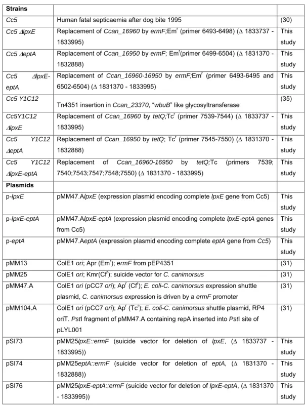
Table 1. Bacterial strains and plasmids used in this study
644
Strains
Cc5 Human fatal septicaemia after dog bite 1995 (30)
Cc5 ΔlpxE Replacement of Ccan_16960 by ermF;Emr (primer 6493-6498) (∆ 1833737 -
1833995)
This study
Cc5 ΔeptA Replacement of Ccan_16950 by ermF; Emr(primer 6499-6504) (∆ 1831370 -
1832888)
This study
Cc5 Δ lpxE-eptA
Replacement of Ccan_16960-16950 by ermF;Emr (primer 6493-6495 and 6502-6504) (∆ 1831370 - 1833995)
This study
Cc5 Y1C12
Tn4351 insertion in Ccan_23370, “wbuB” like glycosyltransferase (35)
Cc5Y1C12
ΔlpxE
Replacement of Ccan_16960 by tetQ;Tcr (primer 7539-7544) (∆ 1833737 - 1833995)
This study
Cc5 Y1C12
ΔeptA
Replacement of Ccan_16950 by tetQ; Tcr (primer 7545-7550) (∆ 1831370 - 1832888)
This study
Cc5 Y1C12
ΔlpxE-eptA
Replacement of Ccan_16960-16950 by tetQ;Tc (primers 7539; 7540;7543;7547;7548;7550) (∆ 1831370 - 1833995)
This study
Plasmids
p-lpxE pMM47.AlpxE (expression plasmid encoding complete lpxE gene from Cc5) This study p-lpxE-eptA pMM47.AlpxE-eptA (expression plasmid encoding complete lpxE-eptA genes
from Cc5)
This study p-eptA pMM47.AeptA (expression plasmid encoding complete eptA gene from Cc5) This
study
pMM13 ColE1 ori; Apr (Emr); ermF from pEP4351 (31)
pMM25 ColE1 ori; Kmr(Cfr); suicide vector for C. canimorsus (31) pMM47.A ColE1 ori (pCC7 ori); Apr (Cfr); E. coli-C. canimorsus expression shuttle
plasmid, C. canimorsus expression is driven by a ermF promoter
(31) pMM104.A ColE1 ori (pCC7 ori); Apr (Tcr); E. coli-C. canimorsus shuttle plasmid, RP4
oriT. PstI fragment of pMM47.A containing repA inserted into PstI site of pLYL001
(31)
pSI73 pMM25lpxE::ermF (suicide vector for deletion of lpxE, (∆ 1833737 - 1833995))
This study pSI74 pMM25eptA::ermF (suicide vector for deletion of eptA, (∆ 1831370 -
1832888))
This study pSI76 pMM25lpxE-eptA::ermF (suicide vector for deletion of lpxE-eptA, (∆ 1831370
- 1833995))
This study
pFR28 pMM25lpxE::tetQ (suicide vector for deletion of lpxE, (∆ 1833737 - 1833995)) This study pFR29 pMM25eptA::tetQ (suicide vector for deletion of eptA, (∆ 1831370 -
1832888))
This study pFR30 pMM25lpxE-eptA::tetQ (suicide vector for deletion of lpxE-eptA, (∆ 1831370 -
1833995))
This study
645 646
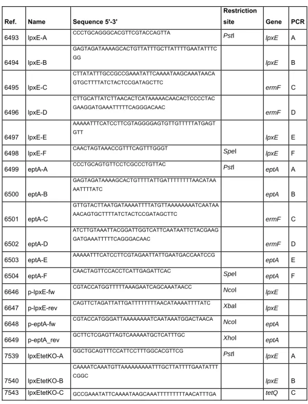
Table 2. Oligonucleotides used in this study
647 648
Ref. Name Sequence 5'-3'
Restriction
site Gene PCR
6493 lpxE-A CCCTGCAGGGCACGTTCGTACCAGTTA PstI lpxE A
6494 lpxE-B GAGTAGATAAAAGCACTGTTATTTGCTTATTTTGAATATTTC GG lpxE B 6495 lpxE-C CTTATATTTGCCGCCGAAATATTCAAAATAAGCAAATAACA GTGCTTTTATCTACTCCGATAGCTTC ermF C 6496 lpxE-D CTTGCATTATCTTAACACTCATAAAAACAACACTCCCCTAC GAAGGATGAAATTTTTCAGGGACAAC ermF D 6497 lpxE-E AAAAATTTCATCCTTCGTAGGGGAGTGTTGTTTTTATGAGT GTT lpxE E 6498 lpxE-F CAACTAGTAAACCGTTTCAGTTTGGGT SpeI lpxE F
6499 eptA-A CCCTGCAGTGTTCCTCGCCCTGTTAC PstI eptA A
6500 eptA-B GAGTAGATAAAAGCACTGTTTTATTGATTTTTTTTAACATAA AATTTTATC eptA B 6501 eptA-C GTTGTACTTAATGATAAAATTTTATGTTAAAAAAAATCAATAA AACAGTGCTTTTATCTACTCCGATAGCTTC ermF C 6502 eptA-D ATCTTGTAAATTACGGATTGGTCATTCAATAATTCTACGAAG GATGAAATTTTTCAGGGACAAC ermF D 6503 eptA-E AAAAATTTCATCCTTCGTAGAATTATTGAATGACCAATCCG eptA E
6504 eptA-F CAACTAGTTCCACCTCATTGAGATTCAC SpeI eptA F
6646 p-lpxE-fw CGTACCATGGTTTTTAAAGAATCAGCAAATAACC NcoI lpxE
6647 p-lpxE-rev CAGTTCTAGATTATTGATTTTTTTTAACATAAAATTTTATC XbaI lpxE
6648 p-eptA-fw CGTACCATGGGATTAAAAAAAATCAATAAATGGACTAACA NcoI eptA 6649 p-eptA_rev GCTTCTCGAGTTAGTCAAAAATGCTCATTTGC XhoI eptA 7539 lpxEtetKO-A GGCTGCAGTTTCCATTCCTTTGGCACGTTCG PstI lpxE A
7540 lpxEtetKO-B
CAAAATCAAATGTTAAAAAAAAATTTGCTTATTTTGAATATTT CGGC
lpxE B
TTTTG 7544 lpxEtetKO-D GATTTTTTTTAACATAAAATTTTATCTTATTTTGATGACATTG ATTTTTGG tetQ D 7541 lpxEtetKO-E CCAAAAATCAATGTCATCAAAATAAGATAAAATTTTATGTTA AAAAAAATC lpxE E 7542 eptAtetKO-F GGACTAGTCAAGGTAAAGCCAATGTTAAGC 1.1.1 SpeI lpxE F 7545 eptAtetKO-A GGCTGCAGTATGGGGAGGAAAGCGTCAATATTG PstI eptA A 7546 eptAtetKO-B CAAAATCAAATGTTAAAAAAAAGCGGTACATTGTTAGTCCA TTTATTG eptA B 7549 eptAtetKO-C CAATAAATGGACTAACAATGTACCGCTTTTTTTTAACATTTG ATTTTG tetQ C 7550 eptAtetKO-D CGGATTGGTCATTCAATAATTTTATTTTGATGACATTGATTT TTGG tetQ D 7547 lpxEtetKO-E CCAAAAATCAATGTCATCAAAATAAAATTATTGAATGACCAA TCCG eptA E 7548 eptAtetKO-F
GGACTAGTCATTAAGTGCTACCCCTATCTTATC SpeI eptA F 649
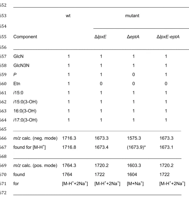
Table 3. MS analysis and interpretation of lipid A variants in wt, lpxE, eptA, and
650
lpxE-eptA deletion mutant strains.
651 ______________________________________________________________________ 652 wt mutant 653 _______________________________________ 654
Component ΔlpxE ΔeptA ΔlpxE-eptA
655 _____________________________________________________________________ 656 GlcN 1 1 1 1 657 GlcN3N 1 1 1 1 658 P 1 1 0 1 659 Etn 1 0 0 0 660 i15:0 1 1 1 1 661 i15:0(3-OH) 1 1 1 1 662 16:0(3-OH) 1 1 1 1 663 i17:0(3-OH) 1 1 1 1 664 __________________________________________________________________ 665
m/z calc. (neg. mode) 1716.3 1673.3 1575.3 1673.3
666
found for [M-H+] 1716.8 1673.4 (1673.9)* 1673.1 667
__________________________________________________________________ 668
m/z calc. (pos. mode) 1764.3 1720.2 1603.3 1720.2
669
found 1764 1722 1604 1722
670
for [M-H++2Na+] [M-H++2Na+] [M+Na+] [M-H++2Na+] 671
__________________________________________________________________ 672
* Ion [M-H+] detected in the neg. ion mode of ΔeptA lipid A was raised from incomplete 673
de-phosphorylated lipid A. The major and representative lipid A molecule of this mutant 674
lacks any charged group and, therefore, its pseudomolecular ion [M+Na+] could only be 675
analyzed in the positive ion mode. 676