HAL Id: hal-01236255
https://hal-amu.archives-ouvertes.fr/hal-01236255
Submitted on 1 Dec 2015
HAL is a multi-disciplinary open access
archive for the deposit and dissemination of
sci-entific research documents, whether they are
pub-lished or not. The documents may come from
teaching and research institutions in France or
abroad, or from public or private research centers.
L’archive ouverte pluridisciplinaire HAL, est
destinée au dépôt et à la diffusion de documents
scientifiques de niveau recherche, publiés ou non,
émanant des établissements d’enseignement et de
recherche français ou étrangers, des laboratoires
publics ou privés.
Prevalence of Mycobacterium lentiflavum in cystic
fibrosis patients, France
Michael Phelippeau, Jean-Christophe Dubus, Martine Reynaud-Gaubert,
Carine Gomez, Nathalie Stremler Le Bel, Marielle Bedotto, Elsa Prudent,
Michel Drancourt
To cite this version:
Michael Phelippeau, Jean-Christophe Dubus, Martine Reynaud-Gaubert, Carine Gomez, Nathalie
Stremler Le Bel, et al.. Prevalence of Mycobacterium lentiflavum in cystic fibrosis patients, France.
BMC Pulmonary Medicine, BioMed Central, 2015, 15 (131), �10.1186/s12890-015-0123-y�.
R E S E A R C H A R T I C L E
Open Access
Prevalence of Mycobacterium lentiflavum in
cystic fibrosis patients, France
Michael Phelippeau
1, Jean-Christophe Dubus
2, Martine Reynaud-Gaubert
3, Carine Gomez
3,
Nathalie Stremler le Bel
2, Marielle Bedotto
1, Elsa Prudent
1and Michel Drancourt
1,4*Abstract
Background: Mycobacterium lentiflavum is rarely isolated in respiratory tract samples from cystic fibrosis patients. We herein describe an unusually high prevalence of M. lentiflavum in such patients.
Methods: M. lentiflavum, isolated from the respiratory tract of cystic fibrosis patients, was identified using both rpoB partial sequencing and detected directly in the sputum by using real-time PCR targeting the smpB gene.
Results: M. lentiflavum emerged as the third most prevalent nontuberculous mycobacterial species isolated in cystic fibrosis patients in Marseille, France. Six such patients were all male, and two of them may have fulfilled the American Thoracic Society clinical and microbiological criteria for M. lentiflavum potential lung infection.
Conclusions: M. lentiflavum was the third most common mycobacteria isolated in cystic fibrosis patients, particularly in six male patients. M. lentiflavum outbreaks are emerging particularly in cystic fibrosis patients.
Keywords: Mycobacterium lentiflavum, Cystic fibrosis, rpoB, smpB Background
Mycobacterium lentiflavumis a fastidious nontuberculous mycobacterium (NTM) isolated from the respiratory tract, urine, lymph nodes and vertebral-bone specimens [1–4]. M. lentiflavumhas seldom been reported in cystic fibrosis patients, at a much lower prevalence than Mycobacterium abscessusand Mycobacterium avium [5–8]. Moreover, its clinical significance is debated because M. lentiflavum is an environmental organism [9].
As we observed an unusual prevalence of M. lentifla-vum isolates in clinical samples taken from patients suffering from respiratory diseases, the objective of this clinical study was to describe the potential opportunistic role of M. lentiflavum in cystic fibrosis.
Methods
Detection and isolation ofM. lentiflavum
Respiratory tract specimens were prospectively collected and analyzed in the Reference Laboratory for Mycobacteria
of the Institut Hospitalo-Universitaire Méditerranée Infec-tion in Marseille, France. After decontaminaInfec-tion using 4 % NaOH-N-acetyl-L-cysteine according to the manufac-turer’s recommendations (MycoPrep, Becton Dickinson, Le Pont-de-Claix, France), each specimen was centrifuged and the pellet was microscopically examined after Ziehl-Neelsen staining. A 500-μL aliquot was simultaneously inoculated into a mycobacterial growth indicator tube (MGIT, Becton Dickinson, Le Pont-de-Claix, France) and onto a Coletsos slant (bioMérieux, La-Balme-les-Grottes, France) incubated at 37 °C in a 5 % CO2
at-mosphere. After Ziehl-Neelsen staining confirmation of positive cultures, the isolates were identified using partial rpoB sequencing [10].
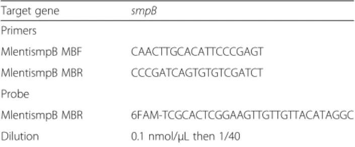
Direct detection of M. lentiflavum in sputum samples was conducted using a specific real-time PCR assay. Briefly, two primers and a probe were designed to specifically hybridize the M. lentiflavum smpB gene (Table 1). The spe-cificity of this assay was checked in silico using the Basic Local Alignment Search Tool (BLAST) [10]. In vitro as-sessment of a collection of sixteen Mycobacterium species (including M. lentiflavum) previously identified by partial rpoB gene sequencing [11], yielded 100 % sensitivity and 100 % specificity for M. lentiflavum (Additional file 1).
* Correspondence:michel.drancourt@univ-amu.fr
1
Aix Marseille Université, URMITE, UMR CNRS 7278, IRD 198, INSERM 1095. Faculté de Médecine, Marseille 13005, France
4
Unité de recherche sur les maladies infectieuses et tropicales émergentes, Faculté de Médecine, 27 Bd jean Moulin, 13385 Marseille, cedex 5, France Full list of author information is available at the end of the article
© 2015 Phelippeau et al. Open Access This article is distributed under the terms of the Creative Commons Attribution 4.0 International License (http://creativecommons.org/licenses/by/4.0/), which permits unrestricted use, distribution, and reproduction in any medium, provided you give appropriate credit to the original author(s) and the source, provide a link to the Creative Commons license, and indicate if changes were made. The Creative Commons Public Domain Dedication waiver (http://creativecommons.org/publicdomain/zero/1.0/) applies to the data made available in this article, unless otherwise stated.
Statistical analysis
Statistical analysis was performed using EpiInfo v3.5.4 software; p < 0.05 was needed for statistical significance.
Ethics
This work was approved by the IFR48 local ethics com-mittee at the Faculty of Medicine, under reference num-ber 07–008. No written consent was needed for this work in accordance with the‘LOI n° 2004–800 relative à la bioéthique’ [Law No. 2004–800 concerning bioethics] published in the Journal Officiel de la République Française on 6 August 2004 because no additional samples were obtained for the study.
Results and discussion
Between January 2010 and September 2014, respiratory tract specimens (sputum, bronchoalveolar lavages and bronchial aspirates) collected from 354 cystic fibrosis pa-tients (235 adults≥18 years and 119 children <18 years) with a female/male ratio of 199/155 (56.2 %) were ana-lyzed for mycobacteria (mean of 13.1 collected speci-men/patient).
In our series, 25/354 (7.1 %) cystic fibrosis patients (twelve children and thirteen adults) had at least one respiratory tract specimen that yielded NTM, including twelve (48 %) patients with M. abscessus complex myco-bacteria, eight (32 %) patients with M. avium complex mycobacteria and six (24 %) patients with M. lentiflavum (Fig. 1); one patient had both M. avium and M. lentifla-vum successively isolated during the study. A total of thirteen M. lentiflavum isolates were identified on the basis of 99.6 % ± 0.003 % similarity with the reference M. lentiflavum CIP 105465T partial rpoB sequence [GenBank:EU109300]. The six M. lentiflavum patients were co-infected by Staphylococcus aureus, Gram-negative bacilli and fungi and were all under azithromycin long-term low-dose prophylaxis (250 mg three times a week). In two patients, M. avium had been previously isolated in 2004 and 2012 from respiratory tract speci-mens (Table 2).
The female/male sex ratio of M. lentiflavum patients (0/6) significantly differed from that of the NTM positive cohort (199/155; 56.2 %) (p = 0.007, Fisher exact test) and from that of patients with another NTM (11/9; 55 %) Table 1 Probe and primer sequences and protocol for real-time
PCR targeting Mycobacterium lentiflavum
Target gene smpB Primers MlentismpB MBF CAACTTGCACATTCCCGAGT MlentismpB MBR CCCGATCAGTGTGTCGATCT Probe MlentismpB MBR 6FAM-TCGCACTCGGAAGTTGTTGTTACATAGGC Dilution 0.1 nmol/μL then 1/40
The extraction of DNA was performed using the EZ1 DNA tissue kit with a Qiagen EZ1 extractor Advanced XL (Qiagen, Courtaboeuf, France) according to the manufacturer’s recommendations. Real-time PCR was performed using a Biorad CFX96 thermocycler with the FAST qPCR MasterMix Plus No ROX kit (Eurogentec, Angers, France) according to the manufacturer’s recommendations: five minutes at 95 °C for activation, followed by 40 cycles of 95 °C for 10 s and 60 °C for 35 secons. Amplification products were analyzed using Biorad software
Fig. 1 Nontuberculous mycobacteria (NTM) isolated from 25/354 cystic fibrosis (CF) patients, January 2010 to September 2014, in Marseille, France. a The number of the patients with at least one respiratory tract specimen that yielded NTM is shown in bars. NTM are color-coded. M. hominissuis, Mycobacterium avium subsp. hominissuis; M. massiliense, Mycobacterium abscessus subsp. massiliense; M. bolletii: Mycobacterium abscessus subsp. bolletii. b Proportion of the pediatric (<18 years) and adult (>18 years) CF patients (nCF= 354) including 25 who yielded NTM including M. lentiflavum, M. avium
complex and M. abscessus complex
(p = 0.02). It further differed from that of M. avium com-plex patients (5/3; 63 %) (p = 0.03) and that of M. absces-suscomplex patients (6/6; 50 %) (p = 0.054) (Fig. 2a). The six M. lentiflavum patients were aged 22.2 ± 11.4 y; the patients with another NTM were aged 22.1 ± 15.1 y; the M. avium complex patients were aged 26.5 ± 19.9 y; and the M. abscessus complex patients were aged 19.2 ± 10.9 y (no statistical significance; p = 0.56; ANOVA) (Fig. 2b).
Two M. lentiflavum patients were clinically stable, had only one positive specimen and were classified as ‘colo-nized’ [12]. The four other M. lentiflavum patients had between two and four positive sputum specimens and in two of them, M. lentiflavum isolation occurred contem-poraneously to the decline of their lung function and thus may fulfill the American Thoracic Society’s (ATS) criteria for NTM lung infection [12] (Additional file 2: Figure S1). Thereafter, the forced expiratory volume improved during antibiotic treatment for M. lentiflavum. One of these two infected patients, aged 17, underwent a double lung-transplant because of poor progression of cystic fibrosis, two years after M. lentiflavum infection had been treated with combined rifabutin, clarithromycin and ethambutol for fourteen months and amikacin for one week. At the four-month follow-up after transplantation,
microbiological surveys did not yield any further M. lentiflavumfrom four separate bronchoalveolar lavages.
In this study, we confirmed the identification of M. lentiflavum, a fastidious organism usually isolated over a period of three weeks, using rpoB partial sequencing [10] and a specific real-time PCR assay targeting the M. lentiflavum smpB gene. Indeed, M. lentiflavum shares <96 % similarity regarding the rpoB gene sequence with closely related species, including Mycobacterium stoma-tepiae DSM 45059, Mycobacterium florentinum DSM 44852, Mycobacterium genavense FI-06288 and Myco-bacterium triplex ATCC 700071 [GenBank:HM022213, HM022205, HM022216 and GQ153311]. Moreover, the routinely used 16S-23S rRNA intergenic spacer sequen-cing [5], hsp65 restriction fragment length polymorph-ism and sequencing [13], and commercial probes for M. avium complex [7, 8, 13] may not be sufficiently dis-criminative as, for example, M. lentiflavum shares > 99 % similarity in the 16S rRNA gene sequence with M. simiae[1].
M. lentiflavumhas emerged over a five-year period as the third most prevalent NTM isolated from the respira-tory tract in our cystic fibrosis cohort. Furthermore, we observed an unexpectedly higher prevalence of patients (6 out of 354; 1.7 %) showing M. lentiflavum isolation than other reported epidemiological surveys. Indeed, only one out of 2912 (0.03 %) French patients [5] and two out of 2970 (0.06 %) American patients [7] were reported in previous studies. In addition, a recent epi-demiology survey of NTM isolated in cystic fibrosis patients in Turkey revealed nine M. lentiflavum isolates collected from one young male teenager out of 130 (0.8 %) cystic fibrosis patients [8]. The reason why M. lentiflavumhas only been isolated in male cystic fibrosis patients remains unexplained. As reported by Bryant et al.[14], patient-to-patient contamination may be sus-pected although this type of cross-contamination is very rare and whole genome sequence analyses would prob-ably be required to satisfactorily conclude a phylogenetic link and track transmission events. Moreover, the six pa-tients described here were treated in two distinct centers (adult and pediatric) for cystic fibrosis. Environmental transmission is another hypothesis for such prevalence.
An in-lab contamination hypothesis has been pro-posed, and eleven non-cystic fibrosis patients yielded M. lentiflavum isolates (out of more than 800 patients (≈1.3 %) who yielded at least one mycobacterial isolate) during the same five-year period. This demonstrates that M. lentiflavum was isolated every two months on aver-age and that the probability of in-lab cross-contamination does exist but remains low.
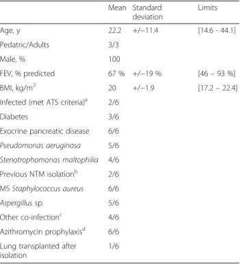
In order to detect M. lentiflavum rapidly in cystic fibrosis patients, we developed an‘in-lab’ real-time PCR targeting the M. lentiflavum smpB gene. This real-time Table 2 Clinical presentation of the six cystic fibrosis patients
who yielded Mycobacterium lentiflavum isolates
Mean Standard deviation Limits Age, y 22.2 +/−11.4 [14.6 - 44.1] Pedatric/Adults 3/3 Male, % 100 FEV, % predicted 67 % +/−19 % [46– 93 %] BMI, kg/m2 20 +/ −1.9 [17.2– 22.4] Infected (met ATS criteria)a 2/6
Diabetes 3/6 Exocrine pancreatic disease 6/6 Pseudomonas aeruginosa 5/6 Stenotrophomonas maltophilia 4/6 Previous NTM isolationb 2/6 MS Staphylococcus aureus 6/6 Aspergillus sp. 5/6 Other co-infectionc 4/6 Azithromycin prophylaxisd 6/6
Lung transplanted after isolation
1/6
FEV forced expiratory volume, ATS American Thoracic Society, BMI body mass index, NTM nontuberculous mycobacteria, MS methicillin susceptible
a
Patients who fulfilled the American Thoracic Society’s microbiological and clinical criteria for NTM pulmonary disease [12]
b
Mycobacterium avium complex
c
Nocardia sp., Penicillium sp., Serratia sp., Achromobacter sp., Scedosporium sp
d
PCR proved its ability to identify all M. lentiflavum isolates specifically. Moreover, our preliminary results indicate that this real-time PCR may be used as a first screening step directly performed on heat-inactivated sputum specimens with good sensitivity and 100 % spe-cificity. These results have to be compared with trad-itional laboratory tools (culture and AFB smears) to clarify its relevance for clinical practice [15] and have to be validated on larger series of prospectively-collected sputum specimens, including from patients who had previously yielded M. lentiflavum in sputum cultures.
M. lentiflavumhad been considered to be a harmless or-ganism. However, this interpretation was recently chal-lenged by the publication of a few cases with disseminated M. lentiflavum infections [16–18]. In one case, hemo-phagocytic lymphohistiocytosis and disseminated M. lentiflavuminfection in a heart-transplanted patient led to the death of this immune-compromised patient within ten days [18].
In the present study, two out of six patients fulfilled the ATS clinical and microbiological criteria for NTM lung disease [12] and had improved respiratory function while receiving specific antibiotic therapy. However, ATS criteria, while they continue to be applied to cystic fibro-sis patients, are far from specific for such patients where radiographic findings, which are often associated with NTM, are commonplace irrespective of colonization. Moreover, clinical decline occurs in cystic fibrosis for a multitude of reasons. The antibiotic therapy our patient
received is, moreover, active against a wide range of respiratory tract pathogens.
In our series, all patients were receiving long-term azithromycin therapy (Table 2). This use of macrolide in CF patients was shown to be a risk factor for NTM infection, especially with M. abscessus [19], by inhibiting intracellular killing of mycobacteria in macrophage by impairing autophagic and phagosomal degradation [20]. Such mechanisms may have played a role in the increase of M. lentiflavum isolation. However, as M. lentiflavum is usually susceptible to clarithromycin [8], the fact that patients were receiving macrolide and M. lentiflavum developed further supports the theory that this is either an environmental contaminant or a transi-ent colonizer which has not been exposed to macrolide for prolonged periods.
Conclusion
M. lentiflavumwas the third most common NTM isolated from male cystic fibrosis patients, although few respiratory cases had been previously reported, particularly in such patients. We propose monitoring cystic fibrosis patients’ respiratory tract samples for mycobacteria detection, to achieve this goal, we propose the use and development of specific molecular tools such as rpoB partial sequencing (or a specific real-time PCR which needs to be fully vali-dated against traditional laboratory tools) to monitor the presence of M. lentiflavum in each cystic fibrosis center and reference laboratories for mycobacteria.
Fig. 2 Sex-ratio (a) and age distribution (b) of the 354 cystic fibrosis (CF) patients and 25 patients who yielded nontuberculous mycobacteria (NTM) including Mycobacterium lentiflavum (n = 6), Mycobacterium avium complex (n = 8) and Mycobacterium abscessus complex (n = 12). * p < 0.05; ** p = 0.053; NS, p > 0.1
Additional files
Additional file 1: Table S1. Mycobacterium species tested for the specificity/sensitivity assay of real-time PCR for M. lentiflavum species. (PDF 43 kb)
Additional file 2: Figure S1. Clinical and microbiological data concerning two cystic fibrosis patients who yielded more than one M. lentiflavum isolate and who may fulfill the American Thoracic Society’s clinical and microbiological criteria for NTM lung infection. BMI: Body Mass Index. (PDF 121 kb)
Abbreviations
AFB:Acid- Fast Bacilli; ANOVA: analysis of variance; ATS: American Thoracic Society; BLAST: Basic Local Alignment Search Tool; BMI: Body Mass Index (Supplementary fugure); CF: cystic fibrosis; NTM: nontuberculous mycobacterium; PCR: polymerase chain reaction.
Competing interests
The authors declare that they have no competing interests. Authors’ contributions
MP performed all assays. MP, JCD, MRG, CG and NSB participated in clinical data reviewing. MP, MB and EP designed the real-time PCR. MD conceived the study. MP and MD participated in its design and coordination and helped to draft the manuscript. All authors read and approved the final version of the manuscript. Authors’ information
Michael Phelippeau is a pulmonologist and resident in infectious diseases medicine with particular interest in tuberculosis and respiratory mycobacterial diseases.
Acknowledgments
We acknowledge URMITE (Unité de Recherche sur les Maladies Infectieuses et Tropicales Emergentes) and Prof. Didier Raoult who contributed to the financial support of this study.
Author details
1Aix Marseille Université, URMITE, UMR CNRS 7278, IRD 198, INSERM 1095.
Faculté de Médecine, Marseille 13005, France.2Centre de Ressource et de Compétences de la Mucoviscidose (CRCM) pédiatrique CHU Hôpital la Timone, Marseille, France.3Centre de Ressource et de Compétences de la
Mucoviscidose (CRCM) adulte; équipe de Transplantation pulmonaire, CHU Hôpital Nord, URMITE - CNRS-UMR 6236 Aix-Marseille Université, Marseille, France.4Unité de recherche sur les maladies infectieuses et tropicales
émergentes, Faculté de Médecine, 27 Bd jean Moulin, 13385 Marseille, cedex 5, France.
Received: 18 May 2015 Accepted: 12 October 2015
References
1. Springer B, Wu WK, Bodmer T, Haase G, Pfyffer GE, Kroppenstedt RM, et al. Isolation an characterization of a unique group of slowly growing mycobacteria: description of Mycobacterium lentiflavum sp. nov. J Clin Microbiol. 1996;34:1100–7.
2. Haase G, Kentrup H, Skopnik H, Springer B, Böttger EC. Mycobacterium lentiflavum: an etiologic agent of cervical lymphadenitis. Clin Infect Dis. 1997;25:1245–6.
3. Cabria F, Torres MV, Garcia Cia JI, Dominguez-Guarrido MN, Esteban J, Jimenez MS. Cervical lymphadenitis caused by Mycobacterium lentiflavum. Pediatr Infect Dis J. 2002;21:574–5.
4. Asiimwe B, Bagyenzi GB, Ssengooba W, Mumbowa F, Mboowa G, Wajja A, et al. Species and genotypic diversity of non-tuberculous mycobacteria isolated from children investigated for pulmonary tuberculosis in rural Uganda. BMC Infect D. 2013;13:88–94.
5. Roux AL, Catherinot E, Ripoll F, Soismier N, Macheras E, Ravilly S, et al. Multicenter study of prevalence of nontuberculous mycobacteria in patients with cystic fibrosis in France. J Clin Microbiol. 2009;47:4124–8.
6. Seddon P, Fidler K, Raman S, Wyatt H, Ruiz G, Elston C, et al. Prevalence of nontuberculous mycobacteria in cystic fibrosis clinics, United Kingdom. Emerg Infect D. 2009;19:1128–30.
7. Olivier K, Weber DJ, Wallace Jr RJ, Faiz AR, Lee JH, Zhang Y, et al. Nontuberculous mycobacteria I: multicenter prevalence study in cystic fibrosis. Am J Respir Crit Care Med. 2003;167:828–34.
8. Satana D, Erkose-Genc G, Tamay Z, Uzun M, Guler N, Erturan Z. Prevalence and drug resistance of mycobacteria in Turkish cystic fibrosis patients. Ann Clin Microbiol Antimicrob. 2014;13:28.
9. Marshall H, Carter R, Torbey MJ, Minion S, Tolson C, Sidjabat HE, et al. Mycobacterium lentiflavum in drinking water supplies, Australia. Emerg Infect Dis. 2011;17:395–402.
10. Cock PJ, Chilton JM, Grüning B, Johnson JE, Soranzo N. NCBI BLAST+ integrated into Galaxy. Gigascience. 2015;4:39.
11. Adékambi T, Colson P, Drancourt M. rpoB-based identification of nonpigmented and late-pigmented rapidly growing mycobacteria. J Clin Microbiol. 2003;41:5699–708.
12. Griffith D, Aksamit T, Brown-Elliott BA, Catanzaro A, Daley C, Gordin F, et al. An official ATS/IDSA statement: diagnosis, treatment and prevention of nontuberculous mycobacterial diseases. Am J Respir Crit Care Med. 2007;175:367–416.
13. Pierre-Audigier C, Sermet-Gaudelus I, Le Bourgeois M, Offredo C, Vu-Thien H, Fauroux B, et al. Age-related prevalence and distribution of
nontuberculous species among patients with cystic fibrosis. J Clin Microbiol. 2005;43:3467–70.
14. Bryant JM, Grogono DM, Greaves D, Foweraker J, Roddick I, Inns T, et al. Whole-genome sequencing to identify transmission of Mycobacterium abscessus between patients with cystic fibrosis: a retrospective cohort study. Lancet. 2013;381:1551–60.
15. Miller K, Harrington SM, Procop GW. Acid-fast smear and histopathology results provide guidance for the appropriate use of broad-range polymerase chain reaction and sequencing for mycobacteria. Arch Pathol Lab Med. 2015. [Epub ahead of print].
16. Nagata N, Honda M, Kobayakawa M, Maeda S, Sakurai T, Akiyama J, et al. Mycobacterium lentiflavum ileitis using aspirated intestinal fluid during endoscopy in HIV-infected patient. Dig Endosc. 2011;23:271–2. 17. Ibanez R, Serrano-Heranz R, Jimenez-Palop M, Roman C, Corteguera M,
Jimenez S. Disseminated infection caused by slow-growing Mycobacterium lentiflavum. Eur J Clin Microbiol Infect Dis. 2002;21:691–2.
18. Thomas G, Hraiech S, Dizier S, Weiller PJ, Ene N, Serratrice J, et al. Disseminated Mycobacterium lentiflavum in a heart-transplanted man responsible for haemophagocyticlymphohistocytosis. J Clin Microbiol. 2014;52:3121–3.
19. Levy I, Grisaru-Soen G, Lerner-Geva L, Kerem E, Blau H, Bentur L, et al. Multicenter cross-sectional study of nontuberculous mycobacterial infections among cystic fibrosis patients, Israel. Emerg Infect Dis. 2008;14:378–84.
20. Renna M, Schaffner C, Brown K, Shang S, Tamayo MH, Hegyi K, et al. Azithromycin blocks autophagy and may predispose cystic fibrosis patients to mycobacterial infection. J Clin Invest. 2014;121:3554–63.
Submit your next manuscript to BioMed Central and take full advantage of:
• Convenient online submission • Thorough peer review
• No space constraints or color figure charges • Immediate publication on acceptance
• Inclusion in PubMed, CAS, Scopus and Google Scholar • Research which is freely available for redistribution
Submit your manuscript at www.biomedcentral.com/submit