dissociable cognitive trajectories in Alzheimer’s disease
The MIT Faculty has made this article openly available.
Please share
how this access benefits you. Your story matters.
Citation
Zhang, Xiuming; Mormino, Elizabeth C.; Sun, Nanbo; Sperling,
Reisa A.; Sabuncu, Mert R. and Yeo, B. T. Thomas. “Bayesian
Model Reveals Latent Atrophy Factors with Dissociable Cognitive
Trajectories in Alzheimer’s Disease.” Proceedings of the National
Academy of Sciences 113, no. 42 (October 2016): E6535–E6544. ©
2016 National Academy of Sciences
As Published
http://dx.doi.org/10.1073/pnas.1611073113
Publisher
National Academy of Sciences (U.S.)
Version
Final published version
Citable link
http://hdl.handle.net/1721.1/108793
Terms of Use
Article is made available in accordance with the publisher's
policy and may be subject to US copyright law. Please refer to the
publisher's site for terms of use.
Bayesian model reveals latent atrophy factors with
dissociable cognitive trajectories in
Alzheimer
’s disease
Xiuming Zhanga, Elizabeth C. Morminob, Nanbo Suna, Reisa A. Sperlingb, Mert R. Sabuncuc,d, B. T. Thomas Yeoa,c,e,1,
and the Alzheimer’s Disease Neuroimaging Initiative
aDepartment of Electrical and Computer Engineering, Agency for Science, Technology and Research-National University of Singapore Clinical Imaging Research Centre, Singapore Institute for Neurotechnology and Memory Networks Program, National University of Singapore, Singapore 117599;bDepartment of Neurology, Massachusetts General Hospital/Harvard Medical School, Charlestown, MA 02129;cMartinos Center for Biomedical Imaging, Massachusetts General Hospital/Harvard Medical School, Charlestown, MA 02129;dComputer Science and Artificial Intelligence Laboratory, Massachusetts Institute of Technology, Cambridge, MA 02138; andeCentre for Cognitive Neuroscience, Duke-National University of Singapore Graduate Medical School, Singapore 169857 Edited by James L. McClelland, Stanford University, Stanford, CA, and approved August 23, 2016 (received for review July 14, 2016)
We used a data-driven Bayesian model to automatically identify distinct latent factors of overlapping atrophy patterns from voxel-wise structural MRIs of late-onset Alzheimer’s disease (AD) demen-tia patients. Our approach estimated the extent to which multiple distinct atrophy patterns were expressed within each participant rather than assuming that each participant expressed a single at-rophy factor. The model revealed a temporal atat-rophy factor (medial temporal cortex, hippocampus, and amygdala), a subcortical atro-phy factor (striatum, thalamus, and cerebellum), and a cortical at-rophy factor (frontal, parietal, lateral temporal, and lateral occipital cortices). To explore the influence of each factor in early AD, atro-phy factor compositions were inferred in beta-amyloid–positive (Aβ+) mild cognitively impaired (MCI) and cognitively normal (CN) participants. All three factors were associated with memory decline across the entire clinical spectrum, whereas the cortical factor was associated with executive function decline in Aβ+ MCI participants and AD dementia patients. Direct comparison between factors revealed that the temporal factor showed the strongest association with memory, whereas the cortical factor showed the strongest association with executive function. The subcortical factor was as-sociated with the slowest decline for both memory and executive function compared with temporal and cortical factors. These results suggest that distinct patterns of atrophy influence decline across different cognitive domains. Quantification of this heterogeneity may enable the computation of individual-level predictions rele-vant for disease monitoring and customized therapies. Factor com-positions of participants and code used in this article are publicly available for future research.
mental disorder subtypes
|
Alzheimer’s disease subtypes|
Alzheimer’s disease heterogeneity|
voxel-based morphometry|
unsupervised machine learningA
lzheimer’s disease (AD) dementia is a devastating neurode-generative disease that affects 11% of individuals over age 65 with no disease-modifying treatment available. Accurate in vivo biomarkers are urgently needed to assist in early detection of at-risk individuals, improve diagnosis, monitor disease progression, and serve as outcome measures in clinical trials.Although AD is typically associated with an amnestic clinical presentation and disruption of the medial temporal lobe (1), it has become increasingly clear that heterogeneity exists within this disease. Specifically, heterogeneity has been observed in the clinical presentation of AD (2) and the spatial distribution of neurofibrillary tangles (NFTs) (3, 4) as well as the presence of comorbid pathologies, such as vascular disease, Lewy bodies, and transactive response DNA binding protein 43 kDa (TDP-43) (5, 6). Interestingly, the spatial distribution of atrophy varies across AD subtypes defined on the basis of NFT distribution (7), suggesting
heterogeneity in AD. Furthermore, although distinct atrophy patterns have been observed in patients who clearly show atyp-ical clinatyp-ical presentations (8), heterogeneity in GM atrophy has also been reported among late-onset AD cases (9). It is, there-fore, likely that the ability to quantify varying patterns of atrophy among AD patients will help inform our understanding of fun-damental disease processes.
In this study, we sought to explore the heterogeneity of atro-phy patterns in late-onset AD using a data-driven Bayesian framework that accounted for and estimated latent AD atrophy factors derived from structural MRI data. The mathematical framework that we used, latent Dirichlet allocation (LDA) (10), has been successfully used to extract overlapping brain networks from functional MRI (11) and metaanalytic data (12, 13). Im-portantly, this approach does not require the atrophy pattern of an individual to be determined by a single atrophy factor. Instead, the model allows the possibility that multiple latent factors are expressed to varying degrees within an individual. For example, the atrophy pattern of a patient might be 90% owing to factor 1
Significance
Alzheimer’s disease (AD) affects 10% of the elderly population. The disease remains poorly understood with no cure. The main symptom is memory loss, but other symptoms might include impaired executive function (ability to plan and accomplish goals; e.g., grocery shopping). The severity of behavioral symp-toms and brain atrophy (gray matter loss) can vary widely across patients. This variability complicates diagnosis, treatment, and prevention. A mathematical model reveals distinct brain atrophy patterns, explaining variation in gray matter loss among AD dementia patients. The atrophy patterns can also explain varia-tion in memory and executive funcvaria-tion decline among dementia patients and at-risk nondemented participants. This model can potentially be applied to understand brain disorders with vary-ing symptoms, includvary-ing autism and schizophrenia.
Author contributions: X.Z., E.C.M., R.A.S., M.R.S., and B.T.T.Y. designed research; X.Z. and N.S. performed research; X.Z., M.R.S., and A.D.N.I. contributed new reagents/analytic tools; A.D.N.I. contributed to design and implementation of the Alzheimer’s Disease Neuroimaging Initia-tive; X.Z. and N.S. analyzed data; and X.Z., E.C.M., M.R.S., and B.T.T.Y. wrote the paper. The authors declare no conflict of interest.
A complete list of the Alzheimer’s Disease Neuroimaging Initiative can be found in
SI Appendix.
This article is a PNAS Direct Submission.
Freely available online through the PNAS open access option.
Data deposition: The Alzheimer’s Disease Neuroimaging Initiative data are publicly avail-able atadni.loni.usc.edu/.
1To whom correspondence should be addressed. Email: thomas.yeo@nus.edu.sg.
PSYCHOLOGI CAL AND COGNITIVE SCIENC ES
and 10% owing to factor 2, whereas the atrophy pattern of another patient might be 60% owing to factor 1 and 40% owing to factor 2. Given that multiple contributors that are not mutually exclusive may influence heterogeneity in AD, such as the spatial location of NFT pathology (3, 4), coexisting non-AD pathologies (5, 14), and genetics (9), we believe that it is more biologically plausible that individuals express varying degrees of distinct atrophy factors rather than one single factor. Thus, the LDA approach is partic-ularly well-suited for these analyses and will provide insight into whether expressing multiple atrophy factors is common among late-onset AD patients.
Most studies investigating the heterogeneity of AD have ex-amined patients soon after AD onset or at advanced AD stages (3, 7, 15–17). However, the pathophysiological processes of AD begin at least a decade before clinical diagnosis (18), suggesting that the emergence of this heterogeneity may occur before the onset of clinical dementia. In this study, we, therefore, examined how distinct atrophy factors identified in AD dementia patients were associated with longitudinal cognitive decline early in non-demented participants who were at risk for AD dementia based on elevated beta-amyloid (Aβ) (19–21).
Our study makes three significant contributions. First, we in-troduced an innovative modeling strategy where expressions of multiple atrophy patterns are estimated rather than assigning each participant to a single subtype. Second, our approach harnesses the rich multidimensional information across all GM voxels, avoiding the need for a priori selection of regions and enabling an in-depth exploration of atrophy patterns. Third, application of this approach to participants spanning the clinical spectrum revealed that latent atrophy factors are associated with distinct memory and executive function trajectories, providing insights into the impact of disease heterogeneity throughout the prolonged course of AD. Results
Overall Approach. Our approach involved three main steps. In step I, we performed LDA (a Bayesian model) (10) to estimate latent atrophy factors in 188 AD dementia patients and used this model to extract factor compositions in two independent samples of nondemented participants: 147 Aβ+ mild cognitively impaired (MCI) and 43 Aβ+ cognitively normal (CN) participants. In step
Step I. Discovering Latent Atrophy Factors in AD Dementia Patients.
We used the Bayesian LDA model (10) to encode our assumption that a patient expresses one or more latent atrophy factors (Fig. 1). The LDA model was applied to the structural MRI of 188 AD dementia patients. Given the voxelwise GM density values derived from structural MRI (FSL-VBM) (22) and a predefined number of factors K, the model is able to estimate the probability that a particular factor is associated with atrophy at a specific spatial location [i.e., Pr(Voxelj Factor) or probabilistic atrophy map of the factor] and the probability that an individual expresses each atro-phy factor [i.e., Pr(Factorj Patient) or atrophy factor composition
Latent Atrophy Factors Brain sMRI
...
Pr(Factor | Patient) Pr(Voxel | Factor)...
Factor 2 Factor 3 Factor 1Voxel 1 Voxel 2 Voxel 3 Voxel 4
Fig. 1. A Bayesian model of AD dementia patients, latent atrophy factors, and brain structural MRI (sMRI). Underpinning our approach is the premise that each participant expresses one or more latent factors. Each factor is associated with distinct but possibly overlapping patterns of brain atrophy. The framework can be instantiated with a mathematical model (LDA) (10), with parameters that can be estimated from the structural MRI data of AD dementia patients. The estimated parameters are the probability that a patient expresses a particular factor [i.e., Pr(Factorj Patient)] and the probability that a factor is associated with atrophy at an MRI voxel [i.e., Pr(Voxelj Factor)].
(A1) Temporal + Subcortical (A1) Temporal + Subcortical
(A2) Cortical (A2) Cortical (B1) Temporal (B1) Temporal (B2) Subcortical (B2) Subcortical (B3) Cortical (B3) Cortical (C1) Temporal (C1) Temporal (C2) Subcortical (C2) Subcortical (C3) Posterior Cortical (C3) Posterior Cortical (C4) Frontal Cortical (C4) Frontal Cortical 0.8e-5
0.8e-5 2.5e-52.5e-5 L R L R K = 2 K = 2 K = 3 K = 3 K = 4 K = 4
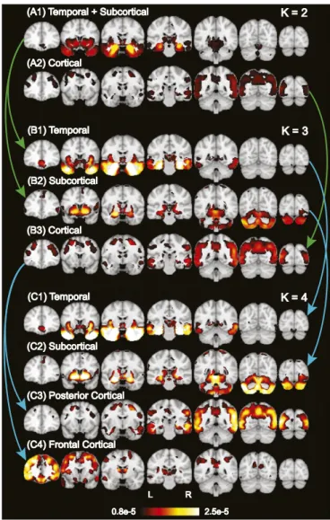
Fig. 2. Hierarchy of latent atrophy factors with distinct atrophy patterns in AD. Bright color indicates higher probability of atrophy at that spatial location for a particular atrophy factor [i.e., Pr(Voxelj Factor)]. Each of the (A) two, (B) three, and (C) four factors was associated with a distinct pattern of brain atrophy and named accordingly. A nested hierarchy of atrophy factors was observed, al-though the model did not mandate such a hierarchy. For example, when going from two to three factors, the temporal+subcortical factor (A1) split into tem-poral (B1) and subcortical (B2) factors, whereas the cortical factor remained the same (A2 and B3). From three to four factors, the temporal and subcortical
of the individual]. Importantly, resulting atrophy factors were not predetermined but estimated from data (Materials and Methods).
An important model parameter is the number of latent atro-phy factorsK. Therefore, we first determined how factor esti-mation changed fromK = 2 to 10. Visual inspection of the spatial distribution of each atrophy factor suggested that factor esti-mates fromK = 2 to 10 were organized in a hierarchical fashion (Fig. 2 andSI Appendix, Fig. S1). For instance, the two-factor model revealed one factor associated with atrophy in temporal and subcortical regions (“temporal+subcortical”) (Fig. 2A1) and another factor associated with atrophy throughout cortex (“cortical”) (Fig. 2A2). The three-factor model resulted in a similar cortical factor (Fig. 2B3 and SI Appendix, Table S1C), whereas the temporal+subcortical factor split into a “temporal” factor associated with extensive atrophy in the medial temporal lobe (Fig. 2B1 andSI Appendix, Table S1A) and a“subcortical” factor associated with atrophy in the cerebellum, striatum, and thalamus (Fig. 2B2 and SI Appendix, Table S1B). Likewise, the four-factor model resulted in the cortical factor splitting into “frontal cortical” and “posterior cortical” factors, whereas the temporal and subcortical factors remained the same (Fig. 2C). Sagittal and axial slices of these probabilistic atrophy maps are available inSI Appendix, Fig. S1.
To quantify the hierarchical phenomenon, we used an ex-haustive search to assess the possibility that two unknown factors in the (K + 1)-factor model were subdivisions of an unknown factor in theK-factor model (whereas the other factors remained the same). The exhaustive search yielded a hypothesized factor hierarchy with associated correlation values quantifying the sub-division quality (SI Appendix, SI Methods). The high correlation values (SI Appendix, Fig. S2) confirmed that additional factors emerged as subdivisions of lower-order factors, corresponding to a nested hierarchy of atrophy factors.
This nested hierarchy suggested that specification of different numbers of estimated factors might yield distinct insights into AD. In the remainder of this paper, we highlighted the results of three-factor model (Fig. 2B), because the emergence of the temporal and cortical factors were consistent with the “limbic-predominant” and “hippocampal-sparing” pathologically defined AD subtypes previously reported (3, 7). We additionally re-peated analyses for two- and four-factor models, which yielded behavioral insights consistent with the three-factor model. These additional results are reported inSI Appendix, Figs. S6 and S8.
To explore the influence of atrophy factors in early AD, proba-bilistic atrophy maps Pr(Voxelj Factor) estimated from the AD dementia patients were used to infer factor compositions Pr(Factorj Participant) of 190 Aβ+ nondemented participants using the
stan-their cerebrospinal fluid (CSF) amyloid data available; 91 of 100 patients were Aβ+ (CSF amyloid concentration <192 pg/mL) (23). We performed LDA on the subset of Aβ+ AD dementia patients (and Aβ+ MCI participants) (SI Appendix, SI Results) and compared atrophy patterns of the resulting factors with those derived using the larger sample (SI Appendix, Fig. S3). Atrophy factors were similar across these methods, with an av-erage correlation across all pairwise comparisons of r = 0.89. Given this similarity and to improve our estimates of the atrophy factors, we elected to use the atrophy factors derived from the larger sample of 188 AD dementia patients for subsequent analyses. Furthermore, resulting atrophy patterns were consis-tent between FreeSurfer (24) and FSL-VBM, suggesting that the atrophy factors were robust to variations in image preprocessing software (SI Appendix, SI Results and SI Methods).
To determine whether expression of atrophy factors remained stable over time, we examined the subset of the Alzheimer’s Disease Neuroimaging Initiative 1 (ADNI 1) participants who had a two-year follow-up scan available (n = 560 of 810). We were specifically interested in whether atrophy factors reflected dif-ferent disease stages rather than difdif-ferent atrophy subtypes (for instance, high expression of the temporal factor may lessen over time with greater expression of the cortical factor). Therefore, we compared factor compositions after two years with baseline compositions. The factor probabilities were positivity correlated and highly consistent (r > 0.85 across all three factors; Fig. 3) (SI Appendix, Fig. S4 shows results by diagnostic group with addi-tional amyloid information), suggesting that these factors do not merely reflect a sequence of atrophy patterns.
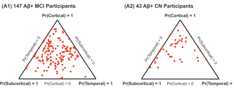
Examination of atrophy factor compositions among AD de-mentia patients revealed that the majority expressed multiple latent atrophy factors rather than predominantly expressing a single atrophy factor (Fig. 4). Examination of factor compositions of 190 Aβ+ nondemented participants revealed a similar pattern, such that the majority of participants expressed multiple atrophy factors (SI Appendix, Fig. S5A). Factor compositions for the two-and four-factor models also suggest that most participants expressed multiple atrophy factors (SI Appendix, Fig. S5 B and C). To understand the association between atrophy factors and de-mographic variables, general linear model (GLM; for continuous Fig. 3. Stability of factor compositions over two years. Each blue dot
repre-sents a participant. For each plot, x and y axes represent the probabilities of factor at baseline and two years after baseline, respectively. In the ideal case, where factor probability estimates remain exactly the same after two years, one would expect a y= x linear fit as well as an r = 1 correlation. In our case, the linear fits were close to y= x with r > 0.85 for all three atrophy factors, sug-gesting that the factor compositions were stable, despite disease progression.
Pr(Cortical) = 1 Pr(Subcortical) = 0 Pr(Cortical) = 0 Pr(T emporal) = 0 Pr(Subcortical) = 1 Pr(Temporal) = 1 Fig. 4. Factor compositions of 188 AD dementia patients. Each patient cor-responds to a dot, with location (in barycentric coordinates) that represents the factor composition. Color indicates amyloid status: red for Aβ+, green for Aβ−, and blue for unknown. Corners of the triangle represent pure factors; closer distance to the respective corner indicates higher probability for the
PSYCHOLOGI CAL AND COGNITIVE SCIENC ES
variables) and logistic regression (for binary variables) were con-ducted in 188 AD dementia patients (SI Appendix, Table S2). Briefly, the response variable was the variable of interest (e.g., age at AD onset), and the explanatory variables consisted of two col-umns encoding participants’ loading on the cortical and subcortical factors. The temporal factor was implicitly modeled, because the factor probabilities summed to one (Materials and Methods).
There were no significant differences in years from AD onset, education, sex, or apolipoprotein E (APOE)e4 loadings across the three factors. Importantly, amyloid level was not significantly different across factors. The cortical factor was associated with significantly younger baseline age than the temporal factor (P = 1e−5) and subcortical factor (P = 2e−6) as well as younger age at AD onset than the temporal factor (P = 3e−4) and subcortical factor (P = 7e−6). In addition, the subcortical factor was associ-ated with higher APOEe2 loading than the temporal factor (P = 0.01) and cortical factor (P = 0.04), but these associations were not significant when corrected for multiple comparisons.
Similar analyses were conducted for the Aβ+ MCI and CN groups. The only significant association was that, among Aβ+ MCI participants, the cortical factor was associated with younger age at baseline compared with the temporal factor (P = 0.05) and subcortical factor (P = 0.02). However, this association did not survive after correcting for multiple comparisons.
Step III. Examining Associations Between Atrophy Factors and Cognition.
We first examined diagnostic group differences in memory (ADNI-Mem) (25) and executive function (ADNI-EF) (26) without con-sidering factor compositions. As expected, cross-sectional memory was worse for AD dementia patients (mean = −0.84) compared with Aβ+ MCI participants (mean = −0.21; t test P = 5e−23). Aβ+ MCI participants had worse memory than Aβ+ CN participants (mean= 0.93; t test P = 2e−26). Likewise, cross-sectional executive function was worse for AD dementia patients (mean = −0.92)
analysis revealed that all factors were associated with baseline memory, and these associations continued to worsen across the disease spectrum (Fig. 5A1). For cross-sectional executive func-tion, there was only an association with the cortical factor, and this association also worsened across the disease spectrum (Fig. 5B1). Next, we examined a linear mixed effects (LME) model pre-dicting longitudinal change in memory and executive function (Fig. 5 A2 and B2). The LME model provides significantly improved exploitation of longitudinal measurements (27) by accounting for both intraindividual measurement correlations and interindividual variability. The model setup was the same as the GLM above, ex-cept that time and its interactions with diagnosis and factor com-positions were included as predictors (SI Appendix, SI Methods).
This analysis revealed that the temporal and subcortical fac-tors exhibited memory decline that began in CN and maintained similar memory decline rates in MCI and AD (Fig. 5A2). In contrast, the cortical factor was not associated with memory decline in CN but showed faster decline in MCI compared with CN and AD compared with MCI (Fig. 5A2). The cortical factor was not associated with executive function decline in CN but showed faster longitudinal executive function decline in MCI compared with CN and AD compared with MCI (Fig. 5B2).
In our final set of analyses examining cognition, we directly compared the three factors. The GLM and LME model were exactly the same as the previous sections, but we instead focused on the contrasts between factors.
For cross-sectional memory, the temporal factor was associ-ated with worse performance than the subcortical (P = 3e−6) and cortical (P = 7e−3) factors among AD dementia patients (Fig. 6A). Similar results were found for Aβ+ MCI participants (SI Appendix, Fig. S7A1). Among Aβ+ CN participants, there
was no memory difference across the atrophy factors (SI Ap-pendix, Fig. S7A1). For cross-sectional executive function, the cortical factor was associated with worse performance than the Fig. 5. Differences by diagnosis and atrophy factor in (1) cross-sectional
baseline and (2) longitudinal decline rates of (A) memory and (B) executive function. Comparisons remaining significant after FDR (q= 0.05) control are highlighted in blue. T, S, and C indicate temporal, subcortical, and cortical factors, respectively. For example, the top left cell of A, 1 suggests that Aβ+ MCI participants with high loading on the temporal factor had worse baseline memory than Aβ+ CN participants with high loading on the same factor (P = 6e−4). However, the bottom left cell of B, 2 suggests that Aβ+ CN participants expressing the cortical factor did not exhibit executive function decline (P= 0.91), whereas the bottom right cell of B, 2 suggests that AD dementia patients expressing the cortical factor showed faster executive function decline than Aβ+ MCI participants expressing the same factor (P = 7e−4).
A
B
Fig. 6. Comparisons of baseline (A) memory and (B) executive function in AD dementia patients across factors. Comparisons remaining significant after FDR (q= 0.05) control are highlighted in blue. T, S, and C indicate temporal, sub-cortical, and cortical factors, respectively. Blue dots are estimated differences between“pure atrophy factors,” and red bars show the SEs (Materials and
temporal (P = 0.01) and subcortical (P = 1e−5) factors among AD dementia patients (Fig. 6B). There was no executive function difference across the factors among Aβ+ CN and MCI partici-pants (SI Appendix, Fig. S7B1).
For longitudinal change in memory (Fig. 7A), the cortical factor was associated with faster longitudinal memory decline than the temporal (P = 1e−4) and subcortical (P = 4e−6) factors among AD dementia patients. Among Aβ+ MCI participants, the subcortical factor was associated with slower decline rate than the cortical (P = 8e−4) and temporal (P = 4e−3) factors. Finally, among Aβ+ CN participants, the cortical factor showed slower memory decline than the temporal (P = 1e−4) and sub-cortical (P = 3e−3) factors.
For longitudinal change in executive function (Fig. 7B), the cortical factor was associated with faster executive function de-cline than temporal (P = 2e−3) and subcortical (P = 2e−4) factors among AD dementia patients. Among Aβ+ MCI partic-ipants, the subcortical factor had slower decline than the cortical (P = 8e−9) and temporal (P = 1e−8) factors. There was no ex-ecutive function decline difference across the factors among Aβ+
results were consistent with the three-factor model (SI Appendix, SI Results). In addition, associations between minimental state examination (MMSE) and the three atrophy factors are reported inSI Appendix, Fig. S7C.
Discussion
In this study, we identified distinct atrophy factors within AD dementia patients using Bayesian LDA modeling of MRI GM density maps. This approach estimated the factor composition of multiple atrophy factors for each participant rather than assuming membership to a single atrophy subtype (Fig. 1). Our analysis yielded a nested hierarchy of atrophy factors (Fig. 2), which cor-responded to distinct trajectories of memory and executive func-tion decline across the disease spectrum (Fig. 8). Overall, these results provide evidence that heterogeneity in patterns of atrophy exists in late-onset AD and that these atrophy patterns are asso-ciated with distinct cognitive trajectories.
A
B
Fig. 7. Comparisons of (A) memory and (B) executive function decline rates across factors by clinical group. Comparisons remaining significant after FDR (q= 0.05) control are highlighted in blue. T, S, and C indicate temporal, subcortical, and cortical factors, respectively. Blue dots are estimated differ-ences between pure atrophy factors, and red bars show the SEs (SI Appendix, SI Methods). For example, the second row in A suggests that AD dementia tients expressing the cortical factor showed faster memory decline than pa-tients expressing the temporal factor (P= 1e−4).
Memory
ADNI-Mem
Disease Stage
Aβ+ CN Aβ+ MCI AD
T < S T < C, S
∆T, ∆S < ∆C ∆C < ∆S, ∆T
ADNI-EF
Disease Stage
Aβ+ CN Aβ+ MCI AD
C < T, S ∆T, ∆C < ∆S Executive Function ∆T, ∆C < ∆S ∆C < ∆S, ∆T Subcortical Cortical Temporal Temporal Cortical Subcortical
A
B
Fig. 8. Schematics of distinct (A) memory and (B) executive function tra-jectories for temporal, subcortical, and cortical atrophy factors. T, S, and C indicate temporal, subcortical, and cortical factors, respectively. Labels on dotted lines indicate cross-sectional differences. For example, T< C, S in A indicates that the temporal factor was associated with the worst baseline memory among AD dementia patients. Labels in the intervals indicate dif-ferences in longitudinal decline rates. For example,ΔT, ΔC < ΔS in B indi-cates that, among Aβ+ MCI participants, the temporal and cortical factors were associated with faster executive function decline than the subcortical factor. The schematics summarize the behavioral results of Figs. 5, 6, and 7 (more discussion is inSI Appendix, SI Results). Within each cognitive domain, the atrophy factors were associated with distinct trajectories across the stages. The trajectories of the cortical and subcortical factors transpose between the
PSYCHOLOGI CAL AND COGNITIVE SCIENC ES
terns, whereas the remaining patterns remained unchanged (SI Appendix, Fig. S2). It is noteworthy that the atrophy patterns extracted usingK = 3 were similar to results from other groups investigating AD subtypes (7, 15, 16), although notable differ-ences did emerge.
Specifically, our three-factor model revealed a temporal factor associated with atrophy in the temporal cortex, hippocampus, and amygdala; a cortical pattern associated with atrophy in the frontal, parietal, lateral temporal, and lateral occipital cerebral cortices; and a subcortical factor associated with atrophy in the cerebellum, striatum, and thalamus (Fig. 2B). Our temporal factor was similar to the previously described limbic-predominant sub-type, whereas the cortical factor was similar to the hippocampal-sparing subtype (3, 7). More specifically, previous pathologically defined subtypes were identified based on the ratio of NFT bur-den in hippocampal subregions versus association cortex, resulting in a limbic-predominant subtype and a hippocampal-sparing subtype. Follow-up VBM analyses (7) suggested GM loss in the temporoparietal cortex, frontal cortex, insula, and precuneus in the hippocampal-sparing subtype, consistent with our cortical at-rophy factor. However, Whitwell et al. (7) identified predominant atrophy in the medial temporal lobe of the limbic-predominant subtype, consistent with our temporal atrophy factor.
A benefit of our approach is that the nested hierarchy of at-rophy patterns was not mandated by our model but completely data-driven. Thus, although not mandated, our results revealed a nested hierarchy in contrast with previous approaches where hierarchy was imposed (15). Specifically, Noh et al. (15) identified three subtypes: a“medial temporal” subtype, a “parietal frontal-dominant” subtype, and a “diffuse” subtype. Our temporal atro-phy factor might correspond to their medial temporal subtype, whereas our cortical factor might correspond to their parietal frontal-dominant subtype, although direct comparison was diffi-cult, because their analyses were restricted to the cerebral cortex. Our model suggests that atrophy patterns in AD patients follow a nested hierarchy structure. Given the nested hierarchy of cognitive functions revealed by a recent large-scale meta-analysis of 10,000 brain imaging experiments (12) as well as brain network analyses (28–31), one might speculate that the nested hierarchy of atrophy factors arises from a natural hierarchy of brain functions and networks.
Atrophy Factors Reflect Subtypes Rather than Disease Stages. A potential pitfall of AD subtype analyses (32) is that the observed heterogeneity might correspond to different disease stages (stage hypothesis) rather than heterogeneity in disease expression (subtype hypothesis). There are various reasons why the atrophy factors discussed in this manuscript likely correspond to subtypes rather than disease stages (33). First, there was not a single factor associated with the worst memory and executive function. Instead, decline trajectories of the temporal and cortical factors varied in their associations with the two cognitive domains (Fig. 8). Fur-thermore, analysis of follow-up MRI scans revealed that factor compositions were stable over time (Fig. 3), suggesting that indi-viduals were not progressing from one factor to another [e.g., from temporal factor to cortical factor as predicted under the Braak staging scheme (34)].
Factor-Dependent Characteristics. There were significant differ-ences across the atrophy factors in baseline age (P = 8e−7) and age at AD onset (P = 1e−5). Baseline age is dependent on study
not simply disease stages. The subcortical factor was associated with a higher prevalence of the APOEe2 allele (P = 0.03; not significant when corrected for multiple comparisons). The protective effects of thee2 allele (35) might potentially contribute to the observation that the subcortical factor was associated with the mildest decline in both memory and executive function across all stages (Fig. 8).
Importantly, a lack of association between each factor and amyloid status suggests that atrophy factors do not merely reflect patterns associated with non-AD dementia patients who may have been “misdiagnosed” as AD dementia within the ADNI dataset (36). However, although repeating our factor estimation with Aβ+ AD dementia patients revealed consistent atrophy patterns with the model using all AD patients, we are not able to determine whether atrophy patterns are a result of Aβ pathology or precede Aβ pathology. For instance, these atrophy patterns may emerge through processes not directly linked to Aβ pathol-ogy but instead, converge with AD patholpathol-ogy to influence disease progression. It is possible that factors, such as comorbid TDP-43 pathology and genetics, as well as development differences con-tribute to this heterogeneity. Along these lines, recent work suggests that different pathologies have distinct impacts on cog-nitive trajectories (37). Interestingly, TDP-43 was shown to have a very early impact on cognitive trajectories compared with other pathologies, such as hippocampal sclerosis and Lewy bodies. Given that TDP-43 is known to impact the medial temporal lobe (6), it is possible that the temporal atrophy factor is influenced by the involvement of this pathology (because the temporal factor shows an early impact on memory among Aβ+ CN in our study). A fundamental question that remains is why the expression of these atrophy patterns varies across individuals, especially be-cause the spatial distribution of Aβ tends to be very diffuse throughout cortex. A similar dissociation is observed among AD patients with atypical clinical presentations, such that, although the spatial pattern of Aβ is diffuse, the underlying pattern of NFTs and GM atrophy aligns with clinical symptoms (4). Future work should investigate the time course of these atrophy patterns using longitudinal MRI as well as longitudinal assessment of Aβ and also investigate the prevalence of atrophy patterns among Aβ− participants to understand whether these patterns are spe-cific for AD or merely converge with AD processes to influence disease progression.
Distinct Memory and Executive Function Decline Trajectories. The behavioral results (Figs. 5, 6, and 7) are summarized in Fig. 8. Overall, we found that the associations between atrophy factors and cognition varied by domain as well as time course in the disease. Specifically, the temporal factor showed the greatest as-sociation with memory, a relationship that emerged early among Aβ+ CN participants and remained consistent in later disease stages. Conversely, the cortical factor was associated with both memory and executive function but exerted greater impact later in the disease among Aβ+ MCI participants and AD patients.
Overall, the trajectories (Fig. 8) revealed several salient points. First, memory decline in the context of late-onset AD occurred earlier than decline in executive function, which is in line with previous studies (38). Second, divergence of memory trajectories among atrophy factors appeared as early as the asymptomatic (CN) stage of the disease, whereas divergence of executive function trajectories was not detectable until the MCI stage (Fig. 8). Specifically, the temporal and subcortical factors showed faster memory decline than the cortical factor among
AD dementia patients expressing the cortical factor exhibited the fastest decline rates in both executive function and memory. Third, the subcortical factor (blue curves in Fig. 8) was the mildest factor in terms of both memory and executive function deterioration. In both Aβ+ MCI and AD dementia participants, the subcortical factor was associated with the best memory and executive function scores as well as the slowest decline rates.
Correspondence and Extensions of AD Heterogeneity Literature.Our results were consistent with the preponderance of literature on heterogeneity among AD dementia patients. For example, our atrophy factors show overlap with the pathologically defined hippocampal-sparing and limbic-predominant subtypes (7) as well as the subtypes described by Noh et al. (15). Our analyses suggested that the cortical factor was associated with faster decline in both memory and executive function than the temporal factor at the dementia stage, which is consistent with the hippocampal-sparing subtype exhibiting faster MMSE decline than the limbic-predominant subtype among AD dementia patients (3). Similarly, our finding that the cortical factor was associated with the most rapid memory and executive function decline among AD dementia patients was also consistent with the work by Byun et al. (16). Among AD dementia patients, the cortical factor was associated with the worst baseline executive function, whereas the temporal factor was associated with the worst baseline memory. This result is consistent with previous work showing that thinning of fron-toparietal cortical regions was associated with nonamnestic presentations and dysexecutive phenotypes (9) and that the “cortical atrophy-only” subtype had worse baseline executive function than the “hippocampal atrophy-only” subtype (16). Thus, our data-driven approach provides additional evidence that distinct atrophy patterns among AD patients impact dif-ferent cognitive domains.
In addition to characterizing heterogeneity among AD de-mentia patients, we extended our approach to participants who were presumably in very early stages of AD development (i.e., Aβ+ but without the clinical symptoms of dementia) (19, 20). By examining earlier stages, we found that the temporal factor showed the greatest association with memory decline among Aβ+ CN participants but that the cortical factor was a stronger predictor of memory decline among AD dementia patients (SI Appendix, Fig. S7A2). Likewise, although the cortical factor was not associated with either cognitive domain among Aβ+ CN participants, this factor was associated with executive function decline in Aβ+ MCI participants and AD patients (Figs. 7B and 8). The impact of these atrophy factors at different points along the clinical spectrum has important implications for measuring decline and understanding the progression of AD. Furthermore, consideration of this heterogeneity may improve the ability to identify individuals most at risk for cognitive decline compared with approaches that measure atrophy using the same regional metric across all participants.
Mixed Membership Modeling and Precision Medicine.One key ad-vantage of our modeling strategy is that individuals can express multiple latent atrophy factors (i.e., mixed membership) rather than being assigned to a single subtype. Therefore, patients clas-sified by Murray et al. (3) as hippocampal-sparing (or limbic-predominant) might correspond to the few patients in our study who predominantly expressed the cortical (or temporal) atrophy factor. Murray et al. (3) also defined a third group of patients who were considered“typical” by virtue of being neither hippocampal-sparing nor limbic-predominant. These typical patients might correspond to the majority of AD dementia patients in our study
For example, consider a hypothetical patient who expressed 50% subcortical, 40% temporal, and 10% cortical factors. In our anal-yses, 50%, 40%, and 10% of the patient’s atrophy map would contribute to the estimation of the probabilistic atrophy maps of the subcortical, temporal, and cortical factors, respectively. This method extends previous approaches (7, 15, 16) that classified each patient into one single subtype and then, performed group com-parisons to obtain differential atrophy patterns, despite the fact that each patient might express multiple latent atrophy factors. Thus, more information about each participant is retained by treating factor compositions continuously rather than assigning participants to a single group.
Similarly, 50%, 40%, and 10% of the hypothetical patient’s cognitive decline rate would contribute to our estimation of the memory decline rates associated with the subcortical, temporal, and cortical factors, respectively. Indeed, when such a patient was simply assigned to a single factor based on the highest probability (i.e., assigned to a pure subtype), the estimated dif-ferences in cognitive decline rates across subtypes were found to be substantially weaker. The reason should be clear when con-sidering the hypothetical patient. Because the patient expressed 50% subcortical, 40% temporal, and 10% cortical factors, one would expect the memory decline rate to be faster than a pure subcortical subtype (and slower than a pure temporal factor). By assigning this patient to be a pure subcortical subtype, one would overestimate the decline rate of the subcortical subtype.
Although we observe some participants with extreme proba-bilities of a single atrophy factor, these participants are infrequent. Instead, the majority of the participants expressed intermediate probabilities across multiple latent atrophy factors. We can po-tentially use the factor decomposition to predict the memory and executive function decline trajectories of individual participants. For example, we might predict the hypothetical patient who expressed 50% subcortical, 40% temporal, and 10% cortical factors to have decline trajectories corresponding to 50% times the blue curve plus 40% times the green curve plus 10% times the red curve from Fig. 8. Therefore, the factor composition can be thought of as an indi-vidualized subtype diagnosis of the participant, representing a small but crucial step toward precision medicine.
Limitations.Our study has multiple limitations. First, direct com-parisons with other subtype studies were difficult because of methodological differences, including the utilization of mixed membership modeling and participant selection. Another limita-tion is the arbitrary choice of the number of latent atrophy factors to estimate using LDA. Given consistency with previous studies and a limited sample size, we focused on K = 2–4 factors, but atrophy factors beyondK = 4 may be biologically relevant. Conclusion
By using a Bayesian modeling framework, our study revealed three latent AD atrophy factors with distinct memory and ex-ecutive function trajectories. Across the clinical spectrum, the cortical atrophy factor was associated with the worst executive function performance, whereas the temporal atrophy factor was associated with the worst memory performance. The subcortical atrophy factor has not been discussed in the literature and was associated with the slowest memory and executive function de-cline. Our approach allowed each individual to express multiple atrophy factors to various degrees rather than assigning the in-dividual to a single subtype. Therefore, each participant exhibited his or her own unique factor composition, which can potentially be exploited to predict individual-specific cognitive decline trajectories, with potential implications for prevention and monitoring disease progression. Finally, our methodological framework is general and
PSYCHOLOGI CAL AND COGNITIVE SCIENC ES
Materials and Methods
Overview. Voxelwise atrophy of 188 AD dementia patients was derived from their structural MRI data (22, 39). Subsequent analyses proceeded in three steps. In step I, a Bayesian model (Fig. 1) (10) was applied to estimate the probabilistic atrophy maps of latent factors Pr(Voxelj Factor) and the factor composition of each patient Pr(Factorj Patient). The probabilistic atrophy maps were then used to infer the factor compositions of 43 Aβ+ CN partic-ipants and 147 Aβ+ MCI participants. In step II, stability of the factor decom-position over a period of two years was analyzed. In addition, characteristics (demographics, age at AD onset, years from AD onset to baseline, amyloid burden, and APOE genotype) of all participants were compared across the factors. Finally, in step III, we analyzed the atrophy factors’ relationships with cross-sectional baseline and longitudinal decline of memory and executive function. Each step is described in detail below.
Data. Data used in this study were obtained from the ADNI database (adni. loni.usc.edu), which was launched in 2003 as a public–private partnership and led by Principal Investigator Michael W. Weiner. The primary goal of the ADNI has been to test whether serial MRI, PET, other biological markers, and clinical and neuropsychological assessment can be combined to measure the progression of MCI and early AD (up to date information is atwww.adni-info. org/). Institutional review boards approved study procedures across partici-pating institutions (the complete list of the institutions is inSI Appendix). Written informed consent was obtained from all participants.
This study considered the structural MRI (T1-weighted, 1.5 T) of 810 participants enrolled in the ADNI 1, comprising 188 AD dementia (at baseline; same hereinafter) patients, 394 MCI participants, and 228 CN participants. Of the 188 AD dementia patients, 100 had their CSF amyloid data available, and 91 of 100 were Aβ+. AD onset was, on average, 3.6 years (SD = 2.5, minimum = 0, maximum= 13) before baseline. Of 394 MCI participants, 197 had their CSF amyloid data available, and 147 of 197 were Aβ+. Of 228 CN participants, 114 had their CSF amyloid data available, and 43 of 114 were Aβ+. The Aβ+ CN elderly participants and the Aβ+ MCI participants are referred to as the Aβ+ nondemented group (n = 190) in this study.
According to the ADNI protocol, AD dementia patients had their cognition examined at baseline and in months 6, 12, and 24. In addition, normal participants were examined in month 36 and annually afterward. MCI par-ticipants underwent another extra examination in month 18. Although this study only considered participants enrolled in the ADNI 1, to increase sta-tistical power, their neuropsychological scores (ADNI-Mem, ADNI-EF, and MMSE) from ADNI Grand Opportunity (GO) and ADNI 2 were also included in the longitudinal analyses of cognitive decline.
Voxel-Based Morphometry. Structural MRI data of all 810 participants were analyzed with FSL-VBM (fsl.fmrib.ox.ac.uk/fsl/fslwiki/FSLVBM) (22), a VBM protocol (40) carried out with FSL tools (41). First, structural images were brain-extracted and GM-segmented before being registered to the Montreal Neurological Institute (MNI152) standard space using affine registration. Second, the affine-registered images were flipped about the x axis and av-eraged to create a left–right symmetric, study-specific affine GM template. Third, the GM images were nonlinearly registered to the affine GM tem-plate, and again, they were flipped and averaged to create a final left–right symmetric, study-specific nonlinear GM template in MNI152 space. Fourth, all native GM images were nonlinearly registered to this final template and modulated to account for local expansion (or contraction) because of the nonlinear component of the spatial transformation. The resulting GM den-sity images were smoothed with a Gaussian kernel of 10-mm FWHM, con-sistent with standard VBM practices (42, 43). Finally, we applied log10to the smoothed GM density images and regressed out possible effects of age, sex, and intracranial volume (ICV) with a GLM estimated from just 228 CN participants.
Quality Control for Voxel-Based Morphometry. The outputs of each VBM step were visually checked by authors X.Z. and N.S. Details are found inSI Ap-pendix, SI Methods.
Bayesian Model. We sought a mathematical model that captured the premise that each AD patient expresses one or more latent atrophy factors, each of
of words. Given a collection of documents, there exist algorithms (10) to es-timate the probability of a dictionary word given a topic Pr(Wordj Topic) and the probability that a topic is associated with a particular document Pr(Topicj Document). The LDA model is useful, because it allows a document to be as-sociated with multiple topics (which can be shared across documents) and each topic to be associated with multiple words (which can be shared across topics). To map the LDA model to the ADNI data, one can think of AD patients as text documents, atrophy factors as topics, and MNI152 voxels as dictionary words. Correspondingly, each patient expresses one or more latent atrophy factors to different extents [Pr(Factorj Patient)], and each factor is associated with atrophy at multiple voxels to different extents [Pr(Voxelj Factor)].
LDA assumes that a document is summarized by the number of times that a dictionary word appears in the document. Because dictionary words corre-spond to MNI voxels, the continuous log-transformed GM density images (in the previous section) were discretized, so that greater atrophy corresponded to larger word counts. More specifically, for each voxel of the log-transformed GM density images, z transformation (with respect to 228 CN participants) was performed for each of 810 participants. Therefore, a z score of<0 at a given voxel of a particular individual would imply above-average atrophy at the voxel relative to the CN participants. z Scores above zero were set to zero, equivalent to regarding the voxels as atrophy-free. Finally, the z scores were multiplied by−10 and rounded to the nearest integer, so that larger positive values (greater word count) indicated more severe atrophy.
The LDA model assumes that the ordering of words within a document is exchangeable. In the context of our application, the corresponding as-sumption is that the ordering of atrophied voxels is exchangeable. Although word order in real documents is important, the ordering of atrophied regions (e.g., prefrontal vs. parietal) reported in an experiment is arbitrary and thus, consistent with the assumption. Consequently, the LDA model appears particularly well-suited for applications in this context.
Given the discretized voxelwise atrophy of 188 AD dementia patients and the number of latent atrophy factors K, the VEM algorithm (www.cs. princeton.edu/∼blei/lda-c/) (10) was applied to estimate Pr(Factorj Patient) and Pr(Voxelj Factor). For each K, the algorithm was rerun with 40 different random initializations, and the solution with the highest likelihood (bound) was selected. The random initializations led to highly similar solutions, sug-gesting that 40 random initializations were sufficient for robust factor estimations.
The probabilistic atrophy maps Pr(Voxelj Factor) estimated from the AD dementia patients were used to infer factor compositions Pr(Factorj Participant) of 190 Aβ+ nondemented participants using the standard VEM algorithm (10). Interpreting Pr(Voxelj Factor) and Pr(Factor j Patient). For a given latent factor, Pr(Voxelj Factor) is a probability distribution over all of the GM voxels, which can be visualized as a probabilistic atrophy map overlaid on the FSL MNI152 template (each row of Fig. 2).
Pr(Factorj Patient) is a probability distribution over latent atrophy fac-tors, representing the factor composition of the patient, and can be visu-alized as a dot inside a“factor triangle” (for K = 3 factors) with barycentric coordinates that equal Pr(Factorj Patient) as shown in Fig. 4 andSI Appendix, Fig. S5A. For example, Pr(Factorj Patient) = [0.7, 0.2, 0.1] implies that the patient expresses a pattern of brain atrophy caused by 70% temporal, 20% subcortical, and 10% cortical factors, respectively, and that the dot repre-senting this patient falls closer to the“temporal corner” of the factor triangle. This approach contrasts with work in the literature that assigns each individual to a single subtype (3, 15, 16).
Quantifying the Nested Hierarchy of Atrophy Factors. An important model parameter is the number of latent factors K. Therefore, we determined how factor estimation changed from K= 2 to 10 factors. The detailed description is inSI Appendix, SI Methods.
Top Anatomical Structures Associated with Each Factor. This manuscript fo-cuses on three atrophy factors. To automatically identify the GM anatomical structures most associated with each atrophy factor, the MNI152 template was first processed using FreeSurfer 4.5.0 (24). The FreeSurfer software au-tomatically segmented the MNI152 template into multiple cortical (44, 45) and subcortical (44, 46) structures, such as the inferior parietal cortex and
Cross-Pipeline Validation of Atrophy Patterns. To ensure that the atrophy factors were robust to choice of VBM software (FSL), we performed post hoc analyses using FreeSurfer. Details are found inSI Appendix, SI Results and SI Methods.
Atrophy Factor Stability. To examine the atrophy factor stability during disease progression, we considered all 810 participants who had their two-year follow-up scans available (n= 560). First, their baseline factor compositions Pr(Factor j Participant) were extracted using their baseline MRI data. Second, VBM was performed on the follow-up structural MRI data using the VBM template previously created with all 810 participants. Subsequent processing (e.g., z normalization) adopted parameters used in processing 810 baseline scans. Factor compositions were then inferred with the processed VBM results (same procedure as inferring factor compositions of Aβ+ CN and MCI participants). The factor stability was visualized with a scatter plot for each factor (Fig. 3 and SI Appendix, Fig. S4). Each participant is represented by a dot with an x co-ordinate that is the factor composition at baseline and a y coco-ordinate that is the factor composition after two years. Therefore, if the factor estimation is stable over disease progression, one would expect a close-to-one correlation coefficient and a y= x linear fit.
Comparing Patient Characteristics by Atrophy Factor. We explored how pa-tient characteristics (baseline age, age at AD onset, years from onset to baseline, education, sex, amyloid, and APOE genotype) varied across the three latent factors (SI Appendix, Table S2) using GLM (and logistic regres-sion for binary variables).
GLM was applied to baseline age, age at AD onset, years from onset to baseline, education, amyloid, and APOE: the characteristic of interest served as response y, and the subcortical factor probability s and cortical factor probability c were included as explanatory variables. Hence, the GLM was y= β0+ βs·s + βc·c + e, where β indicates the regression coefficients, and e is the residual. The temporal factor probability t was implicitly modeled, be-cause t+ s + c = 1. Intuitively, β0reflected the response of the temporal factor,βsreflected the response difference between the subcortical and temporal factors, andβcreflected the difference between the cortical and temporal factors.
Statistical tests of whether the characteristic y varied across factors in-volved null hypotheses of the form Hβ = 0, where β = [β0,βs,βc]T, and H is the linear contrast (47). We first performed a statistical test of overall differences across all factors with H= [0, 1, 0; 0, 0, 1]. We then tested for differences be-tween the factors. For example, H= [0, 1, −1] tested possible differences be-tween the subcortical and cortical factors. H= [0, 1, 0] compared the subcortical and temporal factors. Similarly, H= [0, 0, 1] compared the cortical and temporal factors.
Because sex is a binary variable, logistic regression was applied. In this case, response y was sex (zero for male, and one for female), and explanatory variables consisted of the subcortical factor probability s and cortical factor probability c. Therefore, the regression model was log(μ/(1 − μ)) = β0+ βs·s + βc·c + e, where μ is the probability of female, β indicates the regression co-efficients, ande is the residual. Intuitively, the linear combination β0+ βs·s + βc·c predicts the probability of female (y = 1); exp(β0) reflects the odds ratio for the temporal factor, exp(βs) reflects the ratio of odds ratio between the subcortical and temporal factors, and exp(βc) reflects the ratio of odds ratio between the cortical and temporal factors.
Likelihood ratio test was used to determine whether sex varied across the latent atrophy factors. In short, the test involved comparing the likelihood of an appropriately restricted model with the original model (47). We first performed a statistical test of overall differences across factors. In this case, the restricted model log(μ/(1 − μ)) = β0+ e was fitted to the data, and the resulting likelihood was compared with the likelihood of the original model log(μ/(1 − μ)) = β0+ βs·s + βc·c + e. We then tested for possible differences between atrophy factors. For example, to compare the subcortical and cortical factors, the restricted model was log(μ/(1 − μ)) = β0+ βs·(s + c) + e, becauseβs= βcunder the null hypothesis. To compare the subcortical and temporal factors, the restricted model became log(μ/(1 − μ)) = β0+ βc·c + e, becauseβs= 0 under the null hypothesis. To compare the cortical and temporal factors, the restricted model was log(μ/(1 − μ)) = β0+ βs·s + e, be-causeβc= 0 under the null hypothesis.
General Linear Modeling of Cross-Sectional Cognition Among Aβ+ CN, Aβ+
function (ADNI-EF) (26) and MMSE.
For ease of explanation, we will focus on explaining the GLM for the case of three atrophy factors and ADNI-Mem. Response y of the GLM consisted of 378 (=43 CN + 147 MCI + 188 AD) participants’ baseline ADNI-Mem. Ex-planatory variables consisted of binary MCI group indicator m, binary AD dementia group indicator d, subcortical factor probability s, cortical factor probability c, and interactions between group indicators and factor proba-bilities (i.e., m·s, m·c, d·s, and d·c), whereas nuisance variables consisted of baseline age x1, sex x2, education x3, and total atrophy x4(defined as ICV divided by total GM volume as estimated by FSL).
Therefore, the GLM was y= β0+ βm·m + βd·d + βs·s + βc·c + βms·m·s + βmc·m·c + βds·d·s + βdc·d·c + β1·x1+ β2·x2+ β3·x3+ β4·x4+ e, where β indicates the regression coefficients, ande is the residual. Temporal factor probability t was implicitly modeled, because t+ s + c = 1. Similarly, the CN group in-dicator n was also implicitly modeled, because only one of n, m, and d is one, with the other two being zero. Intuitively,β0reflected the temporal factor’s contribution to ADNI-Mem at the CN baseline (because m= d = s = c = 0), β0+ βmreflected the temporal factor’s contribution to ADNI-Mem at the MCI baseline (because m= 1 and d = s = c = 0), and β0+ βm+ βs+ βmsreflected the subcortical factor’s contribution to ADNI-Mem at the MCI baseline (be-cause m= s = 1 and d = c = 0). With this model setup, variations in age, sex, education, and total atrophy were controlled for across participants.
Statistical tests involved null hypotheses of the form Hβ = 0, where β = [β0, βm,βd,βs,βc,βms,βmc,βds,βdc,β1,β2,β3,β4]T, and H is the linear contrast (47). We tested whether ADNI-Mem deteriorated across disease stages (i.e., from CN to MCI to AD) for each factor. Specifically, for each factor, we tested possible differences in ADNI-Mem between the CN and MCI baselines, be-tween the MCI and AD baselines, and bebe-tween the CN and AD baselines. For example, to test whether ADNI-Mem deteriorated significantly from the CN to MCI baseline for the temporal factor, H was specified, such that Hβ = βm= 0. As another example, Hβ = βd+ βdc– βm– βmc= 0 tested whether ADNI-Mem de-graded greatly from the MCI to AD baseline for the cortical factor. The test results for both memory and executive function are tabulated in Fig. 5 A1 and B1.
To foreshadow the results, the hypothesis tests in the previous paragraph hinted at differences in cross-sectional ADNI-Mem across the factors. Therefore, statistical tests of whether cross-sectional ADNI-Mem y varied across factors at each disease stage were performed. For each stage baseline, we first performed a statistical test of overall differences across all factors and then tested for pairwise differences. Take the AD baseline as an ex-ample. To test whether baseline memory differed across all factors among AD dementia patients, H was specified, such that Hβ = 0 translated to βs+ βds= βc+ βdc= 0. For pairwise comparisons, βs+ βds= 0 tested possible differences between the temporal and subcortical factors at the AD base-line,βc+ βdc = 0 compared the temporal and cortical factors at the AD baseline, andβs+ βds= βc+ βdctested possible differences between the subcortical and cortical factors at the AD baseline.
The results of the above statistical tests are shown in Figs. 5 A1 and B1 and 6 andSI Appendix, Figs. S6 A1 and B1; S7 A1, B1, and C1; and S8 A1 and B1, where (except in Figs. 5 A1 and B1) the blue dots correspond to the esti-mated difference in baseline scores between two“pure factors” after con-trolling for age, sex, education, and total atrophy. For example, when comparing subcortical and cortical factors at the MCI baseline, the estimated difference in baseline cognition is given byβs+ βms– βc– βmc. The red bars correspond to the SE of this estimation given by SD(βs+ βms– βc– βmc). LME Modeling of Longitudinal Cognitive Decline Among Aβ+ CN, Aβ+ MCI, and AD Dementia Participants. To analyze variations in cognitive decline rates across atrophy factors, we used the LME model, which had a setup that was similar to the GLM setup (in the previous section). Details are found inSI Appendix, SI Methods. Results of the LME statistical tests are illustrated in Figs. 5 A2 and B2 and 7 andSI Appendix, Figs. S6 A2 and B2; S7 A2, B2, and C2; and S8 A2 and B2.
False Discovery Rate Correction for Behavioral Tests. Because of the many statistical tests performed in the behavioral analyses, multiple testing was corrected using false discovery rate (FDR) (48) at q= 0.05 for all behavioral comparisons. In detail, included tests are diagnostic group comparisons in memory and executive function regardless of factors as well as all comparisons of baseline and longitudinal decline rates of memory, executive function, and MMSE at all disease stages for K= 2, 3, and 4 factors. In total, we corrected for
PSYCHOLOGI CAL AND COGNITIVE SCIENC ES
Ministry of Education Tier 2 Grant MOE2014-T2-2-016; NUS Strategic Research Grant DPRT/944/09/14; NUS School of Medicine Aspiration Fund R185000271720; Singapore National Medical Research Council Grant CBRG14nov007, NMRC/CG/ 013/2013; NUS Young Investigator Award; and NIH Grants 1K25EB013649-01, 1R21AG050122-01A1, P01AG036694, and F32AG044054. The research also used resources provided by Center for Functional Neuroimaging Technologies Grant P41EB015896 and instruments supported by Grants 1S10RR023401, 1S10RR019307, and 1S10RR023043 from the Athinoula A. Martinos Center for Biomedical Imaging at the Massachusetts General Hospital. Data collec-tion and sharing for this project was funded by the ADNI (NIH Grant U01 AG024904) and the Department of Defense (DOD) ADNI (DOD Grant W81XWH-12-2-0012). The ADNI is funded by the National Institute on Aging and the National Institute of Biomedical Imaging and Bioengineering and through generous contributions from the following: AbbVie, Alzheimer’s As-sociation; Alzheimer’s Drug Discovery Foundation; Araclon Biotech; BioClinica,
Johnson Pharmaceutical Research & Development LLC.; Lumosity; Lundbeck; Merck & Co., Inc.; Meso Scale Diagnostics, LLC.; NeuroRx Research; Neurotrack Technologies; Novartis Pharmaceuticals Corporation; Pfizer Inc.; Piramal Imaging; Servier; Takeda Pharmaceutical Company; and Transition Therapeutics. The Ca-nadian Institutes of Health Research is providing funds to support ADNI clinical sites in Canada. Private sector contributions are facilitated by the Foundation for the National Institutes of Health (www.fnih.org). The grantee organization is the Northern California Institute for Research and Education, and the study is coor-dinated by the Alzheimer’s Disease Cooperative Study at the University of Cal-ifornia, San Diego. The ADNI data are disseminated by the Laboratory for Neuro Imaging at the University of Southern California. Data used in preparation of this article were obtained from the ADNI database (adni.loni.usc.edu). As such, the investigators within the ADNI contributed to the design and implementation of ADNI and/or provided data but did not participate in analysis or writing of this report.
1. Salmon DP, Bondi MW (2009) Neuropsychological assessment of dementia. Annu Rev Psychol 60:257–282.
2. Lam B, Masellis M, Freedman M, Stuss DT, Black SE (2013) Clinical, imaging, and pathological heterogeneity of the Alzheimer’s disease syndrome. Alzheimers Res Ther 5(1):1.
3. Murray ME, et al. (2011) Neuropathologically defined subtypes of Alzheimer’s disease with distinct clinical characteristics: A retrospective study. Lancet Neurol 10(9): 785–796.
4. Ossenkoppele R, et al. (2016) Tau PET patterns mirror clinical and neuroanatomical variability in Alzheimer’s disease. Brain 139(Pt 5):1551–1567.
5. Schneider JA, Arvanitakis Z, Leurgans SE, Bennett DA (2009) The neuropathology of probable Alzheimer disease and mild cognitive impairment. Ann Neurol 66(2):200–208. 6. Josephs KA, et al. (2014) Staging TDP-43 pathology in Alzheimer’s disease. Acta
Neuropathol 127(3):441–450.
7. Whitwell JL, et al. (2012) Neuroimaging correlates of pathologically defined subtypes of Alzheimer’s disease: A case-control study. Lancet Neurol 11(10):868–877. 8. Ossenkoppele R, et al. (2015) Atrophy patterns in early clinical stages across distinct
phenotypes of Alzheimer’s disease. Hum Brain Mapp 36(11):4421–4437.
9. Dickerson BC, Wolk DA; Alzheimer’s Disease Neuroimaging Initiative (2011) Dysex-ecutive versus amnesic phenotypes of very mild Alzheimer’s disease are associated with distinct clinical, genetic and cortical thinning characteristics. J Neurol Neurosurg Psychiatry 82(1):45–51.
10. Blei DM, Ng AY, Jordan MI (2003) Latent Dirichlet allocation. J Mach Learn Res 3: 993–1022.
11. Yeo BTT, Krienen FM, Chee MW, Buckner RL (2014) Estimates of segregation and overlap of functional connectivity networks in the human cerebral cortex. Neuroimage 88:212–227.
12. Yeo BTT, et al. (2015) Functional specialization and flexibility in human association cortex. Cereb Cortex 25(10):3654–3672.
13. Bertolero MA, Yeo BTT, D’Esposito M (2015) The modular and integrative functional architecture of the human brain. Proc Natl Acad Sci USA 112(49):E6798–E6807. 14. Coutu JP, Goldblatt A, Rosas HD, Salat DH; Alzheimer’s Disease Neuroimaging
Ini-tiative (ADNI) (2015) White matter changes are associated with ventricular expansion in aging, mild cognitive impairment, and Alzheimer’s disease. J Alzheimers Dis 49(2): 329–342.
15. Noh Y, et al. (2014) Anatomical heterogeneity of Alzheimer disease: Based on cortical thickness on MRIs. Neurology 83(21):1936–1944.
16. Byun MS, et al.; Alzheimer’s Disease Neuroimaging Initiative (2015) Heterogeneity of regional brain atrophy patterns associated with distinct progression rates in Alz-heimer’s disease. PLoS One 10(11):e0142756.
17. Scheltens NM, et al. (2016) The identification of cognitive subtypes in Alzheimer’s disease dementia using latent class analysis. J Neurol Neurosurg Psychiatry 87(3): 235–243.
18. Villemagne VL, et al.; Australian Imaging Biomarkers and Lifestyle (AIBL) Research Group (2013) Amyloidβ deposition, neurodegeneration, and cognitive decline in spo-radic Alzheimer’s disease: A prospective cohort study. Lancet Neurol 12(4):357–367. 19. Sperling RA, et al. (2011) Toward defining the preclinical stages of Alzheimer’s disease:
Recommendations from the National Institute on Aging-Alzheimer’s Association work-groups on diagnostic guidelines for Alzheimer’s disease. Alzheimers Dement 7(3): 280–292.
20. Albert MS, et al. (2011) The diagnosis of mild cognitive impairment due to Alz-heimer’s disease: Recommendations from the National Institute on Aging-AlzAlz-heimer’s Association workgroups on diagnostic guidelines for Alzheimer’s disease. Alzheimers Dement 7(3):270–279.
21. Rowe CC, et al. (2013) Predicting Alzheimer disease withβ-amyloid imaging: Results from the Australian imaging, biomarkers, and lifestyle study of ageing. Ann Neurol 74(6):905–913.
23. Shaw LM, et al.; Alzheimer’s Disease Neuroimaging Initiative (2009) Cerebrospinal fluid biomarker signature in Alzheimer’s disease neuroimaging initiative subjects. Ann Neurol 65(4):403–413.
24. Fischl B (2012) FreeSurfer. Neuroimage 62(2):774–781.
25. Crane PK, et al.; Alzheimer’s Disease Neuroimaging Initiative (2012) Development and assessment of a composite score for memory in the Alzheimer’s Disease Neuro-imaging Initiative (ADNI). Brain Imaging Behav 6(4):502–516.
26. Gibbons LE, et al.; Alzheimer’s Disease Neuroimaging Initiative (2012) A composite score for executive functioning, validated in Alzheimer’s Disease Neuroimaging Ini-tiative (ADNI) participants with baseline mild cognitive impairment. Brain Imaging Behav 6(4):517–527.
27. Bernal-Rusiel JL, Greve DN, Reuter M, Fischl B, Sabuncu MR; Alzheimer’s Disease Neuroimaging Initiative (2013) Statistical analysis of longitudinal neuroimage data with Linear Mixed Effects models. Neuroimage 66:249–260.
28. Zhou C, Zemanová L, Zamora G, Hilgetag CC, Kurths J (2006) Hierarchical organization unveiled by functional connectivity in complex brain networks. Phys Rev Lett 97(23): 238103.
29. Bassett DS, et al. (2008) Hierarchical organization of human cortical networks in health and schizophrenia. J Neurosci 28(37):9239–9248.
30. Meunier D, Lambiotte R, Fornito A, Ersche KD, Bullmore ET (2009) Hierarchical modularity in human brain functional networks. Front Neuroinform 3:37. 31. Yeo BTT, et al. (2011) The organization of the human cerebral cortex estimated by
intrinsic functional connectivity. J Neurophysiol 106(3):1125–1165.
32. Ritchie K, Touchon J (1992) Heterogeneity in senile dementia of the Alzheimer type: Individual differences, progressive deterioration or clinical sub-types? J Clin Epidemiol 45(12):1391–1398.
33. Young AL, et al.; Alzheimer’s Disease Neuroimaging Initiative (2014) A data-driven model of biomarker changes in sporadic Alzheimer’s disease. Brain 137(Pt 9):2564–2577. 34. Braak H, Braak E (1991) Neuropathological stageing of Alzheimer-related changes.
Acta Neuropathol 82(4):239–259.
35. Corder EH, et al. (1994) Protective effect of apolipoprotein E type 2 allele for late onset Alzheimer disease. Nat Genet 7(2):180–184.
36. Lowe VJ, et al. (2013) Application of the National Institute on Aging-Alzheimer’s Association AD criteria to ADNI. Neurology 80(23):2130–2137.
37. Wilson RS, Capuano AW, Bennett DA, Schneider JA, Boyle PA (2016) Temporal course of neurodegenerative effects on cognition in old age. Neuropsychology 30(5):591–599. 38. Grober E, et al. (2008) Memory impairment, executive dysfunction, and intellectual
decline in preclinical Alzheimer’s disease. J Int Neuropsychol Soc 14(2):266–278. 39. Ashburner J, Friston KJ (2000) Voxel-based morphometry–the methods. Neuroimage
11(6 Pt 1):805–821.
40. Good CD, et al. (2001) A voxel-based morphometric study of ageing in 465 normal adult human brains. Neuroimage 14(1 Pt 1):21–36.
41. Smith SM, et al. (2004) Advances in functional and structural MR image analysis and implementation as FSL. Neuroimage 23(Suppl 1):S208–S219.
42. Dole M, Meunier F, Hoen M (2013) Gray and white matter distribution in dyslexia: A VBM study of superior temporal gyrus asymmetry. PLoS One 8(10):e76823. 43. Pardoe H, Pell GS, Abbott DF, Berg AT, Jackson GD (2008) Multi-site voxel-based
morphometry: Methods and a feasibility demonstration with childhood absence ep-ilepsy. Neuroimage 42(2):611–616.
44. Fischl B, et al. (2004) Sequence-independent segmentation of magnetic resonance images. Neuroimage 23(Suppl 1):S69–S84.
45. Desikan RS, et al. (2006) An automated labeling system for subdividing the human cerebral cortex on MRI scans into gyral based regions of interest. Neuroimage 31(3): 968–980.
46. Fischl B, et al. (2002) Whole brain segmentation: Automated labeling of neuroana-tomical structures in the human brain. Neuron 33(3):341–355.
47. Koch KR (1999) Parameter Estimation and Hypothesis Testing in Linear Models (Springer, Berlin).