HAL Id: inserm-02348651
https://www.hal.inserm.fr/inserm-02348651
Submitted on 5 Nov 2019
HAL is a multi-disciplinary open access
archive for the deposit and dissemination of
sci-entific research documents, whether they are
pub-lished or not. The documents may come from
teaching and research institutions in France or
abroad, or from public or private research centers.
L’archive ouverte pluridisciplinaire HAL, est
destinée au dépôt et à la diffusion de documents
scientifiques de niveau recherche, publiés ou non,
émanant des établissements d’enseignement et de
recherche français ou étrangers, des laboratoires
publics ou privés.
AMPK in skeletal muscle function and metabolism
Rasmus Kjøbsted, Janne Hingst, Joachim Fentz, Marc Foretz, Maria-Nieves
Sanz, Christian Pehmøller, Michael Shum, André Marette, Remi Mounier,
Jonas Treebak, et al.
To cite this version:
Rasmus Kjøbsted, Janne Hingst, Joachim Fentz, Marc Foretz, Maria-Nieves Sanz, et al.. AMPK
in skeletal muscle function and metabolism. FASEB Journal, Federation of American Society of
Experimental Biology, 2018, 32 (4), pp.1741-1777. �10.1096/fj.201700442R�. �inserm-02348651�
THE
JOURNAL • REVIEW • www.fasebj.org
AMPK in skeletal muscle function and metabolism
Rasmus Kjøbsted,*
,1Janne R. Hingst,* Joachim Fentz,* Marc Foretz,
†,‡,§Maria-Nieves Sanz,
{,kChristian Pehmøller,
#Michael Shum,**
,††Andr´e Marette,**
,††Remi Mounier,
‡‡Jonas T. Treebak,
§§Jørgen F. P. Wojtaszewski,* Benoit Viollet,
†,‡,§and Louise Lantier
{{,kk,2*Section of Molecular Physiology, Department of Nutrition, Exercise, and Sports, Faculty of Science, and§§Section of Integrative Physiology, Novo Nordisk Foundation Center for Basic Metabolic Research, Faculty of Health and Medical Sciences, University of Copenhagen, Copenhagen, Denmark;†INSERM, Unit´e 1016, Institut Cochin, Paris, France;‡Centre National de la Recherche Scientifique (CNRS), Unit´e Mixte de Recherche (UMR) 8104, Paris, France;§Universit´e Paris Descartes, Sorbonne Paris Cit´e, Paris, France;{Department of Cardiovascular Surgery, Inselspital, Bern University Hospital, andkDepartment of Biomedical Research, University of Bern, Bern, Switzerland;#Internal Medicine Research Unit,
Pfizer Global Research and Development, Cambridge, Massachusetts, USA; **Axe Cardiologie, Quebec Heart and Lung Research Institute, and
††Institute for Nutrition and Functional Foods, Laval University, Qu´ebec, Canada;‡‡Institute NeuroMyoG`ene, Universit´e Claude Bernard Lyon 1,
INSERM Unit´e 1217, CNRS UMR, Villeurbanne, France; and{{Department of Molecular Physiology and Biophysics andkkMouse Metabolic Phenotyping Center, Vanderbilt University, Nashville, Tennessee, USA
ABSTRACT:
Skeletal muscle possesses a remarkable ability to adapt to various physiologic conditions. AMPK is a
sensor of intracellular energy status that maintains energy stores by fine-tuning anabolic and catabolic pathways.
AMPK’s role as an energy sensor is particularly critical in tissues displaying highly changeable energy turnover. Due
to the drastic changes in energy demand that occur between the resting and exercising state, skeletal muscle is one
such tissue. Here, we review the complex regulation of AMPK in skeletal muscle and its consequences on metabolism
(e.g., substrate uptake, oxidation, and storage as well as mitochondrial function of skeletal muscle fibers). We focus on
the role of AMPK in skeletal muscle during exercise and in exercise recovery. We also address adaptations to exercise
training, including skeletal muscle plasticity, highlighting novel concepts and future perspectives that need to be
investigated. Furthermore, we discuss the possible role of AMPK as a therapeutic target as well as different AMPK
activators and their potential for future drug development.—Kjøbsted, R., Hingst, J. R., Fentz, J., Foretz, M., Sanz,
M.-N., Pehmøller, C., Shum, M., Marette, A., Mounier, R., Treebak, J. T., Wojtaszewski, J. F. P., Viollet, B., Lantier, L.
AMPK in skeletal muscle function and metabolism. FASEB J. 32, 1741–1777 (2018). www.fasebj.org
KEY WORDS:
exercise
•mitochondria
•glucose uptake
•plasticity
•diabetes
One fundamental function of skeletal muscle is to generate
mechanical force to support body posture and to facilitate
a wide variety of movements. Besides this role in body
motility, skeletal muscle has been shown to be
impor-tant for regulating whole-body metabolism. Skeletal
muscle demonstrates high malleability and can adapt
its contractile composition and metabolic properties in
response to a number of physiologic conditions, including
exercise. Such adaptations are reflected by changes in
muscle size, fiber type distribution, contractile velocity,
force production, and endurance capacity, being the result
of the functional demands of the contractile activity (1, 2).
ABBREVIATIONS:4E-BP1, 4E binding protein 1; ACC2, acetyl-CoA carboxylase 2; AICAR, 5-amino-1-b-D-ribofuranosyl-imidazole-4-carboxamide; BDNF, brain-derived neurotrophic factor; C2, 5-(5-hydroxyl-isoxazol-3-yl)-furan-2-phosphonic acid; CaMK, Ca2+/calmodulin-dependent protein kinase; CaMKKb, Ca2+/calmodulin-dependent protein kinase kinase b; CD36, cluster of differentiation 36; DMD, Duchenne muscular dystrophy; ERR, estrogen-related
receptor; FOXO3a, Forkhead box protein O3a; GBD, glycogen-binding domain; b-GPA, b-guanidinopropionic acid; GS, glycogen synthase; HDAC5, class II histone deacetylase 5; HFD, high-fat diet; KD, kinase domain; KO, knockout; LIF, leukemia inhibitory factor; LKB1, liver kinase B1; MAFbx, muscle atrophy F-box; mdKO, muscle-specific double knockout; MEF2, myocyte enhancer factor 2; mTORC1, mammalian target of rapamycin complex 1; MuRF1, muscle RING finger 1; MuSC, muscle stem cell; NAM, nicotinamide; NAMPT, nicotinamide phosphoribosyltransferase; NR4A, nuclear hormone receptor 4A; PDH, pyruvate dehydrogenase; PGC-1a, peroxisome proliferator–activated receptor g coactivator 1a; PKD, protein kinase D; PLIN2, perilipin 2; ROS, reactive oxygen species; SIRT, NAD-dependent sirtuin; SNARK, sucrose nonfermenting AMPK-related kinase; TSC, tuberous sclerosis complex; ULK1, uncoordinated 51-like kinase 1; WADA, World Anti-Doping Agency; WT, wild type; ZMP, 5-aminoimidazole-4-carboxamide ribonucleotide
1Correspondance: Department of Nutrition, Exercise and Sports, Section of Molecular Physiology, Faculty of Science, University of Copenhagen,
Universitetsparken 13, DK-2100 Copenhagen, Denmark. E-mail: [email protected]
2Correspondance: Department of Molecular Physiology and Biophysics, Vanderbilt University, 823 Light Hall, 2215 Garland Ave., Nashville, TN 37232,
USA. E-mail: [email protected]
This is an Open Access article distributed under the terms of the Creative Commons Attribution 4.0 International (CC BY 4.0) (http://creativecommons. org/licenses/by/4.0/) which permits unrestricted use, distribution, and reproduction in any medium, provided the original work is properly cited. doi: 10.1096/fj.201700442R
This plasticity may involve short- and long-term
mecha-nisms, leading to changes in protein abundance and
ac-tivity (1–3). These changes are mediated by activation and
repression of specific intracellular signaling events that
govern effectors involved in metabolic pathways and
transcription/translation processes of exercise-responsive
genes (4). The intracellular signaling mechanisms that
modify skeletal muscle function in response to exercise are
regulated by perturbations in muscle cell homeostasis,
including alterations in tissue perfusion, oxygen tension,
redox state, calcium (Ca
2+) dynamics, and ATP turnover
(5). Evidence suggests that ATP turnover in skeletal
muscle may increase by
.100-fold in response to exercise
(6). Keeping cellular ATP concentrations fairly constant
during such conditions represents a major challenge to the
cell and highlights the vast dynamics of muscle energy
metabolism. Because skeletal muscle ATP consumption
increases during exercise, intracellular AMP
concentra-tions may accumulate as a result of the adenylate kinase
reaction. This increases cellular AMP/ATP and ADP/
ATP ratios, leading to activation of AMPK (7). This kinase
is considered a central sensor of intracellular energy status
and maintains energy stores by regulating anabolic and
catabolic pathways, thereby ensuring a balance between
energy supply and demand (8). In skeletal muscle, acute
pharmacological activation of AMPK has been shown to
promote glucose transport and fatty acid oxidation (9)
while suppressing glycogen synthase activity and protein
synthesis (10, 11). In addition, chronic activation of AMPK
reduces markers of skeletal muscle fragility (12) and
en-hances muscle fiber oxidative capacity by stimulating
mi-tochondrial biogenesis (13–15). These events are initiated by
AMPK downstream phosphorylation of key metabolic
en-zymes as well as transcription factors that modulate cellular
metabolism in order to handle both current and future
metabolic challenges. Several excellent reviews have
exam-ined the role of AMPK in regulating skeletal muscle function
and metabolism (16–49). Therefore, this review addresses
novel concepts and future perspectives of AMPK in skeletal
muscle that need to be experimentally validated and tested.
AMPK STRUCTURE AND EXPRESSION
AMPK is a heterotrimeric protein complex that consists of
a catalytic subunit (a) and 2 regulatory subunits (b and g),
of which several isoforms have been found (a1, a2, b1, b2,
g1, g2, and g3) (50) (Fig. 1). The a subunit contains the
kinase domain, activity of which is highly dependent on
the reversible phosphorylation of a-Thr172 (51–53). The b
subunit acts as a scaffold for binding the a and g subunits
(54) and contains a glycogen-binding domain (GBD) that
likely targets the heterotrimeric complex to glycogen
particles (55, 56). The g subunit functions as a sensor of
intracellular energy status through its direct binding of
adenosine nucleotides (57). Besides these well-established
functions, all 3 subunits contain different domains or
posttranslational modifications that may locate AMPK to
distinct subcellular compartments. In the a subunit, a
nuclear export sequence has been found (58), and
myr-istoylation of the b subunit has been suggested to facilitate
AMPK translocation to perinuclear speckles and
mito-chondrial membranes (59–61). The g subunit isoforms
differ in their N-terminal extensions, which also appear to
determine AMPK localization (62–64). Thus, in skeletal
muscle fibers, the g1 isoform is localized to the z disk,
whereas the g3 isoform is found in the nucleus and along
the z disk and I-band in a pattern that closely resembles the
T-tubule/sarcoplasmic reticulum structures (64). The
functional role of AMPK in different subcellular locations has
not received much attention, but recent findings indicate that
it may represent another way of regulating AMPK activity.
For example, the unspecific AMPK activator PT-1 appears
to only activate g1 complexes in mouse skeletal muscle,
whereas it displays no isoform selectivity in HEK293 cells
stably expressing each of the 3 g isoforms (65). Because PT-1
activates AMPK by inhibiting the mitochondrial respiratory
chain (65), this may signify an important role of the
g1-associated complexes in monitoring ATP synthesis, and the
highly contraction-responsive g3-containing complex may
serve as the major sensor of ATP consumption in skeletal
muscle. On the other hand, the PT-1 concentration
pre-viously used to stimulate isolated skeletal muscle (65) may
have been insufficient to activate g3-containing complexes.
Recent evidence from cell-based studies also indicates that
AMPK subunit composition influences sensitivity to AMP,
which likely contributes to the specialized functions of
AMPK heterotrimeric subtypes (57, 66–68).
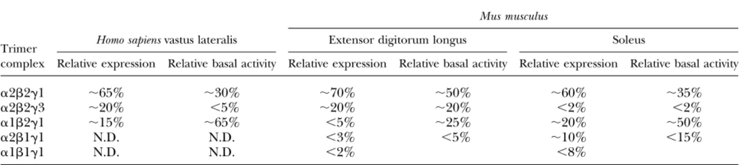
The 7 different AMPK subunit isoforms give rise to 12
heterotrimeric combinations that seem to be expressed in a
tissue-specific manner. Thus, in skeletal muscle
prepara-tions from human and mouse, all subunit isoforms have
been detected, but only a subset of possible heterotrimeric
complexes seems to exist (69, 70). In human skeletal muscle
(vastus lateralis), 3 different complexes have been
de-scribed (a2b2g1, a2b2g3, and a1b2g1) (69), whereas 5
complexes have been identified in mouse skeletal muscle
(a2b2g1, a2b2g3, a2b1g1, a1b2g1, and a1b1g1) (70)
(Table 1). In addition, some AMPK subunits are expressed
in a fiber type–dependent manner (71), which may explain
the relative distribution of complexes between different
muscles (70). Currently, it is not known why mouse
skel-etal muscle appears to express two additional complexes
(b1-associated) compared with human skeletal muscle.
Based on findings in muscle-specific AMPKa and AMPKb
double-knockout (KO) mice (AMPKa mdKO and AMPK
b1b2M-KO mice, respectively) (72–74), the majority of
b1-associated complexes detected in a crude sample of
skel-etal muscle seem to derive from nonmuscle tissue (e.g.,
connective tissue, neuronal cells, adipocytes, endothelial
cells, etc.). This is in line with the notion that the a1b1g1
complex is the most ubiquitously expressed complex of
AMPK (68, 75). Interestingly, the b1 subunit is also
de-tected in sample preparations of human skeletal muscle,
but, in light of findings from coimmunoprecipitations of
AMPKa2, -a1, -g1, and -g3, it does not seem to engage in
stable complex formation or contribute to any measurable
AMPK activity (69, 76). Although it is generally thought
that all 3 AMPK subunits must be present to form a stable
complex, it has been demonstrated in cell models that
stable b1g1 complexes can form in the absence of catalytic
subunits (77). Whether formation of bg heterodimer
complexes also occurs in mature skeletal muscle may be
derived from observations in two muscle-specific AMPK
double-KO mouse models. Thus, in the AMPKb1b2M-KO
model it seems evident that expression of b2 protein is
restricted to the myocytes (74). Interestingly, significant
amounts of b2 protein have been detected in a skeletal
muscle sample preparation from the AMPKa mdKO
mouse model (73), which may suggest the formation of
stable bg complexes in mature skeletal muscle,
assum-ing that sassum-ingle unbound subunits of AMPK are targeted
for degradation. This may also be inferred from the
AMPKb1b2M-KO mouse model, which does not express
a2 protein in skeletal muscle (74), indicating that the b
subunit is vital for maintaining AMPKa muscle protein
expression. Collectively, these observations may suggest
that the AMPK heterodimer (bg) exists in skeletal muscle
tissue and raises the possibility of a regulatory
mecha-nism facilitating the association of catalytic subunits
with regulatory complexes. Alternatively, and somewhat
speculatively, other proteins may bind to the regulatory
heterodimer complex to regulate their activity or cellular
localization. In this context, 12 protein kinases related to
AMPKa1 and AMPKa2 have been detected in the
hu-man kinome (78). These are known as AMPK-related
ki-nases, and, with a single exception, these are activated by
upstream kinase liver kinase B1 (LKB1) (79). Although
green fluorescent protein-transporter associated with
antigen processing–tagged versions of these kinases do
not appear to bind AMPK b and g subunits (80), the
sucrose nonfermenting AMPK-related kinase (SNARK/
NUAK2) is activated in skeletal muscle by
5-amino-1-b-
D-ribofuranosyl-imidazole-4-carboxamide (AICAR),
contraction, and exercise (81, 82), indicating that SNARK
activity is regulated similarly to AMPK. Is it
possi-ble that AMPK bg subunits form heterotrimeric
com-plexes with SNARK, facilitating its regulation by adenine
N C 1 9 4 / 5 8 4 r e S p 2 7 1 r h T p C pSer108 pThr148 D I A D K Myr2 C C N GBD C N CBS1 CBS2 CBS3 CBS4
*
Bateman domain Bateman domain
α1/α2 αH β-ID β1/β2 γ1/γ2/γ3 α-ID γ-ID β-ID
Figure 1. Structure of mammalian AMPK subunits. AMPK is a heterotrimeric protein consisting of 1 catalytic subunit (a subunit)
and 2 regulatory subunits (b and g subunit). The a subunit contains a kinase domain (KD), the activity of which relies on the
phosphorylation of Thr172 by upstream AMPK kinases. The KD is followed by an autoinhibitory domain (AID) and an a-hook
(aH), which seem to be important for AMP-regulated catalytic activity. At the a-C terminus, a b-interacting domain (b-ID) has
been detected that binds to the C-terminal domain of the b subunit. Phosphorylation of Ser485/491 in the b-ID during insulin
stimulation has been suggested to regulate kinase activity. The b subunit is subjected to myristoylation at the N terminus, which
enhances phosphorylation of a-Thr172 by AMP/ADP and facilitates AMPK translocation to specific intracellular compartments.
At the center, the b subunit contains a GBD that causes AMPK to bind to glycogen particles. Within the GBD, 2 phosphorylation
sites have been found (Ser108 and Thr148) that seem to regulate binding capacity to glycogen particles as well as kinase activity.
An a- and g-interaction domain (a-ID, g-ID) is located at the b-C terminus that acts as a scaffold keeping the heterotrimeric
complex together. The g-subunit contains 4 cystathionine–b-synthase (CBS) domains. These occur in tandem pairs, also known
as Bateman domains, and are involved in adenosine nucleotide binding. A b-ID is located close to the g-N terminus. An asterisk
denotes that the 3 g isoforms contain different N-terminal extensions.
TABLE 1. Relative distribution and basal activity of AMPK heterotrimeric complexes detected in human and mouse skeletal muscle
Trimer complex
Mus musculus
Homo sapiens vastus lateralis Extensor digitorum longus Soleus
Relative expression Relative basal activity Relative expression Relative basal activity Relative expression Relative basal activity
a2b2g1
;65%
;30%
;70%
;50%
;60%
;35%
a2b2g3
;20%
,5%
;20%
;20%
,2%
,2%
a1b2g1
;15%
;65%
,5%
;25%
;20%
;50%
a2b1g1
N.D.
N.D.
,3%
,5%
;10%
,15%
a1b1g1
N.D.
N.D.
,2%
,8%
The composition of AMPK heterotrimeric complexes was estimated from immunoprecipitation experiments in extensor digitorum longus and soleus from C57BL/6 mice as well as human male vastus lateralis. Values adapted from references 70, 76, and 487. N.D., nondetectable.
nucleotides? If so, it could be anticipated that
AMPKb-deficient skeletal muscle exhibits a phenotype different
from that of AMPKa-deficient skeletal muscle. Indeed, in
skeletal muscle several phenotypic differences have been
observed between AMPKa mdKO and
AMPKb1b2M-KO mice, including muscle mass, ATP levels,
mitochon-drial DNA and structure, citrate synthase activity, and
peroxisome proliferator–activated receptor g coactivator
1a (PGC-1a) mRNA levels (72, 74). In light of these
ob-servations, it has recently been reported that SNARK may
be involved in the maintenance of muscle mass with age
(83). Assuming that the potential binding of SNARK to
AMPKbg subunits induces an increase in SNARK activity
and/or protects SNARK from degradation and that the
association between SNARK and AMPKbg is enhanced
in AMPKa-deprived muscle, this could explain the
in-crease in muscle mass observed in skeletal muscle
de-prived of AMPKa subunits (72).
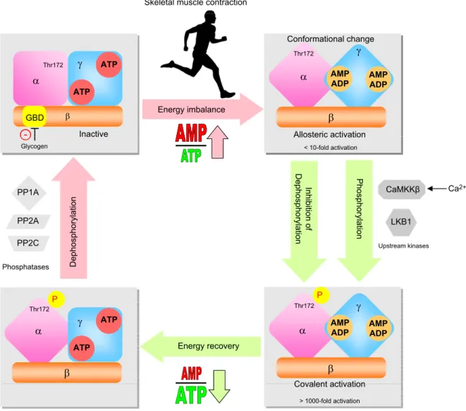
REGULATION AND ACTIVATION OF AMPK IN
SKELETAL MUSCLE
During skeletal muscle contraction, the adenylate energy
charge in muscle is decreased depending on the
dura-tion and intensity of exercise (84, 85). As a result, the
in-tracellular AMP/ATP and ADP/ATP ratios increase,
which leads to activation of AMPK (7). AMPK activation
occurs in two steps: stimulatory allosteric binding of AMP
within the g subunit and covalent activation through
re-versible phosphorylation on Thr172 in the catalytic a
subunit (Fig. 2). AMPK activity is stimulated by AMP and
ADP and inhibited by ATP binding to the two regulatory
Bateman domains of the g subunit. This competitive
binding means that increases in cellular AMP/ATP and
ADP/ATP ratios stimulate AMPK allosterically (86–90).
The allosteric stimulation has a moderate effect on AMPK
activity (,10-fold) (91). More importantly, binding of
Skeletal muscle contraction
ATP Thr172