HAL Id: hal-01809565
https://hal.archives-ouvertes.fr/hal-01809565
Submitted on 30 Nov 2020HAL is a multi-disciplinary open access archive for the deposit and dissemination of sci-entific research documents, whether they are pub-lished or not. The documents may come from teaching and research institutions in France or abroad, or from public or private research centers.
L’archive ouverte pluridisciplinaire HAL, est destinée au dépôt et à la diffusion de documents scientifiques de niveau recherche, publiés ou non, émanant des établissements d’enseignement et de recherche français ou étrangers, des laboratoires publics ou privés.
Kinetic characterization and molecular docking of novel
allosteric inhibitors of aminoglycoside
phosphotransferases
Nadia Leban, Elise Kaplan, Laurent Chaloin, Sylvain Godreuil, Corinne
Lionne
To cite this version:
Nadia Leban, Elise Kaplan, Laurent Chaloin, Sylvain Godreuil, Corinne Lionne. Kinetic character-ization and molecular docking of novel allosteric inhibitors of aminoglycoside phosphotransferases. Biochimica et Biophysica Acta (BBA) - General Subjects, Elsevier, 2017, 1861 (1), pp.3464 - 3473. �10.1016/j.bbagen.2016.09.012�. �hal-01809565�
1
Kinetic characterization and molecular docking of novel allosteric inhibitors of
1
aminoglycoside phosphotransferases
2
3
Nadia Leban a,1, Elise Kaplan a,2, Laurent Chaloin a, Sylvain Godreuil b, Corinne Lionne a,*,3
4
5
a CNRS FRE 3689 – Université de Montpellier, Centre d'études d'agents Pathogènes et
6
Biotechnologies pour la Santé (CPBS), F-34293 Montpellier, France.
7
b INSERM U1058 – Université de Montpellier, Centre Hospitalier Régional Universitaire de
8
Montpellier, Département de Bactériologie-Virologie, Montpellier, France.
9
10
* Corresponding author at: CBS, 29 rue de Navacelles, F-34090 Montpellier, France; Tel.:
11
+33-(0)626-197-417; Fax : +33-(0)467-417-913.
12
E-mail address: corinne.lionne@cbs.cnrs.fr
13
14
1 Present address: Institut Supérieur de Biotechnologie de Monastir, Faculty of Pharmacy,
15
University of Monastir, Tunisia.
16
2 Present address: Department of Pathology, University of Cambridge, Cambridge, United
17
Kingdom.
18
3 Present address: Centre de Biochimie Structurale, CNRS UMR 5048 – Université de
19
Montpellier – INSERM U 1054, Montpellier, France.
20
21
Abbreviations: AME, aminoglycoside-modifying enzyme; AAC, aminoglycoside
N-22
acetyltransferase; ANT, aminoglycoside nucleotidyltransferase; APH, aminoglycoside
O-23
phosphotransferase; GOLD, Genetic Optimization for Ligand Docking.
24
25
Keywords: allosteric inhibitors, antibiotic resistance, docking, inhibition modes, protein
26
dynamics, steady state kinetics.
27
*REVISED Manuscript (text UNmarked)
2
ABSTRACT
28
29
Background: Bacterial antibiotic resistance often leads to treatment failure which may have
30
serious consequences, especially in critically sick patients. Resistance to aminoglycosides is
31
mainly due to the expression of antibiotic-modifying enzymes. One important mechanism of
32
aminoglycoside modification is the ATP/GTP-dependent O-phosphorylation catalyzed by
33
aminoglycoside phosphotransferases, APHs. The aim of this study is to identify specific
34
inhibitors of APHs that could restore bacterial susceptibility to aminoglycosides.
35
Methods: We focused on the search for allosteric inhibitors that bind to small cavities of the
36
protein and block the enzyme function by perturbing its dynamics.
37
Results: From normal mode analysis, a cavity of variable volume belonging to a large groove
38
which splits the protein into two parts was chosen as target. By molecular docking, we
39
screened a large library of commercially available compounds. Seventeen of the highest
40
ranked compounds were tested by in vitro kinetic experiments in order to evaluate their
41
ability to inhibit APHs. Site-directed mutagenesis was carried out with the aim of confirming
42
the inhibition mechanism determined kinetically and the interactions with the protein
43
predicted by in silico studies. These interactions were also confirmed by the use of
44
structurally-related molecules.
45
Conclusions: Two compounds showed interesting inhibition properties, and one was able to
46
block two different classes of APH.
47
General significance: This study gives new insights into the inhibition of APHs by such
48
allosteric inhibitors, and provides the basis for the future development of combined therapies,
49
antibiotic plus APH inhibitor, which may reverse the resistance to aminoglycosides in a
50
clinical context.
51
52
3
1. Introduction
53
54
Aminoglycosides constitute a large family of water soluble, polycationic amino sugars
55
of considerable structural diversity. They are broad spectrum antibacterial agents that are
56
products of bacterial or fungal metabolism. Bacterial resistance to aminoglycosides manifests
57
itself by one of the following mechanisms: (i) the presence of aminoglycoside-modifying
58
enzymes [1]; (ii) the decrease of bacteria membrane permeability towards aminoglycoside
59
uptake into the bacteria or extrusion of the aminoglycosides from the cell by efflux pumps [2];
60
or (iii) the modification of the drug target which can be either mutations that result in
61
structural alterations of the ribosome [3] or methylations by 16S rRNA methyltransferases
62
which interferes with the aminoglycoside binding [4]. Among these mechanisms, inactivation
63
by aminoglycoside-modifying enzymes (AMEs) is the most important both in terms of level
64
and frequency of resistance conferred to the bacteria [5]. There are three types of AMEs,
65
each of which transfers a functional group onto the aminoglycoside structure thereby
66
inactivating the antibiotic: aminoglycoside O-nucleotidyltransferases (ANTs) transfer a
67
nucleoside monophosphate from a nucleoside triphosphate on a hydroxyl group of the
68
antibiotic; aminoglycoside N-acetyltransferases (AACs) transfer an acetyl group from
acetyl-69
CoA on an amine group; and aminoglycoside O-phosphotransferases (APHs) transfer the
-70
phosphate of a nucleoside triphosphate on a hydroxyl group [6]. Each family of enzymes
71
consists of different isoenzymes that differ in substrate specificity and selectivity. At least 50
72
different genes for AMEs have been identified in bacteria [1,7]. A number of bacteria also
73
harbor a bifunctional enzyme that catalyzes both acetylation and phosphorylation [8,9].
74
In bacterial resistant isolates that possess antibiotic-inactivating enzymes, drugs are
75
enzymatically modified when they penetrate into the bacteria. Therefore, the altered drug is
76
unable to interact efficiently with its target, the ribosome. Moreover, in the absence of active
77
aminoglycosides that normally perturb bacterial protein synthesis, the next step of high-rate
78
accumulation of antibiotic does not take place [10,11]. Since the discovery of the
antibiotic-79
inactivating enzymes, many efforts have been carried out in order to chemically modify the
4
existing antibiotics and produce new drugs that retain their antibacterial activity without being
81
inactivated. This strategy has led to considerable success, especially in the fight against
82
penicillin resistance. For instance, the use of methicillin, a derivative of the early penicillins,
83
overcame enzyme-mediated inactivation in most penicillin-resistant, gram-positive organisms
84
[12]. Another strategy to overcome enzyme-associated resistance consists in combining the
85
antibiotics with an inhibitor of the corresponding modifying enzyme. For example
beta-86
lactamase inhibitors, such as clavulanic acid, associated with amoxicillin inhibit most strains
87
of penicillinase-producing S. aureus, H. influenza, E. coli, Klebsiella spp,... [13]. Based on
88
this strategy, several teams have searched for AMEs inhibitors. The new discovery of AAC
89
inhibition by metal salts (mechanism still unknown) may offer a promising therapeutic issue
90
[14,15]. Shi et al. reviewed recent progresses in development of APHs inhibitors [16]. The
91
success in discovering specific inhibitors for AMEs depends on our ability to understand the
92
basic properties of their mode of action and of their substrate specificity. So far, main studies
93
focused on the development of competitive inhibitors that target either the nucleotide binding
94
site, such as protein kinase inhibitors [17,18], or the aminoglycoside binding site, with
95
aminoglycoside analogues [19,20] or non-carbohydrate inhibitors [21]. Using in silico
96
screening of chemical libraries in the aminoglycoside binding site of AAC(6')-Ib, competitive
97
inhibitors were recently identified [22].
98
However, therapeutic enzyme inhibitors are not restricted to active site competitors.
99
Identification of small molecules that stabilize inactive conformations of the protein may
100
represent a powerful starting point for rational drug design. The underlying idea is to complex
101
the enzyme with a molecule that binds to a cavity other than the active site, and stabilizes the
102
enzyme in a non-catalytically competent conformation. For example, the inhibition of
103
APH(3')-IIIa by AR_3a, an ankyrin repeat protein, has been shown to be due to a significant
104
change of the APH conformation upon AR_3a allosteric binding [23]. However, the large size
105
of this modulator makes it hardly druggable.
106
Here, using normal mode analysis on two different APH crystal structures, we
107
identified a common cavity of variable volume and used it as target for screening a library of
5
small commercially available compounds. By molecular docking, several potential allosteric
109
inhibitors against two aminoglycoside-modifying enzymes, aminoglycoside
3'-110
phosphotransferase type IIIa, APH(3')-IIIa, and aminoglycoside 2"-phosphotransferase type
111
IVa, APH(2")-IVa, were identified. We performed kinetic and molecular modelling studies to
112
assess their mechanism of inhibition and to spotlight their binding site in the protein.
113
114
6
2. Materials and methods
115
116
2.1. Normal mode analysis and molecular docking
117
118
Two starting crystal structures of APH(3')-IIIa (PDB ID: 1L8T, including kanamycin A
119
and ADP, from [24]) and APH(2")-IVa (PDB ID: 3SG8, including tobramycin, from [25]) were
120
selected for normal modes computation and cavity search. In these structures, the
121
aminoglycosides were removed to favour the binding of inhibitors near the target cavity. ADP
122
was maintained in 1L8T, except otherwise stated in the text. The first two hundreds normal
123
modes were calculated (all-atoms parameter set 27) using the VIBRAN module of CHARMM
124
program [26,27] allowing to define fifty conformations for each enzyme. Briefly, after addition
125
of hydrogen using the H-build routine, the potential energy of each system was minimized for
126
5,000 steps of conjugate gradient algorithm (tolerance gradient of 0.01 kcal/mol/Å) followed
127
by 50,000 steps of Adopted Basis Newton-Raphson to reach a mean energy gradient of less
128
than 10-5 kcal/mol/Å. Diagonalization of the Hessian matrix and normal modes were
129
computed from these energy-minimized structures. Trajectories of superimposed modes
130
(modes 7 to 200, after removal of the six first modes corresponding to intrinsic translational
131
or rotational motions) were computed with CHARMM to produce fifty frames corresponding to
132
protein conformers (hereafter called "trajectory" allowing to visualize the displacement of the
133
whole coordinates on the superposed modes 7 to 200). Atomic fluctuations averaged by
134
residues were computed from normal modes 7-200 using the fluctuation subroutine
135
implemented in the Vibran module of Charmm. Structure and normal modes trajectories were
136
analyzed with VMD software [28]. From these conformers, all cavities were identified and
137
characterized using MDpocket software [29], and a small cavity fulfilling the criteria for
138
allosteric inhibition (location and variable volume) located behind the kanamycin binding site
139
was selected for subsequent screening.
140
All docking was performed using GOLD (Genetic Optimization for Ligand Docking)
141
program v5.2 [30,31]. Zinc database (http://zinc.docking.org) [32] was queried to find
7
like molecules according to Lipinski's rule-of-five [33]. Briefly, 100,000 compounds from the
143
Zinc database were selected after applying these drug-like filtering criteria, except that logP
144
was defined below or equal to 2 in order to increase the selection of aqueous-soluble
145
compounds. All these molecules were screened by ensemble docking on both APHs using
146
20 genetic algorithm runs within a 15 Å radius of the sphere centered on the targeted site
147
and using Goldscore as scoring function. The docking poses were analyzed by the clustering
148
method (complete linkage) from the rmsd matrix of ranking solutions. A second round of
149
molecular docking (50 genetic algorithm runs) was performed on each APH separately, using
150
the highest ranked compounds (the first hundred) from the ensemble screening. Twenty one
151
compounds corresponding to the highest scores were selected, but finally, only seventeen
152
compounds were commercially available and purchased.
153
Structural analysis and visualization of docking poses were carried out using the
154
PyMOL Molecular Graphics System (version 1.3, Schrödinger, LLC).
155
156
2.2. Kinetic studies
157
158
In all in vitro kinetic experiments, equimolar concentrations of MgCl2 were added to
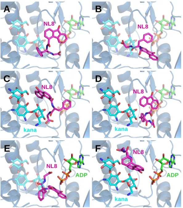
159
ATP. Thus, in the text, ATP and ADP refer to MgATP and MgADP, respectively.
160
Experimental buffer was 50 mM Tris-HCl pH 7.5, 1 mM free MgCl2, 40 mM KCl, and the
161
temperature was 25°C. The APH steady-state activity was evaluated using purified
162
recombinant proteins, produced as previously reported [34], by measuring the time courses
163
of ADP production by two different methods.
164
For a rapid screening of the compounds, the steady-state rate constants, kss, of
165
enzyme activity were monitored in 96-well plates by coupling the release of ADP to a
166
pyruvate kinase/lactate dehydrogenase reaction, as described by McKay et al. [35]. Reaction
167
mixtures contained 0.5 µM APH, 4 mM phosphoenol pyruvate, 280 µM NADH, 20 U/ml
168
pyruvate kinase, 25 U/ml lactate dehydrogenase, 25 or 350 µM ATP and 50 or 100 µM
169
kanamycin A, with APH(3')-IIIa or APH(2")-IVa, respectively. Experiments were carried out in
8
the absence or in the presence of potential inhibitors at 500 µM. Reactions were initiated by
171
the addition of ATP and monitored by reading the absorbance at 340 nm every 11 seconds.
172
Values of kss were determined from the slope of linear phase of the reaction time courses
173
using GraFit 7.0.2 software. The experiments were carried out in triplicate and the averaged
174
inhibition was computed.
175
For the best compounds identified by the previously described enzymatic coupled
176
system, the inhibition constants and inhibition modes were determined by the quench-flow
177
method and HPLC analysis as previously described [36]. Briefly, quench-flow method
178
consists in interrupting enzymatic reactions at different times with a quencher, which is
179
usually a strong acid [37]. Here, 10% perchloric acid was used to stop the reaction every 3–
180
7 s, depending on the enzyme. The reaction was triggered by addition of ATP in a
181
thermostatically-controlled beaker (25°C) containing the enzyme and the aminoglycoside,
182
with or without inhibitor. After quenching, the separation of ADP and ATP was carried out
183
using HPLC (Alliance, Waters) and a partisphere SAX column (AIT France). The mobile
184
phase was 200 mM ammonium phosphate buffer pH 5.5 and 10% acetonitrile. Samples from
185
quench-flow were first spin at 19,000 g for 20 min at 4°C to remove precipitated protein. A
186
volume of 100 µl of supernatant quenched sample was diluted into 900 µl of mobile phase
187
supplemented with KOH to readjust the pH to 5.5. Volumes of 50 or 100 µl of these samples
188
were injected on the HPLC column. Quantification of ADP and ATP was obtained by
189
integrating absorption peaks at 259 nm from the chromatograms. The inhibition mode and
190
inhibition constants were determined by repeating the experiment at different concentrations
191
of ATP (keeping kanamycin A concentration constant, see figure legends) or at different
192
concentrations of kanamycin A (ATP concentration constant). APH concentrations were 0.1–
193
0.5 µM. Fittings were performed using GraFit 7.0.2 software using four different inhibition
194
modes: competitive, non-competitive, uncompetitive and mixed. Only the most appropriate fit
195
(which describes the inhibition with the lowest Chi2) is shown on the figures with the
196
corresponding Lineweaver-Burk representation.
197
198
9
2.3. Site-directed mutagenesis
199
200
R211A mutant of APH(3')-IIIa was constructed by PCR (QuickChange Lightning Multi
201
Site-Directed Mutagenesis Kit) using the two primers :
202
5'-TTTATTGATCTTGGGGCAAGCGGCAGGGCGGAC-3,' and
203
5'-GTCCGCCCTGCCGCTTGCCCCAAGATCAATAAA-3'.
204
Each 50 µl PCR reaction contained 2.5 µl of 10 QuickChange Lightning Multi
205
reaction buffer, 0.75 µl of QuickSolution, 100 ng of DNA template (wild type aph(3')-IIIa in a
206
pET15b plasmid), 1 µl of each mutagenic primer (100 ng), 10 mM dNTP and 2.5 units of
207
QuickChange Lightning Multi enzyme blend. The PCR product was digested by DpnI
208
restriction endonuclease for 1 h at 37°C. Mutant plasmids were recovered after
209
transformation in E. coli BL21 (DE3) chemo-competent cells. Successful introduction of the
210
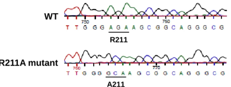
desired mutations was controlled by sequencing the plasmid DNA (Fig. S1).
211
The mutant APH(3')-IIIa was produced in E. coli and purified as the wild type protein.
212
213
10
3. Results
214
215
3.1. Target cavity identification by normal mode analysis
216
217
Here, we focused on the search for APH allosteric inhibitors that bind to small cavities
218
of the protein and block the enzyme function by perturbing its dynamics. Thus, a specific
219
cavity was defined according to the following criteria: the cavity must be present in different
220
APH subfamilies, it must be outside of the substrate binding sites, and lastly, it must undergo
221
obvious volume change during the dynamics of the enzyme, required for its function.
Sub-222
families APH(3') and APH(2") were chosen because of their prevalence [1].
223
From the normal mode analysis, one cavity behind the aminoglycoside binding site
224
was selected because it was fulfilling all the predefined criteria (Fig. 1, Fig. S2 also showing
225
videos). It belongs to a large groove which splits the proteins into two parts. Moreover, the
226
region defining this cavity was found to be flexible according to the fluctuations computed
227
from the superposed normal modes (Fig. 1). It must be emphasized that the observed atomic
228
fluctuations were relatively small and therefore were not considered as the main criteria to
229
select the cavity (commonly, fluctuations of residues located at the interior of the protein are
230
less important compared to external flexible loops). Typically, APH proteins do not require
231
large collective motions like other kinases for their biological function and this feature renders
232
more difficult the search for internal dynamic cavities. Nevertheless and taking into account
233
to these small fluctuations, the target binding pocket was also selected in respect to the
234
volume changes observed along the normal modes. The target cavities were surrounded by
235
residues Gly192 and Arg226 in 1L8T structure and by residues Ser199 and Ser232 in 3SG8
236
(yellow balls in Fig. S2). They are situated at a similar place within the two APHs and their
237
compositions in amino acids were similar but not identical (at least one Phe, Leu, Ile, Ser and
238
Asp found in each case).
239
Figure 1
240
241
11
3.2. Molecular docking
242
243
Using the targeted cavity defined above in APH(3')-IIIa and APH(2")-IVa structures
244
and the ZINC chemicals database [32], a virtual screening was carried out in order to predict
245
and evaluate the potential affinity of these enzymes for drug-like molecules using Gold
246
software. The Goldscore scoring function was used to rank the screened molecules – the
247
highest scores predict highest affinities. In order to favor the binding of inhibitors near the
248
target cavity which is located behind the aminoglycoside binding site, kanamycin A was
249
removed from 1L8T structure (but not ADP) and tobramycin from 3SG8 prior docking.
250
In a first case study using an ensemble docking after overlaying both APH (same
251
center of coordinates defining the target cavity at the same location for the two APHs), the
252
first 100 out of the 100,000 compounds from the drug-like ZINC subset were selected based
253
on their docking scores. Then, a second thorough docking run (50 GA runs) was performed
254
on each APH separately with these selected compounds. We finally selected 21 best ranking
255
ligands (63 < score < 81) after named NL1 to NL21 (Table 1). NL4, NL7, NL10 and NL15
256
were not commercially available, and therefore only 17 NL compounds were purchased for
257
the further experimental assays.
258
259
3.3. Rapid in vitro screening of APH inhibitors using a coupled enzyme kinetic assay.
260
261
To evaluate the potential APH inhibition promoted by the selected molecules, in vitro
262
inhibition studies were performed. The 17 molecules were rapidly screened using an
263
enzymatic coupled-system, as previously described [35]. We used kanamycin A for these
264
studies because both APHs can phosphorylate this aminoglycoside with a good efficiency
265
through a Michaelis-Menten kinetics. Steady-state rate constants (kss) were measured at
266
fixed concentrations of ATP and kanamycin A, in the absence or in the presence of NL
267
compounds (500 µM). Percentages of inhibition by NL compounds were determined and
268
normalized in respect to the activity without compound. The results are summarized in Table
12
1. In these conditions, a 40% or higher inhibition of APH(3')-IIIa activity was observed with
270
compounds NL6 and NL8, and for APH(2")-IVa with compounds NL6, NL9 and NL16. Two
271
compounds seemed particularly interesting: NL8 because it inhibits APH(3')-IIIa by almost
272
80%, and NL6 for its ability to inhibit both APHs.
273
In order to determine the inhibition mode of these two compounds, steady-state
274
kinetic experiments were carried out using a direct quenched-flow method. Results are
275
described below and their mechanism is discussed on the basis of docking experiments.
276
277
3.3.1. Inhibition by NL8 compound.
278
279
NL8 was the most potent inhibitor amongst the 17 molecules tested here by the
280
coupled system, showing a 79% inhibition of APH(3')-IIIa activity at 500 µM. However, it
281
showed little inhibition of APH(2")-IVa activity. The steady-state rate constants, kss, of
282
APH(3')-IIIa were determined at different concentrations of kanamycin A or ATP, and at 0, 50
283
or 100 µM concentrations of NL8 by the quenched-flow method. Double reciprocal plots of
284
1/kss versus 1/[ATP] or 1/[kanamycin A] are shown in Fig. 2.
285
Figure 2
286
The NL8 compound was found to be a competitive inhibitor of APH(3')-IIIa towards
287
ATP with a Ki of 9 ± 2 µM, and a non-competitive inhibitor towards kanamycin A with a Ki of
288
55 ± 2 µM.
289
It should be noted that NL8 was initially identified as potential allosteric inhibitor on
290
the basis of a docking on APH(3')-IIIaADP complex. To explain the unexpected ATP
291
competitive inhibition mode found here, we carried out another docking of NL8 but in the
292
absence of ADP (Fig. S3). In these conditions, NL8 was found to overlap with the nucleotide
293
binding site, especially with the phosphate groups of the nucleotide. Unfortunately, it was
294
difficult to predict with accuracy the binding of NL8 as the molecule presents a high degree of
295
variability, even within repetitions of dockings (several rounds using identical parameters).
13
Similar variabilities were obtained when NL8 was docked in the apo, in the kanamycin-bound
297
or in the ADP-bound structures.
298
NL8 was identified on the basis of an in silico screening in which the cavity of the
299
targeted protein was located behind the aminoglycoside binding pocket (Fig. 1). However,
300
the competition between ATP and NL8 upon binding to APH(3')-IIIa suggests than the
301
inhibitor may bind to a different location, presumably closer to the nucleotide binding pocket
302
without interfering with the aminoglycoside site.
303
304
3.3.2. Inhibition by NL6 compound.
305
306
The coupled enzyme system method showed that NL6 inhibited efficiently both
307
enzymes with 43% and 63% inhibition for APH(3')-IIIa and APH(2")-IVa, respectively.
308
We determined the mode of inhibition of each protein by NL6. Fig. 3A and B show
309
that NL6 is a non-competitive inhibitor of APH(2")-IVa towards both ATP and kanamycin A
310
with Ki of 85 ± 10 µM and 74 ± 4 µM, respectively. Similarly, kinetics with APH(3')-IIIa showed
311
NL6 acts again as a non-competitive inhibitor towards ATP and kanamycin A with Ki of
312
75 ± 3 µM and 76 ± 6 µM, respectively (Fig. 3C and D).
313
Figure 3
314
To further investigate the binding of NL6 to APH(3')-IIIa, molecular docking of NL6
315
was performed in the apo protein, in the ADP-bound structure and in the ternary complex
316
(PDB ID: 1L8T). Although NL6 is acting on both APHs' activity, it was more difficult to predict
317
the binding position of NL6 in APH(2")-IVa. Therefore, we focused on APH(3')-IIIa here. A
318
cluster of the best docking poses is shown in Fig. 4A, B, C for each simulation. Interestingly,
319
if one part of the molecule presents an important degree of flexibility in the apo and in the
320
ADP-bound structures, the dihydro-benzopyran ring of NL6 shows a rigorously identical
321
location in all docking predictions, suggesting the importance of this group for NL6 binding.
322
The interactions occurring between this moiety and the protein are detailed in Fig. 4D.
323
Figure 4
14
All molecular dockings predicted that the dihydro-benzopyran ring is stabilized by the
325
backbone atoms of Leu209 and Gly210 and by a hydrogen bond between the carbonyl near
326
the benzopyran ring and the side chain of Arg211 (Fig. 4D). This latter interaction seems to
327
be of a crucial importance to stabilize the molecule as NL6 presents a completely different
328
predicted location when Arg211 is replaced by an alanine (Fig. 4E). To assess the
329
importance of this interaction for the in vitro binding of NL6, we engineered the R211A
330
mutant of APH(3')-IIIa and performed kinetic studies in the absence or in the presence of
331
NL6. The activity of this mutant was significantly affected: kcat was decreased by 62%.
332
Interestingly, the molecule was completely unable to inhibit the APH mutant (Fig. 4F). This
333
result confirms the binding site of NL6 predicted by in silico studies and highlights the
334
importance of Arg211 for its binding to APH.
335
To further investigate the importance of the two other interactions occurring with the
336
benzopyran ring, we tested the activity of structurally related compounds. Indeed, the
337
benzopyran moiety is stabilized by two hydrogen bonds with the backbones of Leu209 and
338
Gly210, it was thus impossible to modify these interactions by mutations. Consequently, we
339
evaluated the activity of two structurally related molecules. One of them, named NL6-1
340
(Zinc14108604), contains the same dihydro-benzopyran ring than NL6 but with a slightly
341
shorter structure, while the other compound, NL6-2 (Zinc13235262), has a naphthyl group
342
instead. According to ZINC chemical database, the two analogues show more than 70% of
343
structural similarity with NL6 (Fig. 5A).
344
Dockings were achieved in the APH(3')-IIIa with NL6 and its two analogues using the
345
same parameters. According to these in silico studies, the two molecules bind to the same
346
position to that observed for NL6 in the substrate-bound structure of APH(3')-IIIa (Fig. 5C and
347
D). However, the naphthyl group of NL6-2 does not interact with the protein in contrast to the
348
dihydro-benzopyran group NL6-1 which has similar interactions to those of NL6. Similarly,
349
when docked in the apo protein, NL6 and NL6-1 show a strict overlay of their
dihydro-350
benzopyran group, while NL6-2 presents a completely different binding mode (Fig. 5E and
351
F). The naphthyl group of NL6-2 is indeed located at the opposite side from ADP in contrast
15
to the dihydro-benzopyran moiety of NL6 and NL6-1, located near the nucleotide binding site.
353
This suggests that the benzopyran ring is also important for the stabilization of NL6.
354
Figure 5
355
Based on this prediction, these molecules were tested in vitro to estimate their ability
356
to inhibit the APH(3')-IIIa activity. As shown in Fig. 5B, NL6-1 inhibits the activity of the APH.
357
Similarly to NL6, NL6-1 was confirmed to act as a non-competitive inhibitor towards ATP with
358
a Ki of 185 ± 12 µM (data not shown). The 2-fold higher Ki of NL6-1 compared to NL6 can be
359
explained by its shorter structure (20 carbons) compared to NL6 (23 carbons), resulting in a
360
smaller number of interactions with the protein and consequently in a weaker inhibition. In
361
contrast, NL6-2 was completely unable to inhibit the APH activity, confirming the hypothesis
362
that the dihydro-benzopyran group is of major importance for APH inhibition.
363
According to kinetic and molecular docking studies, NL6 is likely to bind between both
364
substrates, without affecting their binding. The presence of NL6 should greatly perturb the
365
phosphotransfer of the γ-phosphate of ATP on the 3'-OH of kanamycin A which could explain
366
the enzyme inhibition.
367
To conclude, the dihydro-benzopyran ring is of crucial importance for the binding of
368
NL6 to APH enzymes. In addition, the two interactions with side chain of Arg211 and
369
backbones of Leu209 and Gly210 were shown to play an essential role in the inhibition of
370
APH.
371
372
373
16
4. Discussion
374
375
The problem of bacterial resistance to antibiotic due to aminoglycoside
376
phosphotransferases APH(2")-IVa and APH(3')-IIIa enzymes could be overcome by the
377
development of novel drugs that target APHs, as suggested by Shi and co-authors in a
378
recent review [16]. For example, because of the structural similarity of the ATP binding
379
domain with protein kinases, APHs can be inhibited by eukaryotic protein kinase inhibitors. In
380
this way, protein kinase inhibitors of the isoquinoline sulfonamide family have been shown to
381
be competitive inhibitors of APH(3')-IIIa towards ATP, although they were unable to restore
382
aminoglycoside susceptibility of strains harboring aph(3')-IIIa gene [38]. In the same manner,
383
Shakya et al. screened a library of known kinase inhibitors and identified pyrido-pyrimidines
384
as selective inhibitors of APH(3')s and flavonoids as general inhibitors of APHs [39]. A crystal
385
structure of the APH(2")-IVakanamycinquercetin complex allowed to confirm that the
386
inhibitor binds to the ATP binding site of the enzyme.
387
The problem with such kinase inhibitors is their intrinsic poor selectivity because they
388
target the ATP binding site of APHs which is similar to that of many eukaryotic important
389
enzymes. However, the recent structure-guided optimization of protein kinase inhibitors
390
constitutes a promising strategy to enhance their specificity for APHs [18]. The originality of
391
our inhibitors, compared to these protein kinase inhibitors, is that they do not target the
392
substrate binding site, hence avoiding or reducing off-target effects. Therefore, they are
393
neither going to compete with the antibiotic itself for the binding to APHs or to the rRNA
394
target, nor with ATP which has a central role in many physiological processes. Instead, they
395
have been selected for their ability to interfere with important dynamic properties of the
396
targeted enzyme.
397
A similar strategy was successful applied to APH with the development of allosteric
398
inhibitors based on engineered ankyrin repeat motives. These compounds inhibited the
399
enzyme activity, both in vitro and in vivo, and were able to restore the bacteria sensitivity to
400
kanamycin and amikacin to a level comparable to that of the corresponding knockout strains
17
[40]. Later, the resolution of the crystal structure of APH(3')-IIIa complexed with one of the
402
most potent ankyrin repeat protein inhibitor has confirmed the allosteric inhibition mechanism
403
[23]. Indeed, the binding of the inhibitor stabilizes the APH in a non-catalytically competent
404
conformation. However, the designed ankyrin repeat protein is a large molecule that is hardly
405
druggable.
406
Here, on the basis of in silico screening of a chemical library, our idea was to find
407
small molecules that are more druggable than large proteins and that inhibit APHs in a
408
similar manner to that of ankyrin repeat protein. Allosteric small-molecule kinase inhibitors
409
are indeed promising alternatives to active site binders, as recently reviewed [41].
410
We have identified twenty one small molecules that are potential allosteric inhibitors
411
of APHs. Out of the seventeen that were evaluated, four molecules were able to inhibit
412
significantly APH activity in vitro. We determined the mode of inhibition for two of them and
413
showed that they are non-competitive towards kanamycin A. One of them, NL8, was a
414
competitive inhibitor of APH(3')-IIIa towards ATP, suggesting that it should bind near the
415
nucleotide binding position, but its precise binding position remains elusive. The fact that it
416
did not inhibit APH(2")-IVa suggests that it does not bind to the nucleotide binding site which
417
is conserved between the two APHs. The most promising molecule, NL6, is able to inhibit
418
non-competitively the two APHs tested. Its binding site was confirmed both by mutating one
419
interacting residue of APH and by the use of NL6 analogues with one missing the
dihydro-420
benzopyran ring required for an efficient binding. As shown in Fig. 6, NL6 binds to a different
421
location in APH(3')-IIIa that of the protein kinase inhibitor CKI, and of the ankyrin-repeat
422
protein inhibitor AR-3A.
423
Figure 6
424
This confirms that non-allosterically modulated proteins such as APHs can be
425
inhibited by small druggable allosteric compounds, which brings new insights for the rational
426
drug discovery of APHs inhibitors.
427
The seventeen compounds will be further tested for their effect on antibiotic
428
susceptibility of different Gram-positive and Gram-negative bacteria strains as well as on
18
their potential cell toxicity. The integration of dynamic motions by using normal mode
430
analysis in combination with molecular docking, enzyme kinetics and antibacterial activity
431
studies, provided important insights into the molecular basis underlying ligand binding and
432
enzyme inhibition. Co-crystallization assays of APHs with these molecules are currently in
433
progress and rational lead optimization processes are considered in order to design new
434
derivatives with higher inhibitory properties. Efforts should be made to increase the affinity of
435
APH for the inhibitor by adding chemical substitutions that would interact with additional
436
residues of the cavity. This should avoid that a single mutation of the protein is enough to
437
prevent the inhibition.
438
439
19
5. Conclusion
440
441
Optimization of the lead compounds identified in this study should help in the design
442
of efficient APHs inhibitors that may overcome APH-dependent bacterial resistance to
443
aminoglycosides. Emergence and frequency of potential resistance mutants will be evaluated
444
in vitro. Moreover, this strategy may be applied to other multi-resistant strains for which the
445
major AMEs have been identified.
446
447
448
20
Acknowledgment
449
450
This work was supported by the Centre National de la Recherche Scientifique
451
(CNRS), the University of Montpellier and the Agence Nationale de la Recherche (ANR
452
Programme Blanc 2011-SIMI7, "cN-II Focus"). France-Tunisia exchange was supported by a
453
CNRS program (EDC25987). Dr. Nadia Leban is grateful to the Infectiopôle Sud Foundation
454
for a post-doctoral fellowship and Dr. Elise Kaplan to the Fondation pour la Recherche
455
Médicale for doctoral fellowship (FDT20140931113).
456
We thank Professor Sergei B. Vakulenko (Notre Dame, USA) and Professor Engin H.
457
Serpersu (Knoxville, USA) for the generous gift of the aph(2")-IVa and aph(3')-IIIa genes. We
458
thank Ms Perrine Lallemand (CPBS) for her help in the APH purification procedure.
459
460
461
21
References
462
463
[1] Ramirez MS & Tolmasky ME (2010) Aminoglycoside modifying enzymes. Drug Resist.
464
Updat. 13, 151–171.
465
[2] Kumar A & Schweizer HP (2005) Bacterial resistance to antibiotics: active efflux and
466
reduced uptake. Adv. Drug Deliv. Rev. 57, 1486–1513.
467
[3] Pfister P, Hobbie S, Vicens Q, Böttger EC & Westhof E (2003) The molecular basis for
468
A-site mutations conferring aminoglycoside resistance: relationship between ribosomal
469
susceptibility and X-ray crystal structures. Chembiochem 4, 1078–1088.
470
[4] Doi Y & Arakawa Y (2007) 16S ribosomal RNA methylation: emerging resistance
471
mechanism against aminoglycosides. Clin. Infect. Dis. 45, 88–94.
472
[5] Vakulenko SB & Mobashery S (2003) Versatility of aminoglycosides and prospects for
473
their future. Clin. Microbiol. Rev. 16, 430–450.
474
[6] Wright GD, Berghuis AM & Mobashery S (1998) Aminoglycoside antibiotics. Structures,
475
functions, and resistance. Adv. Exp. Med. Biol. 456, 27–69.
476
[7] Shaw KJ, Rather PN, Hare RS & Miller GH (1993) Molecular genetics of
477
aminoglycoside resistance genes and familial relationships of the
aminoglycoside-478
modifying enzymes. Microbiol. Rev. 57, 138–163.
479
[8] Ferretti JJ, Gilmore KS & Courvalin P (1986) Nucleotide sequence analysis of the gene
480
specifying the bifunctional 6'-aminoglycoside acetyltransferase 2"-aminoglycoside
481
phosphotransferase enzyme in Streptococcus faecalis and identification and cloning of
482
gene regions specifying the two activities. J. Bacteriol. 167, 631–638.
483
[9] Azucena E, Grapsas I & Mobashery S (1997) Properties of a bifunctional bacterial
484
antibiotic resistance enzyme that catalyzes ATP-dependent 2"-phosphorylation and
485
acetyl-CoA-dependent 6'-acetylation of aminoglycosides. J. Am. Chem. Soc. 119,
486
2317–2318.
487
[10] Dickie P, Bryan LE & Pickard MA (1978) Effect of enzymatic adenylylation on
488
dihydrostreptomycin accumulation in Escherichia coli carrying an R-factor: model
22
explaining aminoglycoside resistance by inactivating mechanisms. Antimicrob. Agents
490
Chemother. 14, 569–580.
491
[11] Höltje JV (1979) Induction of streptomycin uptake in resistant strains of Escherichia
492
coli. Antimicrob. Agents Chemother. 15, 177–181.
493
[12] Bush K & Mobashery S (1998) How beta-lactamases have driven pharmaceutical drug
494
discovery. From mechanistic knowledge to clinical circumvention. Adv. Exp. Med. Biol.
495
456, 71–98.
496
[13] Drawz SM & Bonomo RA (2010) Three decades of beta-lactamase inhibitors. Clin.
497
Microbiol. Rev. 23, 160–201.
498
[14] Chiem K, Fuentes BA, Lin DL, Tran T, Jackson A, Ramirez MS & Tolmasky ME (2015)
499
Inhibition of aminoglycoside 6'-N-acetyltransferase type Ib-mediated amikacin
500
resistance in Klebsiella pneumoniae by zinc and copper pyrithione. Antimicrob. Agents
501
Chemother. 59, 5851–5853.
502
[15] Li Y, Green KD, Johnson BR & Garneau-Tsodikova S (2015) Inhibition of
503
aminoglycoside acetyltransferase resistance enzymes by metal salts. Antimicrob.
504
Agents Chemother. 59, 4148–4156.
505
[16] Shi K, Caldwell SJ, Fong DH & Berghuis AM (2013) Prospects for circumventing
506
aminoglycoside kinase mediated antibiotic resistance. Front. Cell. Infect. Microbiol. 3,
507
22.
508
[17] Fong DH, Xiong B, Hwang J & Berghuis AM (2011) Crystal structures of two
509
aminoglycoside kinases bound with a eukaryotic protein kinase inhibitor. PloS One 6,
510
e19589.
511
[18] Stogios PJ, Spanogiannopoulos P, Evdokimova E, Egorova O, Shakya T, Todorovic N,
512
Capretta A, Wright GD & Savchenko A (2013) Structure-guided optimization of protein
513
kinase inhibitors reverses aminoglycoside antibiotic resistance. Biochem. J. 454, 191–
514
200.
23
[19] Szychowski J, Kondo J, Zahr O, Auclair K, Westhof E, Hanessian S & Keillor JW
516
(2011) Inhibition of aminoglycoside-deactivating enzymes APH(3')-IIIa and AAC(6')-Ii
517
by amphiphilic paromomycin O2"-ether analogues. ChemMedChem 6, 1961–1966.
518
[20] Vong K, Tam IS, Yan X & Auclair K (2012) Inhibitors of aminoglycoside resistance
519
activated in cells. ACS Chem. Biol. 7, 470–475.
520
[21] Welch KT, Virga KG, Whittemore NA, Özen C, Wright E, Brown CL, Lee RE &
521
Serpersu EH (2005) Discovery of non-carbohydrate inhibitors of
aminoglycoside-522
modifying enzymes. Bioorg. Med. Chem. 13, 6252–6263.
523
[22] Lin DL, Tran T, Adams C, Alam JY, Herron SR & Tolmasky ME (2013) Inhibitors of the
524
aminoglycoside 6'-N-acetyltransferase type Ib [AAC(6')-Ib] identified by in silico
525
molecular docking. Bioorg. Med. Chem. Lett. 23, 5694–5698.
526
[23] Kohl A, Amstutz P, Parizek P, Binz HK, Briand C, Capitani G, Forrer P, Plückthun A &
527
Grütter MG (2005) Allosteric inhibition of aminoglycoside phosphotransferase by a
528
designed ankyrin repeat protein. Structure 13, 1131–1141.
529
[24] Fong DH & Berghuis AM (2002) Substrate promiscuity of an aminoglycoside antibiotic
530
resistance enzyme via target mimicry. EMBO J. 21, 2323–2331.
531
[25] Shi K, Houston DR & Berghuis AM (2011) Crystal structures of antibiotic-bound
532
complexes of aminoglycoside 2"-phosphotransferase IVa highlight the diversity in
533
substrate binding modes among aminoglycoside kinases. Biochemistry 50, 6237–6244.
534
[26] Brooks BR, Brooks CL, Mackerell AD, Nilsson L, Petrella RJ, Roux B, Won Y,
535
Archontis G, Bartels C, Boresch S, Caflisch A, Caves L, Cui Q, Dinner AR, Feig M,
536
Fischer S, Gao J, Hodoscek M, Im W, Kuczera K, Lazaridis T, Ma J, Ovchinnikov V,
537
Paci E, Pastor RW, Post CB, Pu JZ, Schaefer M, Tidor B, Venable RM, Woodcock HL,
538
Wu X, Yang W, York DM & Karplus M (2009) CHARMM: the biomolecular simulation
539
program. J. Comput. Chem. 30, 1545–1614.
540
[27] Zhu X, Lopes PEM & Mackerell AD (2012) Recent Developments and Applications of
541
the CHARMM force fields. Wiley Interdiscip. Rev. Comput. Mol. Sci. 2, 167–185.
24
[28] Humphrey W, Dalke A & Schulten K (1996) VMD: visual molecular dynamics. J. Mol.
543
Graph. 14, 33–38, 27–28.
544
[29] Schmidtke P, Bidon-Chanal A, Luque FJ & Barril X (2011) MDpocket: open-source
545
cavity detection and characterization on molecular dynamics trajectories.
546
Bioinformatics 27, 3276–3285.
547
[30] Jones G, Willett P & Glen RC (1995) Molecular recognition of receptor sites using a
548
genetic algorithm with a description of desolvation. J. Mol. Biol. 245, 43–53.
549
[31] Jones G, Willett P, Glen RC, Leach AR & Taylor R (1997) Development and validation
550
of a genetic algorithm for flexible docking. J. Mol. Biol. 267, 727–748.
551
[32] Irwin JJ, Sterling T, Mysinger MM, Bolstad ES & Coleman RG (2012) ZINC: a free tool
552
to discover chemistry for biology. J. Chem. Inf. Model. 52, 1757–1768.
553
[33] Lipinski CA (2004) Lead- and drug-like compounds: the rule-of-five revolution. Drug
554
Discov. Today Technol. 1, 337–341.
555
[34] Lallemand P, Leban N, Kunzelmann S, Chaloin L, Serpersu EH, Webb MR, Barman T
556
& Lionne C (2012) Transient kinetics of aminoglycoside phosphotransferase(3')-IIIa
557
reveals a potential drug target in the antibiotic resistance mechanism. FEBS Lett. 586,
558
4223–4227.
559
[35] McKay GA, Thompson PR & Wright GD (1994) Broad spectrum aminoglycoside
560
phosphotransferase type III from Enterococcus: overexpression, purification, and
561
substrate specificity. Biochemistry 33, 6936–6944.
562
[36] Kaplan E, Guichou J-F, Chaloin L, Kunzelmann S, Leban N, Serpersu EH & Lionne C
563
(2016) Aminoglycoside binding and catalysis specificity of aminoglycoside
2"-564
phosphotransferase IVa: A thermodynamic, structural and kinetic study. Biochim.
565
Biophys. Acta 1860, 802–813.
566
[37] Barman TE, Bellamy SRW, Gutfreund H, Halford SE & Lionne C (2006) The
567
identification of chemical intermediates in enzyme catalysis by the rapid quench-flow
568
technique. Cell. Mol. Life Sci. 63, 2571–2583.
25
[38] Daigle DM, McKay GA & Wright GD (1997) Inhibition of aminoglycoside antibiotic
570
resistance enzymes by protein kinase inhibitors. J. Biol. Chem. 272, 24755–24758.
571
[39] Shakya T, Stogios PJ, Waglechner N, Evdokimova E, Ejim L, Blanchard JE, McArthur
572
AG, Savchenko A & Wright GD (2011) A small molecule discrimination map of the
573
antibiotic resistance kinome. Chem. Biol. 18, 1591–1601.
574
[40] Amstutz P, Binz HK, Parizek P, Stumpp MT, Kohl A, Grütter MG, Forrer P & Plückthun
575
A (2005) Intracellular kinase inhibitors selected from combinatorial libraries of designed
576
ankyrin repeat proteins. J. Biol. Chem. 280, 24715–24722.
577
[41] Wu P, Clausen MH & Nielsen TE (2015) Allosteric small-molecule kinase inhibitors.
578
Pharmacol. Ther. 156, 59–68.
579
580
26
Figure captions
581
582
Fig. 1. Identification of the target cavities using normal mode analysis. (A) Crystal structure of
583
APH(3')-IIIa shown as blue cartoon, complexed with ADP and kanamycin A in stick
584
representation (PDB ID: 1L8T). (B) Detection of all the dynamic cavities, shown in mesh
585
representation, from the fifty frames of the trajectory of the 200 first normal modes of 1L8T
586
structure depleted of ligands. (C) Selected cavity used for virtual screening. (D) Crystal
587
structure of APH(2")-IVa shown as pink cartoon, complexed with tobramycin in stick
588
representation (PDB ID: 3SG8). (E) Detection of dynamic cavities of 3SG8 structure without
589
tobramycin. (F) Selected cavity used for virtual screening. (G) Volume of the target cavities
590
(shown in C and F) showing variation along the normal modes trajectory of APH(3')-IIIa (in
591
blue) and APH(2")-IVa (in red). (H and I) Atomic fluctuations averaged by residue computed
592
from normal mode analysis for APH(3')-IIIa (1L8T) and APH(2")-IVa, respectively. The
593
numbers highlight residues showing significant fluctuations (for more details, see Fig. S2).
594
595
Fig. 2. Determination of APH(3')-IIIa inhibition mode by NL8. (A) Competitive inhibition profile
596
by NL8 towards ATP. Final concentrations were 0.1 µM APH(3')-IIIa, 50 µM kanamycin A
597
and 5–500 µM ATP. (B) Non-competitive inhibition profile by NL8 towards kanamycin A. Final
598
concentrations were 0.1 µM APH(3')-IIIa, 5–200 µM kanamycin A and 25 µM ATP.
599
Lineweaver-Burk representations are shown but the fits were carried out on raw data. NL8
600
concentrations were 0 µM (circles), 50 µM (squares) or 100 µM (triangles).
601
602
Fig. 3. Determination of the inhibition mode of APH(2")-IVa (A, B) or APH(3')-IIIa (C, D) by
603
NL6. Final concentrations were (A) 0.5 µM APH(2")-IVa, 100 µM kanamycin A and 50–500
604
µM ATP; (B) 0.5 µM APH(2")-IVa, 5–150 µM kanamycin A and 400 µM ATP; (C) 0.1 µM
605
APH(3')-IIIa, 50 µM kanamycin A and 5–250 µM ATP; (D) 0.1 µM APH(3')-IIIa, 5–100 µM
606
kanamycin A and 25 µM ATP. NL6 concentrations were 0 µM (circles), 20 µM (triangles) or
607
50 µM (squares).
27
609
Fig. 4. (A, B, C) Clusters of docking poses of NL6 in the apo, in the ADP-bound and in the
610
ternary structures of APH(3')-IIIa, respectively. The stable dihydro-benzopyran group of NL6
611
is shown surrounded by a dashed line. (D) Details of interactions of the dihydro-benzopyran
612
ring of NL6 with IIIa. (E) Docking of NL6 in the substrate-bound structure of
APH(3')-613
IIIa R211A mutant. (F) Steady state activity of APH(3')-IIIa R211A mutant as a function of
614
ATP concentration in the absence (circles) or in the presence of NL6 (squares). Final
615
concentrations were 0.2 µM R211A APH(3')-IIIa, 50 µM kanamycin A, 5-100 µM ATP, and 0
616
or 50 µM NL6.
617
618
Fig. 5. (A) Structure of NL6 and the two selected analogues, NL6-1 and NL6-2. (B)
619
Comparison of APH(3')-IIIa steady state activity as a function of ATP concentration in the
620
absence of NL compound (circles) or in the presence of NL6 (squares), NL6-1 (triangles) or
621
NL6-2 (stars). Final concentrations were 0.1 µM APH(3')-IIIa, 50 µM kanamycin A, 5–100 µM
622
ATP, and 0 or 50 µM NL compounds. (C, D) Overlay of the best docking poses of NL6 (grey)
623
and NL6-1 (pink) or NL6-2 (dark cyan) respectively in the APH(3')-IIIa·ADP·kanamycin A
624
complex. (E, F) Overlay of the best docking poses of NL6 and NL6-1 or NL6-2 respectively in
625
the apo structure of APH(3')-IIIa.
626
627
Fig. 6. (A, B) Crystal structures of the APH(3')-IIIa bound to CKI-inhibitor (yellow sticks, PDB
628
ID: 3Q2J) or to AR_3A inhibitor (green cartoon, PDB ID: 2BKK), respectively. (C) Predicted
629
binding position of NL6 (grey sticks) in APH(3')-IIIa.
630
631
Table 1.
Docking scores and averaged in vitro APH inhibitions of selected NL compounds. Compounds that were not commercially available are
indicated by an asterisk. Zinc numbers are indicated in brackets. Inhibitions of APH activity by 500 µM of NL compound are expressed as
percentage relative to the activity without compound. Inhibitions by 40% or more are highlighted in bold.
APH(3')-IIIa APH(2")-IVa APH(3')-IIIa APH(2")-IVa
Compound
(ZINC number) Structure Score Inhibition Score Inhibition Compound (ZINC number) Structure Score Inhibition Score Inhibition
NL1 (08981682) 71 7 81 10 NL12 (40455179) 75 0 66 17 NL2 (20785546) 73 0 80 26 NL13 (1792615) 75 8 69 27 NL3 (13001091) 77 7 79 9 NL14 (6751595) 61 0 68 0 NL4* (13001089) 78 nd 78 nd NL15* (36046879) 76 nd 69 nd NL5 (22406103) 80 20 77 19 NL16 (13572054) 81 37 63 68 NL6 (13236811) 83 43 77 63 NL17 (14746000) 80 0 63 12 NL7* (52627502) 80 nd 77 nd NL18 (36589946) 79 0 60 12 NL8 (8790387) 81 79 74 7 NL19 (8981627) 75 3 63 0 NL9 (1143187) 80 12 71 45 NL20 (22357542) 73 0 71 14 NL10* (05277506) 71 nd 68 nd NL21 (12636347) 64 37 63 24 NL11 (20737652) 67 2 64 14 Table 1
A
D
B
E
F
C
G
H
I
Figure 11
1/[ATP] (µM-1) -0.05 0 0.05 0.1 0.15 0.2 1/ kss ( s ) 0 10 20 30 1/[kanamycin A] (µM-1) -0.4 -0.3 -0.2 -0.1 0 0.1 0.2 1/ kss ( s ) 0 2 4 6 8 10B
A
Ki = 9 ± 2 µM
Ki = 55 ± 2 µM
Figure 21
1/[ATP] (µM-1) -0.01 0 0.01 0.02 1/ kss ( s ) 0 5 10 15 1/[kanamycin A] (µM-1) -0.2 -0.1 0 0.1 0.2 1/ kss ( s ) 0 2 4 6 8 10B
A
1/[ATP] (µM-1) -0.4 -0.2 0 0.2 1/ kss ( s ) 0 2 4 6 1/[kanamycin A] (µM-1) -0.4 -0.2 0 0.2 1/ kss ( s ) 0 2 4 6D
C
Ki = 85 ± 10 µM Ki = 74 ± 4 µM Ki = 75 ± 3 µM Ki = 76 ± 6 µM Figure 3NL6
A
Leu209 Gly210 Arg211NL6
D
ADP
NL6
B
NL6
kana
ADP
E
1/[ATP] (µM-1) 0 20 40 60 80 100 1/ kss (s ) 0 0.05 0.1 0.15 0.2F
ADP
kana
NL6
C
Figure 4dihydro-benzopyran 2-naphthyl NL6 NL6-1 NL6-2
A
B
C
D
NL6-2
NL6
NL6-1
NL6
dihydro-benzopyran 2-naphthylA
B
E
F
[ATP] (µM) 0 20 40 60 80 100 kss ( s -1) 0 0.1 0.2 0.3 0.4 0.5NL6-1
NL6
NL6-2
ADP
kana
NL6
ADP
kana
Leu209 Gly210 Arg211 Leu209 Gly210 Arg211D
C
Figure 5A
B
C
Figure 6WT R211A mutant R211D mutant D211 A211 R211
Fig. S1. Sequencing result showing the representative part of the aph(3')-IIIa gene where the R211A mutation has been introduced.
Supplementary Material (for online publication)