HAL Id: hal-00282471
https://hal.archives-ouvertes.fr/hal-00282471
Submitted on 28 Apr 2021
HAL is a multi-disciplinary open access
archive for the deposit and dissemination of
sci-entific research documents, whether they are
pub-lished or not. The documents may come from
teaching and research institutions in France or
abroad, or from public or private research centers.
L’archive ouverte pluridisciplinaire HAL, est
destinée au dépôt et à la diffusion de documents
scientifiques de niveau recherche, publiés ou non,
émanant des établissements d’enseignement et de
recherche français ou étrangers, des laboratoires
publics ou privés.
A homologous recombination defect affects
replication-fork progression in mammalian cells.
Fayza Daboussi, Sylvain Courbet, Simone Benhamou, Patricia Kannouche,
Malgorzata Z Zdzienicka, Michelle Debatisse, Bernard S Lopez
To cite this version:
Fayza Daboussi, Sylvain Courbet, Simone Benhamou, Patricia Kannouche, Malgorzata Z Zdzienicka,
et al.. A homologous recombination defect affects replication-fork progression in mammalian cells..
Journal of Cell Science, Company of Biologists, 2008, 121 (Pt 2), pp.162-6. �10.1242/jcs.010330�.
�hal-00282471�
Introduction
Replication forks are routinely arrested by a broad variety of stresses (Hyrien, 2000; Shechter and Gautier, 2004). The relationships between homologous recombination (HR) and DNA replication have been well documented in cells challenged with strong genotoxic stresses. Indeed, HR reactivates replication forks arrested at DNA lesions (Kuzminov, 1995). In mammalian cells, prolonged inhibition of replication progression generates DNA double-strand breaks and stimulates HR (Saintigny et al., 2001). Consistently, the pivotal HR protein RAD51 localizes at arrested replication forks (Sengupta et al., 2003; Sengupta et al., 2004). Finally, treatment with cisplatin reduces replication kinetics, an effect that is abrogated by mutation of the HR gene Xrcc3 (Henry-Mowatt et al., 2003).
By contrast, the impact of HR on the replication dynamics in unchallenged mammalian cells is still unexplored. This prompted us to use the molecular combing approach to measure the rate of fork progression and the density of initiation events in cells affected in HR, in absence of other additional stress. We analyzed three hamster cell lines with different defects in HR: one expresses a dominant-negative form of Rad51 (V79SMRAD51), the other two are mutated either in the Rad51 paralogue Xrcc2 (xrcc2) or in the breast tumor suppressor Brca2 (brca2). The consequences of these genetic modifications for gene conversion and sensitivity to genotoxic stress have been extensively studied in mammalian cells (Daboussi et al., 2005; Johnson et al., 1999; Kraakman-van der Zwet et al., 2002; Lambert and Lopez, 2000; Lambert and Lopez, 2001; Lambert and Lopez, 2002; Liu et al., 1998; Thacker et al., 1995). We also studied complemented cell-lines derived from
Xrcc2 and Brca2 mutant cell lines. Altogether, our results reveal a
novel role for HR in the control of replication dynamics, different from the reactivation of stalled replication forks, and further support the link between HR and replication for the maintenance of genetic stability.
Results and Discussion
Impact of HR deficiency on replication-fork progression Molecular combing assays were carried out in the three different HR-deficient cell lines and their corresponding controls, described in Table 1. Newly synthesized DNA was labeled in vivo by two successive pulses with IdU then CldU (20 minutes each). The combed DNA molecules were uniformly stained blue using anti-DNA antibody and the thymidine analogs were revealed by green and red fluorescence (see Materials and Methods). Analysis of the replication labeling revealed two types of signal: symmetric labeling (equal length of red and green tracks) and asymmetric labeling. The latter pattern most often coincides with the end of the DNA molecules, as indicated by the blue staining and, thus, corresponds to broken molecules (Fig. 1A). We focused on symmetric labeling and, therefore, on full-size replication tracks. Moreover, this allows to specifically measure the kinetics of replication elongation rather than the reactivation of arrested replication forks.
As shown in Fig. 1B, the replication signals appear to be shorter and more frequent in HR-defective cells. In order to quantify this effect, 150 full-size tracks were measured for each cell line. The mean lengths of these tracks were significantly lower in the three HR-deficient cells than in control V79 and V79 Puro cells (P<0.001), indicating that the replication speed slows down in HR mutants. The mean replication speed was between 0.97 and 1.07 Faithful genome transmission requires a network of
pathways coordinating DNA replication to DNA repair and recombination. Here, we used molecular combing to measure the impact of homologous recombination (HR) on the velocity of DNA replication forks. We used three hamster cell lines defective in HR either by overexpression of a RAD51 dominant-negative form, or by a defect in the RAD51 paralogue XRCC2 or the breast tumor suppressor BRCA2. Irrespectively of the type or extent of HR alteration, all three cell lines exhibited a similar reduction in the rate of replication-fork progression, associated with an increase in the density of replication forks.
Importantly, this phenotype was completely reversed in complemented derivatives of Xrcc2 and Brca2 mutants. These data reveal a novel role for HR, different from the reactivation of stalled replication forks, which may play an important role in genome stability and thus in tumor protection.
Supplementary material available online at
http://jcs.biologists.org/cgi/content/full/121/2/162/DC1
Key words: Homologous recombination, Replication, Mammalian cells, Breast cancer, Unchallenged cells
Summary
A homologous recombination defect affects
replication-fork progression in mammalian cells
Fayza Daboussi1, Sylvain Courbet2, Simone Benhamou3, Patricia Kannouche3, Malgorzata Z. Zdzienicka4,
Michelle Debatisse2and Bernard S. Lopez1,*
1UMR 217 CNRS, Institut de Radiobiologie Cellulaire et Moléculaire, 18 route du panorama, 92265, Fontenay aux Roses, Cédex, France
2UMR 7147 CNRS/Institut Curie, 26 rue dʼUlm, 75 248, Paris Cédex 05, France
3FRE 2939, Institut Gustave Roussy, 94800, Villejuif, France
4Department of Molecular Cell Genetics, Nicolaus-Copernicus-University in Torun, ul. Sklodowskiej-Curie 9, 85-094 Bydgoszcz, Poland
*Author for correspondence (e-mail: [email protected]) Accepted 9 October 2007
Journal of Cell Science 121, 162-166 Published by The Company of Biologists 2008 doi:10.1242/jcs.010330
Jour
163
Homologous recombination and replication
Kb/minute in the controls, and between 0.77 and 0.83 Kb/ minute in HR-deficient lines (Fig. 1C). For 20-minute pulse labeling, this corresponds to a difference of ~4 Kb.
To verify that this reduced replication speed actually results from HR alteration, we performed a second set of experiments to compare the velocity of replication forks in Xrcc2- and Brca2-deficient mutants, and in their complemented counterpart. Importantly, the mean replication speeds found in Xrcc2 and Brca2 mutants, as well as in control V79 cells, were not statistically different in the two independent series of experiments (supplementary material Fig. S1). As shown in Fig. 1C, the replication speed was similar in complemented cells, and in control V79 and V79 Puro cell lines (P=0.5 for Xrcc2 complementation;
P=0.42 for Brca2 complementation) but significantly different
from that of their respective parental HR-defective cell line (P<0.001). Hence, the replication phenotype of HR mutants appears fully reversed in complemented cells.
The slow fork progression observed in HR-deficient cells could result either from the accumulation of endogenous damage or from the presence of slow-replicating zones and/or DNA structures that are difficult to replicate. However, the former hypothesis appears unlikely here. First, because we took only symmetrical tracks into account, putative replication stalling should occur on each fork during both pulses (i.e. every 20 minutes) to produce a genome-wide effect, which would correspond to an unlikely high frequency of replication arrests. Second, the extent of replication delay was not correlated with the magnitude of sensitivity to genotoxic stress:
xrcc2- and brca2-defective cells are much more sensitive to
genotoxic stress than SMRad51-expressing cells (Daboussi et al., 2005; Kraakman-van der Zwet et al., 2002). In spite of this, the speed of replication was reduced to similar levels in all cell lines. Third, it has been shown that adducts on DNA decrease replication elongation kinetics and defects in HR restore replication kinetics (Henry-Mowatt et al., 2003). Here, we demonstrate that, in unchallenged cells, defects in HR do not accelerate but rather slow down replication kinetics, suggesting that the defect in replication is not exclusively connected with damaged DNA. Thus, our results strongly suggest that HR exerts a global effect on replication speed rather than a localized rescue of stalled forks. This conclusion is supported in molecular terms by the description of physical interactions between HR proteins (RAD51 and RAD52) and the MCM complex (Shukla et al., 2005). This conclusion does not exclude the possibility that slow replication in HR-deficient cells
could be restricted to a particular subset of genomic loci or in particular population of the cells. If this comes true, this would result in moderate effect on the global measurement of genome replication but would imply that the impact of HR on the subset of genomic loci or in the sub-population is much more important.
It is important to keep in mind that the incorporation of nucleotide analogs might affect replication, which results in a replication stress that could be toxic. In the experiments described here, control and mutant cell lines were submitted – in parallel – to the same conditions, and such toxicity was not observed during the short time course (two 20-minute pulses each) of each experiment. However, it is still possible that the HR defect amplifies and reveals a replication stress generated by the use of nucleotide analogs, which was not detectable in wild-type cells. Nevertheless, the data presented here reveal a novel connection between HR and replication, i.e. a role of HR in the progression of replication forks during elongation. Indeed, this is a new role for HR on replication elongation, different from the well-established role on the reactivation of replication forks that have been blocked.
Defects in HR lead to increased firing of replication origins The slowing down of fork progression has been repeatedly correlated with an increase in the density of initiation events (for reviews, see Gilbert, 2007; Taylor, 1977). Particularly, this association has been observed in different situations, such as perturbation of the nucleotide pools (Anglana et al., 2003), and ATR or Chk1 inactivation (Marheineke and Hyrien, 2004).
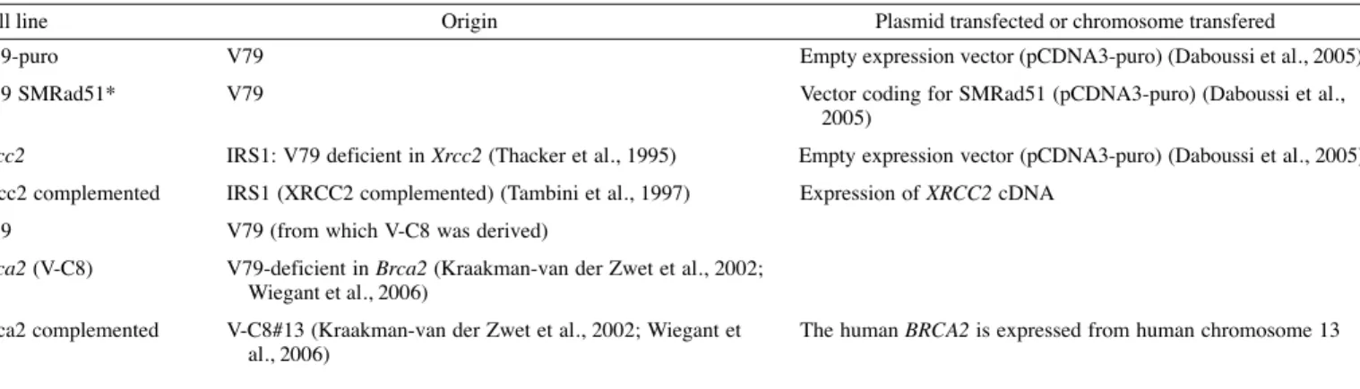
Thus, as a confirmation of the above results, we investigated the impact of HR deficiency on the density of active replication forks. Fork density was determined on 7-10 Mb of DNA fibers, Table 1. Cell lines and derivatives
Cell line Origin Plasmid transfected or chromosome transfered
V79-puro V79 Empty expression vector (pCDNA3-puro) (Daboussi et al., 2005)
V79 SMRad51* V79 Vector coding for SMRad51 (pCDNA3-puro) (Daboussi et al.,
2005)
Xrcc2 IRS1: V79 deficient in Xrcc2 (Thacker et al., 1995) Empty expression vector (pCDNA3-puro) (Daboussi et al., 2005)
Xrcc2 complemented IRS1 (XRCC2 complemented) (Tambini et al., 1997) Expression of XRCC2 cDNA
V79 V79 (from which V-C8 was derived)
Brca2 (V-C8) V79-deficient in Brca2 (Kraakman-van der Zwet et al., 2002;
Wiegant et al., 2006)
Brca2 complemented V-C8#13 (Kraakman-van der Zwet et al., 2002; Wiegant et The human BRCA2 is expressed from human chromosome 13
al., 2006)
*The dominant-negative SMRad51 is a chimera gene made by the fusion of the N-terminal part of yeast RAD51 to the entire mouse Rad51 cDNA (Lambert and Lopez, 2000). V79SMRad51 is a hamster V79 cell line expressing the dominant-negative SMRad51.
Table 2. Density of replication forks
Number of Inter-origin
Cell line forks per Mb distance (Kb)
V79 9.3 43.5
V79 SMRAD51 11.7 31.7
Xrcc2 11.4 31.1
Xrcc2 complemented 8.8 43.9 Fork density was determined on 7-10 Mb of DNA fibers labeled with anti-DNA antibody, bearing at least one elongation signal. Inter-origin distance was measured on fibers bearing at least two signals.
Jour
bearing at least one elongation signal. Our results show a significant increase in the frequency of elongating forks in HR-deficient cell lines, compared with control cell lines (Fig. 1B). The control cell line V79 presented 9.3 forks per Mb, whereas the
Xrcc2 and V79SMRAD51 cell lines presented 11.4 and 11.7 forks
per Mb, respectively (P<0.01) (Table 2). In Xrcc2 complemented cells, the density of replication forks was 8.8 forks per Mb, a value similar to that found in wild-type V79 cells (P=0.3). Reciprocally, we confirmed these data by measuring the intra-origin distances between origins present on a common DNA molecule. Consistently, the inter-origin distance dropped from 43.5 and 43.9 in wild-type and complemented cells, respectively, to 31.7 and 31.1 in SMRad51-expressing and Xrcc2-defective cells,
respectively (Table 2). Taken together, these data show an increase in the frequency of active replication forks in HR-defective cells. Alternatively, it is also possible that the data reflect an increase in the degree of replicon clustering in the HR-defective cells, rather than an actual compensation of the slow replication kinetics. Finally, these results do not support previous work reporting that the frequency of Y-shaped molecules (branched structures at replication forks), as measured by 2D-gel electrophoresis, is not altered in unchallenged Brca2-defective cells (Lomonosov et al., 2003). The authors concluded that defects in BRCA2 per se do not enhance origin firing or replication-fork progression. The differences in our study could result from the fact that molecular combing uses the incorporation of CldU and IdU, which might
Fig. 1. Analysis of replication elongation by molecular combing. (A) Example of labeled molecules with symmetric and asymmetric replication signals. Blue,
anti-DNA; green, anti-IdU; red, anti-CldU. (B) Typical examples of labeled molecules obtained from HR proficient (upper panel) or HR-deficient cells (lower panel). (C) Percentage of forks traveling at the indicated speed (kb/minute) in the three HR-deficient cell lines, their respective parental cell line and complemented cell line derivatives. The analysis was performed on 150 DNA fibers with symmetric labeling. The dotted line corresponds to the mean rate in parental V79 or V79-puro cells. We compared the distributions of V79SM, Xrcc2-defective and Xrcc2-complemented cells with the distribution of V79. Consistently, there were no statistical differences between the complemented and the wild-type parental cell lines (P=0.5 for Xrcc2 and P=0.42 for Brca2). By contrast, the distributions between mutant and parental V79 or V79-puro or complemented derivatives were significantly different (P<0.001). There is no statistical difference between the complemented and parental cell lines (P=0.5 for Xrcc2 and P=0.42 for Brca2).
Jour
165
Homologous recombination and replication
constitute a replication stress that may be amplified by the HR defect, and/or that such particular metabolic conditions would be required to reveal an involvement of HR. In addition, the inconsistencies between the present data and the Y-shaped-molecule analysis could be explained by the fact that their analysis was restricted to the rDNA locus. Here, we studied three different cell lines with different HR defects by using molecular combing, a technique that allowed us to perform a genome-wide study of the replication dynamics. Importantly, the combing technique also permits a more direct measurement of the density of initiation events and of the rate of fork progression.
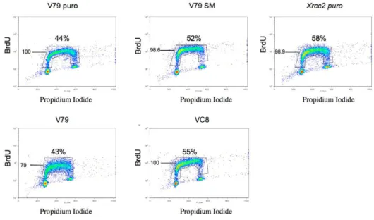
An increase in the density of active replication forks should compensate the lower replication elongation kinetics for the progression through S phase. We thus verified the above data by FACS analysis of control and HR-deficient cell populations that had been pulse-labeled with BrdU (Fig. 2). In eight independent experiments, both the distribution of cells within S phase and the efficiency of BrdU incorporation were not statistically affected by the HR status. Taken together, our results show that the slowing down of replication-fork progression is precisely compensated by the increase in the density of initiation events that take place in HR-defective cells.
In conclusion, it has long been known that HR is required to reactivate previously arrested replication forks (Kuzminov, 1995) and in replication of damaged DNA matrix (Henry-Mowatt et al., 2003). Our data reveal a novel role for HR in the control of replication-fork speed and in the distribution of initiation events.
Materials and Methods Cells and DNA manipulations
Cell lines were maintained at 37°C with 5% CO2in MEM supplemented with 10%
fetal bovine serum, 2 mM glutamine, 200 U/ml penicillin, 200 g/ml streptomycin. All DNA manipulations were performed as described (Ausubel et al., 1999). Cell lines derived from V79 hamster cells (wild-type cells) or from ionising-radiation-sensitive (IRS1) cells, which are xrcc2-defective cells (Thacker et al., 1995). IRS1 cells complemented with XRCC2 cDNA were also used (Tambini et al., 1997). Stable transfectants expressing SMRAD51 were derived from these cells. As controls of selection, V79 and IRS1 cells were stably transfected with the empty expression vector pCDNA-puro (Lambert and Lopez, 2000), namely V79 puro and xrcc2. Finally, we also used the V-C8 cell line, defective in the BRCA2 gene and its counterpart V-C8#13 complemented with chromosome 13 (Kraakman-van der Zwet et al., 2002; Wiegant et al., 2006) or the corresponding V79 parental cell line, corresponding therefore to a second wild-type V79 source.
BrdU incorporation
Cells were incubated for 20 minutes at 37°C with 10 M BrdU. After fixation with 70% ethanol at –20°C, cells were re-suspended in 1 ml of 0.5 mg/ml pepsin in 0.1 M HCl, and 1 ml of 2 M HCl was also added. Cells were incubated for 40 minutes at 37°C, washed and the pellets were then re-suspended in 0.1 g/l anti-BrdU-fluorescein antibody (Becton Dickinson). After 30 minutes, 1 ml of the propidium iodide (PI)/RNase solution was added for a further 30 minutes. Cells (2⫻104) were
analyzed in a FACScalibur (Becton Dickinson). The cell cycle distribution was analyzed by Mod Fit software.
Molecular combing
Molecular combing was performed as described (Michalet et al., 1997). The IdU and CldU labeling (20-minute pulse labeling each) protocol has been described by Anglana et al. (Anglana et al., 2003). The analogs were revealed by incubating coverslips for 1 hour with mouse anti-BrdU FITC antibody (1:5; Becton Dickinson) and rat anti-BrdU antibody (1:25; Seralab). Following washing with 0.5 M NaCl, 20 mM Tris pH 7.8 and 0.05% Tween, coverslips were incubated with secondary antibodies: Alexa-Fluor-488-conjugated goat anti-mouse (1:50; Molecular Probes) and Alexa-Fluor-594-conjugated goat anti-rat (Molecular Probes). After washing, coverslips were sequentially incubated with three antibodies: mouse anti-DNA (1:100; Argene Biosoft), rabbit anti-mouse Alexa-Fluor-350 (1:50; Molecular Probes), and goat anti-rabbit Alexa-Alexa-Fluor-350 (1:25; Molecular Probes). 150 fibers with symmetrical green-red labeling were analyzed for each cell line. To measure the density of replication origins, DNA molecules were uniformly stained with anti-DNA antibody as described previously (Anglana et al., 2003). We focused on molecules bearing at least one symmetrical green-red signal, then added the length of enough fibers to reach a total of 7-10 Mb. We finally counted the number of forks traveling along this DNA length. Images were obtained and processed as described previously (Anglana et al., 2003).
Statistical analysis
Distributions of replication fork velocity and origin firing in HR-defective and wild-type cells were compared by means of Mann and Withney test. The non-parametric Kruskal-Wallis test was used to compare the fluorescence intensity of BrdU between defective and wild-type cells.
We thank M. Jasin for providing the I-SceI expression vector and pDR-GFP, J. Thacker for the IRS1 and the complemented xrcc2 cell lines, A. Bensimon for the coverslips treated for molecular combing. Thanks are due to members of the laboratory and to D. Marsh for helpful discussion and critical reading of the manuscript. This work was supported by La Ligue Nationale contre le Cancer ‘Equipe labellisée’ (B.S.L. and M.D.), the ToxNucE program, ANR, INCA and the European Community under the contract of association between EURATOM and CEA, carried out within the framework of the European Fusion Development Agreement. The views and opinions expressed in this publication do not necessarily reflect those of the European Commission.
Fig. 2. FACS analysis of S-phase progression.
DNA synthesis measured by BrdU incorporation. Graphs are representative of eight independent experiments. The mean BrdU fluorescence intensity and the percentage of cells in S phase are indicated for each cell line. The apparent increase in S-phase cells in HR-defective cells was not statistically significant.
Jour
References
Anglana, M., Apiou, F., Bensimon, A. and Debatisse, M. (2003). Dynamics of DNA
replication in mammalian somatic cells: nucleotide pool modulates origin choice and interorigin spacing. Cell 114, 385-394.
Ausubel, F., Brent, R., Kingston, R., Moore, D., Seidman, J., Smith, J. and Struhl, K.
(ed.) (1999). Current Protocols in Molecular Biology, Vols 1-4. New York: Wiley.
Daboussi, F., Thacker, J. and Lopez, B. S. (2005). Genetic interactions between RAD51
and its paralogues for centrosome fragmentation and ploidy control, independently of the sensitivity to genotoxic stresses. Oncogene 24, 3691-3696.
Gilbert, D. M. (2007). Replication origin plasticity, Taylor-made: inhibition vs recruitment
of origins under conditions of replication stress. Chromosoma 116, 341-347.
Henry-Mowatt, J., Jackson, D., Masson, J. Y., Johnson, P. A., Clements, P. M., Benson, F. E., Thompson, L. H., Takeda, S., West, S. C. and Caldecott, K. W. (2003). XRCC3
and Rad51 modulate replication fork progression on damaged vertebrate chromosomes.
Mol. Cell 11, 1109-1117.
Hyrien, O. (2000). Mechanisms and consequences of replication fork arrest. Biochimie 82, 5-17.
Johnson, R. D., Liu, N. and Jasin, M. (1999). Mammalian XRCC2 promotes the repair
of DNA double-strand breaks by homologous recombination. Nature 401, 397-399.
Kraakman-van der Zwet, M., Overkamp, W. J., van Lange, R. E., Essers, J., van Duijn-Goedhart, A., Wiggers, I., Swaminathan, S., van Buul, P. P., Errami, A., Tan, R. T. et al. (2002). Brca2 (XRCC11) deficiency results in radioresistant DNA
synthesis and a higher frequency of spontaneous deletions. Mol. Cell. Biol. 22, 669-679.
Kuzminov, A. (1995). Collapse and repair of replication forks in Escherichia coli. Mol.
Microbiol. 16, 373-384.
Lambert, S. and Lopez, B. S. (2000). Characterization of mammalian RAD51 double
strand break repair using non lethal dominant negative forms. EMBO J. 19, 3090-3099.
Lambert, S. and Lopez, B. S. (2001). Role of RAD51 in sister-chromatid exchanges in
mammalian cells. Oncogene 20, 6627-6631.
Lambert, S. and Lopez, B. S. (2002). Inactivation of the RAD51 recombination pathway
stimulates UV-induced mutagenesis in mammalian cells. Oncogene 21, 4065-4069.
Liu, N., Lamerdin, J. E., Tebbs, R. S., Schild, D., Tucker, J. D., Shen, M. R., Brookman, K. W., Siciliano, M. J., Walter, C. A., Fan, W. et al. (1998). XRCC2 and
XRCC3, new human Rad51-family members, promote chromosome stability and protect against DNA cross-links and other damages. Mol. Cell 1, 783-793.
Lomonosov, M., Anand, S., Sangrithi, M., Davies, R. and Venkitaraman, A. R. (2003).
Stabilization of stalled DNA replication forks by the BRCA2 breast cancer susceptibility protein. Genes Dev. 17, 3017-3022.
Marheineke, K. and Hyrien, O. (2004). Control of replication origin density and firing
time in Xenopus egg extracts: role of a caffeine-sensitive, ATR-dependent checkpoint.
J. Biol. Chem. 279, 28071-28081.
Michalet, X., Ekong, R., Fougerousse, F., Rousseaux, S., Schurra, C., Hornigold, N., van Slegtenhorst, M., Wolfe, J., Povey, S., Beckmann, J. S. et al. (1997). Dynamic
molecular combing: stretching the whole human genome for high-resolution studies.
Science. 277, 1518-1523.
Saintigny, Y., Delacote, F., Vares, G., Petitot, F., Lambert, S., Averbeck, D. and Lopez, B. S. (2001). Characterization of homologous recombination induced by replication
inhibition in mammalian cells. EMBO J. 20, 3861-3870.
Sengupta, S., Linke, S. P., Pedeux, R., Yang, Q., Farnsworth, J., Garfield, S. H., Valerie, K., Shay, J. W., Ellis, N. A., Wasylyk, B. et al. (2003). BLM
helicase-dependent transport of p53 to sites of stalled DNA replication forks modulates homologous recombination. EMBO J. 22, 1210-1222.
Sengupta, S., Robles, A. I., Linke, S. P., Sinogeeva, N. I., Zhang, R., Pedeux, R., Ward, I. M., Celeste, A., Nussenzweig, A., Chen, J. et al. (2004). Functional interaction
between BLM helicase and 53BP1 in a Chk1-mediated pathway during S-phase arrest.
J. Cell Biol. 166, 801-813.
Shechter, D. and Gautier, J. (2004). MCM proteins and checkpoint kinases get together
at the fork. Proc. Natl. Acad. Sci. USA 101, 10845-10846.
Shukla, A., Navadgi, V. M., Mallikarjuna, K. and Rao, B. J. (2005). Interaction of
hRad51 and hRad52 with MCM complex: a cross-talk between recombination and replication proteins. Biochem. Biophys. Res. Commun. 329, 1240-1245.
Tambini, C. E., George, A. M., Rommens, J. M., Tsui, L. C., Scherer, S. W. and Thacker, J. (1997). The XRCC2 DNA repair gene: identification of a positional
candidate. Genomics 41, 84-92.
Taylor, J. H. (1977). Increase in DNA replication sites in cells held at the beginning of S
phase. Chromosoma 62, 291-300.
Thacker, J., Tambini, C. E., Simpson, P. J., Tsui, L. C. and Scherer, S. W. (1995).
Localization to chromosome 7q36.1 of the human XRCC2 gene, determining sensitivity to DNA-damaging agents. Hum. Mol. Genet. 4, 113-120.
Wiegant, W. W., Overmeer, R. M., Godthelp, B. C., van Buul, P. P. and Zdzienicka, M. Z. (2006). Chinese hamster cell mutant, V-C8, a model for analysis of Brca2
function. Mutat. Res. 600, 79-88.