HAL Id: hal-03085034
https://hal.archives-ouvertes.fr/hal-03085034
Submitted on 22 Dec 2020HAL is a multi-disciplinary open access archive for the deposit and dissemination of sci-entific research documents, whether they are pub-lished or not. The documents may come from teaching and research institutions in France or abroad, or from public or private research centers.
L’archive ouverte pluridisciplinaire HAL, est destinée au dépôt et à la diffusion de documents scientifiques de niveau recherche, publiés ou non, émanant des établissements d’enseignement et de recherche français ou étrangers, des laboratoires publics ou privés.
Squalamine and Aminosterol Mimics Inhibit the
Peptidoglycan Glycosyltransferase Activity of PBP1b
Adrien Boes, Jean-Michel Brunel, Adeline Derouaux, Frederic Kerff, Ahmed
Bouhss, Thierry Touze, Eefjan Breukink, Mohammed Terrak
To cite this version:
Adrien Boes, Jean-Michel Brunel, Adeline Derouaux, Frederic Kerff, Ahmed Bouhss, et al.. Squalamine and Aminosterol Mimics Inhibit the Peptidoglycan Glycosyltransferase Activity of PBP1b. Antibiotics, MDPI, 2020, 9 (7), pp.373. �10.3390/antibiotics9070373�. �hal-03085034�
1
Squalamine and aminosterol mimics inhibit the peptidoglycan
glycosyltransferase activity of PBP1b
Adrien Boes1, Jean-Michel Brunel2, Adeline Derouaux1, Frédéric Kerff1, Ahmed Bouhss3,4,
Thierry Touze3, Eefjan Breukink5, and Mohammed Terrak1
1 InBioS-Centre d’Ingénierie des Protéines, Liège University, B6a, Quartier Agora, allée du
six Août 11, 4000 Liège 1, Belgium
2Aix Marseille Univ, INSERM, SSA, MCT, Marseille, France.
3 Institute for Integrative Biology of the Cell (I2BC), CEA, CNRS, Univ Paris-Sud, Université
Paris-Saclay, Gif-sur-Yvette, France.
4 Laboratoire Structure-Activite des Biomolecules Normales et Pathologiques (SABNP), Univ
Evry, INSERM U1204, Universite Paris-Saclay, 91025 Evry, France
5 Membrane Biochemistry and Biophysics, Department of Chemistry, Faculty of Science,
Utrecht University, The Netherlands.
To whom correspondence should be addressed:
Mohammed Terrak, Centre d’Ingénierie des Protéines, University of Liège, B6a, Quartier Agora, Allée du six Août 11, 4000 Liège 1, Belgium, Tel: +33-4366-3332; E-mail:
mterrak@uliege.be
2
Abstract
Peptidoglycan (PG) is an essential polymer of the bacterial cell wall and a major antibacterial target. Its synthesis requires glycosyltransferase (GTase) and transpeptidase enzymes that, respectively, catalyze glycan chain elongation and their cross-linking to form the protective sacculus of the bacterial cell. The GTase domain of bifunctional PBPs of class A, such as E.
coli PBP1b, belong to the GTase 51 family. They play essential role in PG synthesis and their
specific inhibition by moenomycin lead to bacterial cell death. In this work, we report that the aminosterol squalamine and mimic compounds presented an unexpected additional mode of action consisting in the inhibition of the GTase activity of the model enzyme PBP1b. In addition, selected compounds were able to specifically displace the lipid II from the active site in a fluorescence anisotropy assay suggesting that they act as competitive inhibitors.
3
Introduction
Peptidoglycan (PG) is an essential polymer of the bacterial cell wall and a major antibacterial target; it surrounds the cytoplasmic membrane, determines the cell shape and protects the cell from lysis. The PG structure consists of glycan strands made of alternating β-1,4-linked N-acetylglucosamine (GlcNAc) and N-acetylmuramic acid (MurNAc) residues cross-linked by peptides [1]. It is assembled using the lipid II precursor (undecaprenyl-pyrophosphoryl-MurNAc-(pentapeptide)-GlcNAc) by the glycosyltransferases (GTases) activities of the class A penicillin-binding proteins (aPBPs) and SEDS proteins and cross-linked by the transpeptidases (TPases) activities of aPBPs and class B PBPs (bPBPs) [2–4]. The inhibition of each one of those activities lead to cell lysis and the death of the bacteria [5].
The glycosyltransferases (GTase) domain of bifunction PBPs of class A, such as E. coli PBP1b, belong to the GTase 51 family. The enzymatic cavity of these proteins can be divided into two substrate binding sites: a donor site for the lipid-bound growing glycan chain, and an acceptor site for lipid II [6]. Moenomycin A is the only known natural product that specifically binds to the GTase 51 donor site and competitively inhibits PG synthesis [7]. It is a potent antibiotic that is not used in human therapy but was used in animal feedstock for decades without reported resistance [8].
Squalamine is a cationic aminosterol antibiotic, isolated from the dogfish shark (Squalus
acanthias) [9], exhibiting a broad-spectrum antimicrobial activity against Gram-positive and
Gram-negative bacteria and potentiate the activity of several antibiotic classes at subinhibitory concentrations, against both susceptible and resistant bacteria [10]. Its structure is composed of a steroid linked to a polyamino spermidine moiety, which taken independently exhibit
4
considerably low antibiotic activity and are both required for the observed antimicrobial activity of squalamine [9].
Since more than one decade, numerous aminosterol derivatives of squalamine have been synthesized and characterized [12–16]. Their mode of action was proposed to act by disrupting the outer membrane integrity of Gram-negative bacteria and by depolarizing the cytoplasmic membrane of Gram-positive bacteria [17,18]. Some of them display good membrane selectivity with high activity on bacterial membrane and low hemolytic activity, which make them promising candidate as new antibiotics or as adjuvants to sensitize resistant bacteria to the well-known antibiotics [17,18].
Herein, we will demonstrate that squalamine and other aminosterol mimics present an unexpected additional mode of action, consisting of the inhibition of a bacterial peptidoglycan polymerase. By using in vitro activity assays based on the processing of lipid II substrate into peptidoglycan polymers by E. coli PBP1b, we found that squalamine and six other aminosterol analogs inhibit the GTase activity of this enzyme. In addition, the selected compounds were able to displace the lipid II from the active site in a fluorescence anisotropy assay indicating that they act as competitive inhibitors.
5
Results
Squalamine inhibits the GTase activity of PBP1b
The crystal structure of the glycosyltransferase domain of the class A PBP1a from Aquifex
aeolicus presents a steroidal CHAPS detergent molecule bound to a hydrophobic patch that lead
to the active site [19]. Its side chain was suggested to mimic and partly overlap with diphospholipid chain of the lipid II substrate at the acceptor site. We noticed that the aminosterol antibiotic squalamine and CHAPS have some structural feature in common, consisting essentially of a sterane core (Fig. 2). We were curious to know if squalamine could affect the peptidoglycan polymerase activity of E. coli PBP1b. Surprisingly, by using radioactive lipid II as substrate in an endpoint TLC assay we found that squalamine was able to almost completely inhibit the glycosyltransferase activity of PBP1b (1.1 ± 0.6 % residual activity (RA) at 0.8 mM) (Fig. 1A). Variable concentrations of squalamine were then tested using both an endpoint TLC assay and a continuous fluorescence assay that monitor the consumption of dansyl-lipid II substrate over time. The inhibition result shows concentrations-dependent profile and the efficacy of squalamine was rather modest in the high µmolar range (IC50 value of 291 ± 26 µM) (Fig. 1B, Table 1). When the zwitterionic CHAPS detergent was
tested using the same assay at high concentrations and below the critical micelle concentration (CMC 6-10 mM) value, no inhibition of PBP1b GTase activity was observed up to 5 mM (Fig.
1C). In addition, steroid progesterone11 or octanediamine 12tested alone do not inhibit GTase
activity of PBP1b (Fig. 2, Table 1).
6
Based on these encouraging results we have analyzed twelve additional aminosterol compounds with one or more substitutions at C-3, C-6, C-7 and C-17 positions of the sterane rings. To determine the relative activity of the compounds, they were tested at 400 and 800 µM and ranked according to their effect on the residual activity (RA) of the enzyme. Six compounds were found to inhibit the glycosyltransferase activity of PBP1b. Thus, , the residual GTase activity of PBP1b ranged from 0.3 to 8.5 % and from 1.2 to 39 % by using compound concentrations of 400 and 800 µM, respectively (Fig. 2, Table 1). The active compounds were investigated further to determine the IC50 values (Table 1) and the best derivatives 6 and 2 were
found about three time more efficient that squalamine with an IC50 values of 111 ± 6 µM and
114 ± 6 µM, respectively. Interestingly, compound 9, a close analog of compound 2 which is a stereoisomer at position C-7, was almost inactive (RA 98 % at 800 µM). A second observation that can be made is that the active compounds (except for squalamine) possess nonpolar moiety in C-17. In contrast, the presence of polar groups at this position tends to abolish the activity of the compounds (e.g amino group in 10, 13, 14, 15). Altogether; these results indicate that the observed inhibitions of the GTase activity of PBP1b by squalamine and analogs are specific and exclude the possibility of promiscuous effect of these series of compounds.
Mode of action of aminosterol compounds
The inhibition of the GTase activity can be achieved either by compounds that bind to the active site of the enzyme (e.g. moenomycin A) or through binding to the lipid II substrate (e.g. nisin, ramoplanin). To distinguish between these two mechanisms, we further characterized the mode of action of squalamine, the best inhibitory compound (6) and an inactive compound (15) by studying their interaction with PBP1b and lipid II, using a recently developed fluorescence anisotropy assay based on a fluorescent lipid II as a probe [20]. The assay was previously validated for direct interaction of the probe with PBP1b and with the antibiotics nisin and
7
ramoplanin (FA increase upon binding), as well as the ability of unlabeled-lipid II, nisin or ramoplanin to displace the probe form the enzyme active site (FA decrease in the presence of competitive compound) [20]. While no significant interaction was observed between lipid II and squalamine or compounds 6 and 15, the active compounds were able to compete with lipid II for binding to PBP1b, whereas the inactive compound 15 had no effect. These results clearly indicate that the active aminosterol compounds directly interact with the glycosyltransferase active site of PBP1b (Fig. 3). All together the data reveal that squalamine and active analogs have multiple antibacterial mode of action, in addition to their potent membranes damaging activities, they also have modest cell wall synthesis inhibition activity.
Discussion
The prevalence of antibacterial resistance is increasing and new solutions should be developed urgently to deal with the resistant pathogens. The GTase activity of bifunctional PBPs is essential for bacterial viability and constitute a proven antibacterial target [21,22]. However, only one natural product antibiotic, moenomycin, that specifically bind to the active site of these enzymes has been discovered more than 50 years ago [7,23], and therefore finding a new chemical scaffold able to bind and inhibits this class of enzymes is of great importance. In this regard, the discovery of the natural product squalamine and synthetic analogues that inhibit the GTase activity of the model class A PBP1b enzyme open new perspective for the development of new class of GTase inhibitors. The identification of aminosterol derivatives with improved activity against the GTase, while maintaining some specific bacterial membranes damaging activity, could result in potent antibacterial agents, particularly against resistant Gram-negative bacteria.
Interestingly, FA experiments indicate that these compounds are competitive inhibitors able to displace the lipid II substrate from the GTase active site, but in contrast to moenomycin which
8
bind only to the donor site and shows an increase in FA upon cooperative binding with lipid II substrate to PBP1b [20,24], the titration of the PBP1b-probe complex by aminosterol compounds induce a decrease in FA signal, similar to that observed in the presence of unlabeled lipid II, indicating that they prevent lipid II probe binding to both donor and acceptor sites. The observation that the presence of amino group at the C17 substitution abolish the activity of the compounds (10, 13, 14, 15), may indicate that this part of the molecules interact with the positively charged residues of the active site, potential candidates would be the positive pocket in the donor site formed by residues (K274, R282, R286, R287) [6,25]. Positioning of a CHAPS molecule into the active site of PBP1b by homology to PBP1a-CHAPS structure [19] suggest that this may be the case (Fig 4).
Moenomycin has an excellent antibacterial potency and no reported resistance despite extensive use in animal feeds, but it is not used in human therapy because of it its undesirable physical properties that result in poor bioavailability and long serum half-life, probably due to its lipidic C25 moenocinol moiety [8,26,27]. This lipid tail is necessary for moenomycin activity and its replacement by an alternative one could improve the properties of the resulting molecule. The C25 lipid tail of moenomycin was not resolved in any crystal structure of GTase-moenomycin complexes, indicating that it is not bound in an ordered conformation [6,28]. In contrast, CHAPS was well defined in the structure of the GTase domain of PBP1a from A. aeolicus [19] suggesting that binding of the cyclic steroid-substituted compounds would provide a defined structure and facilitate structure-based optimization of compounds.
Most of GTase inhibitors (moenomycin and synthetics compounds) are inactive against Gram-negative bacteria [22]. On the other hand, aminosterols have the capacity to cause lesions in the outer membrane and either kill or sensitize Gram-negative resistant strains to common antibiotics. Therefore, it is tempting to speculate that aminosterol compounds may be useful substitute to the moenocinol moiety of the moenomycin or analogs (or to the lipid tail of lipid
9
II analogs [29]) to generate useful new hybrid compounds, consisting of the carbohydrate and phosphoglycerate moieties or the minimal moenomycin pharmacophore (disaccharide (EF)-phosphoglycerate [22]) and an aminosterol tail (Fig. 4). Such compounds would have two functions: i) to replace the lipid tail of moenomycin derivatives while maintaining their binding to the GTase active site; and ii) to allow these compounds to cross the outer membrane (via the polyamine cationic moiety) and gain effectiveness against Gram-negative pathogens.
Materiel and Methods
Reagents and protein. Labelled lipid II [N-acetylglucosaminyl-N-acetylmuramoyl
(L-Ala-γ-DGlu-(L)-Lys-(L)-D-Ala-D-Ala)-pyrophosphate-undecaprenol] variants were prepared as previously described: Dansyl-lipid II [30], NBD-lipid II [31], [14C]-Lipid II (0.06 µCi nmol-1) [32].
PBP1bγ(M46-N844) was purified as previously described[33].
Compounds synthesis. The procedures for the synthesis of compounds are described in the
supplementary material section.
All the solvents were purified according to reported procedures, and the reagents used were commercially available. Methanol, ethyl acetate and dichloromethane were purchased from SDS and used without further purification. Column chromatography was performed on SDS silica gel (70-230 mesh). 1H NMR and 13C NMR spectra were recorded in MeOD on a Bruker AC 300 spectrometer working at 300 MHz and 75 MHz, respectively (the usual abbreviations are used: s: singlet, d: doublet, t: triplet, q: quadruplet, m: multiplet). Tetramethylsilane was used as the internal standard. All chemical shifts are given in ppm. Mass spectroscopy analysis have been performed by the Spectropole (Analytical Laboratory) of Aix-Marseille Université (Marseille). The purity of the compounds was checked by analytical HPLC (C18 column, eluent CH3CN-water-TFA (90:10:0.025, v/v/v), 0.5-1 mL/Min) with PDA detector spanning from 210
nm to 310 nm. All compounds possessed purity above 95%, as determined by analytical LCMS Agilent.
10
Octanediamine 11 and progesterone 12 were purchased from Sigma-aldrich (Saint Quentin Falavier, France). Squalamine 1 was prepared according to reported procedures [34,35]. Derivatives 2, 3 and 9 were prepared according [36]. Derivative 4 was prepared according [37]. Derivatives 10, 13 and 14 were prepared according [14]. Claramine A1 8 was prepared according [38].
General procedure for the titanium–mediated reductive amination reaction:
6-(spermine)-cholestan-3-ol 6: A mixture of 6-ketocholestanol (157 mg, 0.39 mmol),
titanium(IV) isopropoxide (573 µL, 2.02 mmol) and spermine (202 mg, 1 mmol) in absolute methanol (5 mL) was stirred under argon at room temperature for 12 h. Sodium borohydride (38 mg, 1 mmol) was then added at -78 ºC and the resulting mixture was stirred for an additional 2 hours. The reaction was then quenched by adding water (1 mL) and stirring was maintained at room temperature for 20 minutes. The resulting inorganic precipitate was filtered off over a pad of Celite and washed with methanol and ethylacetate. The combined organic extracts were dried over Na2SO4, filtered and concentrated in vacuo to afford the expected crude amino
derivative which was purified by flash chromatography affording the expected amino derivative. Purification by column chromatography (silica gel; CH2Cl2/ MeOH/ NH4OH(32%),
7:3:1) afforded a pale yellow solid in 45% yield; this compound can be converted subsequently into its hydrochloride salt as white solid –1H NMR (300 MHz, CD3OD) : δ = 0.67-0.76 (m,
4H), 0.90-1.17 (m, 24H), 1.20-1.46 (m, 7H), 1.47-1.92 (m, 20H), 2.03-2.07 (m, 1H), 2.50-2.54 (m, 1H), 2.63-2.73 (m, 13H), 3.56-3.63 (m, 1H) –13C NMR (75 MHz, CD3OD) : δ = 71.50,
58.96, 56.27, 56.05, 54.78, 49.98, 49.21, 47.99, 47.84, 47.34, 42.62, 40.47, 39.94, 39.49, 39.06, 36.44, 36.14, 35.86, 35.77, 35.63, 33.58, 31.61, 30.45, 28.18, 27.97, 24.37, 23.78, 22.78, 22.52, 21.03, 18.63, 16.30.12.13. C37H72N4O ; MS (ESI) m/z = 589.5 [M+H]+
6-(1,4-diaminobutane)-cholestan-3-ol 5: Purification by column chromatography (silica
gel; CH2Cl2/ MeOH/ NH4OH(32%), 7:3:1) afforded a pale yellow solid in 73% yield; – 1H
NMR (300 MHz, MeOD): = 0.66-3.57 (m, 58H) – 13C NMR (75 MHz, MeOD): = 71.65, 59.88, 58.54, 56.29, 56.04, 54.75, 48.18, 47.29, 42.71, 42.64, 39.94, 39.50, 39.04, 36.16, 35.78, 35.65, 31.56, 31.03, 30.40, 29.67, 27.99, 25.96, 24.35, 23.81, 22.79, 22.54, 21.05, 18.65, 16.33, 14.09, 12.15. C31H58N2O ; MS (ESI) m/z = 475.4 [M+H]+
11
N1,N1'-((3S,7R,10R,13R,17R)-10,13-dimethyl-17-((R)-5-methylhexan-2-yl)-tetradecahydro-1H-cyclopenta[a]phenanthrene-3,7-diyl)bis(pentane-1,5-diamine) 7
Synthesis according a similar protocol than for derivative 5 but using 3,7-diketocholestene and 1,5-diaminopentane as starting materials. Purification by column chromatography (silica gel; CH2Cl2/ MeOH/ NH4OH(32%), 7:3:1) afforded a pale yellow solid in 48% yield; – 1H NMR
(300 MHz, MeOD): = 0.68-0.70 (m, 3H), 0.83-0.87 ( m, 7H), 0.91-0.93 (m, 3H), 1.01-1.03 (m, 3H), 1.06-1.08 (m, 2H), 1.09-1.18 (m, 4 H), 1.19- 1.25 (m, 4H), 1.27-1.35 (m, 8H), 1.36-1.55 (m, 8H), 1.57-2.03 (m, 13H), 2.31-2.37 (m, 1H), 2.59-2.87 (m, 7H), 2.94-3.01 (m, 2H), 5.36-5.38 (m, 1H). – 13C NMR (75 MHz, MeOD): = 140.88, 125.41, 55.81, 55.47, 55.07, 52.16, 47.41, 47.13, 47.02, 42.85, 40.71, 40.61, 40.45, 39.50, 39.40, 38.40, 37.52, 37.25, 36.22, 35.70, 32.97, 32.87, 30.36, 30.26, 29.05, 28.87, 28.19, 24.78, 24.24, 24.14, 24.06, 22.66, 21.32, 18.76, 18.65, 12.35. C36H68N4 ; MS (ESI) m/z = 557.54 [M+H]+ (13S,17S)-17-((4-aminobutyl)amino)-13-methyl-7,8,9,11,12,13,14,15,16,17-decahydro-6H-cyclopenta[a]phenanthren-3-ol 15
Synthesis according a similar protocol than for derivative 5 but using oestrone and 1,4-butanediamine as starting materials. Purification by column chromatography (silica gel; CH2Cl2/ MeOH/ NH4OH(32%), 7:3:1) afforded a pale yellow solid in 37% yield; – 1H NMR
(300 MHz, MeOD): = 0.77-1.58 (m, 17H), 2.02-2.50 (m, 3H), 2.66-2.74 (m, 5H), 3.13-3.30 (m, 3H), 3.43-3.63 (m, 2H), 6.50-7.08 (m, 3H), 8.06 (s, 1H). – 13C NMR (75 MHz, MeOD): = 156.55, 139.17, 132.80, 127.63, 116.65, 114.32, 70.45, 53.94, 45.69, 44.57, 42.29, 40.75, 40.60, 36.24, 31.15, 30.62, 29.13, 28.10, 24.87, 23.05, 20.46, 14.74, 12.77, 12.69. C22H34N2O,
M = 342.53 g.mol-1, Calc. C 77.15, H 10.1, N 8.18, Exp.C 77.12, H 11.3, N 8.21.
Glycosyltransferase assays
Continuous fluorescence assay. The GTase activity of PBP1b was monitored by a continuous
fluorescent assay using dansyl lipid II as substrate. It was performed in a medium binding black 96-well microplate at 30°C (Greiner Bio One) as described [39,40]. The samples (50 µl) contained 10 µM dansyl-lipid II, 50 mM HEPES-NaOH pH 7.5, 200 mM NaCl, 10 mM CaCl2,
0.085% of decyl-PEG, 20 % of dimethylsulfoxide (DMSO), 1 unit of N-acetylmuramidase of
12
were initiated by the addition of 100 nM PBP1b and monitored by following the fluorescence decrease over 15-20 min at 30°C using an Infinite 200 PRO Microplate reader (Tecan, Männedorf, Switzerland) with excitation wavelength at 340 nm and emission at 520 nm.
Glycosyltransferase activity assay using TLC. The end-point assay, based on radiolabeled
lipid II ([14C]lipid II), was performed in 30 µl in the same conditions as the fluorescence assay
using 4 µM [14C]lipid II (0.06 µCi / nmol) instead of the fluorescent substrate and by omitting
the muramidase. The reaction was incubated 15 min at 30°C, stopped by heating at 80°C for 10min and the products were separated by thin-layer chromatography (TLC) on silica gel plates in 2-propanol–ammonium hydroxide (25%)–H2O (6:3:1; V/V/V). The TLC plates were
exposed to a storage phosphor screen (GE Healthcare) for 16 h, and images were revealed using a Typhoon Trio imager and Image Quant TL software (GE Healthcare).
The GTase inhibition assay was carried out by measuring the residual activity (RA) of E. coli PBP1b in the presence of 400 and 800 μM compounds. All the assays were repeated at least three times. The IC50 values were determined for the active compounds using variable
concentrations of inhibitor (0−1000 μM) by fitting the data using GraphPad Prism 6.0 software.
Fluorescent anisotropy (FA) assay
Fixed concentrations of NBD-lipid II (0.33 µM) and PBP1b (~1 µM) in the 25 mM Tris–HCl pH 7.5, NaCl 0.1 M, CHAPS 0.14 % w/v, were mixed with serial dilutions of the compounds in 384-well plates. All the experiments were performed in triplicates and blank samples without proteins were also prepared for background fluorescence estimation. The mixtures (30 µL) were allowed to reach equilibrium for 20 min at room temperature with gentle shaking and the FA signals were recorded at 21°C using an Infinite® F Plex (Tecan, Männedorf, Switzerland) microplate reader equipped with polarization filters with excitation and emission wavelengths at 485 nm and 535 nm respectively. For direct binding evaluation of the compounds with the
13
probe, the experiments were performed in the same condition in the absence of the protein. FA values in millianisotropy units (mA) were plotted as a function of compound concentrations in µM as previously described [20].
Acknowledgements
This work was supported by the “Fonds de la Recherche Scientifique” CDR J.0030.18. MT is a research associate of the FRS_FNRS (Brussels, Belgium), AB is supported by FRIA 1.E.038.17 (Fonds pour la formation à la Recherche dans l'Industrie et dans l'Agriculture) fellowship FRS_FNRS.
References
1. Vollmer, W.; Blanot, D.; de Pedro, M.A. Peptidoglycan structure and architecture.
FEMS Microbiol Rev 2008, 32, 149–167.
2. Sauvage, E.; Kerff, F.F.; Terrak, M.; Ayala, J.A.J.A.; Charlier, P. The penicillin-binding proteins: structure and role in peptidoglycan biosynthesis. FEMS Microbiol.
Rev. 2008, 32, 234–258, doi:10.1111/j.1574-6976.2008.00105.x.
3. Meeske, A.J.; Riley, E.P.; Robins, W.P.; Uehara, T.; Mekalanos, J.J.; Kahne, D.; Walker, S.; Kruse, A.C.; Bernhardt, T.G.; Rudner, D.Z. SEDS proteins are a widespread family of bacterial cell wall polymerases. Nature 2016, 537, 634–638, doi:10.1038/nature19331.
4. Taguchi, A.; Welsh, M.A.; Marmont, L.S.; Lee, W.; Sjodt, M.; Kruse, A.C.; Kahne, D.; Bernhardt, T.G.; Walker, S. FtsW is a peptidoglycan polymerase that is functional only in complex with its cognate penicillin-binding protein. Nat. Microbiol. 2019, 4, 587– 594, doi:10.1038/s41564-018-0345-x.
14
Garner, E.C.; Bernhardt, T.G. Bacterial cell wall biogenesis is mediated by SEDS and PBP polymerase families functioning semi-autonomously. Nat. Microbiol. 2016, 1, 16172, doi:10.1038/nmicrobiol.2016.172.
6. Sung, M.T.; Lai, Y.T.; Huang, C.Y.; Chou, L.Y.; Shih, H.W.; Cheng, W.C.; Wong, C.H.; Ma, C. Crystal structure of the membrane-bound bifunctional transglycosylase PBP1b from Escherichia coli. Proc Natl Acad Sci U S A 2009, 106, 8824–8829, doi:10.1073/pnas.0904030106.
7. Welzel, P. Syntheses around the transglycosylation step in peptidoglycan biosynthesis.
Chem Rev 2005, 105, 4610–4660.
8. Butaye, P.; Devriese, L.A.; Haesebrouck, F. Influence of different medium components on the in vitro activity of the growth-promoting antibiotic flavomycin against
enterococci. J. Antimicrob. Chemother. 2000, 46, 713–6.
9. Moore, K.S.; Wehrli, S.; Roder, H.; Rogers, M.; Forrest, J.N.; Mccrimmon, D.; Zasloff, M. Squalamine: An aminosterol antibiotic from the shark. Proc. Natl. Acad. Sci. U. S.
A. 1993, 90, 1354–1358, doi:10.1073/pnas.90.4.1354.
10. Douafer, H.; Andrieu, V.; Phanstiel, O.; Brunel, J.M. Antibiotic Adjuvants: Make Antibiotics Great Again! J. Med. Chem. 2019, 62, 8665–8681,
doi:10.1021/acs.jmedchem.8b01781.
11. Bellini, A.M.; Mencini, E.; Quaglio, M.P.; Guarneri, M.; Fini, A. Antimicrobial activity of basic cholane derivatives. Part IX. Arch. Pharm. (Weinheim). 1990, 323, 201–5, doi:10.1002/ardp.19903230404.
12. Salmi, C.; Loncle, C.; Vidal, N.; Laget, M.; Letourneux, Y.; Brunel, J.M.
Antimicrobial activities of 3-amino- and polyaminosterol analogues of squalamine and trodusquemine. J. Enzyme Inhib. Med. Chem. 2008, 23, 860–865,
15
13. Kikuchi, K.; Bernard, E.M.; Sadownik, A.; Regen, S.L.; Armstrong, D. Antimicrobial activities of squalamine mimics. Antimicrob. Agents Chemother. 1997, 41, 1433–1438, doi:10.1128/aac.41.7.1433.
14. Djouhri-Bouktab, L.; Vidal, N.; Rolain, J.M.; Brunel, J.M. Synthesis of new 3,20-bispolyaminosteroid squalamine analogues and evaluation of their antimicrobial activities. J. Med. Chem. 2011, 54, 7417–21, doi:10.1021/jm200506x.
15. Choucair, B.; Dherbomez, M.; Roussakis, C.; El Kihel, L. Synthesis of 7α- and 7β-spermidinylcholesterol, squalamine analogues. Bioorganic Med. Chem. Lett. 2004, 14, 4213–4216, doi:10.1016/j.bmcl.2004.06.010.
16. Brycki, B.; Koenig, H.; Pospieszny, T. Quaternary alkylammonium conjugates of steroids: Synthesis, molecular structure, and biological studies. Molecules 2015, 20, 20887–20900.
17. Savage, P.B.; Li, C.; Taotafa, U.; Ding, B.; Guan, Q. Antibacterial properties of cationic steroid antibiotics. FEMS Microbiol. Lett. 2002, 217, 1–7, doi:10.1111/j.1574-6968.2002.tb11448.x.
18. Di Pasquale, E.; Salmi-Smail, C.; Brunel, J.-M.; Sanchez, P.; Fantini, J.; Maresca, M. Biophysical studies of the interaction of squalamine and other cationic amphiphilic molecules with bacterial and eukaryotic membranes: importance of the distribution coefficient in membrane selectivity. Chem. Phys. Lipids 2010, 163, 131–40,
doi:10.1016/j.chemphyslip.2009.10.006.
19. Yuan, Y.; Barrett, D.; Zhang, Y.; Kahne, D.; Sliz, P.; Walker, S. Crystal structure of a peptidoglycan glycosyltransferase suggests a model for processive glycan chain synthesis. Proc Natl Acad Sci U S A 2007, 104, 5348–5353.
20. Boes, A.; Olatunji, S.; Mohammadi, T.; Breukink, E.; Terrak, M. Fluorescence anisotropy assays for high throughput screening of compounds binding to lipid II,
16
PBP1b, FtsW and MurJ. Sci. Rep. 2020, 10, 6280, doi:10.1038/s41598-020-63380-2. 21. Derouaux, A.; Sauvage, E.; Terrak, M. Peptidoglycan glycosyltransferase substrate
mimics as templates for the design of new antibacterial drugs. Front. Immunol. 2013, 4. 22. Sauvage, E.; Terrak, M. Glycosyltransferases and Transpeptidases/Penicillin-Binding
Proteins: Valuable Targets for New Antibacterials. Antibitics 2016, 5, 12, doi:10.3390/antibiotics5010012.
23. Ostash, B.; Walker, S. Moenomycin family antibiotics: chemical synthesis, biosynthesis, and biological activity. Nat Prod Rep 2010, 27, 1594–1617, doi:10.1039/c001461n.
24. Bury, D.; Dahmane, I.; Derouaux, A.; Dumbre, S.; Herdewijn, P.; Matagne, A.; Breukink, E.; Mueller-Seitz, E.; Petz, M.; Terrak, M. Positive cooperativity between acceptor and donor sites of the peptidoglycan glycosyltransferase. Biochem.
Pharmacol. 2015, 93, 141–50, doi:10.1016/j.bcp.2014.11.003.
25. Terrak, M.; Sauvage, E.; Derouaux, A.; Dehareng, D.; Bouhss, A.; Breukink, E.; Jeanjean, S.; Nguyen-Distèche, M. Importance of the conserved residues in the peptidoglycan glycosyltransferase module of the class A penicillin-binding protein 1b of Escherichia coli. J. Biol. Chem. 2008, 283, doi:10.1074/jbc.M803223200.
26. Pfaller, M.A. Flavophospholipol use in animals: positive implications for antimicrobial resistance based on its microbiologic properties. Diagn Microbiol Infect Dis 2006, 56, 115–121.
27. Hentschel, S.; Kusch, D.; Sinell, H.J. [Staphylococcus aureus in poultry--biochemical characteristics, antibiotic resistance and phage pattern (author’s transl)]. Zentralbl.
Bakteriol. B. 1979, 168, 546–61.
28. King, D.T.; Wasney, G.A.; Nosella, M.; Fong, A.; Strynadka, N.C.J.; Peter
17
protein 1B. J. Biol. Chem. 2017, 292, 979–993, doi:10.1074/jbc.M116.718403. 29. Dumbre, S.; Derouaux, A.; Lescrinier, E.; Piette, A.; Joris, B.; Terrak, M.; Herdewijn,
P. Synthesis of modified peptidoglycan precursor analogues for the inhibition of glycosyltransferase. J Am Chem Soc 2012, 134, 9343–9351, doi:10.1021/ja302099u. 30. Breukink, E.; van Heusden, H.E.; Vollmerhaus, P.J.; Swiezewska, E.; Brunner, L.;
Walker, S.; Heck, A.J.; de Kruijff, B. Lipid II is an intrinsic component of the pore induced by nisin in bacterial membranes. J Biol Chem 2003, 278, 19898–19903. 31. van Dam, V.; Sijbrandi, R.; Kol, M.; Swiezewska, E.; de Kruijff, B.; Breukink, E.
Transmembrane transport of peptidoglycan precursors across model and bacterial membranes. Mol. Microbiol. 2007, 64, 1105–14,
doi:10.1111/j.1365-2958.2007.05722.x.
32. Bouhss, A.; Crouvoisier, M.; Blanot, D.; Mengin-Lecreulx, D. Purification and
characterization of the bacterial MraY translocase catalyzing the first membrane step of peptidoglycan biosynthesis. J. Biol. Chem. 2004, 279, 29974–80,
doi:10.1074/jbc.M314165200.
33. Terrak, M.; Ghosh, T.K.; Van Heijenoort, J.; Van Beeumen, J.; Lampilas, M.; Aszodi, J.; Ayala, J.A.; Ghuysen, J.-M.; Nguyen-Distèche, M. The catalytic, glycosyl
transferase and acyl transferase modules of the cell wall peptidoglycan-polymerizing penicillin-binding protein 1b of Escherichia coli. Mol. Microbiol. 1999, 34,
doi:10.1046/j.1365-2958.1999.01612.x.
34. Zhang, X.; Rao, M.N.; Jones, S.R.; Shao, B.; Feibush, P.; Mcguigan, M.; Tzodikov, N.; Feibush, B.; Sharkansky, I.; Snyder, B.; et al. Synthesis of Squalamine Utilizing a Readily Accessible Spermidine Equivalent. ACS Publ. 1998, 63, 8599–8603, doi:10.1021/jo981344z.
18
Spermidine Analogs of the Shark Aminosterol Squalamine. European J. Org. Chem.
2003, 2003, 3897–3907, doi:10.1002/ejoc.200300167.
36. Loncle, C.; Salmi, C.; Vidal, N.; Letourneux, Y.; Brunel, J. Antimicrobial Activities of 7-Aminosterol Squalamine Analogues. Lett. Drug Des. Discov. 2008, 5, 388–393, doi:10.2174/157018008785777306.
37. Loncle, C.; Salmi, C.; Letourneux, Y.; Brunel, J.M. Synthesis of new 7-aminosterol squalamine analogues with high antimicrobial activities through a stereoselective titanium reductive amination reaction. Tetrahedron 2007, 63, 12968–12974, doi:10.1016/j.tet.2007.10.032.
38. Blanchet, M.; Borselli, D.; Rodallec, A.; Peiretti, F.; Vidal, N.; Bolla, J.M.; Digiorgio, C.; Morrison, K.R.; Wuest, W.M.; Brunel, J.M. Claramines: A New Class Of Broad-Spectrum Antimicrobial Agents With Bimodal Activity. ChemMedChem 2018, 13, 1018–1027, doi:10.1002/cmdc.201800073.
39. Offant, J.; Terrak, M.; Derouaux, A.; Breukink, E.; Nguyen-Distèche, M.; Zapun, A.; Vernet, T. Optimization of conditions for the glycosyltransferase activity of penicillin-binding protein 1a from Thermotoga maritima. FEBS J. 2010, 277, 4290–4298. 40. Schwartz, B.; Markwalder, J.A.; Seitz, S.P.; Wang, Y.; Stein, R.L. A kinetic
characterization of the glycosyltransferase activity of Eschericia coli PBP1b and
19
Figure legend
Figure 1.
Inhibition of the GTase activity of PBP1b by squalamine. A) TLC analysis of peptidoglycan
polymerase activity of PBP1b using radioactive lipid II as substrate, in the presence of squalamine and aminosterol compounds. Lanes: 1, lipid II incubated without enzyme; 2-3, lipid II incubated with 100 nM PBP1b with (2) or without (3) addition of methanol (because some compounds tested were solubilized in methanol); and 4-5, the reaction was incubated in the presence of 1 mM squalamine,or compounds 13, 14, 10, respectively. Inhibition of PBP1b GTase activity using increasing concentration of squalamine (200-1000 µM) (B) or CHAPS (1-5 mM) (C), monitored by dansyl-lipid II based continuous fluorescence assay.
Figure 2.
Structures of the aminosterol compounds used in this study.
Figure 3.
Fluorescence anisotropy assay using the NBD-lipid II as a probe. FA (in mA units) is plotted as a function of compound concentrations. The error bars represent the FA values as mean ± s.d. of triplicate experiments. (A) Competition of squalamine and compound 6 for PBP1b/NBD-lipid II binding induce a decrease of the FA of the probe while the inactive compound 15 does not affect the FA. (B) Measurements of direct binding between the probe and the compounds show no significant variation of FA, indicating absence of interactions.
20
Figure 4.
Ligand bound to the GTase domain. Crystal structure of PBP1b GTase domain in complex
with moenomycin A (yellow) with a bound CHAPS molecule (green) in the active site positioned by homology to PBP1a GTase domain of Aquifex aeolicus-CHAPS structure (PDB code 2OQO).
21
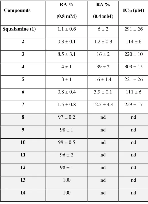
Table 1. GTase inhibition activities of squalamine and aminosterol analogues. RA = the
residual GTase activities values of PBP1b in % of inhibition compared to non-treated control are shown for two compounds concentrations (400 and 800 µM). Both RA and IC50 data
represent mean values of three independent experiments ± s.d.nd, not determined.
Compounds RA % (0.8 mM) RA % (0.4 mM) IC50 (µM) Squalamine (1) 1.1 ± 0.6 6 ± 2 291 ± 26 2 0.3 ± 0.1 1.2 ± 0.3 114 ± 6 3 8.5 ± 3.1 16 ± 2 220 ± 10 4 4 ± 1 39 ± 2 303 ± 15 5 3 ± 1 16 ± 1.4 221 ± 26 6 0.8 ± 0.4 3.9 ± 0.1 111 ± 6 7 1.5 ± 0.8 12.5 ± 4.4 229 ± 17 8 97 ± 0.2 nd nd 9 98 ± 1 nd nd 10 99 ± 0.5 nd nd 11 96 ± 2 nd nd 12 98 ± 1 nd nd 13 100 nd nd 14 100 nd nd
22
15 105 ± 3 nd nd
23
24
25