BIOMECHANICS OF NORTH ATLANTIC RIGHT WHALE BONE: MANDIBULAR FRACTURE AS A FATAL ENDPOINT FOR BLUNT VESSEL-WHALE COLLISION
MODELING by
Regina Campbell-Malone
B.S., University at Buffalo, The State University of New York, 2000
Submitted in partial fulfillment of the requirements for the degree of Doctor of Philosophy
at the
MASSACHUSETTS INSTITUTE OF TECHNOLOGY and the
WOODS HOLE OCEANOGRAPHIC INSTITUTION MASSACHUSTS INSuT
OF TEOHNOLOGY
September 2007
JUN 2 1 2008
© 2007 Regina Campbell-Malone
LIBRARIES
All rights reserved.
The author hereby grants to MIT and WHOI permission to reproduce and to distributeA ' V publicly paper and electronic copies of this thesis document in whole or in part and to
distribute them publicly.
Signature of Author
Joint Pi'ogram in Oceanography/Applied Ocean Science and Engineering Massachusetts Institute of Technology and Woods Hole Oceanographic Institution June 28, 2007 Certified by Michael J. Moore Thesis Supervisor Accepted byZ
(
Edward F. DeLong
Chair, Jbi ommittee for Biological Oceanography Woods Hole Oceanographic Institution
Biomechanics of North Atlantic Right Whale Bone: Mandibular Fracture as a Fatal Endpoint for Blunt Vessel-Whale Collision Modeling
by
Regina Campbell-Malone
Submitted to the MIT Department of Biology and the WHOI Biology Department in partial fulfillment of the requirements for the degree of
Doctor of Philosophy ABSTRACT
The North Atlantic right whale, Eubalaena glacialis, one of the most critically endangered whales in the world, is subject to high anthropogenic mortality. Vessel-whale collisions and entanglement in fishing gear were indicated in 27 (67.5%) of the 40 right whales necropsied between 1970 and December 2006. Of those, at least 9 deaths (22.5%) resulted from blunt contact with a vessel. To reduce the likelihood of fatal collisions, speed restrictions are being considered for vessels traversing critical habitat, although the effects of speed on collision outcomes have not been specifically evaluated from a biomechanics perspective.
The ultimate goal of a larger collaborative project is to evaluate the efficacy of speed restrictions for reducing blunt collision mortality using a multi-scale finite element model. Complete, transverse fracture of the right whale mandible, an injury seen only in right whales killed by vessels, is used as a proxy for mortality in the model. Vital for that model are the material properties and biomechanical behavior of the right whale mandible.
Here, the internal structure and physical properties of right whale jawbone tissue are reported. The average apparent densities, 0.4258 g/cc ±0.0970 and 1.2370 g/cc ±0.0535 for trabecular and cortical bone respectively, indicate that the bone is of relatively low density. Average ash content for trabecular bone (64.38% ±1.1330) is comparable with values from other species, indicating that low density results from a reduction of bone mass, not mineralization.
Mechanical properties of right whale bone (Young's modulus of elasticity and Poisson's ratio) were determined via uniaxial compression testing. These data are incorporated into the finite element model simulating different loading conditions (e.g. vessel speeds) that likely lead to mandibular failure and thereby mortality from blunt vessel collisions. Model results (e.g. risk of fracture) are used to determine the effect of speed restrictions on collision outcomes.
ACKNOWLEDGEMENTS
I am grateful for the opportunity to thank the many individuals and institutions that have supported this endeavor along the way. Without you this volume would not have been possible.
First and foremost, I'd like to express heartfelt thanks to my family for their
unconditional love and support - now, then and always. Madness, Martini and Mandolin - I love you.
I'd also like to thank my advisor, Michael J. Moore for his intellectual and personal contributions toward the development of this body of work. Though I may never learn how to tie your bowline, the lessons I have learned from you will last a lifetime. Special thanks to my labmates, Andrea Bogomolni and Nadine Lysiak for their angelic dispositions. Your patience, warmth and energy are positively contagious.
To Lorna Gibson (MIT) and Igor Tsukrov (UNH) - your technical guidance and personal support were invaluable. I'd like to thank Moira Brown and the right whale research group at the New England Aquarium for providing me with a home and stress-free data access. Your work focuses on an endangered species whose population is rivaled by the number of researchers working to protecting them. Your dedication is an inspiration. Sue Barco, Jerry Conway and Amy Knowlton for making logistical necropsy support look effortless - though we all know better!
Thank you to my MIT/WHOI Joint Program peers, especially Eric Montie, Gareth Lawson, Nathalie Fairbank Goodkin Emami and Benjamin Walther. Special thanks to Andrea Thorrold and Lauren Mullineaux (WHOI) for teaching Communicating Ocean Sciences - I wish I had learned half as much my first time through 4th grade!
I would like to acknowledge the invaluable assistance of contributors without whom this interdisciplinary ship would not have sailed - Bill McLellan (UNCW), Dave Rotstein (UTenn), Pierre-Yves Daoust (UPEI, Canada), Vicke Starczak (WHOI), Roberto Fajardo, John A. Muller, Nipun Patel, and Brian Snyder (Beth Israel Deaconess Medical Center). Technical assistance provided by Darlene Ketten, Julie Arruda and Scott Cramer (WHOI) Robin Fail (University of North Carolina Chapel Hill), Don Duncan, Patrick Barthelemy and Kathy Tinoco (Mercury Computer Systems) was also vital and appreciated. Special thanks to Ken Baldwin, Judson DeCew and Jason Raymond of the University of New Hampshire, Phil Clapham (NOAA Fisheries) and the Cape Cod Stranding Network (Buzzard's Bay, MA).
Personal thanks to Valerie Caron, Marsha Gomes, Shona Vitelli and Julia Westwater for going above and beyond the call of duty. Your dedication and personal touch have taught me that there is nothing that you can't make better with a smile, a hug, and a hot cup of tea. For that
lesson (and so much more) I love you. To Blanche Staton (MIT), Ambrose Jearld (NOAA Fisheries), and Ashanti Pyrtle (USF) for being there in ways that only you could.
Funding for this work was provided by the National Science Foundation (Graduate Research Fellowship Program, Campbell-Malone), the National Oceanic and Atmospheric Administration (Right Whale Grants Program, 2004, PI Campbell-Malone, Award number NA04NMF4720402), the Ocean Life Institute (PI Campbell-Malone and PI Moore), the Quebec
Labrador Foundation (PI Campbell-Malone), WHOI SeaGrant (PI Campbell-Malone), and an MIT (Presidential Fellowship, Campbell-Malone). All right whale samples were collected under National Marine Fisheries Service No. 932-1489-05.
And finally, I would like to thank my husband Kenneth M. Schopf for his unwavering support through a year filled with so much promise, preparation and planning. I am grateful for your patience and feel so blessed to have you in my life.
TABLE OF CONTENTS ABSTRA CT ... ... 3 ACKNOWLEDGEMENTS ... 4 TABLE OF CONTENTS ... ... 5 LIST OF FIGURES ... ... 9 LIST OF TABLES ... 12 LIST OF ABBREVIATIONS ... ... 13 GLOSSARY OF TERMS ... ... 14 CHAPTER 1: INTRODUCTION 1.1 M OTIVA TION ... ... .... ... ... ..21 1.2 INTRODUCTION ... ... 23 1.2.1 Species B ackground ... ... ... 23
1.2.2 Biology, population size and trends ... ... 24
1.2.3 R eproduction ... ... ... 27
1.2.4 Low genetic diversity ... 28
1.2.5 Anthropogenic mortality ... 29
1.2.6 Habitats and distribution ... 32
1.2.7 Protection and management ... 39
1.2.8 American and Canadian recovery plans ... .... 43
1.2.9 Vessel-whale collision modeling...45
1.3 LITERATURE CITED...54
CHAPTER 2: GROSS AND HISTOLOGIC EVIDENCE OF SHARP AND BLUNT TRAUMA IN NORTH ATLANTIC RIGHT WHALES (Eubalaena glacialis) KILLED BY VESSELS ABSTRACT ... ... ... 60
2.1 INTRODUCTION ... 61
2.2 MATERIALS AND METHODS ... 64
2.3 RESULTS.... ... ... . ... ... ... 66
2.3.1 Case Study I - Eg #2143 ... 66
2.3.2 Case Study II -Eg #1909 ... 72
2.3.4 Case Study IV - Eg #2150 ... ... 80
2.4 D ISC U SSIO N ... .. .... ... ... ... 85
2.4.1 Timing of trauma... ... ... 86
2.4.2 Sharp traum a... . ... .. ... 87
2.4.3 Blunt traum a ... 93
2.5 CONCLUSIONS ... ... 97
2.6 ACKNOWLEDGEMENTS ... 100
2.7 LITERATURE CITED... 102
CHAPTER 3: GEOMETRY, INTERNAL MORPHOLOGY AND DENSITOMETRY OF THE RIGHT WHALE MANDIBLE ABSTRACT ... 106
3.1 INTRODUCTION ... ... ... 108
3.1.1 Gross Description ... 109
3.1.2 Mandibular bone tissue structure and physical properties...111
3.2 MATERIALS AND METHODS ... 115
3.2.1 Sample collection and preservation ... ... 15
3.2.2 Morphometric measurements ... ... 116
3.2.3 Computed tomography (CT) ... 116
3.2.4 Quantitative computed tomography (QCT)... 118
3.2 .5 D ensitom etry ... 122
3.3 RESULTS ... ... ... 127
3.3.1 M orphometric measurements ... 127
3.3.2 Computed tomography ... 128
3.3.3 Quantitative computed tomography ... 130
3 .3 .4 D en sitom etry ... 13 1 3.4 DISCUSSION ... ... 134
3.4.1 Right whale bone density and internal structure ... 134
3.4.2 Comparing patterns in bone density - structure vs. function ... 136
3.4.3 Comparison of densitometry techniques ... ... 138
3.4.4 Limitations and caveats ... 145
3.4.5 Implications for fracture risk ... 149
3.5 FUTURE W ORK ... 153
3.6 ACKN OW LEDGEM EN TS ... ... 154
3.7 LITERATURE CITED ... ... 155
CHAPTER 4: MATERIAL PROPERTIES OF THE NORTH ATLANTIC RIGHT WHALE MANDIBLE: A FATAL ENDPOINT FOR VESSEL-WHALE COLLISION MODELING A B STR A C T ... 162
4.1 INTRODUCTION ... 163
4.1.1 Mandibular fracture as a proxy for fatality from vessel-whale collisions... 164
4.1.2 Motivation for present study ... 165
4.1.3 The right whale mandible ... 166
4.1.4 Physical properties as a predictor of material properties in bone ... 167
4.1.5 Apparent density as a predictor of Young's modulus and ultimate compressive strength ... 170
4.1.6 Predicting Young's modulus using quantitative computed tomography (QCT) ... ... 17 1 4.1.7 Investigating anisotropy in right whale bone ... 172
4.1.8 Project objective and justification ... 172
4.2 MATERIALS AND METHODS ... 173
4.2.1 Sample collection and preservation ... 173
4.2.2 Manual densitometry ... 175
4.2.3 Evaluating QCT apparent density as a predictor of apparent density and Young's modulus in right whale trabecular bone ... 176
4.2.4 M echanical testing... ... 177
4.2.5 QCT apparent density and estimated Young's modulus ... 183
4.3 RESU LTS ... 87
4.3.1 D ensitom etry ... 187
4.3.2 M echanical testing... ... 188
4.3.3 QCT apparent density and estimated Young's modulus ... 199
4.4 D ISC U SSIO N ... ... 206
4.4.1 Manual densitometry ... 206
4.4.2 M echanical testing... ... 207
4.4.3 Estimating apparent density and Young's modulus using QCT...211
4.5 C ON C LU SIO N S ... ... 219
4.7 LITERATURE CITED ... ... 223 CHAPTER 5: THESIS SUMMARY
5.1 IN TR O D U CTION ... ... 227 5.2 SUMMARY OF FINDINGS ... ... 230
5.2.1 Chapter 2 - Gross and histologic evidence of sharp and blunt trauma in North Atlantic right whales (Eubalaena glacialis) killed by vessels ... 230
5.2.2 Chapter 3 -Geometry, internal morphology and densitometry of the North Atlantic right w hale m andible ... ... 230 5.2.3 Chapter 4 -Material properties of the right whale mandible: a fatal endpoint for vessel-whale collision modeling ... ... ... 233 5.3 THE FINITE ELEMENT MODEL OF THE MANDIBLE ... 235 5.3.1 Considerations for a vessel-whale collision model ... 235 5.3.2 Finite element model of mandibular failure: design and preliminary results..241 5.3.3 Model limitations ... 249 5.4 C O N CLU SIO N S ... 251 5.5 LITERATURE CITED ... ... 255
LIST OF FIGURES
Figure 1.1. Image of a mature North Atlantic right whale... 25
Figure 1.2. Relative mortality statistics from post-mortem examination of 40 carcasses retrieved between 1970 - December 2006...30
Figure 1.3. Detailed map of North Atlantic right whale distribution in the Western North A tlantic... 36
Figure 1.4. Spatial location of right whales in the Central Early Warning System Survey Area of the Southeastern U.S. critical habitat during the winter 2005-2006 field season
... ... 37
Figure 1.5. Spatial location of commercial vessels >300 gross tons in the Central Early Warning System Survey Area of the Southeastern U.S. critical habitat, collected from AIS beacon signals received over a 64-day period in the winter 2005-2006 field season ... 3 8 Figure 2.1. Characteristic propeller trauma in live and dead whales...65
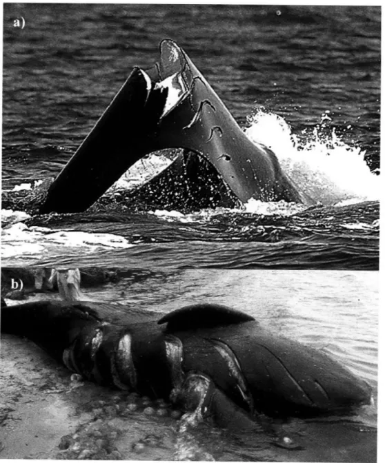
Figure 2.2. Close up of caudal (left) and central (right) propeller scars on left flank of right w hale Eg #2143 ... 69
Figure 2.3. Epithelium from propeller scar... ... 70
Figure 2.4. Dermis from propeller scar...71
Figure 2.5. Extensive soft tissue damage to fluke and peduncle of right whale Eg #1909
... ... 73
Figure 2.6. View of right side of post-cranial cross-section of Eg #1004 after beheading reveals the first signs of extensive blunt trauma... ... 77
Figure 2.7. Schematic representation of sub-dermal hemorrhage and edema in right w hale Eg # 1004... ... 78
Figure 2.8. Evidence of significant blood loss in oral cavity of right whale Eg #1004
... ... 79
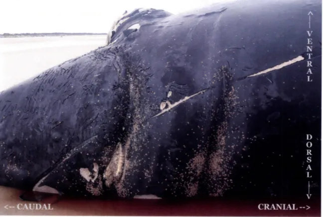
Figure 2.9. Caudal view of skull of right whale Eg #2150 ... 81
Figure 2.10. Internal signs of hemorrhage and a complete fracture of the right mandible of right w hale Eg #2150... 82
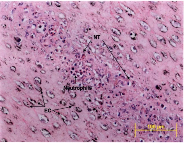
Figure 2.11. Histology of the fracture plane of the jaw bone revealed the presence of woven bone and islands of cartilage consistent with new bone formation in Eg #2150
... ... 8 3
Figure 3.1. Digital reconstruction of the right mandible of right whale Eg #1004...1 11
Figure 3.2. Computed tomography (CT) cross-section of the mandible from Eg #1004
... ... 1 19
Figure 3.3. Calibration curves for CT number (Hounsfield unit)s vs. mineral density (g/cc) ... ... 123
Figure 3.4. Calibration curves for CT number (Hounsfield units) vs. apparent density (g/cc) ... 124
Figure 3.5. The depth of soft tissue overlying the right whale jawbone plotted as a function of distance from the symphysis (yellow triangles) ... 151
Figure 3.6. Close-up of soft tissue stratification overlying the mandible in Eg #1004
... ... 152
Figure 4.1. Right whale mandibles: a) fractured right mandible of Eg# 2150. b) reconstruction of right mandible of Eg# 1004 ... 1...67
Figure 4.2. Schematic of three orthogonal axes of the right whale jawbone... 178
Figure 4.3 Mechanical testing apparatus and sample orientation ... 1....81
Figure 4.4. Engineering stress vs. engineering strain plot from uniaxial compression testing of a representative x-axis sample ... 82
Figure 4.5. Engineering stress vs. engineering strain plots for 16 trabecular bone specimens from the x-axis ... 193
Figure 4.6. Engineering stress vs. engineering strain plots for 24 trabecular bone specimens from the y-axis ... 94
Figure 4.7. Engineering stress vs. engineering strain plots for 36 trabecular bone specimens from the z-axis...195
Figure 4.8. Engineering stress vs. engineering strain plots for 11 cortical bone specimens
... ... 19 5
Figure 4.9. Interspecies comparison of Young's modulus values vs. apparent density ... ... 2 00
Figure 4.10. Interspecies comparison of ultimate compressive strength vs. apparent den sity ... 20 1
Figure 4.11. Apparent density vs. Young's modulus as a function of bone orientation
...2 0 2
Figure 4.12. Apparent density vs. ultimate compressive strength as a function of bone
orientation ... ... 203
Figure 4.13. Graph of apparent density (papp, g/cc) vs. Young's modulus (Eest, MPa) predicted from published regression equations... ... 206
Figure 5.1. Three-dimensional laser scan of right whale mandible produces CAD-compatible point cloud detailing the external geometry of the bone...244
Figure 5.2. Preliminary finite element model of right whale jaw bone ... 245
Figure 5.3. Finite element model simulating loading at 4,000 lbf. ... 246
Figure 5.4. Finite element model simulating loading at 7,000 lbf. ... 247
LIST OF TABLES
Table 2.1. Life history and necropsy findings from four right whales killed by vessels
... ... 67 Table 2.2. Definition and appearance of ante-mortem, perimortem & post mortem pathology of a traumatic origin... ... ... 89 Table 3.1. Morphometric measurements are presented for 5 North Atlantic right whales ... ... 12 9 Table 3.2. Computed Tomography (CT) volume measurements for the right mandible from right whale Eg #1004...130 Table 3.3. Average apparent and mineral densities of five segments of right whale jawbone from Eg #1004 as determined via QCT and physical measurements...132
Table 3.4. The average tissue density, apparent density, percent fat and average ash content measured for mandibular bone extracted from an adult female right whale.133 Table 3.5. Percent difference between average QCT density values and physical measurements of their apparent density and mineral density counterparts ... 133 Table 4.1. Results of manual densitometry of right whale trabecular bone samples from the mandible of Eg# 1004 ... 89 Table 4.2. Results of manual densitometry of right whale cortical bone samples from the m andible of Eg# 1004 ... ... 191 Table 4.3. Material properties of right whale trabecular bone as determined via uniaxial
com pression testing... ... 196 Table 4.4. Material properties of right whale cortical bone determined via uniaxial com pression testing... ... 197 Table 4.5. Average Young's modulus of elasticity and ultimate strength of trabecular and cortical bone from the right whale jaw bone ... 198 Table 4.6. Percent difference between average QCT apparent density and the measured apparent density (papp) from segment B ... ... 204 Table 4.7. Average QCT apparent density for right whale trabecular bone and
LIST OF ABBREVIATIONS c -engineering strain
pCT - microscale computed tomography v -Poisson's ratio
pa -apparent mineral (ash) density Papp -apparent density (g/cc) Pdry - apparent dry density
POCTapp- QCT apparent density (g/cc) Ptissue -tissue density (g/cc)
a -engineering stress
ult -ultimate compressive strength (in MPa) CT -computed tomography
E -Young's modulus of elasticity, as determined via mechanical testing (in MPa)
Eest -estimated Young's modulus of elasticity, as determined by QCT (in MPa)
Eg -Eubalaena glacialis, the scientific name for the North Atlantic right whale
Ftf -force of (tissue) fracture H.U. -Hounsfield units
knot - one nautical mile per hour mash - ash weight
mdry -dry mass
MPa - megapascals, 106 Pascals or 106 N/m2 QCT -quantitative computed tomography
GLOSSARY OF TERMS*
Ante-mortem lesions - lesions occurring more than six hours prior to death, thus allowing sufficient time for initiation of tissue response
Apparent ash density - mineral content of a specimen multiplied by its apparent dry density. Unites are in g/cc.
Apparent density - the mass of a defatted bone specimen divided by the volume of the same defatted bone specimen. The volume of the specimen is calculated from
measurements of the specimen dimension (e.g. for a cubic specimen the volume would equal the length multiplied by the width and the height) which thus includes spaces devoid of mineral. For general use, apparent density equals the mass of an object divided by its volume. Units are in g/cc unless otherwise specified.
Apparent dry density - dry mass of a dehydrated, defatted bone specimen divided by the volume of the specimen. For trabecular bone specimens the volume used to calculate the apparent dry density is the mineral volume as measured using a gas pycnometer. Thus, the volume of inter-trabecular void space in each specimen is not included in this measurement. Units are in g/cc unless otherwise specified.
Ash content - the ash weight divided by the dry mass, then multiplied by 100. This value is given as a percent (%).
Ash fraction - the ash weight (in grams) divided by the dry mass (in grams). This term is also known as the mineral content and may be expressed in grams/gram.
Ash weight - the mass of a bone specimen following dehydration (at 650C) and ashing (at 6000C) until no appreciable change in mass was detectable. Units are in grams unless
otherwise specified.
Autolysis - the disintegration of cells or tissues by endogenous enzymes.t This process is a part of postmortem decomposition.
Boat -A small vessel. The term is often modified to indicate the means of propulsion, such as a motorboat, rowboat, steamboat, sailboat, and sometimes to indicate the intended use, such as lifeboat, fishing boat, etc.tt
Callosities -raised cornified skin patches located in various locations on the heads of right whales, including on the rostrum, behind the paired blowholes, above the eye, on the ventral margin of the lower lip, and along the chin. Callosities form distinctive
*Units given in the glossary of terms are used throughout this document unless otherwise specified.
Cancellous bone - low density bone with large pores and a lattice-work of supportive struts known as trabeculae. Cancellous bone is found internal to the compact bone cortex and at the ends of long bones. Generally, the change from compact to cortical bone occurs over rapidly over a small spatial scale and the demarcation between the two is obvious to the naked eye. The term cancellous bone is used synonymously with
trabecular bone and spongy bone and is often specifically contrasted with compact bone.
Computed tomography (CT) - a radiological imaging technique that produces images of "slices" through [an object].t This technique can be used to determine the internal structure, detailed morphology, relative density, and volume of objects. The CT scanner used for this study was capable of scanning objects up to 1 meter in length and 50 cm in diameter. Objects can be assessed using a slice thickness (inter-slice distance) as low as 0.1 mm.
Compact bone - dense bone with relatively few pores and very low fat/marrow content. In long bones, the diaphysis and the thin cortex of the metaphyses are composed of compact bone. Compact bone is distinguished from trabecular bone based on the relative density of the bone tissue (see definition below).ý The relative density of cortical bone ranges from about 0.7 to 0.95.§ Compact bone is often used synonymously with cortical bone or solid bone.
Cortical bone - the compact bone tissue of the shaft of a bone.t In most mammals, this bone surrounds the marrow cavity of long bones.
Density of the calcified bone matrix -the mass of defatted, hydrated bone divided by the volume of bone tissue that does not include pore spaces. Also known as the mineral matrix density.
Dry mass - the mass of a bone sample dried at 650C until no change in mass due to water loss was observed. Units are in grams unless otherwise stated.
Edema - the abnormal accumulation of fluid in interstitial tissue spaces.
Engineering strain (E) - the change in gauge length of a specimen divided by the initial gauge length of the specimen. In standard notation, the change in gauge length is the length under load minus the original length. Engineering strain is reported here in mm/mm, and may also be considered as a quantity without units (e.g. when determining units for Young's modulus of elasticity).
Engineering stress (a) - the applied load divided by the original cross-sectional area over which it is applied. This is also referred to as the force per unit area. Engineering stress is reported in megapascals (MPa) or 106 Newtons per square meter.
Estimated Young's modulus of elasticity -values for Young's modulus of elasticity
calculated using regression equations. Experimental measurements of Young's modulus are determined during mechanical testing in uniaxial compression. Regression equations are then created, correlating the experimentally measured values for Young's modulus of elasticity (MPa) to physical measurements of apparent density (g/cc). The apparent density of samples not subjected to mechanical testing is then used to predict the Young's modulus using the regression equations. Young's modulus is reported here in
megapascals (MPa) or 106 Newtons per square meter.
Force of (tissue) fracture (Ftf) - The force required to overcome the ultimate compressive strength of a tissue when applied across a given area. This parameter is reported here in Newtons.
Hematoma - a localized collection of extravasated blood, usually clotted, in an organ, space or tissue. Hematomas can occur almost anywhere on the body; they are almost always present with a fracture and are especially serious when they occur inside the skull, where they may produce local pressure on the brain.t
Hemorrhage - the escape of blood from a ruptured vessel. Hemorrhage can be external, internal or into the skin or other tissues.t
Hounsfield units - a normalized unit of measure assigned to a given pixel in a computed
tomography reconstruction. The unit is based on the X-ray attenuation coefficient of the material occupying the physical space in the scanned specimen.
Each pixel is assigned a numerical value (CT number), which is the average of all the attenuation values contained within the corresponding voxel.§" This number is compared to the attenuation value of water and displayed on a scale of arbitrary units named Hounsfield units (HU) after Sir Godfrey Hounsfield. §§
The CT number is K([g-ptw]/gw), where K = a magnifying constant, which depends on the make of CT scanner, g = linear attenuation coefficient of pixel being observed, and
[w = linear coefficient of water."
The calculation of Hounsfield units by the CT scanner and supporting software results in a standardized scale such that Hounsfield units between scanners and between X-ray beam energy spectra are more comparable. The scale spans approximately 2000
Hounsfield units, with -1000 H.U. representing air, 0 H.U. representing water and 1000 H.U. roughly corresponding to compact bone.
Inter-birth interval - the period of time between parturition of one calf and the next.
This parameter has implications regarding the reproductive capacity and growth rate of a population. All else being equal, an increase in the inter-birth interval will result in a decrease in the reproductive potential for a population.
Knot -unit of speed used primarily by mariners. A knot is one nautical mile per hour, which is equal to 1.852 kilometers per hour and 1.150779 statute mile per hour and 0.5144444 meters per second.
Matrix material density - the mass of defatted, hydrated bone divided by the volume of bone tissue that does not include pore spaces. This parameter is also known as the density of the calcified bone matrix.
Mineral content - specific measures of mineral density, defined one of two ways. Mineral content (g/g) can be determined as the ash weight divided by the mass of dry bone (dry mass) and is also referred to as the ash fraction. As calcium is the primary mineral species in bone, mineral content (mg Ca/g dry bone) may also be determined colorimetrically as the mass of calcium (mg) per gram of dried defatted bone. Though they share the same name, it should be noted that these two measures do not produce comparable values and are not generally compared to one another directly in the
literature.
Mineral density - is generally taken as the mass of bone per unit volume of bone. However, there are many measures of "mineral density," with some being measures of mineral per unit volume, while others are measures of mineral (ash) mass per unit dry bone mass. Imaging techniques exist for determining bone mineral density (BMD) as measurements of mineral mass "per unit length (g/cm), area (g/cm2) or volume (g/cm3)" of a bone sample including dual-X-ray absorptiometry and quantitative computed tomography. aa Mineral content (in mg Ca/g dry bone) is another measure of mineral
density and can be determined colorimetrically.
Microscale computed tomography (pCT) - fine-scale computed tomography
performed on objects up to 8mm in length by 36 mm in diameter. Objects can be scanned at slice thicknesses as low as 9 microns. Like quantitative computed tomography,
standards of known density are scanned so that Hounsfield units can be converted to absolute density values.
Necropsy - the systematic dissection of a carcass with the goal of determining the cause of death. Also known as a post-mortem examination.
Peri-mortem lesions - lesions created just prior to or immediately following death
Poisson's ratio (v) - the ratio of transverse strain to axial strain. The ratio is negated to yield a positive value for Poisson's ratio. Poisson's ratio is expressed without units.
Post-mortem lesions - lesions that are created after death and generally lacking evidence of tissue reaction
QCT apparent density - values for apparent density obtained via quantitative computed tomography (QCT). These values are calculated using regression equations created by
correlating the apparent density of known standards to point measurements of x-ray attenuation (in Hounsfield units) from a computed tomography scan of the standards. QCT apparent density is given in grams per cubic centimeter (g/cc).
QCT mineral density -values for mineral density obtained via quantitative computed tomography (QCT). These values are calculated using regression equations created by correlating the mineral density of known standards to point measurements of x-ray attenuation (in Hounsfield units) from a computed tomography scan of the standards. QCT mineral density is given in grams per cubic centimeter (g/cc).
Quantitative computed tomography (QCT) - a calibrated radiological imaging technique. Computed tomography (CT) is conducted on specimens and standards of known density allowing the conversion of the Hounsfield units output by standard CT into absolute physical density values. In this way the absolute physical density of internal structures can be calculated non-invasively in vivo and non-destructively in precious specimens.
Relative density - the ratio of the density of a bone specimen to the density of solid cortical bone. The density of solid cortical bone is taken as 1.8 g/cc. The ratio is reported without units. Trabecular bone has a relative density that ranges from 0.05-0.7 g/cc, while cortical bone relative density ranges from 0.7 - 0.95 g/cc. § There is a rather subjective distinction between low density cortical bone and high-density trabecular bone.
Right whale conservation areas - two regions designated by the Canadian government as right whale habitat. These areas include the Grand Manan Basin of the Lower Bay of Fundy (referred to as Bay of Fundy) and Roseway Basin (an area -50 km south of Nova Scotia).
Right whale critical habitat areas - three regions recognized by the United States government via the National Marine Fisheries Service as current seasonal habitat in U.S. waters. These include":
1) The Great South Channel. The area bounded by 41 40' N/69045' W; 41 00' N/69005' W; 41038'N/68013' W; and 42010' N/68031' W,
2) Cape Cod Bay, Massachusetts. The area bounded by 42004.8' N/70010' W; 42012' N/70015' W; 42'12' N/70030' W; 41046.8' N/70'30' W and on the south and east by the interior shore line of Cape Cod, Massachusetts,
3) Southeastern United States. The coastal waters between 31015' N and 30015' N
from the coast out 15 nautical miles; and the coastal waters between 30015' N and 28000' N from the coast out 5 nautical miles.
Ship - originally a sailing vessel with three or more masts, square-rigged on all. The term is now generally applied to any large, ocean-going vessel, except submarines which are called boats regardless of size. t In 1999, Mandatory Ship Reporting Systems were established by the United States Coast Guard and the National Marine Fisheries Service
requiring all commercial ships, 300 gross tons or larger, entering specified regions in the coastal waters of the United States to report the vessel name, speed, destination and true course (among other data) to land-based stations at the time of entry. As this rule was implemented specifically to address the issue of vessel-whale collisions in right whale critical habitat it is appropriate for research concerning these events to define a ship as any commercial or non-commercial vessel 300 gross tons or larger.
Ship strike - term used as a colloquial synonym for vessel-whale collisions. This term is considered imprecise and inappropriate by the shipping industry as it implies that all vessel-whale collisions involve ships. Strictly defined using the technical definition of a "ship," contact between a whale and a vessel 300 gross tons or larger is considered a ship strike. This document is the first published literature to address this concern and to appropriately draw a distinction between the two terms.
Tissue density - the bulk tissue mass of a specimen (e.g. for bone this includes the mass of fat, water, mineral content as well as mass contributed by organics) divided by the bulk sample volume as measured from the sample dimensions (i.e. including non-mineralized tissues and pore spaces). Tissue density is reported here as grams per cubic centimeter (g/cc).
Trabecular bone - low density bone organized into a lattice of bony struts called trabeculae, with inter-trabecular regions lacking mineral (and in the case of the right whale these regions are filled with inter-trabecular fat). This bone type is also known as cancellous or spongy bone and is found internal to the compact bone cortex and at the ends of long bones. The term trabecular bone is often specifically contrasted with cortical bone.
Ultimate compressive strength (,,it)- the peak engineering stress measured during a compression test. The ultimate compressive strength is also known as the ultimate strength and is reported here in megapascals or 106 Newtons per square meter.
Vessel - Any type of craft which can be used for transportation on water. * Any structure designed to float upon and traverse the water for the carriage of persons or goods; a craft or ship of any kind, now usually one larger than a rowing-boat. I
Vessel-whale collision - contact between a vessel and a whale. Contact may result from blunt impact with the hull or may involve the propeller.
Young's modulus of elasticity (E) - the stress/strain in the linear region of the curve of an ordinary tensile or compressive test. ** This is the equivalent of the slope of the linear region of the stress vs. strain curve in a tensile or compressive test. Uniaxial compressive tests were used here to determine the Young's modulus. Young's modulus is a uniaxial measure of the stiffness of a material. E is reported here in megapascals (MPa) or 106 Newtons per square meter.
Symbols in text denote technical definitions as defined in the following references: - Blood, D.C. and V.P. Studdert, 1999. Saunders Comprehensive Veterinary
Dictionary. 2nd Edition, WB Saunders. New York. 1380 pages.
tt- Bowditch, N., 1995. American practical navigator: an epitome of navigation. Defense Mapping Agency Hydrographic/Topographic Center. Bethesda, MD. §- Huiskes, R. and B. van Rietbergen, 2005. Biomechanics of Bone. In: Mow, V. C.
and Huiskes, R., Eds. Basic Orthopaedic Biomechanics and Mechano-Biology. Lippincott, Williams & Wilkins. New York. pp. 123-179.
§§- Jackson, S. and R. Thomas, 2004. Cross-sectional Imaging Made Easy. Churchill Livingstone.
S- Mull, R. T., 1984. Mass estimates by computed tomography: physical density from CT numbers. American Journal of Roentgenology. 143 (5), pp. 1101-1104. S- Tabensky, A. D., J. Williams, V. DeLuca, E. Briganti and E. Seeman, 1996. Bone
mass, areal, and volumetric bone density are equally accurate, sensitive, and specific surrogates of the breaking strength of the vertebral body: an in vitro study. Journal of Bone and Mineral Research 11 (12), pp. 1981-1988. << - National Marine Fisheries Service, 2005. Critical habitat for northern right
whales. National Oceanic and Atmospheric Administration, Editor. 2005, Department of Commerce. 70 FR 1832 §226.204. p. 189-190.
- Anonymous, 1989. Oxford English Dictionary. Second edition. Oxford University Press.
*- Currey, J.D., 2002. Bones: structure and mechanics. Princeton University Press.
Princeton, NJ. 436 pages.
Species of Interest Balaena mysticetus, Bowhead whale
Balaenoptera acutorostrata, Minke whale Balaenoptera musculus, Blue whale Balaenoptera physalus, Fin whale
Calanusfinmarchicus, Calenoid copepod Dugong dugong, Dugong
Eubalaena australis, Southern right whale Eubalaena glacialis, North Atlantic right whale Eubalaenajaponica, North Pacific right whale Megaptera novaeangliae, Humpback whale Mesoplodon densirostris, Beaked whale Phocoena phocoena, Harbor porpoise Physeter macrocephalus, Sperm whale Trichechus manatus, West Indian manatee
Chapter 1
Introduction
1.1 MOTIVATION
The North Atlantic right whale (Eubalaena glacialis) is a robust baleen whale that
is considered one of the most endangered large whale species (Clapham, et al., 1999,
International Whaling Commission, 2001b). With fewer than 400 individuals remaining
in this well-studied population, a major target for conservation and management is
reducing significant sources of mortality, particularly threats from human activities
(COSEWIC, 2003, Kraus, et al., 2005, National Marine Fisheries Service, 2005a, 2006,
Right Whale Consortium, 2005). Seventy five North Atlantic right whale deaths have
been recorded between 1970 and January 2007 (Knowlton and Kraus, 2001, Moore, et
al., 2005, Right Whale Consortium, 2005. Of these, twenty eight were linked to ship
strikes, eight were attributed to entanglement in fishing gear and eighteen were
considered neonatal deaths. Twenty-one remaining deaths were of unknown cause. The
total number of deaths is unknown as some animals are never seen dead, but are simply
factors (e.g. human activities) are unknown, however, current mortality statistics
represent minimum values.
The most conservative estimates of mortality factors are those that consider cases
that were examined postmortem. Direct threats from anthropogenic activities include
entanglement in fishing gear and collisions with vessels, which together have resulted in
27 (67%) of the 40 mortalities assessed by post-mortem examination of right whale
remains. Of those, 21 (52.5%) of the 40 animals examined post-mortem were killed by
vessels. As such, government agencies charged with species management and population
recovery are considering speed restrictions as a means of reducing the frequency of
vessel-whale collisions and the degree of injury to whales struck by vessels (National
Marine Fisheries Service, 2006).
Data presently used to justify the proposed implementation of speed restrictions
include historical accounts of vessel-whale collisions detailing vessel size, vessel speed
and mortality or injury outcome where available (Laist, et al., 2001, Vanderlaan and
Taggart, 2007). Analysis of these data relies heavily upon circumstantial correlation and
extrapolation from a limited (albeit carefully compiled) dataset which, while appropriate
for elucidating general trends, are insufficient to directly address the influence of vessel
speed on the biomechanics of a dynamic collision (Jensen and Silber, 2003, Laist, et al.,
2001). In addition, few studies have explored the influence of speed on vessel-whale
collision events and no research to date has focused on the biomechanics of vessel-whale
collisions (Knowlton, et al., 1995, Korsmeyer and Hynes, 1995, National Marine
The studies included in this thesis represent a comprehensive analysis of the
biomechanics of vessel whale collisions. The first of the studies detailed here resulted in
the characterization of trauma resulting from vessel-whale collisions and identification of
the right whale mandible as an appropriate fatal endpoint for finite element models of
vessel-whale collisions (Chapter 2). The morphology, internal structure and physical
properties of the right whale jawbone were also investigated (Chapter 3). Mechanical
testing was used to determine the material properties of right whale bone tissue from the
mandible in order to assess how the right whale mandible behaves under loading
(Chapter 4). Finally, thesis findings, preliminary modeling results and the implications of
this work are presented (Chapter 5).
1.2 INTRODUCTION
1.2.1 Species background
The North Atlantic right whale, Eubalaena glacialis, is one of the three right
whale species recognized by the International Whaling Commission following a
comprehensive review of genetic and morphological data in the year 2000 (Rosenbaum,
et al., 2000). The other species include the North Pacific right whale, E. japonica and the
Southern right whale, E. australis.
The North Atlantic right whale population is believed to be divided into two
distinct stocks inhabiting the Eastern North Atlantic and the Western North Atlantic
relatedness (as assessed by mitochondrial DNA), the stocks are managed as two distinct
critically endangered populations (International Whaling Commission, 2001b).
Though dedicated research and sighting efforts have not been conducted in the
Eastern North Atlantic, the rare occurrence of opportunistic sightings of right whales in
this region leaves little doubt that this stock is either severely reduced or extinct (with
sightings representing Western North Atlantic right whales that have strayed from their
known habitat) (Clapham, et al., 1999). As such, subsequent references to the North
Atlantic right whale, Eubalaena glacialis, or 'right whale' in this thesis refer to the
Western North Atlantic stock unless otherwise noted.
1.2.2 Biology, population size and trends
The North Atlantic right whale is a robust mysticete that measures approximately
16 meters in length at maturity (Figure 1.1) (Perrin, et al., 2002). The 200-270 baleen
plates that hang from each side of the upper jaw are used for filtering dense aggregations
of planktonic prey, primarily the copepod (Calanusfinmarchicus), from seawater during
feeding bouts where the mouth remains open for extended periods (Baumgartner, et al.,
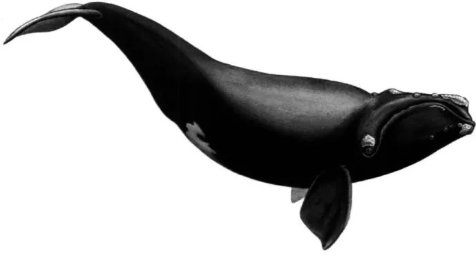
Figure 1.1: Image of a mature North Atlantic right whale. Unique patterns formed by callosities (horny protrusions on the head, upper margin of the lower lip, and chin) permit identification of individuals in this small population. Image by Scott Landry (used with permission).
The right whale has black coloration, though some individuals have patches of white on the ventral surface and/or chin. The upper margin of the lower lip is crenulated and the patterns of lip ridges varies between individuals (Hamilton, et al., 2007). Right whales also have raised cornified skin patches called callosities located in various locations on the heads of individuals, including on the rostrum, behind the paired blowholes, above the eye, on the ventral margin of the lower lip, and along the chin, forming distinctive patterns that are unique to individuals (Hamilton, et al., 2007).
Callosity patterns, along with distinguishing scars, lip ridges and white skin patches, have been used to identify, track, and maintain records from opportunistic
sightings of individuals dating back to 1935 (Kraus and Rolland, 2007a). The
photo-identification and tracking of individuals via systematic aerial and vessel-based surveys
for over 25 years makes this species one of the most well-studied cetacean populations
(Hamilton, et al., 2007).
With such a small population of individually identifiable members under intense
scientific scrutiny, estimating the population size and general trends in growth are
possible. The most recent estimates suggest that the population contains around 400
individuals (Right Whale Consortium, 2005). The highest recorded annual rate of
population growth for this species assessed between 1980-1992 (3.4%) was much lower
than the growth rate seen for Southern right whales (6-7%) (Best, et al., 2001a,
Knowlton, et al., 1994). Estimates of the annual growth rate of the population range from
-2% to 3.4% and average 2.5%, but the consensus is clear - the North Atlantic right
whale is recovering slowly (if at all) from commercial overexploitation (Fujiwara and
Caswell, 2001, Knowlton, et al., 1994).
Several factors are likely limiting the recovery of the species, including intrinsic
factors (e.g. reproduction and low genetic diversity) and extrinsic factors (e.g. direct
anthropogenic mortality from entanglement in fishing gear and vessel-whale collision,
and habitat degradation as a result of pollution and climate change) (COSEWIC, 2003).
While anthropogenic mortality is responsible for the largest identified losses to the
population, each of these factors likely contribute to the lack of recovery of this species
and as such are addressed in subsequent sections (Campbell-Malone, et al., 2007, Moore,
1.2.3 Reproduction
While evidence suggests that Southern right whales have a gestation period of 12
months, the gestation period for the North Atlantic right whale has yet to be determined
convincingly (Best, 1994). Females give birth to a single calf between December and
March (Kraus, et al., 2007). The seasonality of calving suggests that conception or
implantation may also be seasonal events (Parks, 2003). While delayed implantation is a
potential factor confounding accurate estimates, there is no evidence among the balaenids
and thus the gestation period for North Atlantic right whales is presently estimated to be
about twelve months (Best, 1994).
As a result of a relatively long gestation period, lactation for approximately 11
months and an inter-birth interval ranging from 3 to 5 years, maternal investment in
reproduction appears to be substantial (Kraus, et al., 2007). It is likely that costly
metabolic activities such as reproduction and lactation are affected during times of
limited prey abundance, leading to variable reproductive rates and changes in the
inter-birth interval (Moore, et al., 2001). Low genetic diversity, pollutants, marine biotoxins
and infectious diseases may be additional factors directly limiting successful reproduction
of this population (International Whaling Commission, 200 1b, Kraus, et al., 2007).
While some females calve at regular intervals, others successfully produce calves
sporadically while still others have never reproduced despite being considered sexually
produced in a given year is also highly variable, ranging from 1-31 calves between
1980-2004 (Kraus, et al., 2001, National Marine Fisheries Service, 2005a).
This low and variable reproductive output in response to ecological and
environmental parameters would likely have a limited effect on the long-term
contributions of individuals to the population, instead accounting for short term variation
(Kraus, et al., 2007). However, in the face of high anthropogenic mortality, low and
variable reproductive success represents a real challenge to the recovery of the species.
1.2.4 Low genetic diversity
Analyses of the genetic variability of the North Atlantic right whale population
using minisatellite and microsatellite loci, as well as mitochondrial DNA, have led to a
greater understanding of current and historic levels of genetic diversity (Frasier, et al.,
2007). Low levels of genetic variability have been found in extant right whales and it is
clear from recent studies of right whale bones from Basque whaling sites that the
population (or at lease the sub-population taken in the hunting grounds utilized) had
slightly greater genetic variability prior to the Basque whaling era (Frasier, et al., 2007).
Genetic variation is also higher in calves than would be expected under the
assumption of random mating (Frasier, et al., 2007). Comparative genetic analyses of
right whale parents and their offspring revealed that calves have significantly higher
levels of genetic variation than could be explained by random mating (Frasier, et al.,
of genetic variation in right whale calves did not result from selective mating with
"genetically dissimilar individuals" (Frasier, et al., 2007).
It is thus hypothesized that the majority of mating pairs were in fact genetically
similar (as expected due to random mating in a population with low genetic diversity),
which likely results in decreased reproductive success. The current hypothesis regarding
the contribution of low genetic variability to the low reproductive success of the
population is being explored (Frasier, et al., 2007).
1.2.5 Anthropogenic mortality
Entanglement in fishing gear: The two primary sources of anthropogenic
mortality identified in this population include entanglement in fishing gear and
vessel-whale collisions (Anonymous, 2002, Knowlton and Kraus, 2001, Kraus, 1990, Kraus, et
al., 2005, Moore, et al., 2005). Together, these two factors account for 27 (67.5%) of the
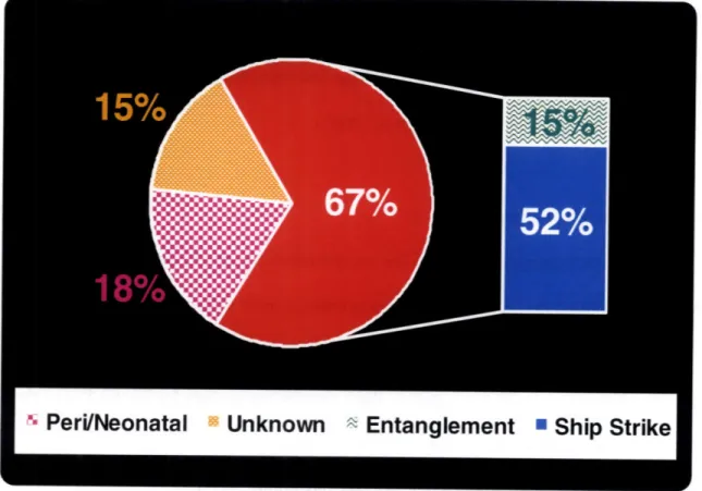
40 deaths investigated post-mortem between 1970-December 2006 (Figure 1.2)
(Campbell-Malone, et al., 2007, Right Whale Consortium, 2005).
Six (15%) of the forty necropsied animals died as a result of entanglement. A far
greater number of animals were last seen entangled and in poor health and if not seen for
six consecutive years they are presumed dead (Knowlton, et al., 1994, Right Whale
Consortium, 2005). These are considered minimum numbers and underestimates of
entanglement mortality, as 31 (43.7%) of the 71 reported right whale carcasses from 1970
Figure 1.2: Relative mortality statistics from post-mortem examination of 40 carcasses retrieved between 1970 - December 2006. Anthropogenic mortality including
entanglement (15%) and vessel-whale collisions (52%) resulted in 67% of deaths investigated. Only 15% of the deaths investigated were classified as unknown cause of death. Cause of death is generally characterized as perinatal or neonatal when involving a newborn calf when no other cause is identifiable.
52
Chronic health effects associated with infection, reduced mobility and impaired feeding result in the metabolism of blubber stores and the loss of positive buoyancy. The body condition of animals with chronic entanglements or poor health result in a
negatively buoyant carcass that is prone to sinking and is thereby likely lost (Knowlton and Kraus, 2001, Moore, et al., 2005, Right Whale Consortium, 2005). As such, statistics
regarding the relative contribution of entanglements to right whale mortality are certainly
underestimated (Moore, et al., 2005).
Sighting data and studies of scarification reveal that many whales become
entangled in fishing gear (Hamilton, et al., 1998, Right Whale Consortium, 2005).
Studies using photographs of extant individuals provide evidence of scars on 220 (61.6%)
of over 350 individuals suggesting that most entangled whales are able to self-disentangle
(Hamilton, et al., 1998). Additional findings indicate that some 62 individuals (17.7%)
had scars from two or more entanglements. While these data suggest that most
entanglements are successfully shed and do little lasting damage, additional data from
observations of entangled animals over time and necropsy data reveal that some
entanglements prove deadly (Moore, et al., 2005).
Entanglement fatality can be acute (i.e. when a whale becomes sufficiently
entangled such that it is prevented from reaching the surface to breathe), or chronic.
Chronic entanglement may result from gear getting entwined through baleen plates, or
wrapped around the peduncle, axilla, or over the rostrum (Moore, et al., 2007). Extremely
serious entanglements result in line embedded below the skin, necrosis, reduced mobility, impaired feeding and infection (Moore, et al., 2005).
Continued growth of the animal, drag on the line from trailing gear, or the
wrapping of multiple extremities in the same gear, can result in line that becomes
embedded beneath the epidermis - sometimes constricting to the point of involving bone
(Moore, et al., 2005). Though the losses to the population from entanglement are
debilitating trauma resulting from human activity is equally significant (Moore, et al.,
2007).
Vessel-whale collisions: The impact of vessel-whale collision on right whales has
been well demonstrated. Massive wounds (e.g., fractured skulls and vertebrae, severed
tails, and propeller cuts) on right whale carcasses show that vessel-whale collisions can
be, and clearly are often, lethal (Campbell-Malone, et al., 2007, Knowlton and Kraus,
2001, Moore, et al., 2005). Vessel-whale collisions are the primary source of right whale
mortality identified by post-mortem examination. These events have resulted in at least
21 (29.6%) of the 71 known deaths since 1970 (Campbell-Malone, et al., 2007). The
direct impacts of vessel-whale collisions on the right whale population are addressed in
Chapter 2.
Anthropogenic mortality from both entanglement and vessel-whale collisions is in
part a result of the substantial overlap between human activities and the coastal
distribution of right whales (Knowlton and Brown, 2007). The discovery of the habitats
utilized by the North Atlantic right whale was a critical step toward determining ways of
reducing the fatal consequences of a shared ocean environment between marine mammal
and man.
1.2.6 Habitats and distribution
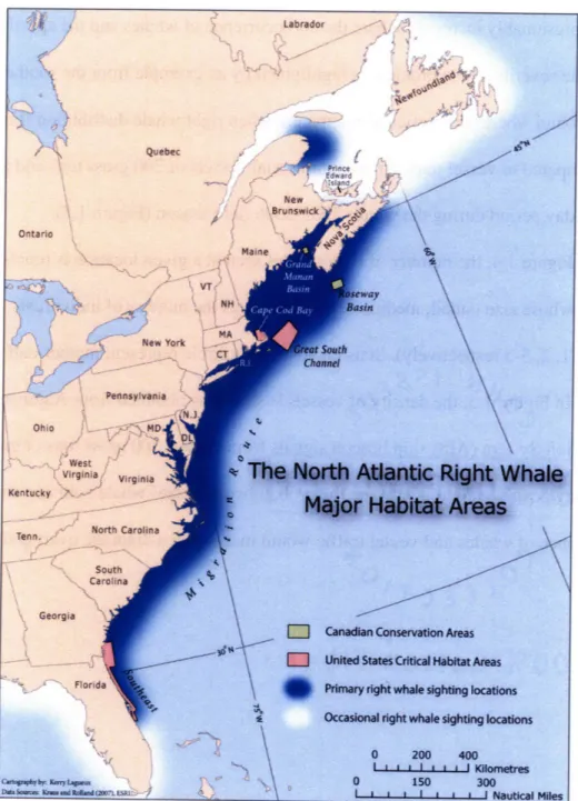
Five regions have been identified as conservation areas or critical habitats by the
fisheries management entities in Canadian and U.S. waters. These are 1) the southeastern
South Channel, northeast of Nantucket 4) Grand Manan Basin in the Lower Bay of
Fundy (between New Brunswick and Nova Scotia) and 5) the Roseway Basin region of
the Scotian Shelf (south of Nova Scotia) (Figure 1.3) (Anonymous, 2004, International
Whaling Commission, 2001b).
The regions within the Exclusive Economic Zone of the United States - including
the southeastern calving grounds, Cape Cod Bay and the Great South Channel -were
designated as critical habitat areas by the United States government in 1994 as required
by the Endangered Species Act (National Marine Fisheries Service, 2005a, United States
Congress, 1973). Right whales utilize the Cape Cod Bay habitat in most months with
peak use occurring between February and May (COSEWIC, 2003). The Great South
Channel is highly utilized in the spring, while the southeastern calving grounds are
utilized by a portion of the population during the winter months (COSEWIC, 2003).
Among the individuals inhabiting the southeastern calving area seasonally are primarily
pregnant females, their calves of the year, and juveniles. Adult males are conspicuously
absent, as are many non-breeding adult females, leaving questions regarding the regions
used by this demographic in the winter months (COSEWIC, 2003).
The regions in the Exclusive Economic Zone of Canada - including the Grand
Manan Basin (Lower Bay of Fundy) and Roseway Basin were given official status as
Right Whale Conservation Areas in 1993 by the Canadian government (COSEWIC,
2003). These habitats are primarily utilized during the summer and autumn when
of Fundy also serves as a summer nursery for many cow/calf pairs that migrate there
from calving grounds (National Marine Fisheries Service, 2005a).
Though these seasonal habitats are used by over 60% of the known population,
questions remain regarding the distribution of the subpopulation that does not migrate to
the winter calving grounds (Kraus and Rolland, 2007a). Similarly approximately
one-third of the females that calve do not bring their offspring to the Bay of Fundy (Kraus and
Rolland, 2007a). Yet the location of this alternate nursery ground is presently unknown.
Genetic evidence from calf paternity analyses suggests that additional right
whales are being "missed" by present surveys of critical habitats. Despite the fact that
nearly 70% of all known males in the population have been sampled, 55% of the calves
genetically tested to infer paternity did not match the subset of sampled fathers (Frasier,
2005). This indicates that additional fathers are contributing to the gene pool slightly
more than the known and sampled males from right whale critical habitats and may be
indicative of a population of males that do not regularly inhabit any of these areas
(Frasier, et al., 2007).
Despite the outstanding questions regarding the winter habitat utilized by much of
the population, the nursery grounds used by cows that do not bring their calves to the Bay
of Fundy and the location/identity of these males, the whereabouts of the majority of the
catalogued population have been well established. The details of their distribution
contribute to the current understanding of the threats that face the population.
The coastal distribution and coastal migration corridor (regions utilized for travel
overlap between right whale habitats and high concentrations of fishing gear and vessels
approaching and departing port or making coastal excursions (Clapham, 1999, Johnson,
et al., 2007, Kraus, et al., 1986). The probability of a whale encountering one of these
threats is presumably increased where the co-occurrence of whales and the specific threat
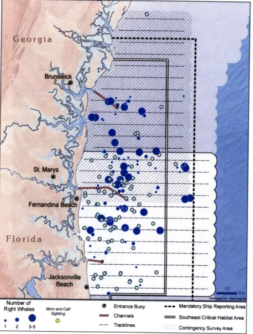
is high. The severity of the problem is highlighted by an example from the southeast U.S.
calving ground where it is particularly apparent when right whale distribution (Figure
1.4) is compared to vessel tracks from commercial vessels of 300 gross tons and above
over a 64-day period during the winter 2005-2006 field season (Figure 1.5).
In Figure 1.4, the number of right whales seen at a given location is represented
by circles whose size (small, medium, large) indicates the number of individuals
observed (1, 2, 3-5 respectively). Stars enclosed by a circle represent mother-calf pair
sightings. In Figure 1.5, the density of vessels is shown as obtained from Automatic
Identification System (AIS) ship beacon signals from vessels 300 gross tons or greater.
Clearly, any positive relation between the probability of vessel-whale collision and the
Figure 1.3: Detailed map of North Atlantic right whale distribution in the Western North Atlantic. Three critical habitats in U.S. waters (Cape Cod Bay, the Great South Channel and the Southeastern U.S. calving grounds) and two conservation areas in Canadian waters (the Lower Bay of Fundy and Roseway Basin) are shown. Adapted from (Kraus and Rolland, 2007b) by Kerry Lagueux, New England Aquarium.
Figure 1.4: Spatial location of right whales in the Central Early Warning System Survey Area of the Southeastern U.S. critical habitat during the winter 2005-2006 field season. Whale distribution is shown as small, medium and large circles representing 1, 2, and 3-5 animals respectively. A star enclosed by a circle represents a mother-calf pair. Image courtesy of Kerry Lagueux, New England Aquarium (Lagueux, et al., 2007).
Right Whales Mom and Calf
Sighting
1 2 3-5
Entrance Buoy Channels Tracklines
S- - Mandatory Ship Reporting Area
Southeast Critical Habitat Area Contingency Survey Area
Figure 1.5: Spatial location of commercial vessels >300 gross tons in the Central Early Warning System Survey Area of the Southeastern U.S. critical habitat, collected from AIS beacon signals received over a 64-day period in the winter 2005-2006 field season. Image courtesy of Kerry Lagueux, New England Aquarium (Lagueux, et al., 2007).
29.9 30.0 59.9 89.8 149.6 269.3 658.1 7,627 Channels Southeast Critical HabitatArea Number of Points / Sq. Kilometer --- Tracklines Contingency Survey Area
1.2.7 Protection and management
At the dawn of the modem whaling era, in the mid to late nineteenth century,
"right whales" (a category at the time including the bowhead whale, Greenland right
whale and pygmy right whale) had already been overexploited and no longer were a
significant portion of the landed take (International Whaling Commission, 2001 a,
Tonnessen and Johnsen, 1982). Initial protection measures, including international limits
on whaling take, were enacted in the early 20th century and were created primarily by
whaling nations in an effort to self-regulate the whaling industry to prevent the collapse
of the lucrative whale fisheries (Tonnessen and Johnsen, 1982).
Unlike its southern hemisphere counterpart, E. australis, the North Atlantic right
whale has failed to recover from severe population depletion due to whaling
(International Whaling Commission, 2001 a). This is true, despite international protection
from whaling since 1935 as per the 1931 Convention for the Regulation of Whaling
(International Whaling Commission, 2001 a). Additional protection for the species has
come in the form of international agreements (including the Convention on International
Trade in Endangered Species of Wild Fauna and Flora 1975, and the International
Convention for the Regulation of Whaling, 1946) and national laws (including the United
States Marine Mammal Protection Act of 1972 and Endangered Species Act of 1973, and
the Canadian Species at Risk Act, 2003) (Kraus and Rolland, 2007a, United States
Monitoring and mortality statistics: In the United States, the Department of
Commerce (DOC) is the agency with the primary responsibility of enforcing the Marine
Mammal Protection Act (United States Congress, 1972). The National Oceanic and
Atmospheric Administration's National Marine Fisheries Service (NMFS) is a division of
the DOC charged with managing marine mammal stocks within the Exclusive Economic
Zone of the United States. Long-term monitoring of important population parameters
including the size, demography, distribution and health of the right whale population is
one of the main action items of the management plan developed by the NMFS (National
Marine Fisheries Service, 2005a). Management approaches have included:
1. conducting aerial surveys (to determine critical habitat use and to monitor
the movements of right whales individually and on the population level),
2. funding scientific research (including acoustic surveys, genetic analyses,
habitat-use monitoring, feeding studies, behavioral observations,
population modeling, and photo-identification of individuals),
3. encouraging the development and testing of fishing gear modifications to
reduce entanglement,
4. seasonal and spatial advisories and restrictions for mariners and
commercial fishermen in areas occupied by right whales, and
5. supporting necropsy efforts to investigate the cause of death following
carcass retrieval (Caswell, et al., 1999, Kenney, 2001, Knowlton and
Together with non-governmental funding and activities which have contributed in
large part to right whale research, these management-approved approaches have
supported the right whale recovery plan by approaching the multi-faceted problem from
various directions. However, it is the latter effort - federal support for post-mortem
examination of retrieved right whale carcasses - that has provided valuable statistics and
estimates regarding the mortality factors facing the right whale.
Examination of carcasses retrieved between 1970 and December 2006 reveals that
vessel-whale collisions were the ultimate cause of death in 21 (52.5%) of the 40 right
whales examined post-mortem (Campbell-Malone, et al., 2007). Even more striking are
statistics for recent years. Between 2001 and 2006, 11 (68.8%) of the 16 right whales
examined were precipitated by a collision with a vessel. These losses, deemed
unsustainable and a threat to the population according to demographic models, lead to a
published call for action from the right whale research community (Fujiwara and
Caswell, 2001, Kraus, et al., 2005).
Necropsy mortality statistics, while incomplete, certainly represent minimum
values regarding the factors responsible for losses in this population. These statistics have
inherent biases that lead to skewed estimates of the proportion of deaths resulting from
the various causes of death (Moore, et al., 2005). The most striking bias stems from the
fact that not all dead right whales are seen, identified or retrieved - making the necropsy
database a subset of individuals that died.
The robust body condition and lack of substantial healing in carcasses hit by