RESEARCH OUTPUTS / RÉSULTATS DE RECHERCHE
Author(s) - Auteur(s) :
Publication date - Date de publication :
Permanent link - Permalien :
Rights / License - Licence de droit d’auteur :
Institutional Repository - Research Portal
Dépôt Institutionnel - Portail de la Recherche
researchportal.unamur.be
University of Namur'Surfactant-free' stable nanoparticles from biodegradable and amphiphilic poly(-caprolactone)-grafted dextran copolymers
Ydens, Isabelle; Degée, Philippe; Nouvel, Cécile; Dellacherie, Edith; Six, Jean Luc; Dubois, Philippe Published in: E-Polymers Publication date: 2005 Document Version
Publisher's PDF, also known as Version of record
Link to publication
Citation for pulished version (HARVARD):
Ydens, I, Degée, P, Nouvel, C, Dellacherie, E, Six, JL & Dubois, P 2005, ''Surfactant-free' stable nanoparticles from biodegradable and amphiphilic poly(-caprolactone)-grafted dextran copolymers', E-Polymers.
General rights
Copyright and moral rights for the publications made accessible in the public portal are retained by the authors and/or other copyright owners and it is a condition of accessing publications that users recognise and abide by the legal requirements associated with these rights. • Users may download and print one copy of any publication from the public portal for the purpose of private study or research. • You may not further distribute the material or use it for any profit-making activity or commercial gain
• You may freely distribute the URL identifying the publication in the public portal ?
Take down policy
If you believe that this document breaches copyright please contact us providing details, and we will remove access to the work immediately and investigate your claim.
e-Polymers 2005, no. 046. http://www.e-polymers.orgISSN 1618-7229
‘Surfactant-free’ stable nanoparticles from biodegradable
and amphiphilic poly(ε-caprolactone)-grafted dextran
copolymers
Isabelle Ydens 1, Philippe Degée 1, Cécile Nouvel 2, Edith Dellacherie 2,
Jean-Luc Six 2, Philippe Dubois 1 *
1 Laboratory of Polymeric and Composite Materials, University of Mons-Hainaut,
20 Place du Parc, 7000 Mons, Belgium; Fax +32(0)-65.37.34.84; [email protected]
2 Laboratoire de Chimie-Physique Macromoléculaire (LCPM), UMR CNRS-INPL
7568, Groupe ENSIC, 1 Rue Grandville, B.P. 451, 54001 Nancy cedex, France
(Received: March 30, 2005; published: July 5, 2005)
Abstract: A variety of poly(ε-caprolactone)-grafted dextran copolymers were syn-thesized with a controlled architecture through a three-step procedure: partial protection of the dextran hydroxyl groups by silylation, ring-opening polymerization of ε-caprolactone initiated from remaining free hydroxyl groups on partially silylated dextran, and silyl ether deprotection under mild conditions. The potential of these amphiphilic grafted copolymers as surfactants was investigated by water/toluene interfacial tension measurements. Since each copolymer showed noticeable sur-factant properties, their ability to form ‘sursur-factant-free’ nanoparticles was evaluated using the nanoprecipitation method. It was found that remarkably stable core (poly-ester) - shell (polysaccharide) nanoparticles with a mean diameter around 200 nm were formed.
Introduction
The development of drug delivery systems aimed at optimizing drug release and targeted therapy has contributed to significant advancements in pharmaceutical engineering of novel formulations such as nanoparticles, which are solid colloidal polymeric carriers less than 1 µm in size. Several publications highlighted the ability of nanoparticles to reduce adverse effects of various drugs released at non-target sites, typically hydrophobic ones [1-5]. Most commonly used methods for preparing polymer-based nanoparticles include emulsion-evaporation [6], in situ monomer poly-merization [7], a method based on the salting out effect [8-9], and nanoprecipitation [10,11]. The latter originally developed by Fessi et al. [11] represents an easy and reproducible technique, which has been quickly and widely explored by several research groups for producing both vesicle and matrix type nanoparticles, also termed nanocapsule and nanosphere, respectively [10,12-13]. The main disad-vantages of conventionally formed nanoparticles are the need for large amounts of surfactants such as poly(vinyl alcohol) [14,15], Tween®, etc. [16,17], to stabilize the suspension, and the difficulty to remove these tensioactive agents at the end of the production process. Moreover, a lot of properties including biodegradability, size
distribution and time-evolution as well as drug release depend on the concentration of residual surfactants [18-20].
For the last decade, surfactant-free nanoparticles formation has been investigated by several authors [11,21-23]. Fessi et al. developed surfactant-free nanocapsules of poly(D,L-lactide) (PLA) by the nanoprecipitation technique [11]. More recently, poly-(lactide-co-glycolide) nanoparticles were prepared by using poly(ethylene glycol)-based block copolymers as substitutes for conventional surfactants [23]. Dialysis post-treatment was also used for producing small and narrowly distributed nano-particles using conventional surfactants or amphiphilic block and graft copolymers [24-26]. The present paper aims at reporting on the exclusive use of amphiphilic, biodegradable poly(ε-caprolactone)-grafted dextran (Dex-g-PCL) enriched in hydro-phobic poly(ε-caprolactone) (PCL) for preparing surfactant-free nanoparticles through the nanoprecipitation technique. Purposely, several well-defined Dex-g-PCL compo-sitions were synthesized according to a three-step procedure already reported by us and depicted in Scheme 1 [27,28]. Beside their macromolecular characterization, the interfacial properties of these amphiphilic graft copolymers were studied by the pendant drop method, as well as the mean diameter and size distribution of the so-formed nanoparticles by using dynamic light scattering.
O CH2 H H OH H OH OH H O H n O CH2 H H OR H OR OR H O H n n O CH2 H H H OR OR H O H O C (CH2)5 O MX O n O CH2 H H H OH OH H O H O C (CH2)5 O H O First step : protection
Where R = Si(CH3)3 or H
Second step : ROP of CL m CL, catalyst (MXn)
Where R = Si(CH3)3
Third step : termination and deprotection
H30+
n-1
m m
Partial and reversible silylation
Scheme 1. Three-step synthetic pathway to dextran-g-poly(ε-caprolactone)
(Dex-g-PCL) copolymers
Experimental part Materials
Dextrans were purchased from Pharmacia Biotech (Mn = 6600 or 21300; Mw/Mn =
procedure recently reported by us and illustrated in Scheme 1 [27,28]. Macromolec-ular characteristics of the Dex-g-PCL graft copolymers used in the present study are shown in Tab. 1 using the following acronyms: Dexx-g-yPCLz, where x and z denote
the molar masses of dextran and poly(ε-caprolactone) grafts (in kg/mol), respectively, and y denotes the average number of PCL grafts per 100 glucose units.
Dextran-g-poly(ε-caprolactone) copolymer (Dexx-g-yPCLz) synthesis [27,28]
The first step consisted of the partial reversible silylation of dextran hydroxyl groups in order to control and limit the number of free hydroxyl groups and to facilitate polysaccharide solubilization in low polar organic solvents suitable to perform the controlled ring-opening polymerization (ROP) of ε-caprolactone (CL). We showed that high silylation yields could be reached without any degradation of dextran by using hexamethyldisilazane in dimethyl sulfoxide (DMSO) while hydroxyl groups located on the third and fourth carbon atoms of glucose repeating units were by far less reactive [30-31]. The second step thus consisted in the regioselective initiation of CL ROP from the hydroxyl groups remaining free along the highly silylated dextran backbone, and previously activated by the addition of triethylaluminium in catalytic amount. The high efficiency of grafting and the control over the PCL grafts’ molar mass and molar mass distribution was evidenced by NMR and size exclusion chromatography (SEC), and attested the ‘living’ character of the coordination-insertion mechanism of the CL ROP. The final step involved the selective hydrolytic deactivation of the propagating active species, together with the removal of the trimethylsilyl protective groups under mild acidic conditions, which did not affect dextran and PCL molecular parameters.
Nanoparticle preparation
Dex-g-PCL graft copolymers were dissolved in DMSO (99+% from Aldrich) at 50°C until complete solubilization (1 h). The organic solutions with concentrations ranging from 0.5 to 2 mg/mL were then added dropwise with vigorous magnetic stirring into a defined volume excess of Millipore water previously thermostated at 50°C (0.03 ≤ DMSO/H2O (v/v) ≤ 0.11). The resulting opalescent colloidal suspension was then
dia-lyzed against Millipore water for 5 days using a 3500 g/mol molecular cut-off dialysis tubular membrane (Spectra/Por® Biotech RC membranes). Finally, the particles suspension was concentrated by water volatilization to a final volume of 9 mL.
Characterization
Interfacial tensions were determined according to the pendant drop method using a Drop Shape Analysis System DSA 10-MK2 tensiometer (from Wilten Fysika) equipped with a thermostated chamber and a circulator Thermo Haake DC10. Practically, a drop of Millipore water was formed at the capillary tip and immersed into a quartz cell containing various Dex-g-PCL copolymer solutions in toluene (99+%, from Devos-François) with concentrations ranging from 10-5 to 10 g/L. Stock amphi-philic graft copolymer solutions were prepared at room temperature by dissolving a known amount of Dex-g-PCL in the appropriate volume of toluene (conc. = 10 g/L). Solutions of lower concentration were obtained by subsequent dilution of the stock solutions. Interfacial tension was determined at 20°C after a time long enough to reach constant values from the shape of the pendant drop by fitting the
Gauss-Laplace equation to the experimental drop shape coordinates [29]. All measurements were triplicate and displayed a maximum variation lower than 2%. Dynamic light scattering measurements were carried out at 25°C using a BI-160 apparatus (Brook-haven Instruments Corporation, USA) with He-Ne laser source operating at 17 mW and delivering a vertically polarized light (λ = 633 nm). The particle size and size distribution were calculated using the CONTIN algorithm. For each sample, the mean diameter and standard deviation were calculated over three measurements.
Tab. 1. Macromolecular characteristics of dextran-g-PCL amphiphilic graft copoly-mers (Dexx-g-yPCLz)
Entry Sample Mn Dex a
/(g·mol-1) y b M n PCL c /(g·mol-1) FwPCL d 1 Dex6,6-g-52PCL1,9 6600 52 1900 0.87 2 Dex6,6-g-76PCL0,2 6600 76 200 0.52 3 Dex21,3-g-48PCL2,9 21300 48 2900 0.89
a Number-average molar mass of dextran as determined by SEC in water at 35°C
with reference to dextran standards. x denotes the molar mass in kg/mol.
b Average number of PCL grafts per 100 glucosidic units as determined by 1H NMR
spectroscopy [27].
c Number-average molar mass of the PCL grafts as determined by gravimetry and 1H
NMR spectroscopy [27]. z denotes the molar mass in kg/mol.
d PCL weight fraction in the copolymer as determined by gravimetry and confirmed by 1H NMR spectroscopy [27].
Results and discussion
Macromolecular characteristics of studied dextran-g-poly(ε-caprolactone) copolymers
Tab. 1 shows the main macromolecular characteristics of the investigated dextran-g-poly(ε-caprolactone) graft copolymers, Dexx-g-yPCLz, where x and z denote dextran
and poly(ε-caprolactone) (PCL) number-average molar masses (in kg/mol), respec-tively, and y denotes the average number of PCL grafts per 100 glucose units. These amphiphilic and biodegradable graft copolymers were synthesized according to a recently reported three-step procedure illustrated in Scheme 1 [27-28,30]. The solubility of the so-formed Dexx-g-yPCLz graft copolymers was shown to strongly
depend on the relative content in PCL grafts, i.e., the weight fraction in PCL (FwPCL).
For instance, limited solubility in water was observed when FwPCL was higher than c. 0.3. PCL-enriched graft copolymers were rather soluble in organic solvents such
as toluene or tetrahydrofuran. When solubilized in DMSO at c. 50°C, which is a common solvent for both native dextran and PCL segments, Dexx-g-yPCLz graft
co-polymers with FwPCL > 0.3 were suspected to adopt a core-shell conformation with an
inner stretched hydrophilic polysaccharide chain surrounded by an outer shell of hydrophobic polyester grafts. 1H NMR spectroscopy in d6-DMSO gave credit to such
a conformation with a screening of polysaccharide protons resulting from a restriction of dextran chain mobility and a decrease of relaxation times [27]. In this further study, it has been decided to investigate the amphiphilic behaviour of these graft co-polymers and their ability to generate stable nanoparticles in aqueous medium.
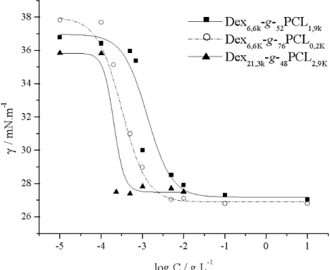
Fig. 1. Semi-logarithmic plot of the concentration dependence of the interfacial tension (γ) for various dextran-g-poly(ε-caprolactone) compositions (Tab. 1)
Amphiphilic character of Dexx-g-yPCLz graft copolymers
In order to get a better insight into the potential of Dexx-g-yPCLz graft copolymers to
behave as surfactants, interfacial tensions were measured by using the pendant drop method applied to drops of water dipped into toluene copolymer solutions of various concentrations. The semi-logarithmic plot of the interfacial tension (γ in mN/m) vs. concentration (in g/L) was typically sigmoidal whatever the studied copolymer compo-sition (Fig. 1). Critical concentrations (Ccrit) were determined from the intersection
between the tangents drawn from higher concentration portions of the sigmoidal plots [32]. For the different copolymers studied, Ccrit is ranging from 0.3 to 2.3·10-3 g/L
(Tab. 2). Critical concentration tends to increase with the content in hydrophobic component (FwPCL) at least for a given dextran molar mass (entries 1 and 2, Tab. 2).
It also comes out that increasing dextran molar mass, while keeping PCL weight fraction and average number of polyester grafts (y) almost constant, contributes to lower Ccrit (entries 1 and 3, Tab. 2). Finally, higher molecular area (A) and
effective-ness (Πcrit = γcrit - γ0 [33]) are reached for Dex6,6-g-76PCL0,2, i.e., for the copolymer
with the highest average number of PCL grafts per 100 glucosidic units, which is in good agreement with observations from other similar amphiphilic systems [34,35]. Interestingly enough, a similar behaviour has been recently reported for dextran-g-polylactide copolymers (Dex-g-PLA) [37]. It was observed that these copolymers showed noticeable surfactant properties whatever their polyester weight fraction. Moreover, depending on their solubility, the self-organization of Dex-g-PLA co-polymer in water or toluene was evaluated using fluorescence spectroscopy. It was proved that these amphiphilic grafted copolymers were able to produce either hydro-phobic or hydrated microdomains in water or toluene solutions, respectively. As a conclusion, these studies confirm that the PCL-grafted dextran copolymers can behave as surfactants at the interface between two immiscible solvents such as toluene and water and prompt us to investigate their ability to form nanoparticles by nanoprecipitation.
Tab. 2. Effect of PCL-grafted dextran copolymer composition on the main interfacial parameters
Entry Dexx-g-yPCLz a FwPCL b Ccrit c
/(mg·L-1) γcrit d /(mN·m-1) A e /nm2 Πcrit f /(mN·m-1) 1 Dex6,6-g-52PCL1,9 0.87 2.3 27.3 0.5 9.3 2 Dex6,6-g-76PCL0,2 0.52 1.3 27.1 0.9 11.0 3 Dex21,3-g-48PCL2,9 0.89 0.3 27.6 0.5 8.3 a Graft copolymer acronyms, where x and z denote the molar masses of dextran and
PCL grafts in kg/mol, respectively, and y denotes the average number of PCL grafts per 100 glucosidic units.
b PCL weight fraction in the copolymer as determined by gravimetry and confirmed by 1H NMR spectroscopy [27].
c Critical concentration determined from the intersection between the tangents drawn
from higher concentration portions of the sigmoidal plots of γ vs. logC/(g·L-1).
d Interfacial tension at the critical concentration.
e Molecular area calculated from the following equations: C T R .303 Γ dlog 2 γ d =− ⋅ ⋅ ⋅ ex⋅ ; N A ⋅ = ex Γ 1
where R is the gas constant, T the absolute temperature, Γex is the surface excess of
surfactant molecules (in mol/area), C is the surfactant concentration, and N is Avo-gadro’s number.
f Π
crit = γcrit - γ0, where γ0 and γcrit stand for the interfacial tensions of the water drop
immersed in pure toluene and a copolymer solution in toluene at critical concen-tration, respectively.
Nanoparticle formation
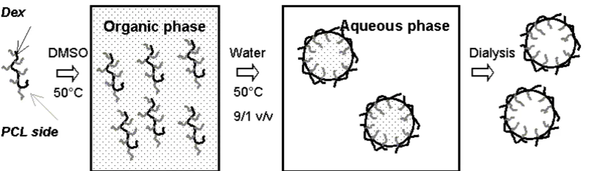
As sketched in Fig. 2, Dexx-g-yPCLz copolymers were first dissolved in DMSO at
50°C until complete solubilization. The organic solution was then added dropwise and with vigorous magnetic stirring into a large volume excess of water previously thermostated at 50°C (see Exptl. part). The suspension rapidly turned opalescent due to colloidal nanoparticle formation. From the aforementioned surface tension properties it can be assumed that the water-soluble dextran backbone is preferably orientated towards the aqueous phase, whereas the hydrophobic PCL side chains concentrate in the dispersed organic phase. To separate DMSO from the medium, the suspension was dialyzed against distilled water using a 3500 g/mol molecular cut-off dialysis tubular membrane. Finally, the resulting suspension was concentrated to a final volume of 9 mL. The influence of the initial DMSO-to-H2O volume ratio (R)
and of the initial copolymer concentration in DMSO on the size distribution of nano-particles was investigated by dynamic light scattering at 25°C. It is worth mentioning that no accurate determination of solution viscosity and differential refractive index was performed so that only apparent mean diameters of nanoparticles can be approached.
Fig. 2. Illustration of the nanoprecipitation technique applied to dextran-g-poly(ε-caprolactone) copolymers 0 100 200 300 400 0.00 0.02 0.04 0.06 0.08 0.10 0.12 R (v/v) d ( nm )
Fig. 3. Influence of the initial DMSO-to-H2O volume ratio (R) on the nanoparticles’
apparent mean size for an initial copolymer concentration in DMSO of 1 mg/mL (Dex6,6-g-52PCL1,9, ; Dex6,6-g-76PCL0,2, ∆) 0 100 200 300 400 500 0 20 40 60 80 100 Intens ity d (nm) First batch Second batch
Fig. 4. Size distribution of nanoparticles as determined by dynamic light scattering in water at 25°C for two individually prepared batches of Dex6,6-g-76PCL0,2 (initial
Effect of the DMSO/H2O volume ratio (R)
Solutions of selected dextran-g-PCL graft copolymers were prepared in DMSO (1 mg/mL) and defined volumes were added into 9 mL of Millipore water at 50°C with vigorous magnetic stirring. As shown in Fig. 3, increasing R from 0.03 to 0.11 contri-buted to increase the apparent mean diameter of the so-generated nanoparticles. Except the specific case of the nanoparticles prepared from Dex6,6-g-52PCL1,9 at high
DMSO-to-water volume ratio (R = 0.11) that display a bimodal size distribution, all other studied compositions yielded monodisperse nanoparticles. In other words, these experiments demonstrate that the smaller the DMSO relative volume compared to water the better defined the nanoparticles’ distribution. From Fig. 4, the reproduci-bility of the nanoprecipitation technique can be appreciated since two individually prepared batches of Dex6,6-g-76PCL0,2 copolymer-based nanoparticles exhibit similar
mean diameter and size distribution for R = 0.11.
0 100 200 300 400 500 0 0.5 1 1.5 2 2 Conc. (mg.mL-1) d ( nm ) .5
Fig. 5. Influence of Dex6,6-g-76PCL0,2 copolymer initial concentration in DMSO on the
apparent mean diameter of nanoparticles as determined by dynamic light scattering (DMSO-to-H2O volume ratio R = 0.11)
Effect of initial copolymer concentration in DMSO and stability of colloidal suspension
This series of experiments was exclusively realized on Dex6,6-g-76PCL0,2 copolymer
as it displayed a monomodal size distribution whatever the R value studied, at least in our prevailing experimental conditions. Practically, 1 mL of Dex6,6-g-76PCL0,2
copolymer solutions in DMSO with concentrations ranging from 0.5 to 2 mg/mL was added to 9 mL of pure water. It comes out that no significant effect of copolymer concentration on the mean diameter can be detected (Fig. 5). Furthermore, the time dependence of the mean size of the recovered nanoparticles has been investigated as a tool to evaluate their stability. Remarkably enough, nanoparticles are stable at least within 3 weeks when stored at room temperature. Indeed, an apparent mean diameter of 201±19 nm is measured after three weeks for Dex6,6-g-76PCL0,2 based
nanoparticles (R = 0.11), which is close to the starting value of 188±16 nm (Fig. 6). It can be concluded that dextran backbones likely located at the surface of nano-particles are able to efficiently stabilize PCL-enriched nanoparticle suspensions in
pure water. Interestingly, these observations are in agreement with recently reported data by Gref et al. [36] on other PCL (low content) graft copolymer based nano-particles prepared by the ‘interfacial migration/solvent evaporation’ method. There is no doubt that such behaviour paves the way to the formulation of drug nanocarriers for controlled release and modulated biodistribution.
0 100 200 300 400 500 0 20 40 60 80 100 Int ens it y d (nm) t0 t0 + 3 weeks
Fig. 6. Apparent size distribution of nanoparticles obtained from Dex6,6-g-76PCL0,2 as
determined by light scattering (initial copolymer concentration in DMSO 1 mg/mL, and DMSO-to-H2O volume ratio R = 0.11)
Conclusion
Well-defined PCL-grafted dextran amphiphilic copolymers have shown high effective-ness in reducing water/toluene interfacial tension. Extending the length of both dextran backbone and PCL grafts allows reducing the critical concentration for a given weight composition. In a next step, these dextran-g-PCL copolymers have been used to readily prepare ‘surfactant-free’ nanoparticles by the nanoprecipitation technique in water. Whatever the composition of PCL-enriched graft copolymers, adequate experimental conditions in terms of copolymer concentration in DMSO and DMSO-to-H2O volume ratio can be found allowing formation of nanoparticles with an
apparent mean diameter close to 200 nm and narrow size distribution. As a rule, the higher the PCL content, the lower should be the relative DMSO volume compared to pure water while the initial concentration of the graft copolymer in DMSO showed no significant effect, at least for concentrations ranging from 0.2 to 0.5 mg/mL. From the time dependence of the nanoparticles’ mean diameter as recorded over a period of three weeks, it can be concluded that stable nanoparticle suspensions have been obtained. Such behaviour is consistent with the formation of a core-shell structure consisting of a hydrophobic polyester core surrounded by a hydrophilic dextran shell, which prevents rapid agglomeration.
Acknowledgement: This work was partially supported by both the Région Wallonne and Fonds Social Européen in the frame of Objectif 1-Hainaut: Materia Nova program. I.Y. is grateful to F.R.I.A. for her Ph.D. grant. This work was also partially supported by a ‘Tournesol’ Program 2000 - 2001 (Program N° 00827WH). LPCM thanks the Belgian Federal Government Office of Science Policy (SSTC-PAI 5/3) for general support.
[1] Couvreur, P.; Roblot-Treupel, L.; Poupon, M. F.; Brasseur, F.; Puiseux, F.; Adv.
Drug Delivery Rev. 1990, 5, 209.
[2] Alléman, E.; Gurny, R.; Doelker, E.; Eur. J. Pharm. Biopharm. 1993, 39, 173. [3] Barichello, J. M.; Morishita, M.; Takayama, K.; Nagai, T.; Drug Dev. Ind. Pharm.
1999, 25, 471.
[4] Chorny, M.; Fishbein, I.; Danenberg, H. D.; Golomb, G.; J. Controlled Release
2002, 83, 389.
[5] Govender, T.; Stolnik, S.; Garnett, M. C.; Illum, L.; Davis, S. S.; J. Controlled
Release 1999, 57, 171.
[6] Zambaux, M. F.; Bonneaux, F.; Gref, R.; Maicent, P.; Dellacherie, E.; Alonso, M. J.; Labrude, P.; Vigneron, C.; J. Controlled Release 1998, 50, 31.
[7] Harmia, T.; Speiser, P.; Kreuter, J.; J. Microencapsul. 1986, 3, 3.
[8] Alléman, E.; Leroux, J. C.; Gurny, R.; Doelker, E.; Pharm. Res. 1993, 10, 1732. [9] Alléman, E.; Gurny, R.; Doelker, E.; Int. J. Pharm. 1992, 87, 247.
[10] Molpeceres, J.; Guzman, M.; Aberturas, M. R.; Chacon, M.; Berges, L.; J.
Pharm. Sci. 1996, 85, 206.
[11] Fessi, H.; Puisieux, F.; Devissaguet, J.-P.; Ammoury, N.; Benita, S.; Int. J.
Pharm. 1989, 55, R1.
[12] Guterres, S. S.; Fessi, H.; Barrat, G.; Devissaguet, J.-P.; Puisieux, F.; Int. J.
Pharm. 1995, 113, 57.
[13] Chacon, M.; Berges, L.; Molpeceres, J.; Aberturas, M. R.; Guzman, M.; Int. J.
Pharm. 1996, 141, 81.
[14] Shakesheff, K. M.; Evora, C.; Soriano, I.; Langer, R.; J. Colloid Interface Sci.
1997, 185, 538.
[15] Lee, S. C.; Oh, J. T.; Jang, M. H.; Chung, S. I.; J. Controlled Release 1999, 59, 123.
[16] Sjostrom, B.; Bergenstahl, B.; Kronberg, B.; J. Pharm. Sci. 1993, 82, 585. [17] Sjostrom, B.; Kronberg, B.; Carlfors, J.; J. Pharm. Sci. 1993, 82, 579.
[18] Landry, F. B.; Bazile, D. V.; Spenlehauer, G.; Veillard, M.; Kreuter, J.;
Bio-materials 1996, 17, 715.
[19] Landry, F. B.; Bazile, D. V.; Spenlehauer, G.; Veillard, M.; Kreuter, J.; J.
Controlled Release 1997, 44, 227.
[20] Lavelle, E. C.; Sharif, S.; Thomas, N. W.; Holland, J.; Davis, S. S.; Adv. Drug.
Delivery Rev. 1995, 18, 5.
[21] Govender, T.; Stolnik, S.; Garnett, M.C.; Illum, L.; Davis, S. S.; J. Controlled
[22] Lemarchand, C.; Couvreur, P.; Besnard, M.; Constantini, D.; Gref, R.; Pharm.
Res. 2003, 20, 1284.
[23] Jeong, Y.-I.; Shim, Y.-H.; Choi, C.; Jang, M.-K.; Shin, G. M.; Nah, J.-W.; J. Appl.
Polym. Sci. 2003, 89, 1116.
[24] Jeong, Y.-I.; Cheon, J. B.; Kim, S. H.; Nah, J. W.; Lee, Y. M.; Sung, Y. K.; Akaike, T.; Cho, C. S.; J. Controlled Release 1998, 51, 169.
[25] Lasic, D. D.; Nature 1992, 355, 279.
[26] Kwon, G. S.; Naito, M.; Yokoyama, M.; Okano, T.; Sakurai, Y.; Kataoka, K.;
Pharm. Res. 1995, 12, 192.
[27] Ydens, I.; Rutot, D.; Degée, Ph.; Six, J.-L.; Dellacherie, E.; Dubois, Ph.;
Macro-molecules 2000, 33, 6713.
[28] Nouvel, C.; Ydens, I.; Degée, Ph.; Dubois, Ph.; Dellacherie, E.; Six, J.-L.;
Polymer 2002, 43, 1735.
[29] Arashiro, E. Y.; Demarquette, N. R.; Mater. Res. 1999, 2, 23.
[30] Nouvel, C.; Dubois, Ph.; Dellacherie, E.; Six, J.-L.; J. Polym Sci., Part A: Polym.
Chem. 2004, 42, 2577.
[31] Nouvel, C.; Dubois, Ph.; Dellacherie, E.; Six, J.-L.; Biomacromolecules 2003, 4, 1443.
[32] Coulembier, O.; Degée, Ph.; Guérin, Ph.; Dubois, Ph.; Langmuir 2003, 19, 8661. [33] Rosen, M. J.; J. Colloid Interface Sci. 1976, 56, 320.
[34] Rouzes, C.; Durand, A.; Léonard, M.; Dellacherie, E.; J. Colloid Interface Sci.
2002, 253, 217.
[35] Wollenweber, C.; Makjevski, A. V.; Miller, R.; Daniels, R.; J. Colloid Interface Sci.
2000, 117, 419.
[36] Rodrigues, J. S.; Santos-Magalhães, N. S.; Coelho, L. C. B. B.; Couvreur, P.; Ponchel, G.; Gref, R.; J. Controlled Release 2003, 92, 103.
[37] Nouvel, C.; Frochot, C.; Sadtler, V.; Dubois, P.; Dellacherie, E.; Six, J.-L.;