HAL Id: inserm-00639292
https://www.hal.inserm.fr/inserm-00639292
Submitted on 8 Nov 2011
HAL is a multi-disciplinary open access
archive for the deposit and dissemination of
sci-entific research documents, whether they are
pub-lished or not. The documents may come from
teaching and research institutions in France or
abroad, or from public or private research centers.
L’archive ouverte pluridisciplinaire HAL, est
destinée au dépôt et à la diffusion de documents
scientifiques de niveau recherche, publiés ou non,
émanant des établissements d’enseignement et de
recherche français ou étrangers, des laboratoires
publics ou privés.
myopathy with prominent nuclear internalization and
large areas of myofibrillar disorganization.
J. A. Bevilacqua, Nicole Monnier, Marcus Bitoun, Bruno Eymard, Ana
Ferreiro, Soledad Monges, Fabiana Lubieniecki, Ana Taratuto, Annie
Laquerrière, Kristin Claeys, et al.
To cite this version:
J. A. Bevilacqua, Nicole Monnier, Marcus Bitoun, Bruno Eymard, Ana Ferreiro, et al.. Recessive
RYR1 mutations cause unusual congenital myopathy with prominent nuclear internalization and large
areas of myofibrillar disorganization.. Neuropathology and Applied Neurobiology, Wiley, 2011, 37 (3),
pp.271-84. �10.1111/j.1365-2990.2010.01149.x�. �inserm-00639292�
Recessive RYR1 mutations cause unusual congenital
myopathy with prominent nuclear internalization and
large areas of myofibrillar disorganization
_271..284J. A. Bevilacqua1,2 , N. Monnier3,4 , M. Bitoun5 , B. Eymard6 , A. Ferreiro6,7 , S. Monges8 , F. Lubieniecki9 , A. L. Taratuto10 , A. Laquerrière11 , K. G. Claeys1,6 , I. Marty4 , M. Fardeau1,6 , P. Guicheney12 , J. Lunardi3,4 and N. B. Romero1,5,6 1
Institut de Myologie, Unité de Morphologie Neuromusculaire, Groupe Hospitalier-Universitaire Pitié-Salpêtrière,
5UPMC-INSERM UMR S974, CNRS UMR 7215, Institut de Myologie, GHU Pitié-Salpêtrière,6Groupe
Hospitalier-Universitaire Pitié-Salpêtrière, AP-HP, Centre de référence des maladies neuromusculaires Paris-Est,
7INSERM UMR S787, CHU Pitié-Salpêtrière,12INSERM UMR S956, CHU Pitié-Salpêtrière, Paris,3Laboratoire de
Biochimie et Génétique Moléculaire and Centre de Référence des Maladies Neuro-Musculaires. CHU Grenoble, Grenoble,
4INSERM U836, Grenoble Institut Neurosciences, La Tronche,11Pathology Laboratory, Rouen University Hospital,
Rouen, France,2Departamento de Neurología y Neurocirugía, HCUCH and Instituto de Ciencias Biomédicas, Facultad de
Medicina, Universidad de Chile, Santiago, Chile, and Departments of8Neuropaediatrics and9Pathology, Garrahan
National Paediatric Hospital, and10Department of Neuropathology, FLENI Institute for Neurological Research, Buenos
Aires, Argentina
J. A. Bevilacqua, N. Monnier, M. Bitoun, B. Eymard, A. Ferreiro, S. Monges, F. Lubieniecki, A. L. Taratuto, A. Laquerrière, K. G. Claeys, I. Marty, M. Fardeau, P. Guicheney, J. Lunardi and N. B. Romero (2011)
Neuropathology and Applied Neurobiology 37, 271–284
Recessive RYR1 mutations cause unusual congenital myopathy with prominent nuclear internalization and large areas of myofibrillar disorganization
Aims: To report the clinical, pathological and genetic findings in a group of patients with a previously not described phenotype of congenital myopathy due to recessive mutations in the gene encoding the type 1 muscle ryanodine receptor channel (RYR1). Methods: Seven unrelated patients shared a predominant axial and proximal weakness of varying severity, with onset during the neonatal period, associated with bilateral ptosis and ophthalmoparesis, and unusual muscle biopsy features at light and electron microscopic levels. Results: Muscle biopsy histochemistry revealed a peculiar morphological
pattern characterized by numerous internalized myonu-clei in up to 51% of fibres and large areas of myofibrillar disorganization with undefined borders. Ultrastructurally, such areas frequently occupied the whole myofibre cross section and extended to a moderate number of sarcom-eres in length. Molecular genetic investigations identified recessive mutations in the ryanodine receptor (RYR1) gene in six compound heterozygous patients and one homozygous patient. Nine mutations are novel and four have already been reported either as pathogenic recessive mutations or as changes affecting a residue associated
Correspondence: Norma Beatriz Romero, INSERM UMR S974, Institut de Myologie, Groupe Hospitalier Universitaire Pitié-Salpêtrière, F-75013 Paris, France. Tel:+33 (0) 1 42 16 22 42; Fax: +33 (0) 1 42 16 22 40; E-mail: nb.romero@institut-myologie.org
JAB and NM contributed equally to this work. The authors have reported no conflicts of interest.
This work was supported by the Institut National de la Santé et de la Recherche Médicale (INSERM), the Association Française contre les Myopathies (AFM), the Association Institut de Myologie (AIM), the National Research Agency (ANR) and The Programme of Collaboration ECOS-SECyT (A02S02), France-Argentina and the Genopole d’Evry. Jorge A. Bevilacqua was supported by the Program Alban, The European Union Program of High Level Scholarships for Latin America, scholarship No.E04E028343CL and the Association Institut de Myologie (AIM), France.
with dominant malignant hyperthermia susceptibility. Only two mutations were located in the C-terminal trans-membrane domain whereas the others were distributed throughout the cytoplasmic region of RyR1. Conclusion: Our data enlarge the spectrum of RYR1 mutations and highlight their clinical and morphological heterogeneity.
A congenital myopathy featuring ptosis and external ophthalmoplegia, concomitant with the novel histo-pathological phenotype showing fibres with large, poorly delimited areas of myofibrillar disorganization and inter-nal nuclei, is highly suggestive of an RYR1-related congenital myopathy.
Keywords: congenital myopathy, myofibrillar disorganization, nuclear internalization, recessive mutations, RYR1 gene
Introduction
The RYR1 gene (OMIM 180901) encodes the ryanodine receptor 1, a Ca2+ channel expressed on sarcoplasmic reticulum membranes at the triad of skeletal muscle fibres. RyR1 mediates the release of Ca2+from intracellular pool in response to nerve stimulation and then plays a crucial role in excitation–contraction coupling [1]. Muta-tions of the RYR1 gene cause well-defined forms of con-genital myopathies, that is, central core disease (CCD; OMIM 117000) and malignant hyperthermia susceptibil-ity (MHS; OMIM 145600), an autosomal dominant phar-macogenetic disease. This gene is also implicated in some cases identified as multi-minicore disease (MmD; OMIM 602771). The RYR1 mutations associated with CCD are usually dominant but recessive inheritance has also been reported, whereas cases identified as MmD are exclusively linked to recessive mutations [2–7] and recently in patients with fibre type disproportion as their only patho-logical feature. [8] Classically in the RYR1 sequence, three hot-spots are considered, two in the large hydrophilic domain of RyR1 and one in the C-terminal hydrophobic domain. Most of the heterozygous dominant CCD muta-tions are mapped to the C-terminal domain, whereas the recessive CCD and MmD mutations are more extensively distributed along the RYR1 sequence. Additionally, a het-erozygous de novo RYR1 mutation in the C-terminal region of the protein has been found in a 16-year-old female patient initially diagnosed with centronuclear myopathy (CNM) with ‘core-like’ lesions and central nuclei in up to 50% of fibres in the muscle biopsy [9], and a heterozygous
de novo RYR1 mutation in the N-terminal domain has
been found in a patient presented with King-Denborough syndrome and MHS [10].
In RYR1-related congenital myopathies, the histological phenotype varies widely. It comprises central and eccen-tric cores, unique and multiple, structured and unstruc-tured, well-delimited cores spanning the entire fibre
length or poorly defined cores that spread only a few sar-comeres, and occasionally a variable degree of sarcomeric disorganization [2,11–13]. These structural abnormali-ties are sometimes associated with an increased number of internal myonuclei (up to 30% of the fibres) and variable degrees of fibrous and adipose tissue replacement [6,14,15]. There also exist biopsies without major alter-ations showing only a type I fibre predominance or unifor-mity [16]. Moreover, a histopathological continuum has been suggested linking the diverse RYR1-related core pathies [17–20]. On the other hand, centronuclear myo-pathies (CNM; OMIM 310400, 160150 and 255200), comprise X-linked recessive, autosomal dominant and autosomal recessive forms, associated, respectively, with myotubularin 1 (MTM1), dynamin 2 (DNM2) and amph-iphysin 2 (BIN1) genes [21–23]. The histopathological presentation of these distinct forms of CNM has been well established [24]; so far, neither cores nor minicores have been described in such genetically determined CNM forms. Here we report clinical, histological and molecular characterization of seven patients initially diagnosed with CNM due to the significantly high number of fibres with internalized nuclei (up to 51% of the fibres). However, the key histopathological feature that led us to screen RYR1 gene for mutations was the invariable presence of large areas of sarcomeric disorganization in the muscle fibres, despite the number and location of internalized nuclei. Thus, RYR1 recessive mutations were found in every patient of the series demonstrating that this peculiar disorder should be classified as a form of RYR1-related congenital myopathy.
Methods
Patients
We retrospectively reviewed the clinical and histological data of patients with an original diagnosis of CNM
without DNM2 mutations. We identified seven unrelated patients (five women and two men) (Table 1) who shared the same morphological findings in the muscle biopsy (see
Results). This study was authorized by the ethical
commit-tee of Pitié-Salpêtrière Hospital (CCPPRB) and the Direc-tion de Recherché Clinique of the Assistance Publique, Hôspitaux de Paris.
Histopathological studies
Skeletal muscle biopsies were obtained from all patients. Age of patient and the biopsied muscles were indicated in Table 1. Histological, histoenzymological and electron microscopic analyses were performed as previously described [25]. Ultrastructural studies were performed in all patients except patient 2. The number of fibres with nuclear centralization (that is, myonuclei in the geometric centre of the fibre) and with nuclear internalization (that is, myonuclei underneath the sarcolemma anywhere within the cytoplasm) were counted in a minimum of 200 adjacent muscle fibres. In each biopsy, the diameter of type 1 and type 2 fibres stained with myosin adenosine triphosphatase (ATPase) 9.4 was measured manually on digital pictures in at least 120 fibres using ImageJ 1.40g® (NIH, Washington, USA).
Molecular genetics studies
Informed consent for genetic analysis was obtained from each patient and their families. RYR1 mutation screening was performed on cDNA obtained after reverse transcrip-tion of total RNA extracted from muscle specimens as previously described [2]. The cDNA was amplified in over-lapping fragments. Sequencing reactions were analysed on an ABI 3130 DNA Analyzer (Life Technologies, Foster City, CA, USA). The presence of the mutations identified in transcripts was confirmed in genomic DNA by direct sequencing of the corresponding exon and intron–exon junctions. None of the novel variants was found in 200 chromosomes from the general population. To evaluate the consequences of the c.8692+131G>A mutation at the transcription level, cDNA fragments encompassing exons 56 and 57 were amplified and cloned using the TOPO TA Cloning®Kit (Invitrogen, Carlsbad, CA,USA). After trans-formation into One Shot Competent DH5a™-T1R cells (Invitrogen), colonies containing the recombinant plas-mids were identified by PCR using RYR1 specific primers, and the cDNA inserts were sequenced.
Protein analysis
To analyse the expression of RyR1, thin slices of frozen muscle biopsies from patients 1 and 6 were homogenized in Hepes 20 mM (pH 7.4), sucrose 200 mM, CaCl2 0.4 mM, Complete Protease Inhibitor® cocktail (Roche, Meylan, France). The amount of RyR1 present in each muscle sample was determined by quantitative Western blot analysis using antibodies directed against RyR1 as described previously [26]. Signals were detected using a chemiluminescent horseradish peroxidase (HRP) sub-strate and quantified using a ChemiDoc XRS apparatus (Biorad, Hercules, CA, USA) and the Quantity 1 software (Biorad).
Results
Patients
The parents of the seven patients were clinically unaf-fected. All patients except patient 6 were born from non-consanguineous families. Patient 1 was the second daughter of a family with two affected and two non-affected children, and her eldest non-affected sister died at 5 months of age due to severe respiratory impairment and weakness; all the other patients were sporadic cases. Pre-natal symptoms were noted only in patient 2 with reduced foetal movements. At birth, the seven patients showed generalized hypotonia, poor spontaneous movements and amyotrophy, together with weak suction and swallowing difficulties. Motor development was delayed in all patients. Poor head control was noted in patients 1 and 2, who required support to sit or walk. Since early childhood, patients showed difficulties in rising up from the floor, climbing stairs and running. Patients progressively improved their motor capabilities and have acquired inde-pendent ambulation with the exception of patient 1. Significant facial involvement (hypomimia, open mouth, facial diplegia and elongated facies) was observed particu-larly in patients 1 and 2, and at a moderate level in the other patients. All patients showed some degrees of ocular involvement consisting of either ptosis or ophthalmopare-sis with limited upward gaze or incomplete eyelid closure. Serum creatine kinase levels were normal or slightly increased. A computed tomography (CT) scan performed to patient 3 showed a discrete symmetric involvement of deltoids and deep muscles of the pelvic girdle, thigh and leg. In patient 4 a CT scan performed at 34 years old
T able 1 . Summar y o f clinical a nd m u scle biopsy d a ta P atient gender , (cur rent age). Origin Onset symptoms Ev olution and cur rent symptom. W alton scor es Biopsied m usc le and ag e at biopsy My ofibrillar/ sar comeric disor ganization Pur ple dusty fibr es % N C % NI %N C + %N I (% MI) Mean fibr e diameter (SD) [R ange] in m m % fibr es T ype I 1. F emale , (15 y ear s). Zaire Neona tal h ypotonia and respir a tor y distress DMM, autonomous g ait nev er achiev ed. Dysmor phism. Ophthalmopareis and ptosis. F acial diplegia, open mouth. Head suppor t. Axial and pro ximal w eakness. OTR a bsent. Hip , knee and ankle contr actures. CPK: nor mal a t bir th; 241 U/l; EMG: m y o g enic . WS: 8. Sev ere . Deltoid (4 months) 0/ ++ 8.7 9.9 18.7 (3.6) 20.1 (⫾ 10.3) [4.7–43.6] 60.8 P a ra v er t (12 y ear s) +++ +++ 1.5 44.9 46.3 (21) 39.7 (⫾ 53.6) [8.5–15.3] 100 Quadriceps (14 y ear s) +++ +++ 4.4 47.0 51.2 (22) 37.8 (⫾ 12.3) [10–64.2] 100 2. F emale , (24 y ear s). T urk ey/F rance (Figure 2 e– h ) R educed foetal mo v ements. Global neona tal h ypotonia DMM. Dysmor phism. Global w eakness, Spor adic w heelchair bound. Ptosis, upper sight limita tion. F acial paresis. Neck, mastica tor s, elbo w and ankle contr actures. RRS. CPK: 204 U/l. WS: 7. Sev ere . Deltoid (21 months) ++ 2.5 1.0 3.4 (0.5) 25.5 (⫾ 7.7) [8.9–60.5] 71.4 Deltoid (12 y ear s) +++ +++ 7.2 23.7 30.9 (9.6) 33.8 (⫾ 13.4) [11.6–76.6] 96.1 3. F emale , (24 y ear s). F rance Suction, sw allo wing and feeding difficulties a t bir th DMM. Independent g ait a t 2 y ear s. Global w eakness. Ey elids w eakness. Nasal v oice . Masseter s contr acture . Cur rentl y sta ble , nor mal dail y lif e acti vities. CPK: nor mal; EMG: m y o g enic . WS: 2. Mild. Deltoid (16 y ear s) ++ 3.3 5.5 8.8 (0.0) 38.8 (⫾ 11.5) [18.4–75] 89.0 4. F emale , (39 y ear s). F rance (Figure 2 a – d ) Neona tal h ypotonia, poor head control DMM. Independent g ait a t 18 months. Global w eakness. Ophthalmoparesis. R etro gna tism, talipes v a rus, thor acic and dor so-lumbar scoliosis. RRS . OTR a bsent. CPK: 26 U/l. WS: 3. Moder a te . Deltoid (37 y ear s) +++ +++ 6.7 29.0 35.7 (10.4) 30.2 (⫾ 9.4) [10.7–63.8] 100 5. Male , (28 y ear s). F rance (Figure 2 i– l) P erina tal/neona tal distress with respir a tor y insufficienc y DMM. Independent g ait a t 18 months. Dysmor phism. Ptosis, ophthalmoparesis and str a bism us. Global m uscle w eakness. Bila ter al Achillean retr action surg er y. WS : 6 . Sev ere Deltoid (21 y ear s) +++ + + 4.8 29.5 34.3 (21.4) 101.8 (⫾ 31.5) [31.3–194.9] 81.6 6. Male (32 y ear s). Cong o Neona tal h ypotonia DMM. Independent g ait a t 2 y ear s. F requent falls, troubles fro climbing stair s and running . Dysmor phism, ophthalmoparesis, incomplete ey elid closing . Global w eakness. CPK: 23 U/l. WS: 4. Moder a te . Deltoid (26 y ear s) ++ + 3.3 9.0 12.3 (2.4) 36.8 (⫾ 13.4) [13.3–76] 100 7. F emale (6 y ear s). Arg entina/Boli via Hypotonia. Suction and sw allo wing difficulties a t bir th DMM. Independent g ait a t 15 months. F requent falls. F acial h ypomimia, mild bila ter al ptosis. Axial and limbs w eakness. Distal h yperlaxity . CPK: 64 U/l; EMG: m y o g enic . WS: 3. Moder a te . Deltoid (5 y ear s) + 0 1.3 1.3 2.7 (0.0) 29.1 (⫾ 13.4) [7.7–70.7] 95.7 Note the increase in all pa tholo gical fe a tures through sequential biopsies in pa tients 1 and 2. % NI, percenta g e of fibres with n uclear inter naliza tion; % NC, percenta g e of fibres with n uclear centr aliza tion; % MI, percenta g e of fibres with m ultipl e inter naliz ed n uclei; DMM, dela y ed motor milestones; RRS ,restricti v e respir a tor y syndrome; OTR, osteotendinous refle x es; CPK, crea tine phosphor kinase; EMG, electrom y o g ra p h y ; W S , W alton score . © 2011 The Authors
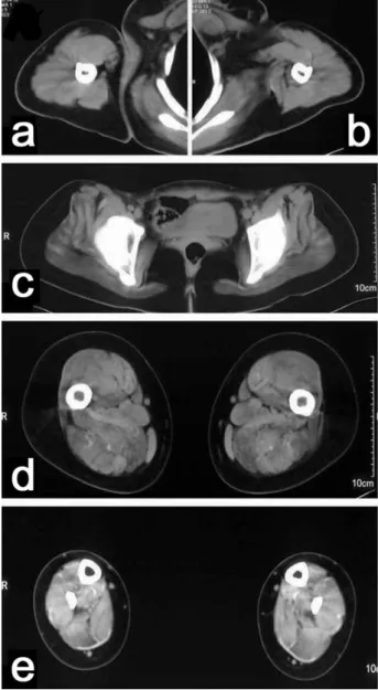
showed a diffuse hypodensity, mainly in the tight and hamstring muscles (Figure 1). Respiratory function was severely affected in patients 1 and 2 early in life but improved slightly; their vital capacities in adolescence or
adulthood were, respectively, 35% and 28% of the theo-retical value (restrictive respiratory syndrome), requiring non-invasive respiratory support. Vital capacities in patients 4 and 6 were 50% and 65% of the theoretical value. Cardiac assessment was normal in all patients.
Morphological studies
Histoenzymological analyses have demonstrated a con-spicuous and reliable morphological pattern on trans-verse muscle cryostat sections consisting of: (i) Large and weakly defined areas devoid of ATPase and oxidative activities observed in some fibres, sometimes covering the majority of the fibre diameter (Figures 2b,f,j and 3g). Such areas were identified as regions of myofibrillar and sarcomeric disorganization, either showing an absence or increased oxidative reactivity (Figures 2c,g,k and 3f). (ii) Several fibres displayed a peculiar ‘purple dusty’ appearance with Gomori trichrome staining, due to a precipitate of numerous small fuchsinophilic particles spreading partially or completely through the fibre cross section (Figures 2d,h,l and 3d,h). These extensive areas of fuzzy reddish aggregates were also evident on haema-toxylin and eosin stained serial sections (Figures 2a,e,i and 3e), corresponding with the zones of abnormal staining on oxidative reactions (Figures 2c,g,k and 3f) and they also lacked ATPase activity (Figures 2b,f,j and 3g). (iii) Type I predominance or type I fibre uniformity and increased variability in fibre size; and (iv) Nuclear internalization and centralization in both fibre types, including frequent multiple internalized nuclei. In addi-tion, a discrete increase of endomysial connective tissue was often observed.
Noticeably, the muscle biopsies performed at the ages of 4 months for patient 1 and 21 months for patient 2, essentially showed type I fibre predominance, increased endomysial connective tissue, significant variation in type I or II fibre size and the presence of some small fibres with central nuclei resembling myotubes. No cores were observed. Thereafter, the muscle biopsies performed at the ages of 12 and 14 years for patient 1 and 12 years for patient 2 showed the peculiar morphological pattern observed in all patients. Nuclear internalization increased with age (Table 1; Figure 3).
Ultrastructural findings
In patients 1 and 3 to 7, ultrastructural analysis of muscle biopsies in longitudinal sections demonstrated large areas Figure 1. Computed tomography limb imaging of patient 4. Upper
and lower limb computed tomography scanner from patient 4 (a to
e) at the age of 34. Axial sections were performed at middle level of the arms (a, b), pelvic girdle (c), upper tight (d) and legs (e). In the arms and pelvic girdle, diffuse atrophy of the muscles is observed without any particular pattern. In the tights (d), fatty infiltration is seen in adductor magnus, semimembranosus, semitendinosus and biceps femoris, with a relative sparing of gracilis, sartorious and anterolateral compartment muscles. In the legs (e), hypodensity of the imaging is prominent in the internal gastrocnemius of the posterior compartment, with a relative normal signal in the other muscle.
of sarcomeric disorganization (Figure 4d). Such areas were present in one or more regions within a fibre, were variable in width and length, frequently covered the entire fibre diameter in cross section (Figures 4a,b) and extended from 2 to 30 sarcomeres in longitudinal sections (Figures 4b,f). Altered fibres often showed one or several misplaced nuclei that were occasionally found at the border of areas of myofibrillar disorganization (Figures 4b,d). Within such disorganized areas, accumu-lation of Z-band proteins, Z-band streaming, enlarged Z-bands and myofibrillar compaction were the most frequent alterations (Figures 4c,e). T-triads-repetitions, honeycomb profiles (corresponding to T-tubules prolifera-tions) and occasional minicore-like lesions (Figure 4f) were also observed amongst other non-specific alter-ations. Mitochondria were present or not in the disorga-nized areas.
Immunohistochemical findings
In order to further study the composition of the disorga-nized intracellular areas, biopsies of patients 2, 3 and 5 were labelled with antibodies to the intermediate filament proteins desmin, aB-crystalline and myotilin. The three markers intensively labelled the disorganized areas, but in serial sections reacting fibres were either labelled with one, two or three of the antibodies used, suggesting a hetero-geneous composition of the disorganized zones (Figure 5).
RYR1 screening
Patient 1 and her deceased sister were c.[10348-6C>G; 14524G>A] + c.[8342_8343delTA] compound heterozy-gous carriers (Table 2). The c.8342_8343delTA frame-shift deletion transmitted by the clinically unaffected Figure 2. Light microscopy of muscle sections. Transverse serial sections of muscle biopsies from patient 4 (a to d), Patient 2 (e to h) and
patient 5 (i to l) stained with haematoxylin and eosin (a, e, i), ATPase 9.4 (b, f, j), nicotinamide adenosine dinucleotide–tetrazolium reductase (NADH-TR) (c, g, k) and Gomori trichrome (d, h, l). Note with haematoxylin and eosin staining the great variability in fibre size; misplaced nuclei and multiple internalized nuclei are observed in many fibres along with increase in endomysial tissue. NADH-TR staining shows unevenness in the sarcoplasmic and mitochondrial distribution. Type I fibre predominance or uniformity is observed in all cases and some fibres show poorly delimited areas devoid of ATPase 9.4 reaction, which correspond to areas presenting sarcomeric disorganization with increased or absence of oxidative reaction. The same fibres contain the purple dusty areas with Gomori trichrome staining described in the text. Note that the two top rows show pictures at¥ 400 magnification, while the bottom row pictures are at ¥ 200 magnification. (See Table 1 for details on the diameter of fibres in each case.) Scale bars: in h= 50 mm, for a to h; and in l = 100 mm, for i to l.
mother introduced a premature stop codon (p.Ile2781ArgfsX49). The two other variants were inher-ited from the clinically unaffected father. The c.10348-6C>G change resulted in a loss of splicing of intron 68 and the introduction of a premature stop codon (p.His3449ins33fsX54). Both unspliced and spliced transcripts were present, thus indicating an incomplete penetrance of this intronic variation. The c.14524G>A change in exon 101 resulted in a p.Val4842Met substitu-tion that mapped to the M8 trans-membrane fragment of the Ca2+pore domain [27]. RyR1 expression analysis did not show truncated proteins but instead a major decrease of the mature protein, indicating the residual presence of a low amount (15⫾ 8%) of mutated Met4842 protein in the proband’s muscle (Figure 6).
Patient 2 was p.[Thr4709Met]+ p.[Glu4181Lys] com-pound heterozygous. The paternal p.Thr4709Met substi-tution, resulting from a c.14126C>T change in exon 96 that affected a conserved threonyl residue located in the Ca2+ pore domain of the protein, has been previously reported in a case of recessive core myopathy [28]. The maternal p.Glu4181Lys novel substitution that resulted from a c.12541G>A transition in exon 90, affected a highly conserved glutamyl residue located in a cytoplas-mic domain of unknown function (Table 2).
Patient 3 was compound heterozygous for the novel p.[Glu4911Lys] and p.[Arg2336Cys] variants. The pater-nal p.Glu4911Lys (c.14731G>A, exon 102) variant affected a highly conserved glutamyl residue that mapped to the M10 trans-membrane fragment of the Ca2+pore domain [27]. The maternal p.Arg2336Cys (c.7006C>T, exon 43) variant also substituted a very well-conserved arginyl residue located in the MH2 domain of the protein, usually associated with malignant hyperthermia domi-nant mutations. However, no anaesthetic history has been reported in the patient or relatives harbouring the p.Arg2336Cys variant (Table 2).
Patient 4 was p.[Pro3202Leu]+ p.[Gly3521Cys] com-pound heterozygous. Both variants are novel and substi-tuted highly conserved residues among species and RYR isoforms. The paternal p.Pro3202Leu (c.9605C>T, exon 65) variant affected a prolyl residue located in a central region of the protein of unknown function. The maternal p.Gly3521Cys (c.10561G>T) variant substituted a glycyl residue located within exon 71 adjacent to the alterna-tively spliced region I (ASI), possibly involved in interdo-main interaction (Table 2) [29].
Patient 5 was p.[Pro3138Leu]+ p.[Arg3772Trp] compound heterozygous. The paternal p.Pro3138Leu (c.9413C>T) variant affected a highly conserved prolyl Figure 3. Evolution of histopathological findings in patient 2. Transverse serial sections of muscle biopsies from patient 2 at 21 months
(a to d), and at 12 years of age (e to h) stained with haematoxylin and eosin (a, e), nicotinamide adenosine dinucleotide–tetrazolium reductase (NADH-TR) (b, f), ATPase 9.4 (c, g) and Gomori trichrome (d, h). Note with haematoxylin and eosin increase in fibre size, number of internalized nuclei and hyaline cytoplasmic protein aggregates. With NADH-TR staining the unevenness in the sarcoplasmic and mitochondrial distribution is recognized in the biopsy at 12 years but was not evident in the firs biopsy. With ATPase 9.4 the type I fibre predominance seen in the first biopsy changes to a type I uniformity and the number of fibres with areas devoid of ATPase 9.4 reaction increase in number and the areas are more conspicuous. Similar increase in the features described is seen with Gomori trichrome. (See also Table 1 for details on the diameter of fibres in each case.) Scale bar= 50 mm.
residue that mapped to exon 63. This variant has not been reported previously. The maternal p.Arg3772Trp (c.11314C>T, exon 79) variant has been recently reported in an MHS patient [30]. The mutation substituted a highly conserved argininyl residue into a nonconservative tryp-tophan located in a cytoplasmic domain of unknown function (Table 2).
Analysis of patient 6’s cDNA revealed the presence of two abnormal transcripts characterized by insertions of 132 bp and 32 bp between exons 56 and 57, and the presence of a normally spliced transcript. Genomic sequencing of intron 56 identified a homozygous c.8692+131G>A change, which revealed a cryptic donor splice site in competition with the physiological splice site (Table 2). Use of this cryptic splice site led mostly to an insertion of 132 bp that introduced 44 amino acids and a premature stop codon between exons 56 and 57 (p.Gly2898GlyfsX36). In addition, the presence of another putative AG dinucleotide splice acceptor site upstream to the cryptic donor splice site, led to an additional alternative frameshift insertion of 32 nucleotides, also leading to a premature stop codon (p.Gly2898AspfsX54) (Figure 7a). However, no truncated proteins were detected on Western blot analysis, suggesting either instability of the cryptic transcripts as a result of a nonsense-mediated mRNA decay process or an early degradation of the truncated
proteins as a result of an unfolded protein response. The residual physiological splicing allowed the production of a low amount of wild-type RyR1 (22⫾ 12%) in the muscle of the patient (Figure 6).
Patient 7 was p.[Pro3202Leu]+ p.[Arg4179His] compound heterozygous. The maternal p.Pro3202Leu (c.9605C>T, exon 65) variant was recurrent in this study (patient 4). The paternal p.Arg4179His (c.12536G>A, exon 90) variant affected a highly conserved arginyl residue that mapped to a cytoplasmic domain of the protein close to the p.Glu4181Lys variant identified in patient 2.
Discussion
We have identified a cohort of seven patients with con-genital myopathy and a peculiar morphological pattern in muscle biopsies associated with recessive mutations in the gene encoding the skeletal muscle ryanodine receptor (RYR1). All the patients showed early onset of the disease, ophthalmoparesis of variable severity and presence of early disabling contractures, especially in the masticators. Rigid spine syndrome was also present in two patients. Otherwise clinical presentation was similar to most con-genital myopathies, showing hypotonia of variable sever-ity, delay in the acquisition of developmental motor Figure 4. Ultrastructural findings in muscle fibres from patients. Electron microphotographs of myofibres from patient 4 (a), patient 6 (b),
patient 5 (c, d, e) and patient 3 (f) show different ultrastructural alterations. Transverse and longitudinal muscle sections are shown in left and right panels, respectively. The lesions cover most of the cross section of affected fibres and are adjacent to internalized nuclei (a, b). The areas of myofibrillar disorganization contain Z band proteins aggregates (c, e). The box framed in c is depicted at higher magnification in e. Two misplaced nuclei are near the border of an area of myofibrillar disorganization, which continues from normally appearing sarcomeres (d). Triad repetitions are seen within a small zone of Z disc streaming (f). Scale bars in a and e= 2 mm, b and c = 10 mm, d = 4 mm and
f= 5 mm.
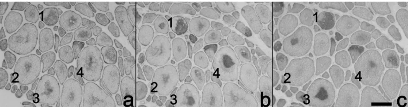
Figure 5. Muscle biopsy immunostaining. Serial sections immunostained with antibodies to desmin (a),aB-crystalline (b) and myotilin (c) showed positive staining for all the markers used. In some cells labelling was similar with the three markers (1); in other, reaction was positive for desmin andaB-crystalline but not myotilin (2), and in a third set of fibres labelling was somehow stronger for myotilin (3), whilst in a fourth group of fibres labelling was stronger foraB-crystalline (4). Scale bar in c = 30 mm.
milestones, axial and proximal limb weakness and restric-tive respiratory syndrome. Cardiac and cognirestric-tive func-tions were invariably spared.
Our data enlarges the histological phenotype associated with RYR1 mutations. Indeed, the areas of sarcomeric/ myofibrillar disorganization are distinguishable from typical cores. On oxidative stains, these areas are large, diffuse and poorly delimited. Ultrastructurally, they are broader than cores in transverse sections, as they fre-quently cover extensive cross-sectional areas of the fibre, often reaching the sarcolemma. They are also shorter than cores, as in longitudinal sections they extend along a relatively small number of sarcomeres. In contrast with cores the presence of mitochondria within the lesions accounts for the excessive oxidative staining in some fibres. On the other hand, ‘purple dusty areas’ correspond-ing to foci of Z line rearrangements are not usually seen in muscle biopsies of patients with classical core myopathies. Interestingly, the first descriptions of congenital myopa-thies with ‘target-like fibres’ as described by Schotland in the 1960s, reported morphological findings close to those described in our patients; retrospectively, these similarities might provide the molecular cause for these earlier observations [31,32].
Moreover, in our series of patients, nuclear misplace-ment affected up to 51% of the fibres. Remarkably, fibres with centralized nuclei ranged from 1 to 9%, while nuclear internalizations were present in up to 47% of the fibre population, of which up to 22% had multiple
T able 2 . Summar y o f molecular g enetic findings P atient Localization Nuc leotide ch anges (cDN A numbering) * Origin of alleles Amino acid changes Expected consequences R ef er ences 1 Intron 6 8 c.10348-6C > G F a ther p .His3449ins33fsX54 Tr unca ted protein Monnier et al. 2008 [19] Ex on 101 c. 14524G > A F a ther p .V al4842Met M issense m u ta tion Monnier et al. 2008 [19] Ex on 53 c. 8342_8343delT A M other p .Ile2781ArgfsX49 Tr unca ted protein This stud y 2 E x o n 9 6 c.14126C > T F a ther p .Thr4709Met M issense m u ta tion Zhou et al . 2006 [28] Ex on 90 c. 12541G > A M other p .Glu4181L ys Missense m u ta tion This stud y 3 E x o n 102 c. 14731G > A F a ther p .Glu4911L ys Missense m u ta tion This stud y Ex on 43 c. 7006C > T M other p .Arg2336Cys M issense m u ta tion This stud y 4 E x o n 6 5 c.9605C > T F a ther p .Pro3202Leu Missense m u ta tion This stud y Ex on 71 c. 10561G > T M other p .Gl y3521Cys M issense m u ta tion This stud y 5 E x o n 6 3 c.9413C > T F a ther p .Pro3138Leu Missense m u ta tion This stud y Ex on 79 c. 11314C > T M other p .Arg3772Tr p M issense m u ta tion L ev ano et al . 2009 [30] 6 Intron 5 6 c.8692 + 131G > A F a ther and mother p .Gl y2898Gl yfsX36 + p .Gl y2898AspfsX54 Tr unca ted proteins This stud y 7 E x o n 6 5 c.9605C > T M other p .Pro3202Leu Missense m u ta tion This stud y Ex on 90 c. 12536G > A F a ther p .Arg4179His M issense m u ta tion This stud y The m u ta tion, origin of m u ta ted R YR1 allele and expected chang e a t the protein lev el are indica ted for each pa tient. See also Figures 4 and 5. *GenBank NM 000540.2 w ith + 1 cor responding to the A o f the A T G tr a nsla tion initia tion codon. P1 kDa RyR 250 150 100 C P6
Figure 6. Western blots analysis of muscle biopsy. Forty
micrograms (40mg) of muscle homogenate from control (C), patient 1 (P1) or patient 6 (P6) have been loaded on a 5–15% acrylamide gel, and after electrotransfert to Immobilon-P, the blot has been incubated with an anti-RyR antibody (Marty et al. [26]) which cross react with the full length protein (the higher molecular weight band in the control) and the degradation fragments (the lower molecular weight bands in the control). Only the full length protein is detected in both patient biopsy, which represent 22%⫾ 12% (P6) and 15% ⫾ 8% (P1), respectively, of the control.
internalized nuclei (Table 1). This contrasts with what is usually observed in DNM2-, BIN1- and neonatal MTM1-related CNM, where fibres with centralized nuclei clearly outnumber fibres with internalized nuclei [24]. In addi-tion, in this set of recessive RYR1-related patients, inter-nalized nuclei are frequently multiple, and are randomly dispersed into the sarcoplasm. As we have stressed in pre-vious reports [24,25,33] and confirmed in the present study, the location of misplaced nuclei (that is, central, random, unique, multiple) is a relevant clue to orientate molecular diagnosis.
Interestingly, a pathophysiological link has been sug-gested between RYR1 and CNM based on the study of a
MTM1 knock out mice, which presented reduced levels of
RyR1 protein and defects in excitation–contraction cou-pling [34]. We assessed MTM1 protein content in muscles
from our recessive RYR1-related patients but no variation was found with respect to control samples (data not shown).
As the areas of myofibrillar disorganization described here in some muscle fibres appear to lack ATPase and oxidative activities, such structural rearrangements could be mistakenly interpreted as similar to the ‘rubbed-out fibres’ usually observed in myofibrillar myopathies, there-fore suggesting a pathological overlap between the two myopathies. However, the structural alterations are differ-ent especially at the ultrastructural level [24,35]. In addition, the clinical, muscle imaging and pathological context of patients should be considered in the differential diagnosis.
The notion that histoarchitectural changes in congeni-tal myopathies evolve according to age is not novel. Figure 7. Schema relating to mutations found in patient 6 and summary of RYR1 mutations. (a) Consequences of the homozygous
c.8692+131G>A mutation in RYR1 intron 56 identified in patient 6. (b) Mapping of the missense mutation in RYR1. MH1 and MH2, hot spots for mutations associated with malignant hyperthermia (MH) susceptibility; TM, transmembrane domains; CaM, calmodulin; 4-CmC, 4-chlorocresol. Seven missense variants were novel (p.Arg2336Cys, p.Pro3138Leu, p.Pro3202Leu, p.Gly3521Cys, p.Arg4179His, p.Glu4181Lys, p.Glu4911Lys); the p.Pro3202Leu variant found in two unrelated patients in this study (patients 4 and 7).
Several reports have addressed the topic, both before and during the molecular genetics era [9,17,20,36,37]. However, the marked alterations described in the biopsies of patients 1 and 2 of this series deserve a special consid-eration, as they may lead to an inappropriate diagnosis. Thereby, after the first years of life, the pattern of alter-ations evolved towards those of a congenital myopathy (that is, type I predominance and hypotrophy, type I uni-formity, low percentage of internalized nuclei), to finally consolidate during the second half of the first decade, into the typical pattern of alterations described herein (core-like lesions, purple dusty fibres, multiple internalized nuclei) (Figure 3). Such considerations are of great relevance for the pathological differential diagnosis. However, the histological hallmark in these cases was the presence of the unclearly delimited areas of myofibrillar disorganization observed with oxidative and ATPase tech-niques. These alterations, which were less conspicuous and affected fewer fibres in younger patients, were none-theless the right clue to direct molecular testing.
Our data significantly enlarges also the spectrum of
RYR1 mutations since; among the 13 variants identified,
nine are novel (Table 2 and Figure 7b). Compound het-erozygous mutations were identified in six unrelated patients and a homozygous mutation in patient 6. Com-pound missense mutations were present in five patients while amorphic/hypomorphic mutations leading to RyR1 depletion were found in two patients (patients 1 and 5). In six patients recessive inheritance was confirmed by familial studies. In patient 6 for whom parental samples were not available, familial consanguinity, homozygosity of the mutation and the absence of familial history were strongly suggestive of a recessive inheritance.
Seven missense variants were novel. All of them were absent in 200 unrelated controls and affected highly con-served residues. The p.Thr4709Met variant has been already reported in a recessive form of core myopathy [28] while the p.Arg3772Trp change has been identified as the single change in RYR1 in an MHS patient [30]. This last variant, which is clearly recessive with respect to the myopathy, could confer dominant MHS susceptibility. This could be also the case of the p.Arg2336Cys variant that mapped to the MH2 domain of the protein, a hot spot for malignant hyperthermia mutations, and whose position has already been involved in a malignant hyperthermia-causing mutation (Arg2336His) [30]. Most of the vari-ants present in this study were located in the cytoplasmic region spanning from the MH2 domain to the Ca2+pore
domain whose functions remain mostly unknown. Moreover, the pathophysiological pathways associated with recessive missense mutations in RYR1 are generally unknown and are likely to be mutation specific [38]. No malignant hyperthermia reactions were documented in these patients or among their relatives; however, in vitro contracture testing was not carried out in this series. Nev-ertheless, awareness about the potential risk of MHS is advisable before affected patients or their possible carrier relatives.
Patient 1 was compound heterozygous for a null muta-tion (c.8342_8343delTA) on one allele and for a hypo-morphic splicing mutation (c.10348-6C>G) associated with a missense variant (p.Val4842Met) on the second allele. Only a low amount of Met4842 mutant RyR1 protein was detected in muscle biopsy. Interestingly, a low amount of Met4842-RyR1 protein has previously been observed in two affected sisters who were com-pound heterozygous for the same missense and other null mutations [c.10348-6C>G, p.Val4842Met] and a c.7324-1G>T [19]. They also presented a severe neonatal form of congenital myopathy. In contrast, patient 6 was homozygous for the hypomorphic c.8692+131G>A mutation. The severity of his disease was relatively mod-erate despite the low amount of RyR1 expressed in the muscle, but it should be noted however that the residual expression corresponded to the wild-type RyR1 protein in this patient. Altogether these data suggest that RyR1 depletion in skeletal muscle is one of the pathophysi-ological mechanisms of the disease as already reported in recessive forms of RYR1-related congenital myopathy [19,28,38–40].
In conclusion, we have identified a specific clinical and histological phenotype associated with recessive RYR1 mutations. Our data clearly show that in this group of patients, the histological phenotype shares features tradi-tionally described in different forms of congenital myopa-thies, namely centronuclear and core myopathies. They strongly support the idea that the presence of disorganized myofibrillar areas with irregular borders in muscle biop-sies from patients with clinical manifestations of congeni-tal myopathy are likely to be due to RYR1 mutations, even in the presence of numerous fibres with internalized nuclei. Hence, this peculiar morphological pattern should be consistently associated with the subgroup of ‘congeni-tal myopathies with cores’. This will improve molecular diagnosis and consequently, genetic counselling and the prognosis given to patients.
Acknowledgements
We are grateful to Professor S. Lyonnet for giving us DNA samples of patient 1. We thank Dr Anna Buj-Bello; Dr R. Peat and Dr Y. Corredoira for proof-reading of the manu-script and helpful advice and L. Manéré, G. Brochier, E. Lacène, M. Beuvin, M.T. Viou, P. Thérier and S. Drouhin for their excellent technical help.
References
1 Takeshima H, Nishimura S, Matsumoto T, Ishida H, Kangawa K, Minamino N, Matsuo H, Ueda M, Hanaoka M, Hirose T, Numa S. Primary structure and expression from complementary DNA of skeletal muscle ryanodine receptor. Nature 1989; 339: 439–45
2 Monnier N, Romero NB, Lerale J, Landrieu P, Nivoche Y, Fardeau M, Lunardi J. Familial and sporadic forms of central core disease are associated with mutations in the C-terminal domain of the skeletal muscle ryanodine receptor. Hum Mol Genet 2001; 10: 2581–92
3 Monnier N, Romero NB, Lerale J, Nivoche Y, Qi D, MacLennan DH, Fardeau M, Lunardi J. An autosomal dominant congenital myopathy with cores and rods is associated with a neomutation in the RYR1 gene encod-ing the skeletal muscle ryanodine receptor. Hum Mol
Genet 2000; 9: 2599–608
4 Jungbluth H, Müller CR, Halliger-Keller B, Brockington M, Brown SC, Feng L, Chattopadhyay A, Mercuri E, Manzur AY, Ferreiro A, Laing NG, Davis MR, Roper HP, Dubowitz V, Bydder G, Sewry CA, Muntoni F. Autosomal recessive inheritance of RYR1 mutations in a congenital myopathy with cores. Neurology 2002; 59: 284–7 5 Davis MR, Haan E, Jungbluth HF, Sewry C, North K,
Muntoni F, Kuntzer T, Lamont P, Bankier A, Tomlinson P, Sánchez A, Walsh P, Nagarajan L, Oley C, Colley A, Gedeon A, Quinlivan R, Dixon J, James D, Müller CR, Laing NG. Principal mutation hotspot for central core disease and related myopathies in the C-terminal trans-membrane region of the RYR1 gene. Neuromuscul Disord 2003; 12: 151–7
6 Romero NB, Monnier N, Viollet L, Cortey A, Chevallay M, Leroy JP, Lunardi J, Fardeau M. Dominant and recessive central core disease associated with RYR1 mutations and fetal akinesia. Brain 2003; 126: 2341–1349
7 Zhou H, Jungbluth H, Sewry CA, Feng L, Bertini E, Bushby K, Straub V, Roper H, Rose MR, Brockington M, Kinali M, Manzur A, Robb S, Appleton R, Messina S, D’Amico A, Quinlivan R, Swash M, Müller CR, Brown S, Treves S, Muntoni F. Molecular mechanisms and pheno-typic variation in RYR1-related congenital myopathies.
Brain 2007; 130: 2024–36
8 Clarke NF, Waddell LB, Cooper ST, Perry M, Smith RLL, Kornberg AJ, Muntoni F, Lillis S, Straub V, Bushby K,
Guglieri M, King MD, Farrell MA, Marty I, Lunardi J, Monnier N, Northet KN. Recessive Mutations in RYR1 Are a Common Cause of Congenital Fiber. Type
Dispropor-tion Hum MutaDispropor-tion 2010; 31: E1544–1550
9 Jungbluth H, Zhou H, Sewry CA, Robb S, Treves S, Bitoun M, Guicheney P, Buj-Bello A, Bönnemann C, Muntoni F. Centronuclear myopathy due to a de novo dominant mutation in the skeletal muscle ryanodine receptor (RYR1) gene. Neuromuscul Disord 2007; 338–45 10 D’Arcy CE, Bjorksten A, Yiu EM, Bankier A, Gillies R,
McLean CA, Shield LK, Ryan MM. King-Denborough syndrome caused by a novel mutation in the ryanodine receptor gene. Neurology 2008; 71: 776–7
11 De Cauwer H, Heytens L, Martin JJ. Central Core Disease.
Neuromuscul Disord 2002; 12: 588–95
12 Sewry CA, Müller C, Davis M, Dwyer JS, Dove J, Evans G, Schröder R, Fürst D, Helliwell T, Laing N, Quinlivan RC. The spectrum of pathology in central core disease.
Neuromuscul Disord 2002; 12: 930–8
13 Romero NB, Herasse M, Monnier N, Leroy JP, Fischer D, Ferreiro A, Viollet L, Eymard B, Laforêt P, Monges S, Lubi-eniecki F, Taratuto AL, Guicheney P, Lunardi J, Fardeau M. Clinical and histopathological aspects of Central Core Diseases associated and non-associated with RYR1 locus.
Acta Myologica 2005; 24: 70–3
14 Ferreiro A, Estournet B, Chateau D, Romero NB, Laroche C, Odent S, Toutain A, Cabello A, Fontan D, dos Santos HG, Haenggeli CA, Bertini E, Urtizberea JA, Guicheney P, Fardeau M. Multi-minicore disease – searching for boundaries: phenotype analysis of 38 cases. Ann Neurol 2000; 48: 745–57
15 Sewry CA. Pathological defects in congenital myopathies.
J Muscle Res Cell Motil 2008; 29: 231–8
16 Sato I, Wu S, Ibarra MC, Hayashi YK, Fujita H, Tojo M, Oh SJ, Nonaka I, Noguchi S, Nishino I. Congenital neuro-muscular disease with uniform type 1 fiber and RYR1 mutation. Neurology 2008; 70: 114–22
17 Ferreiro A, Monnier N, Romero NB, Leroy JP,
Bönnemann C, Haenggeli CA, Straub V, Voss WD, Nivoche Y, Jungbluth H, Lemainque A, Voit T, Lunardi J, Fardeau M, Guicheney P. A recessive form of central core disease, transiently presenting as multi-minicore disease, is associated with a homozygous mutation in the ryanodine receptor type 1 gene. Ann Neurol 2002; 51: 750–9
18 Jungbluth H, Zhou H, Hartley L, Halliger-Keller B, Messina S, Longman C, Brockington M, Robb SA, Straub V, Voit T, Swash M, Ferreiro A, Bydder G, Sewry CA, Müller C, Muntoni F. Minicore myopathy with ophthal-moplegia caused by mutations in the ryanodine receptor type 1 gene. Neurology 2005; 65: 1930–5
19 Monnier N, Marty I, Faure J, Astiglioni C, Desnuelle C, Sacconi S, Estournet B, Ferreiro A, Romero N, Laquerri-ere A, Lazaro L, Martin JJ, Morava E, Rossi A, Van der Kooi A, de Visser M, Verschuuren C, Lunardi J. Null mutations causing depletion of the type 1 ryanodine receptor
(RyR1) are commonly associated with recessive struc-tural congenital myopathies with cores. Hum Mutat 2008; 29: 670–8
20 Taratuto AL, Sacolitti M, Lubieniecki F Monnier N, Romero NB, Richard P. Progressive muscle biopsies mor-phological changes in long-term follow-up of
multimini-core disease related to RYR1 gene (Abstract).
Neuromuscul Disord 2009; 19: 558
21 Laporte J, Hu LJ, Kretz C, Mandel JL, Kioschis P, Coy JF, Klauck SM, Poustka A, Dahl N. A gene mutated in X-linked myotubular myopathy defines a new putative tyrosine phosphatase family conserved in yeast. Nat Genet 1996; 13: 175–82
22 Bitoun M, Maugenre S, Jeannet P-Y, Lacène E, Ferrer X, Laforêt P, Martin JJ, Laporte J, Lochmüller H, Beggs AH, Fardeau M, Eymard B, Romero NB, Guicheney P. Muta-tions in dynamin 2 cause dominant Centronuclear Myopathy. Nat Genet 2005; 37: 1207–9
23 Nicot AS, Toussaint A, Tosch V, Kretz C, Wallgren-Pettersson C, Iwarsson E, Kingston H, Garnier JM, Bian-calana V, Oldfors A, Mandel JL, Laporte J. Mutations in amphiphysin 2 (BIN1) disrupt interaction with dynamin 2 and cause autosomal recessive centronuclear myopa-thy. Nat Genet 2007; 33: 1134–9
24 Romero NB. Centronuclear myopathies: A widening concept [Review]. Neuromuscul Disord 2010; 20: 223–8 25 Bevilacqua JA, Bitoun M, Biancalana V, Oldfors A,
Stol-tenburg G, Claeys KG, Lacène E, Brochier G, Manéré L, Laforêt P, Eymard B, Guicheney P, Fardeau M, Romero NB. “Necklace” fibres, a new histological marker of late-onset MTM1-related centronuclear myopathy. Acta
Neu-ropathol 2009; 117: 283–91
26 Marty I, Robert M, Villaz M, De Jongh K, Lai Y, Catterall WA, Ronjat M. Biochemical evidence for a complex involving dihydropyridine receptor and ryanodine recep-tor in triad junctions of skeletal muscle. Proc Natl Acad Sci
USA 1994; 91: 2270–4
27 Du GG, Sandhu B, Khanna VK, Guo XH, MacLennan DH. Topology of the Ca2+ release channel of skeletal muscle sarcoplasmic reticulum (RyR1). Proc Natl Acad Sci USA 2002; 99: 16725–30
28 Zhou H, Brockington M, Jungbluth H, Monk D, Stanier P, Sewry CA, Moore GE, Muntoni F. Epigenetics allelic silencing unveils recessive RYR1 mutations in core myo-pathies. Am J Hum Genet 2006; 79: 859–68
29 Kimura T, Pace SM, Wei L, Beard NA, Dirksen RT, Dul-hunthy AF. A variably spliced region in the type 1 ryano-dine receptor may participate in an inter-domain interaction. Biochem J 2006a; 401: 317–24
30 Levano S, Vukcevic M, Singer M, Matter A, Treves S, Urwyler A, Girard T. Increasing the number of diagnostic
mutations in malignant hyperthermia. Hum Mut 2009;
30: 590–8
31 Schotland DL. Congenital myopathy with target fibres.
Trans Am Neurol Assoc 1967; 92: 107–11
32 Schotland DL. An electron microscopic study of target fibers, target-like fibers and related abnormalities in human muscle. J Neuropathol Exp Neurol 1969; 28: 214–28
33 Bevilacqua JA, Bitoun M, Maugenre S, Oldfors A, Eymard B, Laforêt P, Olivé M, Colomer J, Lacène E, Rouche A, Brochier G, Fardeau M, Guicheney P, Romero NB. (Abstract). Towards the identification of new morpho-logical subtypes of congenital myopathy. Neuromuscul
Disord 2006; 16: 709–10
34 Al-Qusairi L, Weiss N, Toussaint A, Berbey C, Messaddeq N, Kretz C, Sanoudou D, Beggs AH, Allard B, Mandel JL, Laporte J, Jacquemond V, Buj-Bello A. T-tubule disorga-nization and defective excitation-contraction coupling in muscle fibers lacking myotubularin lipid phosphatase.
Proc Natl Acad Sci USA 2009; 106: 18763–8
35 Claeys KG, Fardeau M, Schröder R, Suominen T, Tolks-dorf K, Behin A, Dubourg O, Eymard B, Maisonobe T, Stojkovic T, Faulkner G, Richard P, Vicart P, Udd B, Voit T, Stoltenburg G. Electron microscopy in myofibrillar myopathies reveals clues to the mutated gene.
Neuromus-cul Disord 2008; 18: 656–66
36 Fardeau M. Congenital myopathies. In Skeletal Muscle
Pathology. Eds FL Mastaglia, Sir John Walton. London:
Churchill Livingston, 1982; 161–203
37 Fardeau M, Tome F. Congenital myopathies. In Myology 2nd edn. Eds AG Engel, C Franzini-Armstrong. New York: McGraw Hill, 1994; 1500–24
38 Zhou H, Yamaguchi N, Xu X, Wang Y, Sewry C, Jung-bluth H, Zorzato F, Bertini E, Muntoni F, Meissner G, Treves S. Characterization of recessive RYR1 mutations in core myopathies. Hum Mol Genet 2006; 18: 2791–803 39 Monnier N, Ferreiro A, Marty I, Labare-Vila A, Mezin P, Lunardi JA. homozygous splicing mutation causing a depletion of skeletal muscle RYR1 is associated with MmD congenital myopathy with ophthalmoplegia. Hum
Mol Genet 2003; 12: 1–8
40 Monnier N, Laquerrière A, Marret S, Goldenberg A, Marty I, Nivoche Y, Lunardi J. First genomic rearrange-ment of the RYR1 gene associated with an atypical pre-sentation of lethal neonatal hypotonia. Neuromuscul
Disord 2009; 19: 680–4
Received 18 August 2010 Accepted after revision 22 October 2010 Published online Article Accepted on 10 November 2010