HAL Id: hal-02453710
https://hal.univ-reims.fr/hal-02453710
Submitted on 4 Jun 2020
HAL is a multi-disciplinary open access
archive for the deposit and dissemination of sci-entific research documents, whether they are pub-lished or not. The documents may come from teaching and research institutions in France or abroad, or from public or private research centers.
L’archive ouverte pluridisciplinaire HAL, est destinée au dépôt et à la diffusion de documents scientifiques de niveau recherche, publiés ou non, émanant des établissements d’enseignement et de recherche français ou étrangers, des laboratoires publics ou privés.
sternal replacement
Francois Bertin, Alessandro Piccardo, Eric Denes, Gonzagues Delepine,
Jeremy Tricard
To cite this version:
Francois Bertin, Alessandro Piccardo, Eric Denes, Gonzagues Delepine, Jeremy Tricard. Porous alu-mina ceramic sternum: A reliable option for sternal replacement. Annals of Thoracic Medicine, Medknow Publications, 2018, 13 (4), pp.226. �10.4103/atm.ATM_80_18�. �hal-02453710�
Porous alumina ceramic sternum:
A reliable option for sternal
replacement
François Bertin, Alessandro Piccardo, Eric Denes1, Gonzagues Delepine2, Jeremy Tricard
Abstract:
CONTEXT: To date, there is no gold standard technique for sternum replacement. Current techniques rely on metallic prosthesis, meshes and bars, or bone grafts. However, they have several pitfalls. AIMS: The aim of this article is to report the results of sternal replacement with a porous alumina ceramic sternum.
SETTINGS AND DESIGN: Surgeries were performed in two teaching hospitals in France.
METHODS: We designed a porous alumina ceramic prosthesis which possesses interesting characteristics for this surgery such as great biocompatibility, a certain level of bacterial resistance, radiolucency, and compatibility with radiotherapy. The implant is stitched to the ribs with suture thread and does not require osteosynthesis material.
RESULTS: Six patients with a mean age of 60.6 years received this prosthesis. Indication was tumor in five cases and mediastinitis in one case. The mean follow‑up is 20 months (3–37 months). No major complication occurred and healing was fine for all patients. Patients did not complain of breathing discomfort or pain related to the prosthesis.
CONCLUSIONS: This new technique is promising even if there are only six patients in this study. Keywords:
Alumina, cancer, ceramic, prosthesis, replacement, sternum
T
o date, there is no gold standard technique for sternal replacement when it is destroyed during an infection such as deep wound sternal infection or when there is a need for a resection during cancer, either for primary cancer, metastasis, or radio‑induced tumor. Several techniques are reported in the literature such as bone graft, mesh and metal osteosynthesis, or metallic prosthesis. Characteristics of these materials include synthetic or organic, rigid or semi‑rigid, and absorbable or nonabsorbable. However, none satisfies all ideal requirements such as sufficient rigidity to prevent paradoxical chest motion andprotect mediastinal organs, biologically inertness, with a low infection risk, radiolucency with consequent facilitation of follow‑up imaging, easy to implant, reproducibility, or limited cost.
We developed a porous alumina ceramic prosthesis (I.Ceram, Limoges, France) for sternal replacement surgery, with the scope to comply with the above‑mentioned requirements. We present the results of the first implantations.
Methods
Alumina ceramic is a bioinert, biocompatible, and biomaterial. Alumina is widely used in orthopedic surgery in its dense form for prosthetic femoral head, for example. Address for correspondence: Dr. François Bertin, Service de Chirurgie Thoracique, CHU Dupuytren, 87000 LImoges, France. E‑mail: francois.bertin@ chu‑limoges.fr Submission: 14‑03‑2018 Accepted: 22‑04‑2018 Department of Cardiothoracic Surgery, Limoges Teaching Hospital, Limoges, 2Department of Cardiothoracic Surgery, Reims Teaching Hospital, Reims, 1Department of
R and D, I.Ceram, Limoges, France
Access this article online Quick Response Code:
Website:
www.thoracicmedicine.org
DOI:
10.4103/atm.ATM_80_18
How to cite this article: Bertin F, Piccardo A,
Denes E, Delepine G, Tricard J. Porous alumina ceramic sternum: A reliable option for sternal replacement. Ann Thorac Med 2018;13:226‑9. This is an open access journal, and articles are
distributed under the terms of the Creative Commons Attribution-NonCommercial-ShareAlike 4.0 License, which allows others to remix, tweak, and build upon the work non-commercially, as long as appropriate credit is given and the new creations are licensed under the identical terms.
Bertin, et al.: Alumina ceramic sternum
Annals of Thoracic Medicine ‑ Volume 13, Issue 4, October‑December 2018 227
I.Ceram produces a porous form of this ceramic implanted for >20 years, for example, as cervical cages or tibial osteotomy wedges. This ceramic possesses several interesting characteristics for sternal replacement: osteoconduction and osseointegration, biocompatibility, a certain level of bacterial resistance, radiotransparency, and mechanical strength.[1]
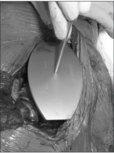
The collaboration between surgeon (FB) and the manufacturer led to the design of a sternal implant [Figure 1]. This prosthesis is anchored to the ribs thanks to nonresorbable suture threads [Figure 1] passed thru predrilled holes in the prosthesis. This binding allows the stability of the chest wall that is reinforced by bone cells which colonize the porous structure from surrounding bone and cartilage and thus create a link between tissues and prosthesis. No metallic osteosynthesis is needed. This prosthesis is developed in five sizes. Three sizes of sternal implants are delivered to the surgeon in order to optimize the replacement because of a possible difference between imaging evaluation and surgical debridement, depending on the evolution of the disease. Implants are provided with metallic trial implant to choose the best fitting ceramic implant after debridement [Figure 2]. Partial sternectomy is also possible with a “half” sternum prosthesis (e.g., manubrium replacement).
The first implantation was approved by the Ethics Committee of the Limoges Teaching Hospital in February 2015, and all the surgeries were allowed by the French Agency for Health Security (Agence Nationale de la Sécurité du Médicament et des produits de santé).
Results
There were three men and three women with a mean age of 60.6 years (54–79 years). There were five replacements for cancer and one for deep sternal wound infection
(DSWI). The mean follow‑up is 20 months (3–37 months). Characteristics of patients and surgery are summarized in Table 1. After the debridement step and the removal of the tumor or the infected and necrotized tissues, implants were attached either to costal bone stumps or to cartilages. Closing of the surgical wound was performed as usually. There is no evaluation of surgery time as most of the operating time was dedicated to debridement. However, the implantation time was short as the prosthesis is ready‑to‑use, that the size is chosen with the trial implant and that there is no need for modeling or adapting the prosthesis. The stitching with suture threads circumvents the preparation of the ribs, the use of screws, or other osteosynthesis material. To date, the follow‑up did not identify complication due to the prosthesis. Complications which occurred during the follow‑up were related to surgery with one hematoma and two surgical wound infections without prosthesis infection and which did not required prosthesis removal. The radiologic follow‑up is simplified as the image of prosthesis disappears after 1 month even if ceramic is nonresorbable [Figure 3]. All patients returned home and get back to their normal activities. None of them complained about pain or breathing discomfort. Computed tomography (CT) scan follow‑up showed a close contact between rib and prosthesis after 4 months [Figure 4].
Discussion
We propose a new material for thoracic wall reconstruction and particularly for sternum. Several drawbacks have been reported for biomaterials used in chest wall reconstruction over the past 30 years:[2]
erosion of adjacent structures, rupture and migration, infection, or immunological reaction. During the past 5 years, custom‑made titanium sternal implants, sternal allografts, and mesh have been associated with advantages over other materials. Nonetheless, all requirements of the ideal sternal replacement
Figure 2: Testing metallic implant Figure 1: Anchored prosthesis with sutures
device have not been met. For le Roux and Shama, ideal characteristics for a prosthetic material for chest wall reconstruction are rigidity, malleability, radiolucency, and inertness.[3] This ceramic prosthesis
almost presents all these requirements as it is
rigid, radiolucent, and totally inert. The material is not malleable; however, the stitching and the osseointegration allow certain malleability after its implantation. Titanium has no osteoconduction or osseointegration properties, while the porous ceramic prosthesis showed osseointegration after 4 months on CT scan [Figure 4]. Titanium has been recently associated with increased risk of infection; sternal allografts require storage within tissue banks and are limited by paucity of donors. Conversely, the porous ceramic prosthesis is immediately available in several sizes, and intraoperative sizing is done with dedicated calibrators. Implantation of a sternal graft requires a more complex surgical procedure (using titanium plates and screws for fixation). However, the structure and angulation of the ceramic sternum were intended to optimize its stability through enhanced contact surface and complementarity with bone tissue. No osteosynthesis material is needed, so the procedure is simplified as there are no preparation of the ribs and no need to screw metal parts. The surgical technique appears to be reproducible and fast.
Figure 4: Computed tomography scan done 4 months after surgery, showing
a close contact between the rib and the prosthesis revealing the beginning of osseointegration
Figure 3: Chest X‑ray. (a) Few days after surgery showing the prosthesis (b) 1 month after surgery, the prosthesis is no more visible (c) three‑dimensional reconstruction of
computed tomography scan showing the prosthesis after implantation
c b
a
Table 1: Characteristics of patients
Sex Age (years) Indication Risk factors ASA
score BMI Type of surgery Complications Follow-up (months)
1 Female 55 Radio‑induced sarcoma Malignancy 3 22.5 Complete
replacement None 37
2 Female 54 Breast cancer
metastasis and skin localization following a biopsy
Malignancy, diabetes
mellitus 3 24.6 Complete replacement Hematoma without infection 30
3 Male 61 Sternal disunion
after aortic valve replacement
Diabetes mellitus, active smoker (55PA), COPB, lung cancer with radiotherapy including sternal area (2007)
4 26.3 Complete
replacement Surgical wound infection without prosthesis infection
32
4 Male 79 Manubrial thyroid
cancer metastasis None 2 27.9 Half‑sternum replacement Surgical wound infection without prosthesis infection
11
5 Female 57 Breast cancer
metastasis Breast cancer surgery, obesity 2 38.5 Complete replacement None 9
6 Male 57 Low‑grade sternal
chondrosarcoma None 2 Complete replacement None 3
Bertin, et al.: Alumina ceramic sternum
Annals of Thoracic Medicine ‑ Volume 13, Issue 4, October‑December 2018 229
Radiolucency allows a simplified follow‑up, particularly for patients with cancer. Even if titanium does not create artefact, metal parts can mask mediastinal structures on chest X‑ray in case of recurrence.
It seems that there is no more complication than with the other techniques even if it is too early, with a low number of patients to draw this conclusion. During the immediate follow‑up, we observe three minor complications: one hematoma and two surgical site infections. None required a new surgery or the prosthesis removal. The follow‑up allowed saying that the ceramic sternum was not involved as no infection occurred. Even if a direct comparison is difficult as fortunately, the number of patients requiring this type of surgery is low (only a mean of 2.3 patients per year per department in the literature[4‑6]), the number
of complications observed in this study is approximately the same as those found in the literature. Zhang found one hematoma in eight patients receiving a titanium mesh.[7] With the use of titanium plates, Vos observed
25% of superficial infection (SSI) and 15% which needed a material removal.[8] In another study, comparing rigid
to nonrigid material for chest wall reconstruction, SSI was found in 5.3% with a need of removal 3.8%. Three hematomas were also observed.[9] The number seen here is
then in the same order of magnitude than in other studies and those not seem to be in relation with the ceramic. To avoid this type of complication, we developed an antibiotic‑loaded sternum which was not used in this study. The loading is done making use of the porosity of the ceramic. The goal is to protect implantation for patients presenting a high risk of infection (cancer, antitumor chemotherapy, diabetes mellitus, and obesity) or to replace sternum for patient presenting refractory deep sternal wound infection. The sternum is loaded with gentamicin. To date, three patients received this loaded sternum, but it is too early to draw conclusions even if no infections occurred during the follow‑up of these patients with a high risk of prosthesis infection.
Conclusions
This new type of prosthesis seems to be a reliable technique for the replacement of sternal defect. A longer
follow‑up with more patients is needed to confirm these first conclusions. To enhance our knowledge about this prosthesis, a clinical trial is ongoing (ClinicalTrials.gov. identifier: NCT02683590).
Financial support and sponsorship
Nil.
Conflicts of interest
E. Denes is an employee of I.Ceram and hold stocks; François Bertin is a member of the scientific committee of I.Ceram. There are no conflicts of interest for the other authors.
References
1. Denes E, Barrière G, Poli E, Lévêque G. Commentary: Bioceramics and scaffolds: A winning combination for tissue engineering. Front Bioeng Biotechnol 2017;5:15.
2. Butterworth JA, Garvey PB, Baumann DP, Zhang H, Rice DC, Butler CE, et al. Optimizing reconstruction of oncologic sternectomy defects based on surgical outcomes. J Am Coll Surg 2013;217:306‑16.
3. le Roux BT, Shama DM. Resection of tumors of the chest wall. Curr Probl Surg 1983;20:345‑86.
4. Kaláb M, Karkoška J, Kamínek M, Matějková E, Slaměníková Z, Klváček A, et al. Reconstruction of massive post‑sternotomy defects with allogeneic bone graft: Four‑year results and experience using the method. Interact Cardiovasc Thorac Surg 2016;22:305‑13.
5. Galbis Caravajal JM, Yeste Sánchez L, Fuster Diana CA, Guijarro Jorge R, Fernández Ortiz P, Deaville PJ, et al. Sternal resection and reconstruction after malignant tumours. Clin Transl Oncol 2009;11:91‑5.
6. Girotti P, Leo F, Bravi F, Tavecchio L, Spano A, Cortinovis U, et al. The “rib‑like” technique for surgical treatment of sternal tumors: Lessons learned from 101 consecutive cases. Ann Thorac Surg 2011;92:1208‑15.
7. Zhang Y, Li JZ, Hao YJ, Lu XC, Shi HL, Liu Y, et al. Sternal tumor resection and reconstruction with titanium mesh: A preliminary study. Orthop Surg 2015;7:155‑60.
8. Vos RJ, Jongbloed L, Sonker U, Kloppenburg GT. Titanium plate fixation versus conventional closure for sternal dehiscence after cardiac surgery. Thorac Cardiovasc Surg 2017;65:338‑42. 9. Weyant MJ, Bains MS, Venkatraman E, Downey RJ,
Park BJ, Flores RM, et al. Results of chest wall resection and reconstruction with and without rigid prosthesis. Ann Thorac Surg 2006;81:279‑85.