TRENDS
Circulating miRNAs: a new generation of anti-doping
biomarkers
Nicolas Leuenberger&Neil Robinson&Martial Saugy
Received: 4 July 2013 / Revised: 27 August 2013 / Accepted: 2 September 2013 / Published online: 28 September 2013 # Springer-Verlag Berlin Heidelberg 2013
Abstract MicroRNAs (miRNAs) are small non-coding RNAs that regulate a variety of biological processes. Cell-free miRNAs detected in blood plasma are used as specific and sensitive markers of physiological processes and some diseases. Circulating miRNAs are highly stable in body fluids, for example plasma. Therefore, profiles of circulating miRNAs have been investigated for potential use as novel, non-invasive anti-doping biomarkers. This review describes the biological mechanisms underlying the variation of circulating miRNAs, revealing that they have great potential as a new class of biomarker for detection of doping substances. The latest de-velopments in extraction and profiling technology, and the technical design of experiments useful for anti-doping, are also discussed. Longitudinal measurements of circulating miRNAs in the context of the athlete biological passport are proposed as an efficient strategy for the use of these new markers. The review also emphasizes potential challenges for the translation of circulating miRNAs from research into practical anti-doping applications.
Keywords Doping . Circulating microRNAs . Biomarkers . Athlete biological passport
Introduction
A major challenge in anti-doping is identification of specific and sensitive non-invasive biomarkers that can be routinely
measured in easily accessible samples. MicroRNAs (miRNAs) are a particularly promising class of biomarker. miRNAs are small (19–25 nucleotides), non-encoding RNAs that enable post-transcription regulation of gene expression by suppres-sion of specific target messenger RNAs (mRNAs) [1]. Since their discovery in the early 1990s, these small molecules have been revealed to have important regulatory functions in a wide range of biological and pathological processes. To date, more than 1800 miRNAs have been identified in humans [2]. The number of miRNAs in our species therefore seems to be smaller than the number of conventional mRNAs, estimated to be 30,000 [3].
Since 2008, miRNAs have been detected in serum, plas-ma, urine, saliva, and other body fluids [4]. Although most RNA molecules are unstable, circulating miRNAs are high-ly stable and readihigh-ly detectable. Cell-free miRNAs in body fluids are stable under harsh conditions, including boiling, low and high pH, extended storage, and multiple freeze– thaw cycles [5]. In contrast, synthetic or purified miRNAs added to serum or plasma are quickly degraded by high levels of RNase activity in plasma [6]. Thus, circulating miRNAs are not intrinsically resistant to endogenous RNase activity.
Stability of circulating miRNAs
The molecular basis of the high stability of circulating miRNAs has been investigated in different laboratories. One hypothesis suggests that circulating miRNAs are protected by microvesicles, for example exosomes and microparticles; al-ternatively, circulating miRNAs may be associated with spe-cific protective proteins.
Recently, different groups have found that most circulating miRNAs co-fractionate with protein complexes [6,7]. Arroyo et al. [6] found that miRNAs are sensitive to protease Published in the topical collection Anti-doping Analysis with guest editor
Christopher Harrison.
N. Leuenberger (*)
:
N. Robinson:
M. SaugySwiss Laboratory for Doping Analyses, University Center of Legal Medicine, Centre Hospitalier Universitaire Vaudois and University of Lausanne, Lausanne, Switzerland
e-mail: Nicolas.leuenberger@chuv.ch DOI 10.1007/s00216-013-7340-0
treatment of plasma, indicating that protein complexes pro-tect circulating miRNAs from plasma RNases. Investigation of the protective protein complexes revealed that Argonaute 2 (Ago2), a protein component of the RNA-induced silenc-ing complex, is present in human plasma and elutes with plasma miRNAs in size-exclusion chromatography [6, 8]. The extracellular stability of Ago2–miRNA complexes has also been observed in cell culture. Both Ago2 protein and miRNAs remain stable for several weeks in cell lysates, even in the absence of protease inhibitors [7]. Immunopre-cipitation assays performed on plasma revealed that Ago2 readily recovers non-vesicle-associated plasma miRNAs. In these studies, most miRNAs tested co-purified with the Ago2 ribonucleoprotein complex; however, only a minority of specific miRNAs were associated with vesicles [6]. These observations revealed the existence of two popula-tions of circulating miRNAs, and suggested that circulating Ago2 complexes are most probably the reason for the stability of plasma miRNAs [6, 7]. These studies have important implications for the development of biomarker approaches based on sampling and analysis of circulating miRNAs.
Secretion mechanism and origin of circulating miRNAs The presence of miRNAs in the extracellular environment has led researchers to develop two main hypotheses regard-ing systems for miRNA export from cells and tissues [9]. One hypothesis is that all types of circulating miRNA in biological fluid are by-products of cellular activity and cell death [7]. This “by-product release” hypothesis could ex-plain multiple observations that, after toxicity occurs in some tissues, the levels of miRNAs specific for the affected tissue increase in the plasma [10–12]. The other hypothesis is that secretion of miRNAs is caused by an increase in cellular activity leading to the release of miRNAs, either in Ago2–miRNA complexes or, for a minority of miRNAs, packaged in microvesicles, for example exosomes.
Recently, more attention has been devoted to under-standing the origin of circulating miRNAs and its effect on biomarker specificity [9, 13, 14]. Because blood cells are in extensive contact with plasma, they should be major contributors to the extracellular miRNA con-tent of plasma [15, 16]. This idea is supported by observations that perturbations in blood-cell counts and haemolysis can alter plasma miRNAs; furthermore, ul-tracentrifugation steps that remove intact cells and de-bris reduce the level of miRNAs in plasma [16]. How-ever, because tissue-specific mRNAs from liver, muscle, heart, brain, lungs, and placenta have been detected in plasma [10–13, 17], it is probable that other organs contribute to extracellular miRNA. Many groups have
observed that tumours secrete miRNAs, and have de-tected cancer tissue-specific miRNAs in the circulatory system [18]. These observations support the theory that circulating blood cells are not the only source of circu-lating miRNAs.
Circulating miRNAs as disease biomarkers
In healthy individuals levels of cell-free miRNAs present in serum are stable, and the plasma and/or serum miRNA profile is similar to that of circulating blood cells. Altered serum miRNA levels may be indicative of physiological or pathological changes, and might therefore be used as surrogate biomarkers.
Lawrie et al. were the first to discover tumour-specific deregulation of circulating miRNAs [19]. Their study revealed that miR-21 is abundant in the sera of diffuse large B-cell lymphoma patients. Since then, many other investigations have revealed the diagnostic and prognostic value of measur-ing miRNAs in body fluids in the context of a variety of solid cancers [20, 21]. In 2008, Mitchell et al. reported that miRNAs derived from epithelial tumours are also rapidly released into the bloodstream [5]. Shen et al. [22] revealed that plasma miRNA expression profiles could be useful for discriminating non-small cell lung cancer (NSCLC) patients from healthy controls and patients with chronic obstructive pulmonary disease (COPD).
Regarding non-neoplastic changes, tissue-specific miRNAs have been analysed in the bloodstream as markers of myocar-dial [11] and liver injury [12]. For example, myocardial infarc-tion induced an increase in the plasma level of miR-133a, a muscle-specific miRNA [23]. Several studies have revealed that liver injury is associated with increased plasma levels of miR-122 and miR-192 [12].
Physiological changes, for example pregnancy, increase the level of several miRNAs in plasma [13]. miR-517a and miR-526a undergo substantial changes that could be used to distinguish pregnant from non-pregnant women with high accuracy.
Circulating miRNAs as anti-doping biomarkers
In the same way that some biomarkers are used to detect the biological signs of disease, doping biomarkers have been used for some time in anti-doping tests to detect the biological signs of doping. The potential of circulating miRNAs for use as specific anti-doping biomarkers has been revealed by our laboratory and others. We found that plasma miRNA expres-sion profiles can be useful in detecting erythropoiesis-stimulating agents (ESA) [24]. That study revealed that levels of a specific circulating miRNA, miR-144, increase in plasma samples after a single methoxy polyethylene glycol-epoetin
beta (Mircera) injection. This miRNA has been reported to be essential for erythropoiesis in a variety of organisms, and our finding revealed the potential of miRNAs as circulating bio-markers for ESA detection. Another study revealed that cir-culating miRNAs could be used as biomarkers to detect recombinant human growth hormone (rhGH) abuse: Kelly and colleagues identified and confirmed four miRNAs that were differently expressed in all individuals using therapeutic replacement doses of rhGH, relative to normal controls or individuals with naturally high levels of GH [25].
Recently, we found that circulating miRNAs could be used to detect autologous blood transfusion [26]. Transfusion of autologous blood induced an increase in the plasma levels of specific circulating miRNAs compared with a non-transfused control group. Because blood transfusion causes substantial changes in miRNA levels in the lung, the origin of these miRNAs was related to pulmonary tissue. We also observed partial suppression of erythropoietin (EPO) after transfusion. A combination of miRNAs and EPO measurement in a math-ematical model increased the efficiency of detection of autol-ogous transfusion via miRNA analysis.
Methods for analysis of circulating miRNAs
Three matrices have been studied in the context of miRNA profiling in blood: serum, plasma, and whole blood. The choice of matrix has been reported to have a significant effect on miRNA concentration. Serum has lower levels of circulat-ing miRNAs than plasma [27]. Because of the large number of blood analyses required for the athlete biological passport (ABP), EDTA–plasma is the matrix most widely used in anti-doping. Because blood variables and miRNAs are mea-sured in the same matrix, a single EDTA–plasma sample would be sufficient to perform the test, and plasma would therefore seem to be the best material for miRNA analysis in anti-doping laboratories. However, plasma samples contain-ing heparin as an anticoagulant should not be used because this substance can interfere with subsequent assays, for exam-ple the reverse transcription reaction of RT–PCR.
One critical factor that might affect the results of miRNA quantification is the presence of haemolysis. Several studies have observed that mature erythrocytes contain abundant and diverse miRNA. In a recent study, Blondal et al. suggested different procedures and tests for assessment of haemolysis [28]; one simple and cost-effective way is to use a spectrophotometer and measure oxyhaemoglobin absorbance atλ =414 nm. Other methods, based on delta calculation between erythrocyte-enriched miR-451 and miR-23a, which is not affected by haemolysis, have been proposed as indicators of haemolysis.
Because of the small amount of circulating miRNA and the large amount of proteins and lipids, miRNA extraction from blood samples is technically challenging. Moreover, plasma
and serum contain large amounts of reverse transcription and PCR inhibitors. To overcome these challenges, several phe-nol–chloroform-based procedures are available for extracting miRNA from biological fluid [29].
Most of these methods combine phenol–guanidine-based lysis of samples and silica-membrane-based purification of total RNA (Fig.1). The lysis reagent is a solution of phenol and guanidine thiocyanate, designed to facilitate lysis, to de-nature protein complexes and RNases, and to remove most of the residual DNA and proteins from the lysate via organic extraction. After addition of chloroform, the lysate is separated into aqueous and organic phases by means of centrifugation. The aqueous phase is extracted and the sample is then applied to the silica membrane-based spin column, where the RNA binds to the membrane but phenol and other contaminants are efficiently washed away. To increase the efficiency of phenol– chloroform extraction, Andreasen et al. revealed that addition of a small carrier RNA before total RNA extraction improved the yield and reproducibility of separate RNA extractions [29]. Commercially available kits without acid-phase separation can also be used for isolation of miRNA from body fluids (Fig.1). The general principle of extraction is similar to that of phenol–chloroform extraction, but the non-phenol lysis solu-tion contains only guanidine thiocyanate, and salt solusolu-tion is added to denature and precipitate proteins. The phenol –chlo-roform procedure yields more miRNA, reduces the variance between assays [27], and could be fully automated, and this procedure will therefore usually be the best method for extracting miRNAs from biofluids. Nevertheless, different extraction methods should be tested for each study to establish the optimum procedure.
One of the main problems associated with circulating miRNA extraction and comparison of collected samples is quantification of the miRNA. Because the miRNA content of plasma is low, it can only just be determined by use of spec-trophotometers. Efficiency of extraction of circulating miRNA could be assessed by spiking plasma with known quantities of non-human miRNAs (e.g. that of Caenorhabditis elegans ). Synthetic spike-in miRNAs should be added to the denatur-ation buffer (e.g. Trizol or lysis buffer). These miRNAs go through the entire extraction process and are measured in the final miRNA eluate by use of qRT–PCR, providing an internal reference against which to assess the efficiency of extraction. Addition of synthetic spike-in miRNA can also be used to assess the reverse transcription reaction.
Profiling new circulating miRNA anti-doping biomarkers When searching for circulating miRNA-based doping bio-markers, different profiling methods can be used. Profiling enables us not only to discover changes in miRNA levels after treatment, but also to find stable miRNAs that could be used as
endogenous controls. Technological advances have resulted in many miRNA-profiling techniques.
High-throughput quantitative reverse transcription (qRT)– PCR remains the most sensitive and reliable method for detecting circulating miRNAs. A positive aspect of this ap-proach is its ease of incorporation into the workflow of labo-ratories that are already familiar with real-time PCR. For large-scale miRNA profiling (which involves hundreds of miRNAs) by use of qRT–PCR, commercially available pre-plated PCR primers, typically distributed across multi-well dishes, are available [3]. To prevent variation of primer hybridisation among the hundreds of PCR assays, locked nucleic acids (LNA) are incorporated into primers [30].
Microarrays were among the first methods to be used for parallel analysis of large numbers of miRNAs, and several variations of the approach have been developed [3]. miRNA microarrays have the advantage of being less expensive than other profiling methods. However, several limitations have been described, including a restricted linear range of quantification, low sensitivity, and imperfect specificity for some miRNAs.
A deep-sequencing approach has been investigated in sev-eral studies with the objective of evaluating the potential use of circulating miRNAs as disease biomarkers [13]. The major advantages of next-generation sequencing for miRNA profil-ing are the detection of both novel and already known miRNAs and the precise identification of miRNA sequences. Potential limitations of next-generation sequencing include high cost, although this is dropping with the introduction of new instru-ments and the use of DNA“barcoding”. For example, use of barcoded adapters enables multiplexing of 20 samples in a single sequencing reaction, dramatically reducing the cost of sequencing five million reads per sample to 150 USD [13].
Strategies for identifying circulating miRNA-based anti-doping biomarkers
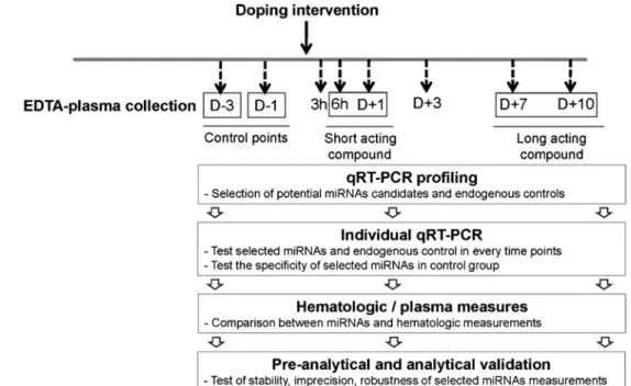
In the process of searching for blood miRNA-based doping biomarkers, a high-throughput RT–qPCR strategy was used to identify plasma miRNA-based biomarkers for detecting au-tologous blood transfusion [26]. In this strategy, multiplex real-time RT–qPCR is used to identify a set of specific circu-lating miRNAs. RT–qPCR was chosen because it is a faster, simpler, and more sensitive technique than sequencing or use of microarrays.
As depicted in Fig.2, the strategy was separated into several steps:
1. profiling with pre-plated PCR primers, using subjects who have undergone treatment including blood transfu-sion, to find a potential profile of circulating miRNAs and endogenous controls;
2. individual RT–qPCR validation of selected miRNAs obtained at every time point before and after doping intervention (from three days before treatment, D− 3, to 10 days afterward, D + 10);
3. comparison with variations in blood properties; and 4. pre-analytical and analytical characterisation of selected
miRNAs.
In general, initial screening by use of the high-throughput technique is intended to identify differently expressed plasma miRNAs in artificially doped volunteers. However, once we know the specific miRNAs linked to doping, we can directly determine their levels by use of individual qRT–PCR.
The detection window for the panel of differently expressed miRNAs from the screening study was determined Fig. 1 Schematic outline of two
miRNA extraction procedures. The commercial kit does not use phenol–chloroform to degrade protein from plasma samples
by longitudinal measurement using a maximum number of time points. The stabilities of non-differently expressed miRNAs at every time point were also investigated, and were used as endogenous controls. The screened endogenous controls and artificial spike-in miRNAs were used for normalisation.
For decades, haematological variables have been common-ly used as reference values in clinical and anti-doping studies. For example, haemoglobin has been tested to detect autolo-gous blood transfusion [31]. Correlation between blood-marker variables and circulating miRNAs could increase the efficiency of result interpretation. In our study the combina-tion of circulating miRNAs and other variables, including EPO, resulted in greater discriminative power for the detection of autologous blood transfusion [26].
Providing proof of principle for a reliable miRNA doping test is a long process. Many pre-analytical and analytical variables that might affect the measurement of miRNAs in anti-doping applications have yet to be studied in detail. McDonald et al. proposed a method for validating circulating miRNAs used in clinical diagnosis [27]. The relative contri-bution to assay imprecision from components including intra and inter-assay imprecision, miRNA extraction, reverse tran-scription, real-time PCR, and normalisation to spiked or internal-control miRNAs has yet to be established.
Potential use of circulating miRNAs in the athlete biological passport
The fight against doping is mainly based on direct detection of a prohibited substance in an athlete’s biological sample. Some methods, including the ABP, also use indirect markers [32].
The ABP is a new tool, which has immense potential in the current climate of rapidly advancing biomarker discovery [32]. Doping induces physiological changes that enhance performance. In the same way that disease-related biomarkers are invaluable tools that assist physicians in the diagnosis of pathology, specific biomarkers can be used to detect doping.
In 2008, the haematological module of the ABP was the first to be implemented by the International Cycling Union [33]. Biomarkers related to the haematopoietic system (haemoglobin concentration, reticulocytes) are monitored over time and analysed by use of mathematical models that quantify individual variation to identify patterns that raise suspicion of blood doping (e.g. ESA and autologous blood transfusion) [31].
The underlying principle of the ABP is the use of informa-tion from biological tests as indirect evidence to detect doping. Fig. 2 Suggested strategy for
investigating use of circulating miRNA-based blood biomarkers to detect abuse of doping compounds
Table 1 Advantages of using circulating miRNAs in the athlete biolog-ical passport
Step Advantages of circulating miRNAs Collection Similar to guidelines used for the
haematological module of the ABP Transport High stability during transport Analysis Long time period for detection
Correlation with blood-property variables qPCR technology enables multiplexing analysis Multiple quality controls could be easily added Storage High stability during storage in EDTA–plasma
Not sensitive to unregulated room-temperature storage (up to 10 years)
For this reason, a stringent process of sample collection, transport, and data analysis has been put into place to guaran-tee objective and reliable use of this tool [34]. This is of particular importance because blood is a living tissue that undergoes permanent changes over time; these changes must be limited as much as possible, either by using standardised procedures or by taking these changes into account in evalu-ation of the data.
Circulating miRNAs have been investigated as potential biomarkers for detection of blood doping, and could therefore be incorporated into the adaptive model of the ABP. Among the advantages of miRNAs as biomarkers is their high stability in blood (Table1), which enables detection over a longer time period. Furthermore, in contrast with haematological vari-ables, miRNAs are not affected by environmental factors, for example inadequate storage during transport of the blood samples [35]. Thus, use of miRNAs could reduce difficulties caused by the extensive documentation regarding transport and storage of samples. cDNA samples are also highly stable under different storage conditions [36].
Blood samples included in haematological modules of the ABP are analysed in WADA-accredited laboratories, where strict quality-control criteria are applied to provide analysis of forensic quality. Blood-variable analyses are performed by use of automated blood-cell counters, and haematological vari-ables could be correlated with miRNA measurements. Circu-lating miRNAs could be analysed by use of qPCR. miRNA qPCR is a well-established, robust, and reproducible method with several important advantages, including high sensitivity and specificity, potential for target multiplexing, and low RNA input requirements, all of which facilitate expression analysis, even for anti-doping samples with a limited amount of material [28]. This method has already been implemented in many diagnostic laboratories, and routine procedures are well established. Quality control could easily be included at every step to monitor sample haemolysis and extraction, re-verse transcription, and qPCR efficiency, and such controls could be used as normalisation factors [28]. Incorporation of quality controls eliminates within-subject variation, increasing the efficiency of the evaluation of biological data.
To validate and implement new biomarkers in the ABP, a rigorous procedure must be followed. For the haematological module of the ABP it has been possible to take advantage of experience gained over many years with biomarkers including haematocrit and haemoglobin. Schumacher et al. revealed that haemoglobin concentration has significant diurnal and exercise-related variation [37]. The same group observed that a percentage of reticulocytes is modified by both long-term and short-term exercise [38]. All these observations are al-ready used for interpreting athlete blood-profile analyses. For circulating miRNA, few data exist regarding external and environmental factors that could interfere with miRNA mea-surements in blood. Nevertheless, Baggish et al. revealed that
exercise can affect some types of circulating miRNA, includ-ing those involved in inflammation (miR-146). Interestinclud-ingly, miRNAs involved in heart and skeletal muscle physiology 133a) and adaptation to hypoxia and ischaemia (miR-210) did not vary in response to exercise [39].
Other confounding factors in the interpretation of miRNA measurements should be investigated. As an example, the effect of altitude and of use of hypoxic chambers on circulat-ing miRNA levels should be studied, and the effect of the circadian cycle on miRNA measurements should be investi-gated. Unlike haemoglobin, circulating miRNAs were discov-ered in 2008. Therefore additional studies from different dis-ciplines, for example diagnosis of diseases for which circulat-ing miRNAs have been tested as blood-related biomarkers, should be used in the interpretation of longitudinal measure-ments of miRNAs in the context of the ABP.
Outlook
Many elements contribute to the enormous potential of circu-lating miRNAs as a class of ideal anti-doping biomarkers. Circulating miRNAs are very stable molecules, which are well preserved under harsh conditions and resistant to RNase ac-tivity. They are easily accessible, can be sampled in a relative-ly non-invasive manner, and can be readirelative-ly measured by use of simple qPCR technology. Longitudinal measurement of circulating miRNAs in the context of the ABP should be a suitable way of using these new biomarkers in anti-doping. Circulating miRNA research is still in its early stages, and further work must be done to characterise the potential con-founding factors affecting these biomarkers. As a result of the plethora of data supporting the clinical use of miRNAs as biomarkers for diseases, robust and reproducible tests for use in patient treatment decisions are currently being developed. The increasing clinical knowledge of these next-generation biomarkers could be easily extrapolated to anti-doping. Acknowledgements We acknowledge Yorck Olaf Schumacher and Torben Pottgiesser for critical reading of the manuscript. Our research on circulating miRNAs was supported by Partnership for Clean Competition (PCC), the World Anti-Doping Agency, and Exiqon grant program.
References
1. Fabian MR, Sonenberg N, Filipowicz W (2010) Regulation of mRNA translation and stability by microRNAs. Annu Rev Biochem 79:351– 379
2.http://www.mirbase.org/cgi-bin/mirna_summary.pl?org=hsa
3. Pritchard CC, Cheng HH, Tewari M (2012) MicroRNA profiling: approaches and considerations. Nat Rev Genet 13:358–369 4. Weber JA, Baxter DH, Zhang S, Huang DY, Huang KH et al (2010)
5. Mitchell PS, Parkin RK, Kroh EM, Fritz BR, Wyman SK et al (2008) Circulating microRNAs as stable blood-based markers for cancer detection. Proc Natl Acad Sci U S A 105:10513–10518
6. Arroyo JD, Chevillet JR, Kroh EM, Ruf IK, Pritchard CC et al (2011) Argonaute2 complexes carry a population of circulating microRNAs independent of vesicles in human plasma. Proc Natl Acad Sci U S A 108:5003–5008
7. Turchinovich A, Weiz L, Langheinz A, Burwinkel B (2011) Characterization of extracellular circulating microRNA. Nucleic Acids Res 39:7223–7233
8. Turchinovich A, Burwinkel B (2012) Distinct AGO1 and AGO2 associated miRNA profiles in human cells and blood plasma. RNA Biol 9:1066–1075
9. Turchinovich A, Weiz L, Burwinkel B (2012) Extracellular miRNAs: the mystery of their origin and function. Trends Biochem Sci 37:460– 465
10. Bala S, Petrasek J, Mundkur S, Catalano D, Levin I et al (2012) Circulating microRNAs in exosomes indicate hepatocyte injury and inflammation in alcoholic, drug-induced, and inflammatory liver diseases. Hepatology 56:1946–1957
11. Li C, Pei F, Zhu X, Duan DD, Zeng C (2012) Circulating microRNAs as novel and sensitive biomarkers of acute myocardial Infarction. Clin Biochem 45:727–732
12. Starkey Lewis PJ, Dear J, Platt V, Simpson KJ, Craig DG et al (2011) Circulating microRNAs as potential markers of human drug-induced liver injury. Hepatology 54:1767–1776
13. Williams Z, Ben-Dov IZ, Elias R, Mihailovic A, Brown M et al (2013) Comprehensive profiling of circulating microRNA via small RNA sequencing of cDNA libraries reveals biomarker potential and limitations. Proc Natl Acad Sci U S A 110:4255–4260
14. Ma R, Jiang T, Kang X (2012) Circulating microRNAs in cancer: origin, function and application. J Exp Clin Cancer Res 31:38 15. Pritchard CC, Kroh E, Wood B, Arroyo JD, Dougherty KJ et al
(2012) Blood cell origin of circulating microRNAs: a cautionary note for cancer biomarker studies. Cancer Prev Res (Phila) 5:492–497 16. Duttagupta R, Jiang R, Gollub J, Getts RC, Jones KW (2011) Impact
of cellular miRNAs on circulating miRNA biomarker signatures. PLoS One 6:e20769
17. Laterza OF, Lim L, Garrett-Engele PW, Vlasakova K, Muniappa N et al (2009) Plasma MicroRNAs as sensitive and specific biomarkers of tissue injury. Clin Chem 55:1977–1983
18. Redova M, Sana J, Slaby O (2013) Circulating miRNAs as new blood-based biomarkers for solid cancers. Future Oncol 9:387–402 19. Lawrie CH, Gal S, Dunlop HM, Pushkaran B, Liggins AP et al
(2008) Detection of elevated levels of tumour-associated microRNAs in serum of patients with diffuse large B-cell lymphoma. Br J Haematol 141:672–675
20. Shen J, Stass SA, Jiang F (2013) MicroRNAs as potential biomarkers in human solid tumors. Cancer Lett 329:125–136
21. Brase JC, Wuttig D, Kuner R, Sultmann H (2010) Serum microRNAs as non-invasive biomarkers for cancer. Mol Cancer 9:306
22. Shen J, Liu Z, Todd NW, Zhang H, Liao J et al (2011) Diagnosis of lung cancer in individuals with solitary pulmonary nodules by plasma microRNA biomarkers. BMC Cancer 11:374
23. Kuwabara Y, Ono K, Horie T, Nishi H, Nagao K et al (2011) Increased microRNA-1 and microRNA-133a levels in serum of patients with cardiovascular disease indicate myocardial damage. Circ Cardiovasc Genet 4:446–454
24. Leuenberger N, Jan N, Pradervand S, Robinson N, Saugy M (2011) Circulating microRNAs as long-term biomarkers for the detection of erythropoiesis-stimulating agent abuse. Drug Test Anal 3:771–776 25. Kelly BN, Haverstick DM, Lee JK, Thorner MO, Vance ML, et al.
(2013) Circulating microRNA as a biomarker of human growth hor-mone administration to patients. Drug Test Anal, Mar 12 doi: 101002/ dta1469 [Epub ahead of print]
26. Leuenberger N, Schumacher YO, Pradervand S, Sander T, Saugy M et al (2013) Circulating microRNAs as Biomarkers for Detection of Autologous Blood Transfusion. PLoS One 8(6):e66309
27. McDonald JS, Milosevic D, Reddi HV, Grebe SK, Algeciras-Schimnich A (2011) Analysis of Circulating MicroRNA: Preanalytical and Analytical Challenges. Clin Chem 57(6):833–840
28. Blondal T, Jensby Nielsen S, Baker A, Andreasen D, Mouritzen P et al (2013) Assessing sample and miRNA profile quality in serum and plasma or other biofluids. Methods 59:S1–6
29. Andreasen D, Fog JU, Biggs W, Salomon J, Dahslveen IK et al (2010) Improved microRNA quantification in total RNA from clin-ical samples. Methods 50:S6–9
30. Jacobsen N, Andreasen D, Mouritzen P (2011) Profiling microRNAs by real-time PCR. Methods Mol Biol 732:39–54
31. Pottgiesser T, Sottas PE, Echteler T, Robinson N, Umhau M et al (2011) Detection of autologous blood doping with adaptively evalu-ated biomarkers of doping: a longitudinal blinded study. Transfusion 51:1707–1715
32. Sottas PE, Vernec A (2012) Current implementation and future of the Athlete Biological Passport. Bioanalysis 4:1645–1652
33. Schumacher YO, Saugy M, Pottgiesser T, Robinson N (2012) Detection of EPO doping and blood doping: the haematological module of the Athlete Biological Passport. Drug Test Anal 4:846– 853
34. Robinson N, Sottas PE, Pottgiesser T, Schumacher YO, Saugy M (2011) Stability and robustness of blood variables in an antidoping context. Int J Lab Hematol 33:146–153
35. Grasedieck S, Sorrentino A, Langer C, Buske C, Dohner H et al (2013) Circulating microRNAs in hematological diseases: principles, challenges and perspectives. Blood :Apr 2 [Epub ahead of print] 36. Mraz M, Malinova K, Mayer J, Pospisilova S (2009) MicroRNA
isolation and stability in stored RNA samples. Biochem Biophys Res Commun 390:1–4
37. Schumacher YO, Wenning M, Robinson N, Sottas PE, Ruecker G et al (2010) Diurnal and exercise-related variability of haemoglobin and reticulocytes in athletes. Int J Sports Med 31:225–230 38. Schumacher YO, Sahm D, Baumstark MW, Pottgiesser T (2010)
Reticulocytes in athletes: Longitudinal aspects and the influence of long- and short-term exercise. Drug Test Anal 2:469–474
39. Baggish AL, Hale A, Weiner RB, Lewis GD, Systrom D et al (2011) Dynamic regulation of circulating microRNA during acute exhaus-tive exercise and sustained aerobic exercise training. J Physiol 589: 3983–3994