HAL Id: hal-02069829
https://hal.archives-ouvertes.fr/hal-02069829
Submitted on 15 Mar 2019
HAL is a multi-disciplinary open access
archive for the deposit and dissemination of sci-entific research documents, whether they are pub-lished or not. The documents may come from
L’archive ouverte pluridisciplinaire HAL, est destinée au dépôt et à la diffusion de documents scientifiques de niveau recherche, publiés ou non, émanant des établissements d’enseignement et de
Injectable Hybrid Hydrogels, with Cell-Responsive
Degradation, for Tumor Resection
Giuseppe Alonci, Federica Fiorini, Pietro Riva, Francisco Monroy, Ivan
López-Montero, Silvana Perretta, Luisa de Cola
To cite this version:
Giuseppe Alonci, Federica Fiorini, Pietro Riva, Francisco Monroy, Ivan López-Montero, et al.. In-jectable Hybrid Hydrogels, with Cell-Responsive Degradation, for Tumor Resection. ACS Applied Bio Materials, ACS Publications, 2018, 1 (5), pp.1301-1310. �10.1021/acsabm.8b00189�. �hal-02069829�
Injectable hybrid hydrogels, with cell-responsive
degradation, for tumor resection
Giuseppe Alonci‡, Federica Fiorini‡, Pietro Riva‡, Francisco Monroy, Ivan López-Montero
Silvana Perretta, Luisa De Cola*
‡These authors contributed equally to the work G. Alonci, Dr. F. Fiorini, Prof. L. De Cola
Institut de Science et d'Ingénierie Supramoléculaires Université de Strasbourg
8 rue Gaspard Monge, 67000 Strasbourg France
E-mail: [email protected]
P. Riva, Prof. S. Perretta IHU Strasbourg
1 place de l’Hôpital, 67000 Strasbourg France
Prof. F. Monroy, I. López-Montero Univeridad Compluense de Madrid
Avda. Complutense s/n 28040 Madrid
Spain and
Instituto de Investigación Sanitaria Hospital 12 de Octubre (imas12), Avda. Córdoba s/n
28041 Madrid Spain
KEYWORDS
Injectable hydrogels, breakable nanocapsules, polyamidoamines, endoscopic submucosal dissection, tumor resection
ABSTRACT
Biocompatible soft materials have recently found applications in interventional endoscopy to facilitate resection of mucosal tumors. When neoplastic lesions are in organs that can be easily damaged by perforation, such as stomach, intestine and esophagus, the formation of a submucosal fluid cushion (SFC) is needed to lift the tumor from the underlying muscle during the resection of neoplasias. Such procedure is called endoscopic submucosal dissection (ESD).
We describe an injectable, biodegradable, hybrid hydrogel able to form a SFC and to facilitate ESD. The hydrogel, based on polyamidoamines, contains breakable silica nanocapsules covalently bound to its network, and able to release biomolecules. To promote degradation, the hydrogel is composed of cleavable disulfide moieties that are reduced by the cells through secretion of glutathione. The same
stimulus triggers the breaking of the silica nanocapsules; therefore, the entire hybrid material can be completely degraded and its decomposition depends entirely on the presence of cells.
Interestingly the hydrogel precursor solution showed rapid gelation when injected in vivo and afforded a long-lasting high mucosal elevation, keeping the cushion volume constant during the dissection. This novel material can provide a solution to ESD limitations and promote healing of tissues after surgery
1. Introduction
Gastrointestinal (GI) tract cancers are amongst the most aggressive tumors, with an estimated worldwide mortality per year of over 1.75 million people.1 Endoscopic early detection and surgical treatment could
potentially lead to a reduction of mortality, especially for early gastrointestinal neoplasms or precancerous lesions. Nowadays, endoscopic dissection is the first-line treatment granting lower morbidity and mortality rates than surgery with equal oncological results and better quality of life. Endoscopic submucosal dissection (ESD) is a minimally invasive procedure now accepted worldwide as the preferred treatment modality for the removal of large and early gastric epithelial lesions.2
ESD, developed in Japan in the mid-1990s,3-5 consists in the injection of a solution or a gel between the
mucosal lesion and the muscularis,6-8 in order to obtain submucosal tissue elevation by generating a
submucosal fluid cushion (SFC). The “cushioned” submucosa forms a safety margin to the underlying muscularis, thus minimizing the risk of perforation during ESD.9-10
Additionally, this procedure allows an accurate histological assessment, therefore lowering the possibility of neoplastic recurrence.11
For gastrointestinal (GI) tumors especially, due to the low thickness of the GI wall and the use of electrocautery, there is a high risk of perforation.12-13 ESD is indeed a complex procedure requiring a
improve the efficacy and safety of endoscopic submucosal dissection techniques, the quality and duration of the submucosal lift are crucial.7-8
Several injection solutions have been developed for this purpose, but normal saline solution (NS) is the most commonly used, because of its low cost and ease of use. However, simple NS injection suffers from low mucosal elevation, fast diffusion of the solution in the tissues, requiring multiple injections, often resulting in longer operative time, multiple instrument exchange and poor visualization, which could led to bleeding and perforation.14
Viscous solutions such as glycerol,15-16 hydroxypropyl methylcellulose,17 chitosan,18 and hyaluronic
acid19 have been proposed to achieve sustained mucosal elevation and avoid injuries to the muscularis.
Although hyaluronic acid solution is one of the best options, it has been shown to stimulate the growth of residual tumors in animal models.20 Moreover, a large amount of hyaluronic acid is necessary to create
a SFC and its use is associated with high costs and a general lack of availability.11
To be clinically applicable, the lifting solution should be biocompatible, easily injectable, able to provide a prolonged and thick SFC to make ESD procedure safer, more cost-effective, and preserve tissue specimens for precise pathologic staging.10
Additionally, desirable features are controlled and tunable biodegradability and the possibility of releasing active components to assist the mucosal healing process after the resection, avoiding scarring and stenosis.
Finally, injectable biomaterial systems with a liquid-to-gel transition, such as hydrogels, are more promising in providing extended and thicker submucosal lift and easiness of handling. Using a hydrogel with tunable mechanical and degradation properties, which remains in place even after the procedure is
In the last years, injectable hydrogels have brought a shift in the search for the optimal SFC material towards the development of solutions that rely on in situ gel formation. For example, a photo-crosslinked chitosan hydrogel has been recently reported as a submucosal injection agent:21 mucosal elevation was
created after the injection of the chitosan viscous solution, which was crosslinked in situ via UV irradiation, resulting in an insoluble hydrogel. However, the use of UV light for the photoinitiated radical polymerization may be difficult to perform in certain organs and for large areas.22 Moreover, as the
authors mentioned, UV irradiation may be associated with inflammation of the residual tissue.
To replace light with heat, thermoresponsive polymers, or thermogels, have been investigated, such as the recently proposed water solution of a PEG/PLGA-based temperature-sensitive polymers.23 However,
many of these materials have been shown to clog inside long delivery tools at normal body temperature.24
Until now, several of the in situ forming hydrogels proposed are hindered by administration difficulties, and adequate materials for ESD are still missing.7
An ideal hydrogel for in vivo applications should have a liquid–solid transition at room temperature, must solidify in few seconds without any catalyst or heat and be biodegradable. Herein we report on an injectable hybrid hydrogel, based on degradable polyamidoamines, dPAA, with the aim of meeting all the requirements for an ESD material, in order to address the limitations previously listed. This class of hydrogels, pioneered by Ferruti et al.,25 has very attractive characteristics: they solidify in water at room
temperature, without the need of a catalyst, their mechanical and chemical properties are easily tunable and their high porosity is compatible with cellular colonization and nutrients diffusion.
We have already demonstrated that polyamidoamines-based hydrogel have excellent biocompatibility and even stem cells can proliferate into the porous scaffold.26 A similar scaffold, developed and
optimized for tissue engineering applications, was also applied for the first time as adsorbent material for wastewater treatment, widening the biomedical application to environmental technology.27
The hydrogel developed for this work, namely dPAA, is composed of amino-functionalized breakable28
silica-shell nanocapsules, BNCs, covalently bound to the polyamidoamines-based hydrogel network. Such network contains cross-linkers bearing a disulfide bond, to impart breakability to the material upon exposure to cell-released glutathione. Therefore, the hydrogel degradation is promoted by the proliferation of cells onto the scaffolds. By focusing on the reduction-triggered degradation at low glutathione level, which mimics the rarely investigated extra-cellular reducing conditions,29-31 we present
here a cell-responsive degradable hydrogel for in vivo applications.
Moreover, by injecting the developed hydrogel in vivo when still in its fluid phase, the gelation and formation of a stable and long-lasting submucosal fluid cushion was achieved in situ in less than three minutes. This allowed performing ESD with a single injection of material, thus greatly improving the procedure outcome.
2. Results and discussion
2.1 Materials synthesis and characterization
A degradable nanocomposite hydrogel, dPAA, was synthetized using the general procedure shown in Figure 1. The hybrid hydrogel is composed of a polyamidoamines-based network with covalently linked breakable silica nanocapsules, BNCs. The hybrid material is formed in water at room temperature using a catalyst-free Michael-type addition reaction.
To achieve a degradation that could be triggered by cells proliferating onto the material, without the need of any additional stimulus, we have incorporated a disulfide crosslinker in the polymeric network of the hydrogel, i.e. cystamine. Disulfide bonds are susceptible of thiol exchange in the presence of reducing agents, such as glutathione (GSH), which is a cell metabolite. Redox-responsive breakable hybrid nanocapsules, BNCs, and in particular disulfide containing organosilica have been reported by our group.32 This material is composed of a silica shell able to encapsulate functional proteins in their active
folding and it is engineered to break when a reducing agent, such as GSH, is present, with a complete release of the loading.
Thus, we decided to exploit these BNCs to construct hybrid hydrogels able to release active biomolecules during the degradation of the material. The BNCs were functionalized with 3-aminopropyltriethoxysilane, to be able to covalently link them to the polymeric hydrogel network. The surface functionalization was confirmed by the shift from negative to positive values of the ζ-potential, from -10.5 mV of the pristine nanocapsules to + 2.2 mV. A scheme of the synthesis and functionalization,
Figure 1 Scheme of the synthesis and functionalization of BNCs containing disulfide moiety in the framework and loaded
with Cyt-C inside the silica capsule (a); SEM image of the monodispersed functionalized BNCs, in the insert SEM picture of a naked nanoparticle (b); degradation of the BNCs (c).
as well as the SEM of the pristine and functionalized BNCs and of the degradation via GSH is displayed in Figure 2.
The amino groups on BNCs were then reacted with the unsaturated moiety of methylene bisacrylamide (MBA) through a one-pot Michael poly-addition in water, conducted at room temperature without any additional catalyst, and the reaction resulted in the formation of transparent hydrogel in 48 h (Figure 2a, b). An initial water suspension of 1 mg/ml of BCNs was found to be the optimal particles concentration in order to obtain a homogeneous material, as confirmed by rheological analysis where there is no indication of phase separation. A higher concentration (e.g. more than 2 mg/ml) led indeed to visible aggregates in the initial water dispersion and thus was discarded.
Gelation was confirmed by the absence of gravitational flow when the test tubes containing the hydrogels were inverted, through the so called “inverted test tube method”. A full rhelogical characterization is reported below.
Figure 2. Synthesis of dPAA, embedding BNCs (a); scheme of the network (b); FTIR trace of dPAA (c); SEM
The formation of a crosslinked network was further established by Fourier-transformed infrared spectroscopy (FTIR), and the resulting spectrum is shown in Figure 2c. Peaks at 1640 and 1530 cm-1 are
typical for the absorption of the amides carbonyl (st and of C=O), while the 3260 cm-1 band is attributed
to the N-H stretching (st NH2). Furthermore, the absorption band at 1039 cm-1, which was ascribed to the
vibration of C-S-S-C bond, confirmed the successful incorporation of the disulfide cross-linker in the polymeric network. Since the samples were lyophilized to allow a correct peak determination, the presence of adsorbed water cannot be seen from FTIR spectrum, apart from a weak residual shoulder at 3430 cm-1, which is related to the OH angular deformation of water.
The morphological analysis of the obtained hydrogel was assessed via scanning electron microscopy (SEM) of the lyophilized scaffolds. SEM images showed a highly porous structure, with pore diameter in the range of 40 to 100 m, as can be seen in Figure 2d.
The swelling behavior of the hybrid hydrogel was analyzed and suggests that the matrix can contain up to 93% of water, and in the absence of reducing agent is stable for weeks.
2.2 dPAA degradation in presence of GSH
For ESD application, hydrogels should maintain the required mucosal elevation for the whole time of the procedure (i.e. 1 hour) and degrade, in few days, into small fragments in order to have a complete clearance from the body.
The degradation kinetics of the dPAA was examined by measuring swelling ratio variations as function of time in the presence of a low concentrated GSH solution (i.e. 10 M GSH solution in PBS), mimicking the extracellular environment. Hydrogels, incubated in PBS in the absence of GSH, were used as control. The swelling ratio curve of dPAA showed an initial phase where a clear increase in sample mass was observed, followed by a rapid downward phase (Figure 3a). The imbibing of the solvent into the hydrogel caused the initial increasing phase. This was then quickly outweighed by the cleavage of the disulfide bonds, leading to the complete degradation of the hydrogel.
The dPAA equilibrated in PBS showed instead a first phase of swelling followed by a plateau that was reached after 24 hours, demonstrating that the material is stable in absence of reducing agents. The swelling was followed for 6 days.
To evaluate the effect of the disulfide crosslinker density on the degradation kinetics of the hydrogel nanocomposite, other two samples were synthetized, containing lower and a higher amount of cystamine, 10-wt% and 40-wt%, compared to the dPAA hydrogel which had 20 wt%.
The degradation profiles of the three samples are reported in Figure S2. The degradation time was found to be proportional to the amount of disulfide crosslinker.
Figure 3. Degradation of the dPAA hydrogel with 10
µM GSH (solid line) and with culture media (dashed line) (a). Breakability of the BNPs before (i) and after exposure to GSH (ii) (b).
As previously mentioned, the degradable hydrogel is decorated with breakable nanocapsules able to degrade with the same mechanism of the hydrogel, through the reduction of the disulfide bonds, and able to release their content, as already reported by our group.31 The fragmentation of the BNCs in presence
of the reducing GSH after 72 hours was further confirmed by scanning transmission electron microscope (Figure 3b).
2.3 In vitro and ex-vivo analyses
2.3.1 Cell-mediated degradation
Since the degradation of dPAA could be achieved at low GSH concentration, such as the extra-cellular one, this prompted us to test the degradation of the scaffold in the presence of cells. In particular, Human Dermal Fibroblast (HDFa) cells were chosen for this study because fibroblasts residing within the extracellular matrix in the body are critical for matrix synthesis and repair. Upon injury or wound formation, these cells migrate to the wound site to repair the damaged tissue.33 Thus, to simulate the
cell-mediated degrading conditions in vivo, we selected HDFa.
The dPAA hydrogels for this test were synthetized in an 8 mm diameter and ~1 mm height disc shape and. 2.5 x 10⁵ HDFa cells were seeded onto the hydrogels and cultured in the corresponding growth
medium; hydrogels without cells, acellular, incubated in growth medium were used as control.
AlamarBlue assay indicated that the majority of the encapsulated cells were viable and proliferating onto the scaffold (Figure S3a) up to 4 days. This is consistent to what has been observed for similar polyamidoamines-based scaffolds and it confirmed that dPAA containing disulfide moieties supported cell encapsulation and viability. Erreur ! Source du renvoi introuvable. S3b displays an image of the 3D proliferation of the cells stained in red (Vibrant DiD stain) for better visualization, indicating that they
permeate in the depth of the scaffold. A 3-channel visualization of the surface of the dPAA is reported in Figure S3c and is indicative of the growth of the cells onto the scaffold.
As hypothesized, the hydrogel underwent degradation responding to cell-secreted GSH, in the absence of any external stimulus. In addition, many cell surface molecules contain thiol groups and thus could contribute to the cleavage of the disulfide bonds of the network.34-36
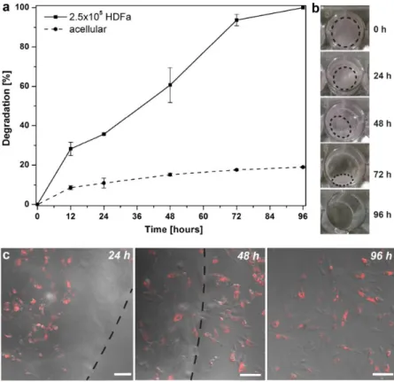
The scaffold resulted largely reduced in size and weight after 72 hours and the complete degradation was achieved after 96 hours (Figure 4a,b), demonstrating a good agreement with the GSH degradation tests. It was observed that the degradation process resulted into a gradual movement of the cells from the
nanocomposite to the bottom of the well containing the scaffold (Figure 4c). The viability of HDFa measured after the degradation of the scaffold showed that 87 % of the cells were viable, thus confirming that the degradation products were non-toxic.
Previous studies on linear
polyamidoamines systems have shown that the degradation products were completely non-cytotoxic, since the degradation produced oligomers.37
The acellular hydrogels used as control
Figure 4. HDFa-mediated degradation as function of time of dPAA
hydrogels, solid line, containing 2.5x10⁵ cells and control (acellular dPAA), dashed line (a); macroscopic pictures showing the cell-mediated degradation of the dPAA (b). Overlay images (i.e brightfield and DiD channel) of the HDFa growing onto the scaffold (24 h) and
the course of the studies (Figure 4a, dashed line). This small degradation (17 %) is probably due to the presence of fetal bovine serum in the culture medium, which contains various proteins and amino acids with thiol groups.
The results obtained confirmed that the dPAA hydrogels were prone to HDFa-mediated degradation through thiol reductive exchange, therefore showing potential for in vivo applications requiring degradation of the scaffold.
2.3.2 Mechanical properties of dPAA and mechanical degradation in presence of GSH
The mechanical properties of dPAA were characterized in terms of shear rheological behavior (Figure 5 and Supplementary Information). Specifically, dPAA is a quite soft hydrogel characterized by a shear modulus ca. 6 kPa, in agreement with other injectable hydrogels for drug delivery, allowing their mechanical properties to be matched with different soft tissues in the human body. The soft character of dPAA is particularly evident as regards the relative high fluidity of the hybrid hydrogel, characterized by a small value of the loss modulus G’’ ≈ 100 Pa. Although this parameter corresponds to a high viscosity as compared to a typical fluid, the fact that the viscous losses of the material are much lower than its effective rigidity (G’’ << G’) converts the system in a quite resilient material with a relatively high mechanical compliance. In effect, the soft mechanical nature of dPAA (a soft hydrogel with a pasty-like fluidity) makes it especially well-suited to mimic the mechanical response of muscular tissue, being structurally resilient but compliant to deformation.
After GSH addition, dPAA hydrogel did not show a clear degradation in terms of G’ and G’’ during the first hour of incubation. However, both G’ and G’’ values decreased down to 25% and 1% of their initial values respectively and eventually stabilize after 5 hours of GSH addition. The mechanical degradation causes a shear softening of dPAA and a concomitant shear fluidization of the hydrogel. Thus, dPPA hydrogel can simultaneously maintain the structural integrity but facilitate the content release of BNCs through a less viscous media under reducing conditions provided by the extracellular environment, here mimicked by the GSH solution.
2.3.3 dPAA: injectability and formation of SFC
Since the ESD procedure requires the injection of the hydrogel solution underneath the mucosa, we first optimized the synthetic procedure of the hydrogel to obtain a material that could be delivered in vivo via injection, and then rapidly jellify inside the body.
Polyamidoamine-based hydrogels have the great advantage of allowing network formation under physiological conditions.38-40 This approach has been already investigated in the past years for various
Figure 5. Shear storage modulus (G’) and shear loss modulus (G’’) of dPAA hydrogel as measured by oscillatory shear
commercially available precursors, forming polymer networks with minimal structural deficiencies within a surgically relevant timeframe, while avoiding the production of free radicals.42
When mixed all the components start to react after 30 minutes as indicated by the formation of a clear solution.
The ability of this hydrogel solution to flow through a disposable 23-gauge catheter injection needle was then examined. The dPAA solution was able to flow under hand pressure and the maximum needle injection pressure was found to be comparable to saline solution (Figure S4a). At the temperature to 37 °C, we observed the formation of a self-standing hydrogel in 18 hours. Such long time is not suitable for injectable hydrogels but knowing that the submucosa is rich in type I collagen and that this presents amino and hydroxyl groups as side groups, we postulated the possible formation of hydrogen bonds that could lead to a faster gelation kinetic in vivo.
We therefore tested this hypothesis by injecting the dPAA solution, colored in blue with methylene blue, in the submucosa of a porcine stomach, ex vivo. The injection was performed on the tissue at 37 °C and the gelation, with formation of a SFC, was immediately observed (Figure S4b). There was no leakage of the solution from the injected site and, 5-10 min after injection the tissue was cut revealing the formation of the solid phase of the dPAA (Figure S4c).
Interestingly the nanocomposite hydrogel was found completely adherent to the submucosal layer and it had to be removed with scissors and tweezers. Moreover, it was confirmed that there had not been diffusion of the solution into the surrounding tissues.
The investigation of injectability and gelation time was then performed in vivo. The dPAA showed a gelation time of approximately 3 minutes when injected in the submucosal layer in a living pig.
It is undeniable that the physiological conditions of temperature (37 °C) and pH (7.4), contribute to the faster gelation than what observed in vitro, as already reported for similar systems.43 However, tests done in vitro at 37 °C and pH 7.4 did not give the same results. This suggests the contribution of additional
factors related to the intermolecular interactions between the hydroxyl and amino groups of the collagen side chains with amide groups and side chains of dPAA that further crosslink the polymer network and facilitate the covalent polymerization. The formation of mechanical entanglements between the collagen fibers and the dPAA backbone could also contribute to the formation of an interpenetrated hydrogel network, favoring the faster formation and adhesion of a stable and elastic hydrogel in situ. This morphological change was observed in the SEM of the explanted tissues, which indeed showed interactions between the hydrogel scaffold and collagen fibers (Figure 6a).
d
Figure 6. SEM image of the explanted tissue and hydrogel, showing collagen fibers within the hydrogel matrix (a), scale
bar is 100 m; endoscopy images of the dPAA stained with Methylene Blue formed in vivo (b) and close up (c) showing with fibrous formation within the hydrogel matrix. Change in mucosal elevation as a function of time after the injection of NS or dPAA into a resected porcine stomach (d). Methylene blue was mixed as color agent. Height values (black bar) obtained for NS were 6.7 mm, 4.2 mm and 2.9 mm after 10 sec, 10 min and 1 h respectively; for dPAA were 8.3 mm, 6.4 mm, 5.8 mm after 10 sec, 10 min and 1 h respectively.
In vivo images of the dPAA formed in situ (Figure 6b) and stained with Methylene Blue also displayed
fibrous formation within the hydrogel matrix (Figure 6c). However, a deep investigation is currently ongoing to explain this behavior.
As already mentioned in ESD procedures the ability of mucosal lifting and its maintenance are essential to avoid perforation and safely complete the en bloc resection of the lesion.
We examined the formation and lasting of a SFC ex vivo comparing the dPAA with an isotonic saline solution. In particular, fresh resected porcine stomachs were used, and 2 ml of NS or DPAA solution were injected. Protrusions appeared at the injection site and the height changes in submucosal elevation were recorded after 10 seconds, 10 minutes and 1 hour, to cover the whole time of the ESD procedure, which is approximately 40-50 minutes (Figure 6d). Although both the examined solution and the hydrogel led to the mucosal elevation right after the injection, the dPAA nanocomposite displayed higher mucosal lifting: 8.3 mm vs 6.7 mm, for the dPAA and the NS respectively, with the same amount of solution injected. This showed that already part of the NS solution was absorbed by the surrounding tissues after 10 seconds. After the injection of the hydrogel solution, the formation of a solid SFC was detected, which showed only a small change in size over 1 hour, i.e. from 8.3 mm to 5.8 mm. No significant change in shape or consistency of mucosal lifting was observed. This behavior was due to the formation of the dPAA hydrogel under the submucosa.
In contrast, the elevation created with NS gradually collapsed, showing a reduction of 37% in size already after 10 minutes and of more than half after 1 hour (i.e. height from 6.7 mm to 2.9 mm).
2.4 In vivo ESD procedure
The higher performance of the dPAA nanocomposite in ex vivo was therefore evaluated in vivo in a living pig. We first set appropriate lesions sizes of approx. 3 cm in diameter in the porcine stomach and then 8 ml of the hydrogel solution were injected in the submucosa.
The ESD procedure was performed in triplicate in different areas of the same porcine stomach; NS solution was used as control.
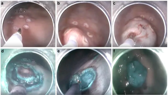
In Figure 7a is reported the endoscopic view of the stomach at time 0 before the injection of the pre-hydrogel solution. It shows the set of an appropriate lesion of approx. 3 cm in diameter. We observed that in all the cases the injected dPAA solutions in the submucosa led to the gelation of the material in less than 3 minutes, which thus formed a clear and stable mucosal elevation (Figure 7b,c).
A comparison with the normal saline solution generally used showed initially no significant difference compared to the SFC formed by dPAA. However, the elevation of the SFC formed by NS had obviously reduced after 15 min, due to quick diffusion of the NS at the target site and absorption of the liquid by the tissue, thus it was necessary to repeat the injections to keep the submucosa lifted and be able to finish the surgery.
In contrast, the mucosal lifting obtained with dPAA allowed the surgeon to perform the entire ESD procedure (40 min) without requiring a second injection, therefore significantly simplifying the procedure and avoiding large injection of liquids.
The presence of the hydrogel allowed the use of the common electrocautery settings and the long-lasting conservation of the mucosal elevation created with the dPAA enabled the surgeon to smoothly accomplish the submucosal dissection (Figure 7d). We can conclude that no significant complications or perforation occurred during the procedure due to the reliable and long-lasting mucosal lifting achieved with the dPAA.
Then, the lesion was resected en bloc without any sign of mucosal or muscularis damage, confirming that the dPAA hydrogel formed in situ was able to “dissect” the submucosal layer (Figure 7e). Intact mucosal specimens were conveniently achieved via the performed ESD procedures. This is essential, as a definite resection provides accurate histological assessment and thus, can reduce the risk of neoplastic recurrence. To prove that fast gelation can occurs also in other tissues and that the procedure can be applied for other lesions the same procedure was also performed in the colon and in the esophagus with excellent results (Figure S5). Besides aiding the surgery, the protecting layer created with the gels can
Figure 7. Endoscopic views of the different steps of the ESD procedure performed using the dPAA stained
with Methylene Blue. Setting of the lesion, approx. 3 cm in diameter (a); injection of the dPAA solution (b); formation of the SFC after gelation of the dPAA (c); circumferential cutting (d); complete resection with protective layer of dPAA that remained adhered to the muscolaris (e); wound left after ESD with layer of the dPAA (f).
protect the layer underneath (Figure 7f). The possibility of releasing active components from the breakable capsules is highly beneficial at the end of such delicate procedure. Biomolecules, such as adrenaline, proton-pump inhibitors, corticoids or antibiotics could potentially be efficiently delivered to assist the cauterization of the resected tissue or the prevention of inflammations.
3. Conclusions
A degradable hybrid hydrogel was successfully developed by embedding breakable nanocapsules into a disulfide-containing polyamidoamines-based hydrogel.
We demonstrated that the network can be completely cleaved in 3 days upon incubation in a GSH solution mimicking the extra-cellular concentration (10 M). Most importantly, the obtained dPAA sustained the proliferation of HDFa and underwent complete degradation in response to cell-secreted molecules from HDFa seeded onto the scaffold, without any external stimulus.
The obtained dPAA can be delivered to the desired tissue by facile injection through a 23-gauge needle. To prove its applicability in real models, the hydrogel solution was injected in the submucosa of a porcine stomach in vivo. It formed an elastic hydrogel in 3 minutes, most likely due to temperature increase and interaction with collagen fibers.
Such an important result, together with the low-cost starting materials employed for the synthesis, motivated us to employ the dPAA hydrogel as a novel submucosal injection agent in ESD. The hydrogel formed a reliable SFC in vivo, enabling a long-lasting mucosal elevation, which was superior to commonly used NS. This facilitated en bloc resection of the lesion, which was successfully accomplished
Moreover, part of the in situ formed hydrogel adhered tightly to the muscolaris, under the resected mucosa, allowing protection of the membrane during the procedure and after it.
The possibility to release active molecules and to biodegrade the hybrid hydrogel in few days opens interesting perspective for a faster regeneration of the tissue.
4. Experimental Section
Synthesis of dPAA: In a 5 ml round bottom flask methylenbisacrylamide (MBA; 200 mg) and
N,N-dimethylethylenediamine (70 l) were added to a water solution of amino-functionalized BNCs freshly sonicated (1.5 ml; 1 mg/ml). Then, cystamine (65 mg) was added to the mixture that was stirred vigorously for 1 hour at room temperature, after which a homogeneous and clear pre-hydrogel solution was obtained. When the hydrogels were needed for in vitro experiments (i.e. GSH degradation and cellular viability and degradation), the obtained solution was transferred to glass vials (500 l per vial) and allowed to react in static conditions at r.t. Glass vials with inner diameter of 8 mm were used as molds. The hydrogels were obtained after 48 hours.
Once obtained, the disk-shaped hydrogels were freeze-dried and weighted. Dried hydrogels were used to study the swelling ratio at different pH and the degradation kinetics with different concentrations of GSH. This step allowed us as well to sterilize the materials for in vitro experiments. Sterile and ultrafiltered water was used during hydrogel preparation for in vivo tests; the synthesis was carried out in closed sterile vials and protected from bacteria contamination, thus we assumed that the final product was free of bacterial contamination.
To assess the presence of unreacted MBA, the hydrogels were washed several times; the extracted washing solutions were then freeze-dried and analyzed by H-NMR spectroscopy, which confirmed that
no unreacted monomers were present. We concluded that the final hydrogel composition was not significantly different from the feeding starting monomer mixture, as elsewhere observed.26
Cell-mediated degradation of dPAA: Hydrogels nanocomposites were freeze-dried and weighed (Wi).
Then 2.5x10⁵ HDFa were seeded onto the samples (see paragraph 5.6.5). The cell-laden samples were
collected at pre-determined time points and were freeze-dried to obtain their dry weight after degradation (Wf).
The cell-mediated degradation of the hydrogels, D, was calculated using the following equation:
𝐷 (%) = 𝑊𝑖 − 𝑊𝑓
𝑊𝑖 × 100
Acellular hydrogels were used as degradation control.
Creating submucosal cushion and performing ESD in a living pig: The pig was fasted for 1 day before
operation. A standard endoscope (Karl Storz, Tuttlingen, Germany) was used in the pig under general anesthesia. Both the dPAA solution and the NS used as control contained a small amount of Methylene Blue as a color agent in order to facilitate visualization of the SFC.
After setting appropriate lesion sizes of approx. 3 cm in diameter in the porcine stomach, 8-10 ml of dPAA solution and NS were injected in the stomach submucosa through the endoscope accessory channel using a 23-gauge injection needle.
The mucosal elevation due to the injected dPAA at the target site was observed endoscopically before starting the ESD. It was compared under direct view with the elevation caused by NS during the procedure. After injection, the ESD was performed and a circumferential mucosal incision was accomplished using a TT knife (Olympus, Tokyo, Japan). Injection of dPAA and ESD were repeated three times. The animal was euthanized after completion of experiments; the whole procedure was
The main outcome measures were (1) the rapid gelation of dPAA when injected into the submucosa and (2) the long-lasting SFC formed; (3) the feasibility of the dissection procedure during ESD; (3) the adhesion of dPAA to the muscolaris layer and thus the increase of protection during the procedure and after it.
ASSOCIATED CONTENT
The following files are available free of charge:
Supporting information (PDF), including details of the synthesis and functionalization of BNCs, degradation kinetic in presence of GSH, mechanical characterization of dPAA by shear rheology,
in-vitro cell culturing, cell staining and viability studies, evaluation of the gelation and formation of SFC ex
vivo.
AUTHOR INFORMATION Corresponding Author
* Prof. L. De Cola, Institut de Science et d'Ingénierie Supramoléculaires, Université de Strasbourg, 8 rue Gaspard Monge, 67000 Strasbourg, France
E-mail: [email protected]
Author Contributions
The manuscript was written through contributions of all authors. All authors have given approval to the final version of the manuscript. ‡These authors contributed equally to the work.
ACKNOWLEDGMENT
F.F. and L.D.C. acknowledge the European Research Council (ERC), grant no. 2009-247365 titled “MaGIC”, for financial support.
G.A. and L.D.C. acknowledge the European Union Horizon 2020 research and innovative programme under the Marie Sklodowska-Curie grant agreement No 642192.
I. L-M acknowledge the ERC Starting Grant MITOCHON” (ERC-StG-2013-338133).
I. L-M. and F.M. acknowledge Spanish Ministry of Economy MINECO (FIS2015-70339-C2-1-R) The authors gratefully acknowledge use of the services and facilities of the IRCAD and the Institute of Image Guided Surgery IHU-Strasbourg.
REFERENCES
(1) Ferlay, J.; Shin, H. R.; Bray, F.; Forman, D.; Mathers, C.; Parkin, D. M., Estimates of Worldwide Burden of Cancer in 2008: Globocan 2008. Int. J. Cancer 2010, 127 (12), 2893-917.
(2) Coda, S.; Lee, S.-Y.; Gotoda, T. Endoscopic Mucosal Resection and Endoscopic Submucosal Dissection as Treatments for Early Gastrointestinal Cancers in Western Countries. Gut and Liver 2007, 1 (1), 12-21.
(3) Ono, H.; Yao, K.; Fujishiro, M.; Oda, I.; Nimura, S.; Yahagi, N.; Iishi, H.; Oka, M.; Ajioka, Y.; Ichinose, M.; Matsui, T. Guidelines for Endoscopic Submucosal Dissection and Endoscopic Mucosal Resection for Early Gastric Cancer. Digestive Endoscopy 2016, 28 (1), 3-15.
(4) Hosokawa, K.; Yoshida, S. Recent Advances in Endoscopic Mucosal Resection for early Gastric Cancer. Gan To Kagaku Ryoho 1998, 25 (4), 476-483.
(5) Hirao, M.; Masuda, K.; Asanuma, T.; Naka, H.; Noda, K.; Matsuura, K.; Yamaguchi, O.; Noriyuki Ueda. Endoscopic Resection of Early Gastric Cancer and other Tumors with local injection of Hypertonic Saline-Epinephrine. Gastrointestinal endoscopy 1988, 34 (3), 264-269.
(6) Ishihara, M.; Kumano, I.; Hattori, H.; Nakamura, S. Application of Hydrogels as Submucosal Fluid Cushions for Endoscopic Mucosal Resection and Submucosal Dissection. Journal of Artificial
Organs 2015, 18 (3).
(7) Yu, L.; Xu, W.; Shen, W.; Cao, L.; Liu, Y.; Li, Z.; Ding, J. Poly(lactic acid-co-glycolic acid)– poly(ethylene glycol)–poly(lactic acid-co-glycolic acid) Thermogel as a Novel Submucosal
various Submucosal Injection Solutions for maintaining Mucosal Elevation during Endoscopic Mucosal Resection. Endoscopy 2004, 36 (7), 579-583.
(9) Oka, S.; Tanaka, S.; Kaneko, I.; Mouri, R.; Hirata, M.; Kawamura, T.; Yoshihara, M.; Chayama, K. Advantage of Endoscopic Submucosal Dissection Compared with EMR for early Gastric Cancer. Gastrointest. Endosc. 2006, 64 (6), 877-883.
(10) Gotoda, T.; Yamamoto, H.; Soetikno, M. R. Endoscopic Submucosal Dissection of Early Gastric Cancer. J. Gastroenterol. 2006, 41 (10), 929-942.
(11) Committee, A. T.; Maple, J. T.; Abu Dayyeh, B. K.; Chauhan, S. S.; Hwang, J. H.; Komanduri, S.; Manfredi, M.; Konda, V.; Murad, F. M.; Siddiqui, U. D.; Banerjee, S. Endoscopic Submucosal Dissection. Gastrointestinal endoscopy 2015, 81 (6), 1311-25
(12) Fujishiro, M.; Yahagi, N.; Kashimura, K.; Matsuura, T.; Nakamura, M.; Kakushima, N.; Kodashima, S.; Ono, S.; Kobayashi, K.; Hashimoto, T.; Yamamichi, N.; Tateishi, A.; Shimizu, Y.; Oka, M.; Ichinose, M.; Omata, M. Tissue Damage of Different Submucosal Injection Solutions for EMR. Gastrointest. Endosc. 2005, 62 (6), 933-942.
(13) Cho, K. B.; Jeon, W. J.; Kim, J. J. Worldwide Experiences of Endoscopic Submucosal Dissection: Not just Eastern acrobatics. World Journal of Gastroenterology : WJG 2011, 17 (21), 2611-2617. (14) Tran, R. T.; Palmer, M.; Tang, S.-J.; Abell, T. L.; Yang, J. Injectable drug-eluting Elastomeric
Polymer: a novel Submucosal Injection Material. Gastrointest. Endosc. 2012, 75 (5), 1092-1097. (15) Uraoka, T.; Fujii, T.; Saito, Y.; Sumiyoshi, T.; Emura, F.; Bhandari, P.; Matsuda, T.; Fu, K.-I.;
Saito, D. Effectiveness of Glycerol as a Submucosal Injection for EMR. Gastrointest. Endosc. 2005, 61 (6), 736-740.
(16) Fujishiro, M.; Kakushima, N.; Kodashima, S.; Kashimura, K.; Matsuura, T.; Muraki, Y.; Tateishi, A.; Omata, M. Appropriate mixture of Hyaluronic Acid, Glucose and Glycerin for a Submucosal Fluid Cushion during Endoscopic Submucosal Dissection in the Dog Stomach. Digestive
Endoscopy 2007, 19 (1), 26-31.
(17) Feitoza, A. B.; Gostout, C. J.; Burgart, L. J.; Burkert, A.; Herman, L. J.; Rajan, E. Hydroxypropyl Methylcellulose: A better Submucosal Fluid Cushion for Endoscopic Mucosal Resection.
Gastrointest. Endosc. 2003, 57 (1), 41-47.
(18) Hattori, H.; Tsujimoto, H.; Hase, K.; Ishihara, M. Characterization of a water-soluble Chitosan derivative and its potential for Submucosal Injection in Endoscopic Techniques. Carbohydrate
polymers 2017, 175, 592-600.
(19) Yoshida, N.; Naito, Y.; Kugai, M.; Inoue, K.; Uchiyama, K.; Takagi, T.; Ishikawa, T.; Handa, O.; Konishi, H.; Wakabayashi, N.; Yagi, N.; Kokura, S.; Morimoto, Y.; Kanemasa, K.; Yanagisawa, A.; Yoshikawa, T. Efficacy of Hyaluronic Acid in Endoscopic Mucosal Resection of Colorectal Tumors. J. Gastroenterol. Hepatol. 2011, 26 (2), 286-291.
(20) Matsui, Y.; Inomata, M.; Izumi, K.; Sonoda, K.; Shiraishi, N.; Kitano, S. Hyaluronic Acid stimulates Tumor-cell Proliferation at wound sites. Gastrointest. Endosc. 2004, 60 (4), 539-543. (21) Hayashi, T.; Matsuyama, T.; Hanada, K.; Nakanishi, K.; Uenoyama, M.; Fujita, M.; Ishihara, M.;
Kikuchi, M.; Ikeda, T.; Tajiri, H. Usefulness of Photocrosslinkable Chitosan for Endoscopic Cancer Treatment in Alimentary Tract. Journal of Biomedical Materials Research Part B: Applied
Biomaterials 2004, 71B (2), 367-372.
(22) Kumano, I.; Ishihara, M.; Nakamura, S.; Kishimoto, S.; Fujita, M.; Hattori, H.; Horio, T.; Tanaka, Y.; Hase, K.; Maehara, T. Endoscopic Submucosal Dissection for Pig Esophagus by using Photocrosslinkable Chitosan Hydrogel as Submucosal Fluid Cushion. Gastrointest. Endosc. 2012,
(23) Cao, L.; Li, Q.; Zhang, C.; Wu, H.; Yao, L.; Xu, M.; Yu, L.; Ding, J. Safe and Efficient Colonic Endoscopic Submucosal Dissection Using an Injectable Hydrogel. ACS Biomaterials Science &
Engineering 2016, 2 (3), 393-402.
(24) Ifkovits, J. L.; Burdick, J. A. Review: Photopolymerizable and Degradable Biomaterials for Tissue Engineering Applications. Tissue Eng. 2007, 13 (10), 2369-2385.
(25) Ferruti, P. Poly(amidoamine)s: Past, present, and perspectives. Journal of Polymer Science Part
A: Polymer Chemistry 2013, 51 (11), 2319-2353.
(26) Fiorini F.; Prasetyanto E. A.; Taraballi F.; Pandolfi L.; Monroy F.; López-Montero I.; Tasciotti E.; De Cola L. Nanocomposite Hydrogels as Platform for Cells Growth, Proliferation, and Chemotaxis. Small, 2016, 12 (35), 4881-4893.
(27) Rizzi V.; Fiorini F.; Lamanna G.; Gubitosa J.; Prasetyanto E.A.; Fini P.; Fanelli F.; Nacci A.; De Cola L.; Cosma P., Polyamidoamine‐Based Hydrogel for Removal of Blue and Red Dyes from Wastewater. Adv. Sustainable Syst. 2018, 2, 1700146. DOI: 10.1002/adsu.201700146.
(28) Maggini, L.; Cabrera, I.; Ruiz-Carretero, A.; Prasetyanto, E. A.; Robinet, E.; De Cola, L. Breakable Mesoporous Silica Nanoparticles for targeted Drug Delivery. Nanoscale 2016, 8 (13), 7240-7247. (29) Yang, F.; Wang, J.; Peng, G.; Fu, S.; Zhang, S.; Liu, C. PEG-based bioresponsive Hydrogels with
Redox-mediated Formation and Degradation. J. Mater. Sci. Mater. Med. 2012, 23 (3), 697-710. (30) Kar, M.; Vernon Shih, Y.-R.; Velez, D. O.; Cabrales, P.; Varghese, S. Poly(ethylene glycol)
Hydrogels with Cell Cleavable Groups for autonomous Cell Delivery. Biomaterials 2016, 77, 186-197.
(31) Huang Y.; Luo Q.; Zha G.; Zhang J.; Li X.; Zhao S.; Li X., Biomimetic ECM Coatings for Controlled Release of rhBMP-2: Construction and Biological Evaluation, Biomater. Sci., 2014, 2 (7), 980-989
(32) Prasetyanto, E. A.; Bertucci, A.; Septiadi, D.; Corradini, R.; Castro-Hartmann, P.; De Cola, L. Breakable Hybrid Organosilica Nanocapsules for Protein Delivery. Angew. Chem. Int. Ed. 2016,
55 (10), 3323-3327.
(33) Smithmyer, M. E.; Sawicki, L. A.; Kloxin, A. M. Hydrogel Scaffolds as in vitro Models to study Fibroblast Activation in Wound Healing and Disease. Biomaterials Science 2014, 2 (5), 634-650. (34) Kagatani, S.; Sasaki, Y.; Hirota, M.; Mizuashi, M.; Suzuki, M.; Ohtani, T.; Itagaki, H.; Aiba, S.
Oxidation of Cell Surface Thiol groups by Contact Sensitizers triggers the Maturation of Dendritic Cells. The Journal of investigative dermatology 2010, 130 (1), 175-83.
(35) Sahaf, B.; Heydari, K.; Herzenberg, L. A.; Herzenberg, L. A. Lymphocyte Surface Thiol levels.
Proceedings of the National Academy of Sciences of the United States of America 2003, 100 (7),
4001-5.
(36) Donoghue, N.; Yam, P. T.; Jiang, X. M.; Hogg, P. J. Presence of Closely Spaced Protein Thiols on the Surface of Mammalian Cells. Protein science: a publication of the Protein Society 2000, 9 (12), 2436-45.
(37) Ferruti, P.; Bianchi, S.; Ranucci, E.; Chiellini, F.; Caruso, V. Novel Poly ( amido-amine ) -Based Hydrogels as Scaffolds for Tissue Engineering. Macromol. Biosci. 2005, 613-622.
(38) Peng, B.; Lai, X.; Chen, L.; Lin, X.; Sun, C.; Liu, L.; Qi, S.; Chen, Y.; Leong, K. W. Scarless Wound Closure by a Mussel-Inspired Poly(amidoamine) Tissue Adhesive with Tunable Degradability. ACS Omega 2017, 2 (9), 6053-6062.
(39) Wang, J.; He, H.; Cooper, R. C.; Yang, H. In Situ-Forming Polyamidoamine Dendrimer Hydrogels with Tunable Properties Prepared via Aza-Michael Addition Reaction. ACS applied materials &
(41) Lutolf, M. P.; Hubbell, J. A. Synthesis and Physicochemical Characterization of End-Linked Poly(ethylene glycol)-co-peptide Hydrogels Formed by Michael-Type Addition.
Biomacromolecules 2003, 4 (3), 713-722.
(42) Pritchard, C. D.; O’Shea, T. M.; Siegwart, D. J.; Calo, E.; Anderson, D. G.; Reynolds, F. M.; Thomas, J. A.; Slotkin, J. R.; Woodard, E. J.; Langer, R. An Injectable Thiol-Acrylate Poly(ethylene glycol) Hydrogel for sustained release of Methylprednisolone sodium succinate.
Biomaterials 2011, 32 (2), 587-597.
(43) Nguyen, M. K.; Park, D. K.; Lee, D. S. Injectable Poly(amidoamine)-poly(ethylene glycol)-poly(amidoamine) Triblock Copolymer Hydrogel with Dual Sensitivities: pH and Temperature.
Biomacromolecules 2009, 10 (4), 728-731.