ARTICLE
PARANASAL SINUS SYSTEM OF GEOSAURUS ARAUCANENSIS AND THE HOMOLOGY
OF THE ANTORBITAL FENESTRA OF METRIORHYNCHIDS
(THALATTOSUCHIA: CROCODYLOMORPHA)
MARTA S. FERNA´ NDEZ*,1and YANINA HERRERA2
1
Departamento Paleontologı´a Vertebrados, Museo de La Plata, Paseo del Bosque s/n, (1900) La Plata, Argentina, CONICET, martafer@fcnym.unlp.edu.ar;
2
Departamento Paleontologı´a Vertebrados, Museo de La Plata, Paseo del Bosque s/n, (1900) La Plata, Argentina, yaninah@fcnym.unlp.edu.ar
ABSTRACT—Metriorhynchids have been interpreted as the only archosaurs entirely adapted to pelagic marine life, given the deep morphological modifications of their skeletons. The most conspicuous feature in the skull involves the “fenestra and fossa antorbitalis complex.” Exceptionally preserved natural cast of snout cavities of Geosaurus araucanen-sis, found in the Late Jurassic of the Neuque´n Basin (northwestern Patagonia, Argentina), allow exploration of the rostral anatomy of this metriorhynchid. The presence of a paranasal sinus system, hypothetically reconstructed by other authors using EPB method approach, can now be confirmed based on direct morphological evidence. We propose that the openings classically identified in the literature as internal and external antorbital fenestrae of metriorhynchid have been misidentified; the preorbital opening of metriorhynchids is a neomorph associated with a novel salt gland that evolved independent of the antorbital fenestra, and that the true antorbital cavity of G. araucanensis, as well as this cavity in all other metriorhynchids, is internalized. Although this hypothesis could be considered as unorthodox, it is the one that requires the least ad-hoc assumptions to explain observations. Many phylogenetic studies depict the Thalattosuchia nested within Neosuchia. In these trees, the successive outgroups of Thalattosuchia are Dyrosaurids/Pholidosaurids, followed by derived Neosuchians (Goniopholids, Bernissartia, Eusuchians). All these taxa have a closed antorbital fenestra. Within this phylogenetic scenario, the internalization of the antorbital cavity did not occur in the ancestor of thalattosuchians, but in the ancestor of a much more inclusive clade of neosuchians.
INTRODUCTION
Evolutionary history of archosaurs, which spans 250 million years, occurred mainly on the continental realm. Although throughout this long history the fossil record documents several attempts of marine colonization since at least the Middle Trias-sic (Li et al., 2006), only metriorhynchid crocodylomorphs can be defined as completely adapted to pelagic marine life (e.g., capable of drinking sea water and eating osmoconforming preys). The fossil record suggests that metriorhynchids achieved the highest degree of marine adaptation, at least since the Mid-dle Jurassic (Ferna´ndez and Gasparini, 2008). Their skeletons display unique modifications such as a streamlined skull and body, loss of bony armor, short and paddle-like forelimbs and a hypocercal tail. Within the skull, one of the most conspicuous reorganizations involves the antorbital fenestra and fossa com-plex. In metriorhynchids, the internal antorbital fenestra is reor-iented so that it is hidden in lateral view or can be seen in anterolateral view. Contrarily, in other crocodylomorphs with external and internal antorbital fenestra exposed, the internal antorbital fenestra can be seen in lateral view, even in forms with an elongate external antorbital fenestra such as Calsoyasu-chus valliceps Tykoski et al., 2002. In metriorhynchids, bony recesses, principally on the maxilla and nasal, form an elongate, low, and obliquely oriented antorbital fossa, occurring anteriorly to a reduced internal antorbital fenestra. Recent phylogenetic revisions indicate that the Metriorhynchidae are a well-suppor-ted clade within Thalattosuchia (Gasparini et al., 2006; Pol and
Gasparini, 2009). Among the synapomorphies supporting this clade, three comprise the fenestra and fossa antorbitalis (i.e., elon-gate and low antorbital fenestra oriented obliquely, nasals des-cending on lateral surface of skull with extensive participation in antorbital fossa and fenestra, and jugal participating in the antor-bital fossa). Despite the fact that antorantor-bital fenestra and fossa in metriorhynchids are quite peculiar in comparison with other Cro-codylomorpha and have been extensively used to resolve phyloge-netic relationships of thalattosuchians (e.g., Clark, 1994; Gasparini et al., 2006; Young, 2006; Pol and Gasparini, 2009), no explicit discussions have concentrated on the primary homology of these structures in thalattosuchians in general, and in metriorhynchids in particular.
Although the fenestra and antorbital cavity are among the most frequently cited synapormophies of archosaurs, their func-tion remained obscure for many years. Three main hypotheses concerning the antorbital cavity function have been proposed in the literature: the antorbital cavity houses a gland, part of the jaw adductor musculature, or an air sac. In his classic paper, Witmer (1997) used the Extant Phylogenetic Bracket approach (EPB, Witmer, 1995a) to test the hypotheses, using birds and crocodilians as extant brackets. The only hypothesis that sur-vived testing with very little speculation (level I inference) is the last: the antorbital cavity houses an epithelial air sac in all archosaurs.
Exceptionally preserved natural casts of the snout cavity of the metriorhynchid Geosaurus araucanensis Gasparini and Dellape´, 1976, reveal for the first time, internal details of the snout anatomy. Examination of these natural casts permits test-ing of hypothetical reconstructions based on the EPB approach (Witmer, 1997), as well as hypotheses concerning bony snout
*Corresponding author.
Journal of Vertebrate Paleontology 29(3):702–714, September 2009 #2009 by the Society of Vertebrate Paleontology
structure homologies. Those discussed here include: soft struc-tures preserved as natural casts (i.e., glands and paranasal diver-ticula), and bony structures related to them (i.e., antorbital fossa and fenestra). As Rieppel and Kearney (2002) pointed out, in morphology-based phylogenetic analyses, a tendency toward in-creasing the amount of data could discourage in depth compara-tive anatomical studies. These authors proposed that primary conjectures of homology need to be testable and potentially refutable in their own right. In this context, the objectives of this study are to discuss primary conjectures concerning homology (i.e., homology statements before phylogenetic reconstructions) of the antorbital fenestra and fossa complex of metriorhynchids and to explore how soft tissue reconstructions and skeletal fea-tures can be integrated to test conjecfea-tures of bony structure homologies in extinct forms.
Institutional Abbreviations—MDA, Museo del Desierto de Atacama, Chile; MGHF, Museo Fuenzalida at the Universidad Cato´lica de Chile; MHNSR, Museo de San Rafael, Argentina; MLP, Museo de La Plata, Argentina; MOZ, Museo Olsacher, Zapala, Argentina.
Anatomical Abbreviations—afo, antorbital fossa; asin, antor-bital sinus; cnp, nasal cavity proper; co, convexity; csin, cavicon-chal sinus; div 1, diverticulum 1; div 2, diverticulum 2; eaf, external antorbital fenestra; f, frontal; font a o, fonticulus antor-bitalis; fpn, postnasal fenestra; gd, duct of the gland; gl, gland; gr, groove; iaf, internal antorbital fenestra; la, lacrimal; lwdiv2, lat-eral wall of diverticulum 2; mx, maxilla; n, nasal; ngl, nasal gland; npdu, nasopharyngeal duct; oasin, opening of the antorbital si-nus; ogl, opening for salt gland drainage; or, orbit; pch, primary choana; pfr, prefrontal; pl, palatine; pfrp, prefrontal pillar; pmx, premaxilla; preo, preorbital opening; sdac, subsidiary diverticu-lum in accessory cavity; sdiv, suborbital diverticudiverticu-lum; se, septum; snf, suture between nasal and frontal; ve, vessel; vo, vomer.
MATERIAL AND METHODS Materials
Materials used to analyze the snout anatomy of Geosaurus araucanensis Gasparini and Dellape´, 1976, include five three-dimensionally preserved skulls including the holotype (MLP 72-IV-7-1, MLP 72-IV-7-2, MLP 72-IV-7-3, MLP 72-IV-7-4, MLP 86-XI-5-7) and four specimens (MLP 76-XI-19-1, MLP 84-V-1-1, MLP 86-XI-10-6, MLP 86-XI-10-7), in which part of the filling of soft component of the snout are preserved as natural casts. All the specimens were recovered from Tithonian beds (Late Juras-sic) of the Vaca Muerta Formation exposed in the Neuque´n basin, northwestern Patagonia, Argentina. Other metriorhynchids with 3-D preserved skulls were used for comparison (Metriorhynchus casamiquelai MGHF 1-08573, 181097; Metriorhynchus wester-manni MDA 1, and Dakosaurus andiniensis MHNSR 344, MOZ 6146).
The reconstruction of soft structures within the snout is mainly based on two specimens of G. araucanensis (MLP 76-XI-19-1, 84-V-1-1). In these specimens the bones of the snout have been naturally removed by weathering leaving exposed the natural cast of the major soft components of this region (Figs. 1, 2, 3A– C). One of the natural casts (MLP 84-V-1-1) was accidentally broken into three sections during extraction (I, II and III, Fig. 2A), exposing the internal arrangement of the major com-ponents of the nasal cavity for study.
Methods
Reconstruction of soft organs—In order to reconstruct the soft organs, we first described the soft structures preserved as natural casts and the relationship between them and the skull bones and cavities. As the scars of the bony sutures are preserved on the external surface of the natural casts, the reconstruction of soft
tissues could be superimposed on the image of the G. araucanen-sis holotype skull (MLP 72-IV-7-1) to identify the exact relation-ship between the soft organs and the bones covering them. Secondly, homologies of the soft structures preserved in the natural casts were hypothesized by topological correspondence with the facial structures described in extant archosaurs (Witmer, 1995b). Thirdly, the osteological correlates of soft tissues hypothesized in G. araucanensis were explored in other metriorhynchids. A posteriori to the application of this protocol, reconstructions based on the natural casts were used to test the reconstructions of facial structures of extinct archosaurs based on the Extant Phylogenetic Bracket approach (EPB) (Witmer, 1995a).
To explore internal anatomical details of the snout, MLP 76-XI-19-1 and MLP 72-IV-7-1 were subjected to X-ray compu-tered tomographic (CT) scanning at the Hospital Paroissien in La Matanza, Buenos Aires, Argentina. Slice thickness for MLP 76-XI-19-1 and MLP 72-IV-7-1 were 1 mm and 2 mm, respec-tively. In both cases, CT scanning provided good resolution be-tween bone and rock matrix. The anatomical terminology used herein follows Witmer (1995b).
Homology—Establishment of homology has been proposed as a two-step independent procedure (de Pinna, 1991). The first step comprises the hypothesis of structural correspondences (i.e., primary homology) based on comparative biology. In the second step, character hypotheses are subjected to the test of congruence (optimized onto the optimal phylogenetic trees). This test equates homology to synapormorphy (i.e., secondary homology). As these two steps are independent, the optimiza-tion procedure does not involve any alteraoptimiza-tion of the correspon-dences that were previously determined as primary homologies (Ramirez, 2007). Rieppel and Kearney (2002) proposed that primary conjectures of homology need to be testable, and poten-tially refutable, in their own right. In this context, the focus of this contribution is related to the first step of this procedure. Primary homologies are hypothesized and tested based on mor-phological evidences (i.e., topological equivalence, or position and connections); and when more than one hypothesis of struc-tural correspondences can be constructed, the one that requires the least ad-hoc assumptions to explain observations is pre-ferred. This approach is close in spirit to the ideas proposed by Ramı´rez (2007), who considered homology as a parsimony problem.
After the formulation and test of the hypotheses of primary homology of the antorbital fenestra, the hypothesis that survived testing was mapped on a generalized thalattosuchian tree that summarizes published cladograms (Clark, 1994; Mueller-To¨we, 2005; Gasparini et al., 2006).
SNOUT MORPHOLOGY OF GEOSAURUS ARAUCANENSIS
Soft Organs Preserved as Natural Casts
General Features—The most complete natural casts corre-spond to two adult specimens of G. araucanensis (MLP 76-XI-19-1 and 84-V-1-1) (Figs. 1, 2, 3A–D). In both specimens most of the nasal cavity has been preserved as natural casts.
The soft structures that can be observed and externally out-lined on the natural casts are glands, components of the cartilag-inous nasal capsule, nasopharyngeal ducts, and diverticula of the nasal cavity. Geodes formed on the internal walls of some of the structures (i.e., nasopharyngeal ducts and diverticula) in MLP 84-V-1-1. A common genetic aspect of the geode formation is the emplacement by precipitation from aqueous solution (authi-genesis) within cavities (e.g., cavities within fossils) (Milliken, 2003). In the MLP 84-V-1-1, cavities such as the nasopharyngeal ducts and diverticula could become the locus of geode formation (Fig. 3A, B).
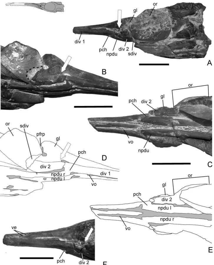
FIGURE 1. Natural cast of Geosaurus araucanensis (MLP 76-XI-19-1). A, left lateral view; B, right lateroventral; C, ventral view; D, line drawings of B; E, line drawings of C; F, left laterodorsal view. White arrow shows the location of the opening classically identified as internal antorbital fenestra. Black dashed line on B, posterior extension of suborbital diverticulum. Line drawing on the left upper corner shows the part preserved as natural cast. Scale bars equal 5 cm.
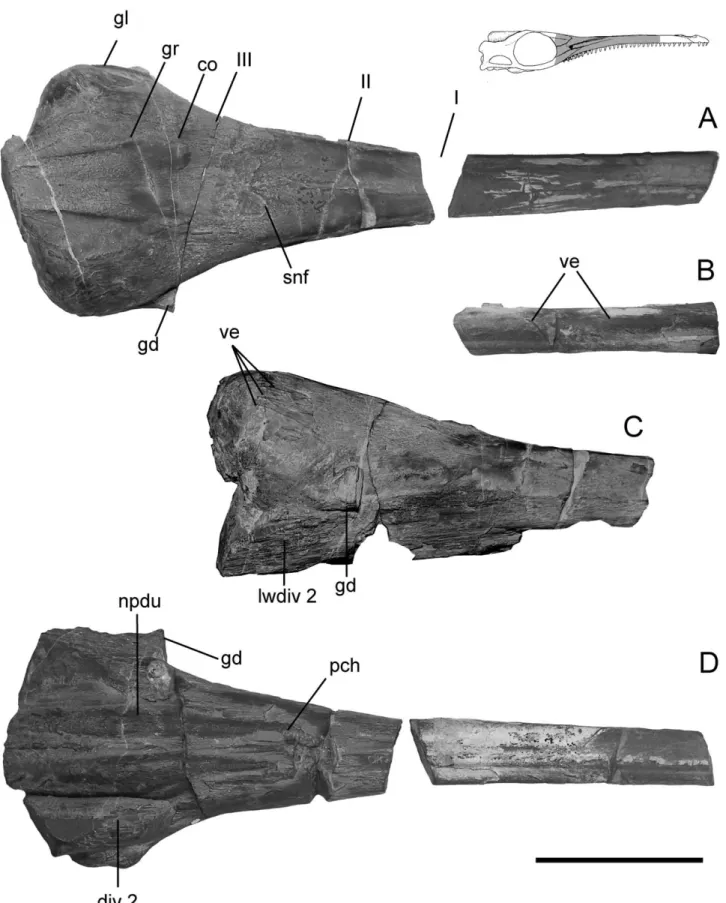
FIGURE 2. Natural cast of Geosaurus araucanensis (MLP 84-V-1-1). A, dorsal view; B, anterior fragment in left lateral view; C, right lateral view; D, ventral view. Roman numbers show the position of the planes of fracture. Line drawing on the right upper corner shows the part preserved as natural cast. Scale bar equals 5 cm.
Nasal Cavity—In crocodilians the nasal cavity can be divided into three major components, the vestibule, the nasal cavity proper, and nasopharyngeal ducts (Parsons, 1970). As out-growths of the nasal cavity beyond the cartilaginous nasal cap-sule, paranasal sinuses or diverticula occur, which are closely related to bony recesses. Witmer (1995b) identified five major recesses and air sacs associated with the nasal cavity of extant crocodilians, among which the caviconchal sinus is proposed as the homologue to the antorbital sinus of birds and other osaurs. The osteological correlate of this sinus is, in living arch-osaurs, a bony aperture (internal antorbital fenestra) through which passes the paranasal air sac (Witmer, 1997).
Of the three major divisions of the nasal cavity, enough mor-phological evidence is preserved to reconstruct most of the nasal cavity proper and nasopharyngeal ducts, as well as paranasal diverticula. Approximately 60% of the infilling of the nasal cavi-ty proper is preserved in MLP 76-XI-19-1, and 80% in MLP 84-V-1-1 (Figs. 1, 2).
The anterior part of the nasal cavity proper is roughly quad-rate in cross section, with vertical lateral margins. The external
surface of the laterally surrounding bones (premaxilla and max-illa) is convex. Posteriorly, the nasal cavity is dorsoventrally depressed with a slightly concave dorsal surface. The cross sec-tion of the rostrum corresponds to the narrow platyrostral con-dition (Busbey, 1995).
The texture of the roof of the nasal capsule and lateral walls is relatively smooth, and impressions of blood vessels have been preserved on both specimens. The smooth texture is only marked by fine and subtle striations, principally orientated ante-roposteriorly. The location of the septum nasale is also clearly marked in both specimens, although preservation is slightly dif-ferent: on MLP 76-XI-19-1 the location of the septum is marked by a low ridge, whereas on the anterior part of MLP 84-V-1-1 the septum is similarly marked, except that posteriorly, a 3 mm wide rugose texture occurs. On both sides of the septum, the roof of the nasal capsule is slightly convex.
On the left lateral aspect of the natural cast of MLP 76-XI-19-1, the infilling of a major vessel extends posteriorly from the later-odorsal portion of the lateral wall, becomes slightly inclined ventrally, and splits into caudodorsal and caudoventral branches
FIGURE 3. A–C, Natural cast of Geosaurus araucanensis (MLP 84-V-1-1). A, Anterior view at the plane fracture III; B, anterior view at the plane fracture II; C, posterior view. See Figure 2 for position of plane fractures. D, CT slice of MLP 76-XI-19-1 through rostrum in transverse plane; E, posterior view of MLP 86-XI-10-7; F, G. araucanensis (MLP 76-XI-19-1) showing the relative positions of the slice plane shown in D. Scale bars equal 5 cm.
(Fig. 1F). On the right side, one blood vessel impression has been preserved and has the same course as the caudodorsal branch of the left view.
On the dorsum of both specimens, the suture between the anterior process of the frontal and nasals is marked by a low ridge (Fig. 2A). Here, the texture is more rugose and coarse. Posteriorly between the salt glands in MLP 84-V-1-1, the texture is coarsely striate and fibrous. Also in this area, two anteropos-teriorly elongate convexities were preserved in both natural casts (Fig. 2A). These convexities correspond, in both form and location, to the osseous concavities on the ventral surface of the frontal of Metriorhynchus superciliosus identified as the osteolo-gical correlates of the olfactory bulbs by Wenz (1968). Laterally, a groove separates these convexities from the space containing the casts of both left and right glands (Fig. 2A, 3B). This groove can also be observed in the CT scan of the holotype (Fig. 4D).
Nasopharyngeal Ducts—Both nasopharyngeal ducts are com-pletely preserved in MLP 76-XI-19-1 (Fig. 1B). The right duct is also completely exposed, whereas the posterior portion of the left nasopharyngeal duct is covered by matrix and can only be observed in the CT scan images. In MLP 84-V-1-1, the filling of the ducts are preserved from their origin up to the front margin of the orbit (Fig. 2D). The accidentally broken specimen allows observation of a transverse section with small inwardly pointing crystals distributed on the duct walls in the form of geodes (Fig. 3A). Impressions of blood vessels have been preserved on the external surface of both specimens. Two major branches, almost parallel to each other, extend anteroposteriorly along the ventral surface of the right nasopharyngeal duct of MLP 76-XI-19-1. A short vessel branches anteriorly at the point where the duct enters the nasal cavity of the left nasopharyngeal duct.
In ventral and lateral views of both specimens, the anterior opening of the nasopharyngeal duct into the nasal cavity (i.e., primary choana) can be clearly identified (Figs. 1B, 2D). The anterior half of the ducts within the snout are anteriorly curved upward and almost parallel to one another.
Glands—Lobulated structures in the posterodorsal portion of the snout of Geosaurus araucanensis (Fig. 1B, 2A, 5B) have been interpreted as the casts of hypertrophied glands for salt excre-tions (Ferna´ndez and Gasparini, 2000). Although lobulaexcre-tions can be caused by air sinuses or by glands, the morphology of the lobes in each case is different. Whereas pneumatic cells of air sinuses are separated by bones and resulting lobulations are irregular, glandular lobes are separated by connective tissue pro-ducing rounded lobes with a relatively regular pattern (Fig. 5B, C). In G. araucanensis rounded lobes are arranged in rows forming approximately concentric semi-circles, and no traces of bones separating the lobes have been identified. The gross anatomy of G. araucanensis glands has been described elsewhere (Ferna´n-dez and Gasparini, 2000, 2008). In this contribution, two topics will be emphasized: one concerning the gland blood supply and the other, to the gland drainage ducts.
In dorsal view, the glands are nearly triangular with the apex pointing anteriorly (Fig. 2A). Based on the texture, the main body of the glands is divided into two principal areas: the anteri-or panteri-ortion, with coarsely striate and fibrous texture with small and subtle lobules, and the posterior portion, smoother in tex-ture and with large and well marked lobules. The anterior por-tion is covered by the nasal, and the posterior porpor-tion, by the prefrontal and lacrimal bones.
In extant birds, the salt gland is supplied by a complex system of large anastomosed arteries (Peaker and Linzell, 1975). A rich blood supply to the glands can be inferred in G. araucanensis based on the impressions of vessels preserved on the natural cast. Vessels are best preserved on the external surfaces of MLP 84-V-1-1. In left and right lateral views, short and rather straight fillings of vessels have been preserved on the anterior apex of the gland cast. In dorsal view, the lateral and posterior borders
of the posterior portion of the gland are rounded. From this rounded margin, relatively straight vessels diverge anteriorly and dorsoanteriorly (Fig. 2C). On the lateral surface of the right gland the vessels are more clearly marked. There are three main vessels, one of which branches, forming a sharp, backward-pointing V. On the external surface of MLP 76-XI-19-1, these vessels were not preserved. However, on the ventrolateral wall of the right gland, a vessel travels dorsoposteriorly from the anterior part of the lateral wall of diverticulum 2 (see below) onto the ventrolateral wall of the gland, diverging dorsally into two branches and forming a distorted Y.
A significant topic related to the glands refers to the identifi-cation of the ducts through which the glands drain. In extant sauropsids with nasal glands modified for salt excretion, the ducts differ. In birds one or two ducts occur on each side for drainage of nasal fluids, whereas in lizards single short duct exists (Peaker and Linzell, 1975).
On the lateral view of MLP 84-V-1-1, a short single duct exits the gland (Figs. 2C; 3A, B). The duct of the right gland is more completely preserved than its contralateral counterpart. The duct originates on the ventral portion of the lateral wall of the main body of the gland, and passes through the bony opening identified as internal antorbital fenestra in former contributions (Gasparini and Dellape´, 1976; Ferna´ndez and Gasparini, 2000). On MLP 76-XI-19-1, the ducts are not so well preserved. How-ever, on the left lateral side, a thin fragment of the maxilla and lacrimal marks the ventral and posterior borders of the structure identified as the duct (Fig. 1F). Also, on a third specimen of Geosaurus araucanensis (MLP 86-XI-10-6) is preserved a short duct exiting the left gland (Fig. 5B).
Ferna´ndez and Gasparini (2000) suggested that the glands drained through the “internal antorbital fenestra,” but at that time, no direct evidence was available to support this contention; however, examination of MLP 84-V-1-1 and further preparation of MLP 86-XI-10-6 provides compelling morphological evidence in support of the hypothesis. Gandola et al. (2006) described teardrop-shaped depressions formed principally by the ventral side of the prefrontals in Metriorhynchus sp., which putatively housed nasal glands. They also described: “on the ventral side of the skull roof, narrow grooves lead away from these depressions in an anterolateral direction towards the margin of the snout (...) these grooves form the dorsal margin of ducts that open into the posterior end of the antorbital fossa” (Gandola et al. 2006:1009). The relatively clear osteological correlates of a well developed salt gland in Geosaurus araucanensis are produced by the gland ducts. That is, the osteological correlates that can be used as proxies for well developed salt glands in Geosaurus araucanensis are the opening on the lateral wall of the snout, formed mainly by the lacrimal and nasal, facing anteriorly or anterolaterally, and located on the posterior portion of an elongate and narrow fossa on the lateral wall of the snout. One specimen of Metrior-hynchus westermanni (MDA 1, Gasparini et al., 2008), on which the internal view is exposed, preserves a well-developed depres-sion on the internal surface of the prefrontal and lacrimal where salt gland natural casts have been identified in G. araucanensis. Probably, the body of the gland also produced similar depres-sions to house enlarged salt glands, but no skull material with an appropriate internal view of G. araucanensis and other metrior-hynchids exists to test this contention.
Diverticula Associated with the Nasal Cavity—The infilling of two main paired diverticula related to the nasal cavity have been preserved in MLP 76-XI-19-1 and 84-V-1-1. The hollow nature of both pairs is inferred from the formation of geodes on the internal wall of the right diverticula 1 and 2 of MLP 84-V-1-1. CT scans of MLP 76-XI-19-1 and of G. araucanensis holotype (MLP 72-IV-7-1) show that diverticula 2 (left and right) were only partially included within bony recesses. Thus, the fine preser-vation of the natural casts allows elaboration of their morphology.
Diverticulum 1 is a long, tube-like structure trending ventro-laterally to the nasal cavity (Figs. 1A, B; 3B). The lateral and ventral walls of the diverticulum are covered by the lateral wall of the maxilla, and medially by the medial lamina of the ascend-ing process of the maxilla. More distally, at the anterior border of the primary choana, diverticulum 1 is covered medially by the vomer, and ventrally and laterally by the maxilla. At the level of the primary choana, diverticulum 1 extends posteriorly and dor-solaterally to the nasopharyngeal duct.
In MLP 84-V-1-1, on the lateroventral aspect of diverticulum 1, at the level of the anterior half of the nasal-maxillary suture, two main vessel fillings are preserved. Both have a similar course: they gently curve ventrally, extending anteroposteriorly from the dorsolateral view of the lateral wall of the diverticulum to the ventrolateral view (Fig. 2B). Their locations and courses are similar to the branches of the dorsal maxillary artery described in Alligator by Sedlmayr (2002). Diverticulum 1 could also be identi-fied in the holotype of Dakosaurus andiniensis (Fig. 6B).
Diverticulum 2 is nearly conical in lateral aspect and lies just ventral to the cast of the gland, extending laterally to the filling of the nasopharyngeal duct (Fig. 1A–C). In MLP 84-V-1-1, geodes were formed on the inner wall of the right diverticulum 2 (Fig. 3A, B). The apex of the diverticulum points anteriorly. The left diver-ticulum 2 of MLP 76-XI-19-1 is the most completely preserved, and its most anterior preserved part occurs lateral to the primary choana (Fig. 1C). As it can be observed on Figure 1B, the anterior part of diverticulum 2 projects anteriorly, passing ventrally to the opening classically identified as the “internal antorbital fenestra.”
The chamber containing paranasal diverticulum 2 is marked dorsally by a thin septum. This septum is very thin as to be hardly observable in CT scan images (Fig. 4D, F). The same septum is also observed in Metriorhynchus casamiquelai (Fig. 6D). The bony recess that housed diverticulum 2 is formed ventrally and ventrolaterally by the maxilla, laterally by the lacrimal, and medially by the palatine. The posterior portion of diverticulum 2 passes through the postnasal fenestra into the orbital cavity forming a suborbital diverticulum. The relation-ships of diverticulum 2 are similar to those of the postconchal cartilage of extant crocodilians, but diverticulum 2 and cartilage postconcha differ because the external surface of the cartilage postconcha obliterates most of the postnasal fenestra whereas diverticulum 2 evaginates posteriorly crossing the postnasal fe-nestra and forms a suborbital diverticulum.
The infilling of a suborbital diverticulum has been preserved on the left side of MLP 76-XI-19-1. Although the posterior part of the left diverticulum is covered by the filling of the orbital cavity (Fig. 1C), the extension of the diverticulum within the orbit can be inferred because of a subtle semicircular mark on the sediment filling the right orbit (Fig. 1B). In birds, the subor-bital diverticulum of the antorsubor-bital sinus exits the antorsubor-bital cavity posteriorly through the postnasal fenestra. The suborbital diverticulum is absent in any crocodyliform (Witmer, 1997) but has been inferred in extinct non-avian archosaurs (e.g., thero-pods). The analysis of the putative functional significance of this diverticulum in G. araucanensis is beyond the scope of this paper and will be explored in a future contribution.
Bony Structures
Antorbital Fenestra and Fossa—The antorbital fenestra of archosaurs is surrounded by the maxilla, the lacrimal and, in
some groups, the jugal and the nasal. The bones surrounding the fenestra may be excavated forming an antorbital fossa, and thus bounding an external and an internal antorbital fenestra. The antorbital fenestra and fossa complex is associated with a paranasal air sinus (Witmer, 1997). Classically, these structures have been recognized on the lateral wall of the snout of metriorhynchids (e.g., Wenz, 1968:fig. 4; Witmer, 1997:fig. 27). Principal features of the antorbital fenestra and fossa complex of metriorhynchids can be summarized as follows: the external antorbital fenestra is the extensive opening formed by the na-sal, maxilla, lacrimal and jugal; excavations of the nana-sal, maxil-la and maxil-lacrimal form an elongate, low, and obliquely oriented antorbital fossa; and within the antorbital fossa the internal antorbital fenestra is the small medioposterior opening formed mostly by the nasal and the lacrimal, with a reduced participa-tion of the maxilla at its ventral edge (Fig. 7A). The elongated and obliquely oriented antorbital fossa has been recognized as a synapomorphy of the Metriorhynchidae (Gasparini et al., 2006: Character 246).
Based on the assumption of the classical homology of these openings and fossa on the lateral walls of a metriorhynchid skull, in Geosaurus araucanensis the length of the external antorbital fenestra represents approximately 25% of the rostral length.
In the G. araucanensis holotype, the fossa is deeper on the posterior half where its bony rim, the “external antorbital fenes-tra,” is clearly marked. The anterolateral surface of the lacrimal is depressed forming the posteromost portion of the antorbital fossa. Anteriorly, the antorbital fossa vanishes forming a subtle groove excavated on the lateral surface of the maxilla up to approximately half of the snout length. In all the specimens of G. araucanensis examined (MLP 72-IV-7-2, MLP 72-IV-7-3, MLP 72-IV-7-4, MLP 86-XI-5-7), the morphology of the antor-bital fenestra and fossil is consistent with that observed in the holotype.
Another peculiarity of the internal antorbital fenestra of metriorhynchids is its position within the posterior part of the antorbital fossa. The lacrimal forms a stout column which can completely hide the internal antorbital fenestra in lateral view (e.g., G. araucanensis). This peculiarity had been previously noted in Metriorhynchus superciliosus by Witmer (1997:fig. 27).
The dorsal border of the external antorbital fenestra is formed anteriorly by the maxilla, and posteriorly by the nasals. In G. araucanensis, as well as M. casamiquelai and D. andiniensis (Fig. 6A), the surface texture of the bones forming the antorbital fossa is smooth.
Postnasal Fenestra—The antorbital cavity of archosaurs com-municates with the orbit through the postnasal fenestra (Witmer, 1995b, 1997). In G. araucanensis this aperture is formed laterally and dorsolaterally by the lacrimal, dorsally and dorsomedially by the prefrontal pillar, and medioventrally by the palatines (Fig. 3E). In extant archosaurs several structures (e.g., nerves, vessels, mus-culature, pneumatic diverticula, ducts of glands) pass through the fenestra (Witmer, 1997).
Based on the natural casts of MLP 76-XI-19-1, compelling morphological evidence supports the occurrence in G. arauca-nensis of a suborbital expansion of the paranasal diverticulum 2 extending from the antorbital cavity into the orbit through the postnasal fenestra (Fig. 1B). In extant crocodilians the post-concha bulges posterolaterally through the postnasal fenestra and is part of the origin of the dorsal pterygoideus muscle (Witmer, 1995b).
/ FIGURE 4. A, B embryos of Alligator mississippiensis modified from Witmer, 1995b:figs. 15 and 17. A, rostral part of the chondrocranium of a 34-day embryo in left lateral view; B, transverse sections at the level of the concha of a 36-day embryo. Not to scale. Arrow in B shows direction of nasal rotation; C–F, slices through rostrum of Geosaurus araucanensis in transverse plane of MLP 72-IV-7-1 (C, D) and of MLP 76-XI-19-1 (E, F); G, G. araucanensis (MLP 72-IV-7-1) showing the relative positions of the slice planes shown in C–F. Scale bar equals 5 cm.
HOMOLOGY Soft Organs
Salt Glands—In marine sauropsids, different cephalic glands may function as “salt glands.” The capability for salt excretion by “salt glands” evolved independently in crocodylomorphs at least twice: in metriorhynchids (Ferna´ndez and Gasparini, 2000) and crocodylids (Taplin and Griggs, 1981; Jackson et al., 1996). In extant sauropsids, the glands modified for salt excretion, and thus candidates for the “salt gland” in any extinct sauropsids, are: nasal (in marine “lizards” and birds), lacrimal (in marine turtles), posterior sublingual (hydrophid sea snakes), premaxil-lary (in the homalopsid snake Cerberus) and lingual glands (in estuarine crocodilians) (Minnich, 1982).
As the gland of metriorhynchids is directly associated with the nasal cavity, the nasal gland is the best candidate for the “salt gland” in this group, as previously proposed (Ferna´ndez and Gasparini, 2000). In all extant sauropsids, nasal glands are extracapsular, and this condition has been inferred for extinct sauropsids as well (Witmer, 1995b). Based on direct evidence from natural casts of Geosaurus, the extracapsular nature of the glands inferred by Witmer (1995b, 1997) based on EPB approach can be confirmed. Nevertheless, the hypothesis that “salt glands” of metriorhynchids were nasal is problematic, as nasal glands of all sauropsids drain into the nasal vestibule, whereas evidence from natural casts of Geosaurus suggests that salt glands in this species drain through the “antorbital fenestra.”
Paranasal Diverticula—In extant crocodilians, Witmer (1995b) identified five types of diverticula within the nasal cavity proper and associated recesses: the caviconchal recess (homologous with the antorbital sinus of birds), the postvestibular recess, the maxil-lary cecal recesses, the posterolateral recess of the palatine, and the prefrontal recess.
Of these five structures and based on its location within the maxilla, the major candidate for the soft organ described as diverticulum 1 is a sinus housed in a maxillary cecal recess. Maxillary cecal recesses occur as blind lateral evaginations of the nasal cavity into the maxilla. Although in Crocodylus ssp., recesses are variable in number and position, normally forming a row that extends anteriorly for most of the length of the maxilla (Witmer, 1995b). In Geosaurus araucanensis, diverticulum 1 occurs as a single lateral blind evagination of the nasal cavity within the maxilla.
Diverticulum 2 is connected to the nasal cavity lateral to the primary choana and has a subsidiary diverticulum that exits the antorbital cavity posteriorly via the postnasal fenestra situated anteriorly and ventrally to the orbit. The only parana-sal air sac that is evaginated to form a suborbital diverticulum is the antorbital sinus of extant birds, which is absent in extant crocodilians and inferred for theropod dinosaurs (Witmer, 1997).
Of the five major candidates for diverticulum 2, the antorbital sinus is the most likely, based on the location of its ostium directly opposite the posteromost portion of the primary choana and evagination into the orbital cavity forming a suborbital di-verticulum. If the identification of diverticulum 2 as the antorbital sinus is correct, its topographic relationships in G. araucanensis resemble those of extant crocodile embryos and birds. During early ontogeny of crocodilians and before nasal rotation (Meek, 1911; Witmer, 1995b), the caviconchal sinus (= antorbital sinus) expands within the antorbital cavity in the area of the fontanelle (fonticulus antorbitalis sensu Witmer, 1995b) between the developing maxilla, lacrimal, nasal, and prefrontal (Fig. 4A, B). The similarity among embryos of extant crocodiles and adult metriorhynchids is not surprising, as the occurrence of paedo-morphosis is widespread among secondary aquatic reptiles (Carroll, 1997), but primarily affects the appendicular skeleton
FIGURE 5. Geosaurus araucanensis (MLP 86-XI-10-6). A, left latero-dorsal view; B, left lateral view; C, snout of Caiman latirostris in lateral view, showing lobulations of the paranasal air sinuses after latex injec-tion. Line drawing on the right upper corner shows the part preserved as natural cast. Scale bar equals 5 cm.
(Rieppel, 1989); paedomorphic features have been also recog-nized in the basicranium of plesiosaurus (O’Keefe, 2006).
Bony Structures
Antorbital Fenestra—There are two hypotheses of homology of the antorbital fenestra of G. araucanensis. The classical one, in which the opening formed by the lacrimal, nasal and maxilla is the internal antorbital fenestra, is based on skull features (hy-pothesis 1) (Fig. 7A). The alternative hy(hy-pothesis, in which the antorbital cavity is located completely internally and the open-ing formed on the lateral wall of the snout is not the internal antorbital fenestra (hypothesis 2), is based on evidence drawn from natural casts of the snout (Fig. 7C–E).
When skull features and soft organ evidences are integrated to accept hypothesis 1 (i.e., the opening formed by the lacrimal, nasal and maxilla is the internal antorbital fenestra), the follow-ing ad-hoc assumptions must be made: (1) in G. araucanensis the internal antorbital fenestra is displaced posteriorly and is not
located lateral to the primary choana but is re-directed so as to be hidden in lateral view of the skull and is formed principally by the lacrimal and nasal with a reduced contribution of the maxilla; (2) in G. araucanensis the internal antorbital fenestra is related to a gland and not to a paranasal diverticulum (i.e., air sac); (3) in G. araucanensis a new diverticulum of the nasal capsule (diverticulum 2) is located in the same position of the antorbital sinus of birds and crocodile embryos but is not the antorbital sinus (= caviconchal sinus) as it does not pass through the internal antorbital fenestra.
An alternative explanation (hypothesis 2) could be that in G. araucanensis diverticulum 2 corresponds to the antorbital sinus, and the bony aperture on the lateral view of the snout and its related fossa are neomorphs related to salt gland drain-age. This proposal implies that the external antorbital fenestra (homologous with the external antorbital fenestra of all other archosaurs) is closed and the antorbital cavity is internalized. It is noteworthy that the external antorbital fenestra indepen-dently closes in at least ten different clades of archosaurs, and
FIGURE 6. A, B Dakosaurus andiniensis. A, MOZ 6146 in right lateral view showing the relative position of the plane of fracture shown in B; B, MHNSR 344 in posterior view; C–D Metriorhynchus casamiquelai (MGHF 181097) in posterior (C) and left lateroventral view (D). Scale bars for B and C equal 5 cm, A and D not to scale.
the reduction and eventual closing of this fenestra has been proposed as a conspicuous facial trend in Crocodylomorpha (Witmer, 1997).
If hypothesis 2 is accepted, the antorbital sinus and the inter-nal antorbital fenestra of Geosaurus araucanensis follow the same general course as in all other archosaurs. Thus, the antor-bital sinus passes laterally from the nasal cavity through a bony aperture located lateral to the primary choana (i.e., internal antorbital fenestra) to occupy a bony cavity bounded principally by the maxilla (Fig. 7E).
According to hypothesis 2, the reconstruction of the antorbital sinus in G. araucanensis agrees with the reconstruction in anoth-er extinct archosaur, Allosaurus fragilis, based on the EPB ap-proach (Witmer, 1997). Primary differences between these two taxa are the result of different skull architecture. In G. arauca-nensis, as the rostrum is low and elongate, and the nasal gland is enlarged, the sinus antorbitalis is not expanded as in A. fragilis (Fig. 7F).
Nevertheless, hypothesis 2 requires, as an ad-hoc assumption, the formation of a neomorphic preorbital opening and the exca-vation of its surrounding bones on the lateral wall of the skull. These neomorphic structures are not known in any other croco-dylomorphs or archosaurs and seem to be functionally related to salt gland drainage. In spite of their absence in any other arch-osaurus, Metriorhynchidae is the only archosaur clade character-ized by a truly pelagic lifestyle. The extreme degree of marine adaptation comprises the enlargement of nasal glands for salt excretion (Ferna´ndez and Gasparini, 2008). The enlargement implies a rearrangement of internal components of the snout in order to generate free space to house enlarged glands. Other sauropsids with nasal salt glands enlarged the region of the skull containing the glands (in “lizards”), or the body of the glands lie outside the snout (in birds). Thus, Uta tumidarostra, U. encan-tada and U. loweii have enlarged rostral regions containing nasal glands which are five times larger than nasal glands of other Uta (Hazard et al., 1998); whereas in most marine birds the glands are above the eyes on supraorbital depressions (Marples, 1932; Technau, 1936). In both cases, glands drain through one or two ducts into the nasal vestibule, and evaporation tends to form salt encrustations. To avoid the risk of salt encrustations blocking the passage, lizards sneeze to expel salt encrustation, whereas birds shake their heads (Peaker and Linzell, 1975; Withers, 1992). Considering the location of the gland in Geosaurus, if salt glands drained into the nasal vestibule, a long duct must have connected the gland (e.g., in the holotype of G. araucanensis the duct must have been at least 270 mm long). This fact would have increased the risk of salt encrustations, and the two known cleansing mechanisms seem quite improbable for Geosaurus. In this context, a solution for the gland’s drainage could be a new opening with a shorter duct (Fig. 7E).
If we accept hypothesis 2, the specified osteological correlates of the antorbital cavity internalization (e.g., the closure of the external antorbital fenestra) could easily be searched among other Thalattosuchia as it is a conspicuous external feature. Al-though no complete agreement exists about the phylogenetic interrelationships of Thalattosuchia (Clark, 1994; Mueller-To¨we, 2005; Gasparini et al., 2006; Pierce and Benton, 2006; Young, 2006; Pol and Gasparini, 2009), all authors agree with the mono-phyly of metriorhynchids, and the basal position of Pelagosaurus within Thalattosuchia clade (Fig. 7B). Pelagosaurus typus
exhib-its an extensive reduction of the external antorbital fenestra (Pierce and Benton, 2006). Witmer (1997), based on a conical paranasal chamber within the maxilla, proposed that in this tax-on the antorbital cavity is internalized. Also, Steneosaurus spp depicted a reduced antorbital fenestra, which could be complete-ly closed (Westphal, 1962). Many phylogenetic studies depict Thalattosuchia nested within Neosuchia (Clark, 1994; Wu et al., 1997; Gasparini et al., 2006; Pol and Gasparini, 2009). Gasparini et al. (2006) and Pol and Gasparini (2009) propose as successive outgroups of Thalattosuchia, the Dyrosaurids/Pholidosaurids and advanced Neosuchians (Goniopholids, Bernissartia, Eusu-chians). All these taxa have a closed antorbital fenestra (Gaspar-ini et al., 2006:Character 673,). Within this phylogenetic scenario, the internalization of the antorbital cavity and the reduction, and eventual closure, of the external antorbital fenestra could not have occurred in the ancestor of thalattosuchians but in the ancestor of a much more inclusive clade of neosuchians, whereas a new preorbital opening characterises metriorhynchids. A more detailed study of this region in Pelagosaurus and the broad di-versity of “teleosaurids” are critical to test this proposal, but these topics are beyond the scope of this contribution.
The rearrangement of snout anatomy and the development of enlarged salt glands may represent a key innovation that facili-tated the utilization of a novel diet (osmoconforming preys and drinking sea water, Ferna´ndez and Gasparini, 2008), ultimately leading to the unparalleled marine specialization of metrior-hynchids among archosaurs.
Whether or not the homologies proposed herein are accepted, the natural casts of Geosaurus araucanensis reveal a paranasal diverticula associated with the nasal cavity proper, forming a paranasal sinus system in this extinct archosaur. Geode forma-tions on the internal walls of paranasal diverticula provide addi-tional support to the hollow nature of these structures connected to the nasal cavity. Such a system in extinct archosaurs was pre-dicted by Witmer (1997) by using EPB approach and is now supported by direct evidences. The agreement between natural cast morphology and Witmer’s hypothetical reconstruction high-light the predictive value of the EPB method.
CONCLUSIONS
The natural cast of snout cavities of Geosaurus araucanensis shows a paranasal sinus system as proposed by Witmer (1997) based on the EPB approach.
As the paranasal air system is closely related to the antorbi-tal fenestra, primary homology hypotheses of paranasal sinuses (described as diverticulum 1 and 2) involve homology correla-tions of this bony structure. Diverticulum 2, hypothesized to be homologous with the antorbital sinus, has a suborbital divertic-ulum. The location of diverticulum 2 is consistent with the antorbital sinus of birds and caviconchal sinus of embryonic crocodiles. Two alternative hypothesis have been tested: hy-pothesis 1, the opening on the lateral sides of the snout of metriorhynchids, formed principally by the nasal and the lacri-mal and located on the posterior portion of an elongate fossa, are the antorbital fenestra and the antorbital fossa, respec-tively; and hypothesis 2, the preorbital opening and fossa are neomorphs related to the drainage of salt glands. This last hypothesis implies the internalization of the antorbital cavity in G. araucanensis.
/ FIGURE 7. Graphic representation of hypothesis 1 (A), map of hypothesis 2 on the thalattosuchian cladogram (see text for discussion) (B), and hypothesis 2 (C–E). A, left anterolateral view of Geosaurus araucanensis holotype (MLP 72-IV-7-1); C, scanned image of MLP 76-XI-19-1 in ventral view; D, scanned image of MLP 76-XI-19-1 in left anterolateral view; E, reconstruction of soft components of the snout based on MLP 76-XI-19-1 and MLP 84-V-1-1 superimposed on the MLP 72-IV-7-1 skull profile; F, reconstruction of paranasal sinus system in Allosaurus fragilis modified from Witmer (1997:fig. 6C). White arrow in D shows the location of the opening classically identified as internal antorbital fenestra. A, C–F not to scale.
Although hypothesis 2 could be considered as unorthodox, it requires the least ad-hoc assumptions to explain observations, and so is the one preferred herein.
ACKNOWLEDGMENTS
H.-D. Sues, and two reviewers provided thorough and helpful reviews of this manuscript. We are particularly grateful to L. Witmer for stimulating discussions on the paranasal sinus system of thalattosuchians and for sharing unpublished data, and to Z. Gasparini for constructive suggestions on an early version of the manuscript. M. Cigliano provided discussion on homology concepts. M. Griffin and J. Martin improve the English of the text. We are especially grateful to R. A. Alvarez del Rivero and O. Salvador of the Unidad Tomografı´a Computada at the Hospi-tal Interzonal de Agudos de la Matanza “Dr. Diego Pairoissien” (La Matanza, Argentina) for providing access to CT facilities and assistance with CT scanning. J. Gonzalez drawn Figure 7E. M. Iberlucea did latex injection of paranasal sinuses of Caiman latirostris. This research has been supported by the following grants: Agencia Nacional de Promociones Cientı´ficas y Tecnolo´gicas de Argentina (PICT 25276), Consejo Nacional de Investigaciones Cientı´ficas y Tecnolo´gicas (PIP 0426), and Subsidios Automa´ticos de la Universidad Nacional de La Plata (N463).
LITERATURE CITED
Busbey, A. B. 1995. The structural consequences of skull flattening in crocodilians; pp. 173–192 in J. J. Thomason (ed.), Functional mor-phology in vertebrate paleontology. Cambridge University Press, New York.
Carroll, R. L. 1997. Mesozoic marine reptiles as models of longterm, large-scale evolutionary Phenomena; pp. 467–489 in J. M. Callaway and E. L. Nicholls (eds.), Ancient marine reptiles. Academic Press, San Diego.
Clark, J. M. 1994. Patterns of evolution in Mesozoic crocodyliformes; pp. 84–97 in N. C. Fraser and H.-D. Sues (eds.), The shadow of the dinosaurs. Early Mesozoic tetrapods. Cambridge University Press, New York.
dePinna, M. C. C. 1991. Concepts and tests of homology in the cladistic paradigm. Cladistics 7:367–394.
Ferna´ndez, M., and Z. Gasparini. 2000. Salt glands in a Tithonian metriorhynchid crocodyliform and their physiological significance. Lethaia 33:269–276.
Ferna´ndez, M., and Z. Gasparini. 2008. Salt glands in the Jurassic metriorhynchid Geosaurus: implications for the evolution of osmo-regulation in Mesozoic marine crocodyliforms. Naturwissenschaften 95:79–84.
Gandola, R., E. Buffetaut, N. Monaghan, and G. Dyke. 2006. Salt glands in the fossil Crocodile Metriorhynchus. Journal of Vertebrate Pale-ontology 26:1009–1010.
Gasparini, Z., and D. Dellape´. 1976. Un nuevo cocodrilo marino (Tha-lattosuchia, Metriorhynchidae) de la Formacio´n Vaca Muerta (Jur-a´sico, Titoniano) de la provincia de Neuque´n. Actas I Congreso Geolo´gico Chileno 1:C1–C21.
Gasparini, Z., D. Pol, and L. Spalletti. 2006. An unusual marine Croco-dyliform from the Jurassic–Cretaceous boundary of Patagonia. Sci-ence 311:70–73.
Gasparini, Z., A. Paulina-Carabajal, and G. Chong. 2008. Un nuevo especimen de cocodrilo marino del Jura´sico Medio del norte de Chile: revalidacio´n de Metriorhynchus westermanni (Crocodyli-formes: Metriorhynchidae). Revista Geolo´gica de Chile 35:335–346. Hazard, L. C., V. H. Shoemaker, and L. Lee Grismer. 1998. Salt gland secretion by an intertidal lizard, Uta tumidarostra. Copeia 1:231–234. Jackson, K., D. G. Butler, and D. R. Brooks. 1996. Habitat and phylogeny influence salinity discrimination in crocodilians: implications for os-moregulatory physiology and historical biogeography. Biological Journal of the Linnean Society 58:371–383.
Li, C., X. Wu, Y. Cheng, Y. T. Sato, and L. Wang. 2006. An unusual archosaurian from the marine Triassic of China. Naturwissenschaf-ten 93:200–206.
Marples, B. J. 1932. The structure and development of the nasal glands in birds. Proceedings of the Zoological Society of London 102:829–844.
Meek, A. 1911. On the morphogenesis of the head of the crocodile (Crocodylus porosus). Journal of Anatomy and Physiology 45:357–377. Milliken, K. L. 2003. Geodes; pp. 306–308 in G. V. Middleton, M. V. Church, M. Coniglio, L. A. Hardie, and F. J. Longstaffe (eds.), Encyclopedia of Sediments & Sedimentary Rocks. Springer. Minnich, J. E. 1982. The use of water; pp. 325–393 in C. Gans and
H. Pough (eds.), Biology of the Reptilia 12. Academic Press, London. Mueller-To¨we, I. J. 2005. Phylogenetic relantioships of the
Thalattosu-chia. Zitteliana A45:211–213.
O’Keefe, F. R. 2006. Neoteny and the Plesiomorphic Condition of the Plesiosaur Basicranium; pp. 391–409 in M. T. Carrano, T. J. Gaudin, R. W. Blob, and J. R. Wible (eds.), Amniote Paleobiology. Univer-sity of Chicago Press, Chicago.
Parsons, T. S. 1970. The nose and Jacobson’s organ; pp. 99–191 in C. Gans and T. S. Parsons (eds.), Biology of the Reptilia. Vol. 2. Academic Press, New York.
Peaker, M., and J. L. Linzell (eds.). 1975. Salt glands in birds and reptiles. Cambridge University Press, London, 307 pp.
Pierce, S. E., and M. J. Benton. 2006. Pelagosaurus typus Bronn, 1841 (Mesoeucrocodylia: Thalattosuchia) from the Upper Lias (Toarcian, Lower Jurassic) of Somerset, England. Journal of Vertebrate Pale-ontology 26:621–635.
Pol, D., and Z. Gasparini. 2009. Skull anatomy of Dakosaurus andiniensis (Thalattosuchia: Crocodylomorpha) and the phylogenetic position of Thalattosuchia. Journal of Systematic Paleontology 7:163–197. Ramı´rez, M. J. 2007. Homology as a parsimony problem: a dynamic
homology approach for morphological data. Cladistics 23:1–25. Rieppel, O. 1989. Helveticosaurus zollingeri Peyer (Reptilia, Diapsida)
skeletal paedomorphosis, functional anatomy and systematic affi-nities. Palaeontographica Abt. A 208:123–152.
Rieppel, O., and M. Kearney. 2002. Similarity. Biological Journal of the Linnean Society 75:59–82.
Sedlmayr, J. C. 2002. Anatomy, evolution, and functional significance of cephalic vasculature in Archosauria. Ph.D. thesis Ohio University, Athens, Ohio, 398 pp.
Tapplin, L. E., and G. C. Griggs. 1981. Salt glands in the tongue of the Estuarine Crocodile, Crocodylus porosus. Science 212:1045–1047. Technau, G. 1936. Die Nasendru¨se der Vo¨gel. Journal fu¨r Ornithologie
84:511–617.
Tykoski, R., T. Rowe, R. A. Ketcham, and M. W. Colbert. 2002. Cal-soyasuchus valliceps, a new crocodyliform from the Early Jurassic Kayenta Formation of Arizona. Journal of Vertebrate Paleontology 22:593–611.
Wenz, S. 1968. Contribution a l’e´tude du genere Metriorhynchus: craˆne et moulage endocranien de Metriorhynchus superciliosus. Annals de Pale´ontologie 54:148–191.
Westphal, F. 1962. Die Krokodilier des deutschen und englischen oberen Lias. Palaeontographica Abt. A 118:23–118.
Withers, P. C. 1992. Comparative Animal Physiology. 949 pp. Saunders College Publishing, Sydney.
Witmer, L. M. 1995a. The extant phylogenetic bracket and the impor-tance of reconstructing soft tissue in fossils; 19–33 in J. J. Thomason (ed.), Functional morphology in vertebrate paleontology. Cam-bridge University Press, New York.
Witmer, L. M. 1995b. Homology of facial structures in extant archosaurs (Birds and Crocodilians), with special reference to paranasal pneu-maticity and nasal conchae. Journal of Morphology 225:269–327. Witmer, L. M. 1997. The evolution of the antorbital cavity of
Archo-saurs: a study in soft-tissue reconstruction in the fossil record with an analysis of the function of pneumaticity. Society of Vertebrate Paleontology Memoir 3:1–73.
Wu, X.-C., H.-D. Sues, and Z.-M. Dong. 1997. Sichuanosuchus shuhanen-sis: a new? Early Cretaceous protosuchian (Archosauria: Crocodyli-formes) from Sichuan (China), and the monophyly of Protosuchia. Journal of Vertebrate Paleontology 17:89–103.
Young, M. T. 2006. Evolution and taxonomic revision of the Mesozoic marine crocodyliforms Metriorhynchidae, a phylogenetic and mor-phometric approach. M.S. thesis University of London, London, 76 pp. Submitted October 22, 2008; accepted December 8, 2008.