HAL Id: hal-01960535
https://hal.archives-ouvertes.fr/hal-01960535
Submitted on 17 Apr 2019
HAL is a multi-disciplinary open access
archive for the deposit and dissemination of
sci-entific research documents, whether they are
pub-lished or not. The documents may come from
teaching and research institutions in France or
abroad, or from public or private research centers.
L’archive ouverte pluridisciplinaire HAL, est
destinée au dépôt et à la diffusion de documents
scientifiques de niveau recherche, publiés ou non,
émanant des établissements d’enseignement et de
recherche français ou étrangers, des laboratoires
publics ou privés.
Effect of 3 Preservation Methods (Freezing,
Cryopreservation, and Freezing plus Irradiation) on
Human Menisci Ultrastructure An Ex Vivo Comparative
Study With Fresh Tissue as a Gold Standard
Christophe Jacquet, Roger Erivan, Jean-Noël Argenson, Sebastien Parratte,
Matthieu Ollivier
To cite this version:
Christophe Jacquet, Roger Erivan, Jean-Noël Argenson, Sebastien Parratte, Matthieu Ollivier.
Ef-fect of 3 Preservation Methods (Freezing, Cryopreservation, and Freezing plus Irradiation) on
Hu-man Menisci Ultrastructure An Ex Vivo Comparative Study With Fresh Tissue as a Gold Standard.
American Journal of Sports Medicine, SAGE Publications (UK and US), 2018, 46 (12), pp.2899-2904.
�10.1177/0363546518790504�. �hal-01960535�
Effect of 3 Preservation Methods
(Freezing, Cryopreservation,
and Freezing 1 Irradiation) on Human
Menisci Ultrastructure
An Ex Vivo Comparative Study
With Fresh Tissue as a Gold Standard
Christophe Jacquet,*
yMD, Roger Erivan,
zMD, Jean-Noe¨ l Argenson,*
yMD, PhD,
Sebastien Parratte,*
yMD, PhD, and Matthieu Ollivier,*
y§MD, PhD
Investigation performed at the Institute of Movement and Locomotion,
Department of Orthopedic Surgery and Traumatology, St Marguerite Hospital, Marseille, France
Background: Three main meniscus preservation methods have been advocated: freezing (–80°C), freezing with gamma irradia- tion (–80°C 1 25 kGy), and cryopreservation (–140°C).
Hypothesis: All preservation methods will result in structural and architectural properties similar to those of fresh meniscus, defined as the gold standard.
Study Design: Controlled laboratory study.
Methods: Five human intact menisci were collected from 5 patients undergoing total knee arthroplasty. The inclusion criteria were patients \70 years old with primary unilateral (medial) femorotibial knee osteoarthritis and without surgical or traumatic history on the operated knee. Four cubes (9 mm3) were cut inside of the white, or avascular, area of each specimen’s middle horn and
divided into 4 groups: ‘‘fresh’’ control, frozen (–80°C), cryopreserved (–140°C), and frozen 1 irradiated (–80°C 1 25 kGy). Speci- mens of the control group were evaluated at day 1, and specimens from the frozen, cryopreserved, and frozen 1 irradiated groups were evaluated after 1 month of storage. Evaluation was performed with electron microscopy according a validated pro- tocol to analyze (1) mean diameters of the collagen fibers in longitudinal and transverse sections in 5 points per section and (2) validated architectural scores.
Results: No significant difference was found between the control and cryopreserved groups regarding mean transverse and lon- gitudinal diameters (transverse: 95.39 6 15.87 nm vs 99.62 6 19.23 nm, P = .1; longitudinal: 96.31 6 13.96 nm vs 94.57 6 16.42 nm, P = .1). Significant differences were found between the control and frozen groups (transverse: 95.39 6 15.87 nm vs 70.20 6 13.94 nm, P \ .001; longitudinal: 96.31 6 13.96 nm vs 71.28 6 10.64 nm, P \ .001) and the control and frozen 1 irradiated groups (transverse: 95.39 6 15.87 nm vs 63.1 6 15.57 nm, P \ .001; longitudinal: 96.31 6 13.96 nm vs 60.9 6 14.8 nm, P \ .001). Regarding architectural score calculation, there were significant differences between the control and frozen groups (4.5 6 1.3 vs 2.3 6 1.4, P = .02) and the control and frozen 1 irradiated groups (4.5 6 1.3 vs 1.4 6 0.9, P = .02).
Conclusion: Cryopreservation is the only method that preserves fresh meniscus architectural specificities. Freezing and freezing 1 irradiation methods modify histologic properties of meniscal allograft. Irradiation deeply alters diameters and the organization of collagen fibers, and this method should be used with caution to preserve and sterilize meniscus tissue.
Clinical Relevance: The results of our study exhibited detrimental effects of simple freezing and freezing 1 irradiation on the col- lagen network of sample meniscus. If those effects occur in menisci prepared for allograft procedures, important differences could appear on the basis of the preservation procedure in terms of the graft’s mechanical properties and, thus, the patient’s outcomes. Keywords: meniscus allograft; irradiation; cryopreservation; freezing; histologic; collagen
The American Journal of Sports Medicine 1–6
DOI: 10.1177/0363546518790504 © 2018 The Author(s)
The long-term deleterious effects of total meniscectomy include pain, potential instability, and osteoarthritis.10,11,14
To treat these painful issues, menisci allografts are advocated
2 Jacquet et al The American Journal of Sports Medicine
to restore knee joint mechanics and potentially slow the onset of osteoarthritis.15 Verdonk et al19 described midterm results
showing a significant improvement in patients’ pain associ- ated with 5-year allograft survival .85%.18,19,21 As those
interesting results imply a meniscal graft equivalent to the native fibrocartilage, graft preservation methods play a vital role in the clinical and biological success of meniscal allograft techniques. Three main menisci preservation methods were advocated: freezing, freezing with gamma irradiation, and cryopreservation.17 Simple freezing brings the material to
280°C, but deep modification of the collagen network and global architecture was reported with this preservation.6,9
Freezing associated with irradiation allows a deep steriliza- tion of the graft but also compromises allograft microarchitec- tural and biomechanical properties.5,17 Cryopreservation
techniques require cryoprotectants (dimethylsulfoxide) and bring the tissue to 2145°C. Gelber et al7 demonstrated that
cryopreservation was superior to freezing to maintain the meniscus ultrastructure during the storage process. To date, studies investigating human meniscus preservation had 2 main limitations: first, they did not compare preservation pro- cesses with the same meniscus sample; second, conserved tis- sues are not compared with fresh meniscus samples. As meniscus ultrastructure depends on various demographic and pathologic confounding factors, comparing potential detri- mental effects of various conservation processes should be per- formed on ‘‘identical’’ tissue samples.1,3,8,12 Therefore, we
aimed to analyze and compare 3 preservation processes with a ‘‘fresh tissue’’ control group using samples harvested from the same human meniscus. We hypothesized that all preser- vation methods would result in structural and architectural properties similar to those of fresh meniscus, defined as the gold standard. We aimed to estimate the effects of these meth- ods on meniscus ultrastructure by using electronic microscopy to compare collagen fiber diameters in longitudinal and trans- verse sections and by calculating a validated architectural score per sample.6,7
METHODS
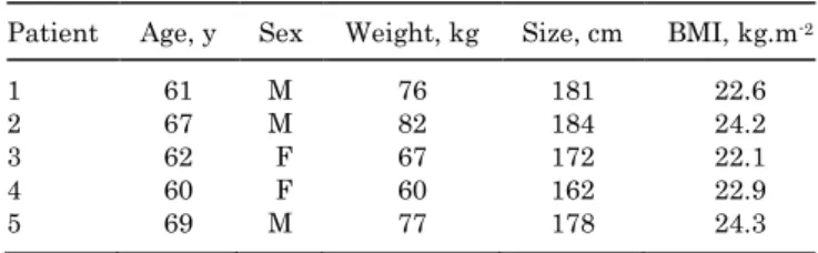
After local review board approval, 5 human lateral menisci were collected from patients who had total knee arthroplasty between September and October 2017. All patients signed an informed consent form before their inclusion in our study. Inclusion criteria were as follows: patient aged \70 years undergoing total knee arthroplasty because of isolated inter- nal femorotibial arthritis or femoropatellar and internal fem- orotibial joint degeneration (but with an external femorotibial compartment graded Kellgren and Lawrence \2)13 and no
surgery, trauma, or developmental disease of the operated knee. Table 1 summarizes patients’ characteristics.
TABLE 1 Patients’ Demographicsa
Patient Age, y Sex Weight, kg Size, cm BMI, kg.m-2
1 61 M 76 181 22.6
2 67 M 82 184 24.2
3 62 F 67 172 22.1
4 60 F 60 162 22.9
5 69 M 77 178 24.3
aBMI, body mass index.
Sample Creation
The anterior and posterior horns were sectioned to retain only the median horn of the meniscus. Four cubes (9 mm3)
were cut inside of the white, or avascular, area of each spec- imen and divided into 4 groups: ‘‘fresh’’ control, freezing, cryopreserved, and freezing 1 irradiation. Histologic fixa- tion of the control specimens was performed directly in the operating theater under a hood, which consisted of the following steps based on a previously validated protocol: 1. Five-minute rinsing with 0.1M cacodylate buffer. 2. Fixation in a solution of 2.5% glutaraldehyde in cacody-
late buffer for 1 hour.
3. Rinsing 3 times for 10 minutes with 0.1M cacodylate buffer.
4. Postfixation with a solution of 2% osmium tetroxide in 0.1M cacodylate buffer for 30 minutes.
5. Rinsing 3 times for 15 minutes with 0.1M cacodylate buffer.
6. Progressive dehydration of samples ranging from 50% to 100% ethanol before inclusion in Spurr.20
7. Resin fixation for transmission electron microscopy Control specimens were analyzed immediately after those steps in a delay \6 hours. The other 3 fragments were placed in physiologic saline in a cryokit and kept at 8°C until their transport to our local tissue bank (Etablisse- ment Franxcais du Sang) (\6 hours). The 3 samples were then prepared with the following steps: (1) graft reception in clean room (controlled atmosphere zone), (2) decontam- ination of the graft with an antibiotic solution (rifampicin 1 thiamphenicol), (3) rinsing with 0.1M cacodylate buffer for 5 minutes, and (4) bacteriologic sampling. Then, the 3 conservation methods were applied (1 sample for each). For the cryopreservation group, cryoprotective solution (10% of DMSO 1 SCOT 30) was added, and the bag was vacuumed to extract the residual air and the temperature progressively decreased (staring at 24°C and then decreas- ing at 22°C per minute to 240°C and then 25°C per minute to 2140°C). Samples were stored in a nitrogen tank in vapor phase at 2145°C. For the freezing group, a simple freezing
§Address correspondence to Matthieu Ollivier, MD, PhD, Institute of Movement and Locomotion, Department of Orthopedics and Traumatology, St Mar-
guerite Hospital, 270 Boulevard Sainte Marguerite, BP 29 13274 Marseille, France (email: ollivier.matthieu@yahoo.fr). *Aix-Marseille Universite´ , CNRS, ISM UMR 7287, Marseille, France.
yInstitute of Movement and Locomotion, Department of Orthopedics and Traumatology, St Marguerite Hospital, Marseille, France. zUniversite´ Clermont Auvergne, CHU Clermont-Ferrand, CNRS, SIGMA Clermont, ICCF, Clermont-Ferrand, France.
One or more of the authors has declared the following potential conflict of interest or source of funding: M.O. is a consultant for Stryker and Nex-clip. J.N.A. is a consultant for Zimmer. S.P. is a consultant for Zimmer, Arthrex, and Nex-clip.
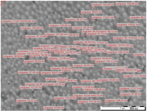
Figure 1. Example of longitudinal diameter measurement (cryopreserved sample diameter estimated on a picture at 40,0003 magnification).
process was used by progressively decreasing the tempera- ture (staring at 24°C and then decreasing at 22°C per min- ute to 240°C and then 25°C per minute to 280°C). For the freezing 1 irradiation group, a simple freezing process with a progressive decrease in temperature was performed (start- ing at 24°C and then decreasing at 22°C per minute to 240°C and then 25°C per minute to 280°C). The grafts were then transported in a dry ice–controlled container (stored at 280°C) to be irradiated by gamma ray by IONI- SOS. The doses received ranged between 23.9 and 26.5 kGy (2.4-2.6 Mrad). After this treatment, the samples were again stored at 280°C until analysis.
After 1 month, the samples were transported to the elec- tronic microscopy laboratory to be analyzed. The fixing steps (steps 1-7) were the same as those described for the control group. For all samples, ultrafine 60-nm sections were made with an Ultracut ultramicrotome (Reichert- Jung); contrast of the sections was made with uranyl ace- tate and lead citrate. Ultrastructure pictures were obtained with a transmission electron microscope (JEM 1400; JEOL) at 80 kV with a Megaview III camera and iTEM Five soft- ware (SIS Imaging). For each sample, we took 10 pictures with a magnification of 60003 and 40,0003. The longitudi- nal (Figure 1) and transverse diameters of the collagen fibers were measured at 70 points on a picture taken at 40,0003 magnification. The collagen meniscal architecture scoring system was calculated for each sample on 5 pictures with 60003 magnification (Table 2).7
Statistics
Before the initiation of the study, a sample analysis esti- mated that 5 samples for each group was necessary to be
TABLE 2
Collagen Meniscal Architecture Scoring System
0 Point 1 Point 2 Points
Disrupt/periodicity Mild Moderate Severe
Intrafibrillary edema No Yes —
Packing High density Intermediate Low density
Banding Yes No —
Fibril size variability Low High —
powered to distinguish D 20 6 10 nm regarding collagen transverse or longitudinal diameters.4,5,7
Patient characteristics were expressed with the appropri- ate descriptive statistics for the type of variables. Descriptive statistics included mean with SD or median with interquar- tile range as appropriate for continuous variables. The intra- class correlation coefficient with 95% CIs was calculated to assess intra- and interobserver reproducibility for the trans- verse and longitudinal diameter values. Student t tests were used to compare the distribution of continuous parameters between groups (or the Mann-Whitney test when the data were not normally distributed or when the homoscedasticity assumption was rejected). All reported P values were 2-sided, with a significance threshold of .05. Statistical analyses were performed with SPSS/JMP software (v 13; Microsoft). RESULTS
Transverse Diameter
No significant difference was found between the control and cryopreserved groups regarding mean transverse
4 Jacquet et al The American Journal of Sports Medicine
TABLE 3
Collagen Fiber Transverse Diameter Comparison Among Groups
Group Comparison Mean Difference, nm 95% CI, nm P Value
Control Cryopreserved 4.22 1.36-7.08 .1 Control Irradiated 32.27 29.55-34.99 \.001 Control Frozen 25.191 22.61-27.77 \.001 Cryopreserved Frozen 29.413 26.5-32.2 \.001 Cryopreserved Irradiated 36.4 33.34-39.65 \.001 Frozen Irradiated 7.08 4.64-9.52 .023 TABLE 4
Collagen Fiber Longitudinal Diameter Comparison Among Groups
Group Comparison Mean Difference, nm 95% CI, nm P Value
Control Cryopreserved 1.738 0.55-4.03 .1 Control Irradiated 35.39 32.79-37.98 \.001 Control Frozen 25.02 22.86-27.19 \.001 Cryopreserved Frozen 23.29 20.87-25.70 \.001 Cryopreserved Irradiated 33.65 30.99-36..31 \.001 Frozen Irradiated 10.36 8.24-12.49 .011 diameter (95.39 6 15.87 nm vs 99.62 6 19.23 nm, P = .1). There was a significant difference between the control and frozen groups (95.39 6 15.87 nm vs 70.20 6 13.94 nm, P \ .001) and between the control and frozen 1 irradiated groups (95.39 6 15.87 nm vs 63.1 6 15.57 nm, P \ .001) (Table 3).
The mean difference between the frozen and frozen 1 irradiated groups was also significant: D 7.1 nm (95% CI, 4.6-9.5 nm; P \ .023).
Longitudinal Diameter
We found no significant difference in mean longitudinal diameter between the control and cryopreserved groups (96.3 6 14 nm vs 94.6 6 16.4 nm, P = .1). There was a signif- icant difference in mean longitudinal diameter between the control and frozen groups (96.3 6 14 nm vs 71.3 6 10.6 nm,
P \ .001) as well as the frozen 1 irradiated groups (96.3 6
14 nm vs 60.9 6 14.8 nm, P \ .001) (Table 4).
The mean difference between the frozen and frozen 1 irradiated groups was also significant: D 10.6 nm (95% CI, 8.2-12.5 nm; P \ .011).
Collagen Meniscal Scoring System
The mean values of the collagen meniscal architecture scoring system are summarized in Table 5. No difference was found between the control and cryopreserved groups (4.5 6 1.3 vs 4.3 6 1.6 points, P = .9). There were signifi- cant differences between the control and frozen groups (4.5 6 1.3 vs 2.3 6 1.4 points, P = .02) and between the con- trol and frozen 1 irradiated groups (4.5 6 1.3 vs 1.4 6 0.9,
P = .02).
DISCUSSION
The main finding of the current study was that cryopreser- vation preserves meniscus histologic ultrastructure, unlike simple freezing or freezing 1 irradiation. We rejected our hypothesis that all preservation methods will result in structural and architectural properties similar to those of fresh meniscus. Cryopreservation does not entail a signifi- cant modification in terms of collagen fiber diameter or architectural organization as compared with fresh tissue. We did find, however, significant differences regarding those 2 measurements when we compared freezing and freezing 1 irradiation processes with fresh tissue or cryo- preserved samples. Irradiation was the more detrimental process in terms of tissue preservation, as all of our meas- urements were inferior to those taken for fresh tissue, cryo- preservation, and freezing samples.
Our study is limited in that our patients are older than usual donors2 (mean age: 63.8 years in our study vs 53.5 in
the register). Because of this, menisci evaluated during our analyses might have been altered by aging and degeneration processes. We tried to avoid limitations related to this meth- odological bias by studying nonarthritic joints (lateral com- partment) from subjects having only medial femorotibial degeneration. Moreover, our direct comparative design allowed us to think that potential degenerative changes would have similarly affected our evaluation of the 3 preser- vation methods, since the compared samples were created from the same meniscus. Our relatively short storage period before the analysis could also be considered a limitation. We supposed that most of the ultrastructure alteration related to the preservation method occurred during the decreas- ing-of-temperature steps, including direct chemical effects, ice formation, and dehydration, as described by Pegg.16
TABLE 5
Collagen Meniscal Architecture Scoring Comparison Between Preserved and Control Samples
Group Score, Mean 6 SD P Value
Control vs 4.5 6 1.3 —
Cryopreservation 4.3 6 1.6 .9
Frozen 2.3 6 1.4 .02
Irradiation 1.4 6 0.9 .02
collagen fiber diameters; as such, 1 senior technician trained in transmission electron microscopy carried out the entire collection of data and was blinded regarding the preservation process of the specimens. The measurements were always made at the same magnification (40,0003) and at 70 random points of the picture, with consideration to only the most circular fibers for the transverse diameter and the most linear for the longitudinal diameter. Our pro- tocol did not aim to investigate cell viability. It was impossi- ble for us to perform both ultrastructure analysis and flow cytometric cell counts on the same sample. Gelber et al7
were the first to demonstrate that cell survival was possible in cryopreserved samples, but in a study of 15 meniscal goat allografts, Fabbriciani et al4 showed that even if cryopreser-
vation made it possible to maintain partial cell viability in the tissue, the morphologic and biochemical characteristics of the graft were not improved. Finally, our sample size might seem low, but the numbers of specimens included was decided before the initiation of the study to compare groups upon collagen thickness evaluation.
Despite these limitations, our study is the first to directly compare the influence of preservation processes on 4 samples prepared from the same meniscus, permitting us to assert that the differences observed are mostly related to preservation methods and their specificities more than demographic confounding factors.
We used the collagen meniscal scoring system by Gelber et al6,7 to compare our results with the existing literature.
Gelber et al6,7 advocated the superiority of cryopreservation
on meniscus ultrastructure as compared with freezing, with 2 studies corroborating our results, even if they did not use a control group. In another study, Vangsness et al17 demon-
strated that the use of gamma irradiation caused clear alter- ation of musculoskeletal tissues mechanical properties.
Bone sample load-to-failure behavior was significantly lower when exposed to .3 Mrad of irradiation.17 Fideler
et al5 determined that .2 Mrad of irradiation of bone–
patellar tendon–bone allografts adversely affected 4 of the 7 structural properties that were analyzed. Thus, they found that all structural parameters were deeply affected at irradiation levels of 3.0 and 4.0 Mrad. This con- clusion is supported by our data: we found the highest rate of collagen disorganization for samples exposed to
2.5 Mrad (25 kGy: freezing 1 irradiation group). Advan- tages in terms of sterilization allowed by gamma irradia- tion should be balanced with those deep architectural and potentially mechanical consequences.
With regard to the meniscus, Lewis et al15 studied 7
human menisci and demonstrated that samples that
underwent a single freeze-thaw cycle had a significantly higher Young modulus than did those undergoing multiple freeze-thaw cycles (Young modulus: 1.2 3 107 for 1 cycle vs
8.5 3 106 for multiple cycles).
Our histologic results must be confirmed by mechanical trials to better understand potential consequences of ultra- structure alteration on meniscal graft mechanical proper- ties. In the same manner, only in vivo studies will be able to assess the real clinical relevance of our ex vivo conclusion. CONCLUSION
Cryopreservation is the only method that preserves fresh meniscus architectural specificities. Freezing and freezing 1 irradiation methods modify histologic properties of menis- cal allograft. Irradiation deeply alters diameters and organi- zation of collagen fibers, and this method should be used with caution to preserve and sterilize menisci tissue. ACKNOWLEDGMENT
The authors thank Alexandre Altie for his support during electron microscopy analysis.
REFERENCES
1. Choi Y-H, Seo Y-J, Ha JM, Jung KH, Kim J, Song SY. Collagenous ultrastructure of the discoid meniscus: a transmission electron microscopy study. Am J Sports Med. 2017;45(3):598-603. 2. Cohen J, Bistritz Y, Ashkenazi T. Deceased organ donor characteris-
tics and organ utilization in Israel, 2004-2013. Isr Med Assoc J. 2015;17(6):365-369.
3. Delrio AN, Marceddu S, Montella A, Gasparini G, Gulisano M. Struc- tural organization of the menisci of the human knee: scanning elec- tron microscopy study [in Italian]. Boll Della Soc Ital Biol Sper. 1992;68(6):359-364.
4. Fabbriciani C, Lucania L, Milano G, Schiavone Panni A, Evangelisti M. Meniscal allografts: cryopreservation vs deep-frozen technique. An experimental study in goats. Knee Surg Sports Traumatol Arthrosc. 1997;5(2):124-134.
5. Fideler BM, Vangsness CT, Lu B, Orlando C, Moore T. Gamma irra- diation: effects on biomechanical properties of human bone–patellar tendon–bone allografts. Am J Sports Med. 1995;23(5):643-646. 6. Gelber PE, Gonzalez G, Lloreta JL, Reina F, Caceres E, Monllau JC.
Freezing causes changes in the meniscus collagen net: a new ultra- structural meniscus disarray scale. Knee Surg Sports Traumatol
Arthrosc. 2008;16(4):353-359.
7. Gelber PE, Gonzalez G, Torres R, Garcia Giralt N, Caceres E, Monllau JC. Cryopreservation does not alter the ultrastructure of the menis- cus. Knee Surg Sports Traumatol Arthrosc. 2009;17(6):639-644. 8. Ghadially FN, Lalonde JM, Wedge JH. Ultrastructure of normal and
torn menisci of the human knee joint. J Anat. 1983;136(pt 4):773-791. 9. Giannini S, Buda R, Di Caprio F, et al. Effects of freezing on the bio- mechanical and structural properties of human posterior tibial ten- dons. Int Orthop. 2008;32(2):145-151.
10. Higuchi H, Kimura M, Shirakura K, Terauchi M, Takagishi K. Factors affecting long-term results after arthroscopic partial meniscectomy.
Clin Orthop Relat Res. 2000;377:161-168.
11. Horsky´ I, Huraj E, Huraj E, Sklovsky´ A. Degenerative changes in the knee joint after meniscectomy [in Slovak]. Acta Chir Orthop Trauma-
tol Cech. 1987;54(6):517-521.
12. Katsuragawa Y, Saitoh K, Tanaka N, et al. Changes of human menisci in osteoarthritic knee joints. Osteoarthritis Cartilage. 2010;18(9):1133-1143.
6 Jacquet et al The American Journal of Sports Medicine
13. Kohn MD, Sassoon AA, Fernando ND. Classifications in brief: Kellgren-Lawrence classification of osteoarthritis. Clin Orthop Relat
Res. 2016;474(8):1886.
14. Kru¨ ger-Franke M, Siebert CH, Kugler A, Trouillier HH, Rosemeyer B. Late results after arthroscopic partial medial meniscectomy. Knee
Surg Sports Traumatol Arthrosc. 1999;7(2):81-84.
15. Lewis PB, Williams JM, Hallab N, Virdi A, Yanke A, Cole BJ. Multiple freeze-thaw cycled meniscal allograft tissue: a biomechanical, bio- chemical, and histologic analysis. J Orthop Res. 2008;26(1):49-55. 16. Pegg DE. Principles of cryopreservation. Methods Mol Biol.
2015;1257:3-19.
17. Vangsness CT, Garcia IA, Mills CR, Kainer MA, Roberts MR, Moore TM. Allograft transplantation in the knee: tissue regulation, procurement, processing, and sterilization. Am J Sports Med. 2003;31(3):474-481.
18. Verdonk PCM, Demurie A, Almqvist KF, Veys EM, Verbruggen G, Verdonk R. Transplantation of viable meniscal allograft: survivorship analysis and clinical outcome of one hundred cases. J Bone Joint
Surg Am. 2005;87(4):715-724.
19. Verdonk PCM, Verstraete KL, Almqvist KF, et al. Meniscal allograft transplantation: long-term clinical results with radiological and mag- netic resonance imaging correlations. Knee Surg Sports Traumatol
Arthrosc. 2006;14(8):694-706.
20. Wallis MA, Griffin RL. A routine method for embedding animal tissues in Spurr resin for electron microscopy. J Clin Pathol. 1973;26(1):77- 78.
21. Wirth CJ, Peters G, Milachowski KA, Weismeier KG, Kohn D. Long- term results of meniscal allograft transplantation. Am J Sports
Med. 2002;30(2):174-181.