HAL Id: hal-01325648
https://hal.inria.fr/hal-01325648
Submitted on 2 Jun 2016
HAL is a multi-disciplinary open access
archive for the deposit and dissemination of
sci-entific research documents, whether they are
pub-lished or not. The documents may come from
teaching and research institutions in France or
abroad, or from public or private research centers.
L’archive ouverte pluridisciplinaire HAL, est
destinée au dépôt et à la diffusion de documents
scientifiques de niveau recherche, publiés ou non,
émanant des établissements d’enseignement et de
recherche français ou étrangers, des laboratoires
publics ou privés.
Distributed under a Creative Commons Attribution - NonCommercial - ShareAlike| 4.0
International License
Resource-Centered Distributed Processing of Large
Histopathology Images
Daniel Salas, Jens Gustedt, Daniel Racoceanu, Isabelle Perseil
To cite this version:
Daniel Salas, Jens Gustedt, Daniel Racoceanu, Isabelle Perseil. Resource-Centered Distributed
Pro-cessing of Large Histopathology Images. 19th IEEE International Conference on Computational
Sci-ence and Engineering, Aug 2016, Paris, France. �hal-01325648�
ISSN 0249-6399 ISRN INRIA/RR--8921--FR+ENG
RESEARCH
REPORT
N° 8921
June 2016 Project-Team CamusResource-Centered
Distributed Processing of
Large Histopathology
Images
Daniel Salas
Jens Gustedt
RESEARCH CENTRE NANCY – GRAND EST
615 rue du Jardin Botanique CS20101
54603 Villers-lès-Nancy Cedex
Resource-Centered Distributed Processing of
Large Histopathology Images
Daniel Salas
∗†‡Jens Gustedt
†‡Daniel Racoceanu
§Isabelle Perseil
∗Project-Team Camus
Research Report n° 8921 — June 2016 — 11 pages
Abstract: Automatic cell nuclei detection is a real challenge in medical imagery. The Marked Point Process (MPP) is one of the most promising methods. To handle large histopathology images, the algorithm has to be distributed. A new parallelization paradigm called Ordered Read-Write Locks (ORWL) is presented as a possible solution for solving some of the unwanted side effects of the distribution, namely an imprecision of the results on the internal boundaries of partitioned images. This solution extends a parallel version of MPP that has reached good speedups on GPU cards, but was not scaling to complete images as they appear in practical data.
Key-words: parallelization; parallel computing; distributed computing; marked point process; ordered read-write locks; cell nuclei recognition; histopathology
∗ Inserm CISI, Paris, France †INRIA, Nancy – Grand Est, France
‡ICube – CNRS, Universit´e de Strasbourg, France §Universit´e Pierre et Marie Curie, Paris, France
Calcul distribu´
e centr´
e resources
pour de larges images histopathologiques
R´esum´e : La d´etection automatique de noyaux cellulaires est un vrai chal-lenge pour l’imagerie m´edicale et la lutte contre le cancer. L’un des axes de recherche les plus prometteurs est l’utilisation de Processus Ponctuels Marqu´es (PPM). L’algorithme tir´e de cette m´ethode a ´et´e parall´elis´e et atteint de bonnes performances d’acc´el´eration sur carte GPU. Cependant, cette parall´elisation ne permet pas de traiter une lame compl`ete issue d’un pr´el`evement de biopsie. Il est ainsi n´ecessaire de distribuer les calculs. Cette distribution entraˆıne toute-fois des pertes de pr´ecision au niveau des axes de coupe de l’image. Un nouveau paradigme de parall´elisation appel´e Ordered Read-Write Locks (ORWL) est une solution possible `a ce probl`eme.Mots-cl´es : parall´elisation; calcul parall`ele; calcul distribu´e; processus ponctuel marqu´e; ordered read-write locks; d´etection de noyaux cellulaires; histopathologie
Resource-Centered Distributed Processing of Large Histopathology Images 3
1
Introduction
Breast cancer is the third common cancer all over the world with 1.677 mil-lion cases per year [1]. Both its detection and treatment are important concern for public health. E.g, pathologists of the Paris hospital La Piti´e Salp´etri`ere have to analyze more than 2000 stained biopsies per day. They evaluate cancer gradation on a scale from 1 to 3 according to 6 criteria. One of the most impor-tant criteria is the nuclei size atypia. Along with the cancer progression, nuclei sizes are growing until becoming abnormally big. In 2012, the International Conference on Pattern Recognition organized a contest [2]. Pathologists had to analyze and grade images. A free database of commented slides results from this contest. This is a great opportunity for designing and testing tools that could offer pathologists a second opinion.
Within this context, a team of the Laboratoire d’Imagerie Biom´edicale1 (LIB) has begun to test techniques to automate cell nuclei detection. An origi-nal approach in observing cell nuclei atypia is the Marked Point Process, MPP. In collaboration with the Laboratoire d’Informatique de Paris 62 (LIP6), an algorithm has been implemented and then parallelized on CPU with OpenMP and on GPU with CUDA. An acceleration of 22 has been reached with the GPU version. But unfortunately a single GPU is not able support the analysis of a complete slide and so we have to investigate different ways of distributing the algorithm. Section 2 discusses MPP, its existing parallelization and its limits.
In Section 3, we then discuss a conventional distribution method with MPI and a new one using Ordered Read-Write Locks (ORWL) and the benefits of the latter. Section 4 concludes and gives an outlook to future work.
2
The Marked Point Process for Cell Nuclei
Recog-nition in Breast Cancer Images
2.1
Marked Point Process Algorithm
A team from LIP6 has implemented a discretization of the Marked Point Pro-cess [3]. The global algorithm is based on a simulated annealing [4] proPro-cess. The solution tested on each run is the result of a Birth and Death process. The steps of this process are as follows:
Initialization: Image load and resource allocation
Birth Step: A randomly distributed configuration of points is generated. A probability coefficient of birth is decreased in each iteration, to favor con-vergence.
Mark Step: The best approaching form of a cell nuclei is an ellipse. Points previously created are therefore marked with a wide axis, a short axis
1See: https://www.lib.upmc.fr/ 2See: http://www.lip6.fr/
Resource-Centered Distributed Processing of Large Histopathology Images 4
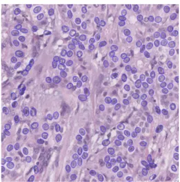
Figure 1: Parallel Birth and Death algorithm result
and an inclination angle. From these parameters, fidelity to the data (also called attachment) is computed, namely the ellipse’s accuracy with respect to the underlying image. The Bhattacharyya distance [5] is used to compare the intensity difference between the dark border and the inner cell nuclei.
Neighbors step: A neighbor map is built with the data fidelity values of each pixel. For every pixel of every ellipse, if the map’s pixel value is greater than the ellipse’s data fidelity value, then the map is updated.
Death Step: A first selection is made between overlapping ellipses. Only the best attached survive. Then a death rate filter is applied on ellipses created from the beginning. This filter is based on the data fidelity value and a death probability. This coefficient is increased in each iteration to favor convergence.
Convergence Test: Convergence is reached when all objects created in the birth phase are killed in the death phase.
2.2
A Parallel Algorithm for MPP
LIP6 has also parallelized this algorithm on CPU with OpenMP (see a processed image in Figure 1) and on GPU using CUDA. In each step, the parallelization is straightforward. The loops used to iterate over pixels are distributed to available threads. An initial parallelization problem concerning the death step has been solved using the principle of the neighbors map to test and set the minimal data fidelity values.
Reported parallelization speedups [6] are shown in Table 1.
Resource-Centered Distributed Processing of Large Histopathology Images 5
Sequential 4 OpenMP threads GPU Time 200 seconds 60 seconds 10 seconds
Speedup - 3.32x 22 x
Table 1: MPP results on a 1024 by 1024 pixels image
2.3
Parallel Birth and Death Limitations
The PBD (Parallel Birth and Death) algorithm is reaching a good speedup on GPU devices. But it is limited to the memory size of the GPU. A complete slide size may be up to 100,000 by 100,000 pixels at full resolution. Modern GPU cards offer 12GiB memory which is almost the size of our slide. In addition to the image, the application allocates 12 times the number of pixels in arrays of floating points. The total memory needed is about 450 GiB. This is far more than the available memory of our GPU.
Therefore, the PBD algorithm has to use subdivisions of the complete slide. On our 12GiB memory GPU, we could maximally take in charge an image side of about 16,000 px. If we consider analyzing images with a side length of 10,000 pixels, a complete slide would be composed of a hundred images. This leads us to another kind of problem. Subdividing the slide will have the side effect of truncating cell nuclei. The truncated ellipses would not be taken into account for the diagnostic. For an image of x40 magnification, if we consider that our cell nuclei is 80 pixels long, the band of image data that would not be considered correctly would represent a surface of about 3,000,000 px. For the total image this surface represents about 3.2%. If we look at the criteria of nuclei size for cancer gradation [2] (see Table 2),
grade % of atypic nuclei 1 0 to 30% nuclei are bigger 2 30 to 60% nuclei are bigger 3 more than 60% nuclei are bigger
Table 2: Breast cancer gradation for atypic nuclei criteria
the unconsidered 3% of nuclei are sufficient for establishing a diagnostic of grade 1. A cancer may be therefore detected in its early stage. In the following we present a strategy for taking into account carefully the boundary pixels by using a new distributed and parallel computation model.
3
Distributed PBD Process
In distributed computing, each processor has its own private memory. This adaptable memory size enables us to manage bigger issues and in particular to analyze bigger images.
Resource-Centered Distributed Processing of Large Histopathology Images 6
0 1 2
3 4 5
6 7 8
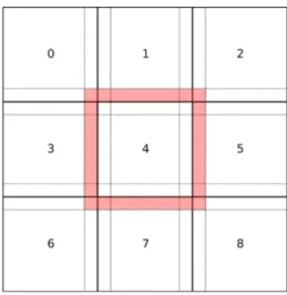
Figure 2: Image distribution strategy
3.1
Implementation strategy
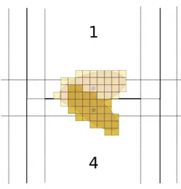
The main problem for distributing the algorithm is the neighbor step. For each ellipse data fidelity has to be taken into account to classify for the best attached values. A global map could be distributed among all nodes, according to Fig-ure 2. Every processor could then compute its part of the image locally. At the end of the neighbor step, when the local neighbor map has been drawn, the processors could send each other messages to detect crossing ellipses, according to Figure 3. In the example, Processor 4 sends a message to Processor 1 and informs him of the position and data fidelity value of its ellipses. Processor 1 should be listening asynchronously for this possible message. To avoid deadlock situations, the sends and receives should be non blocking. Then, synchroniza-tion barriers must be use to guarantee the coherence of the execusynchroniza-tion. Such barriers would force the first processor to wait until the last has finished. So, the whole execution would be as slow as the slowest processor. If the work load is not completely balanced, this would add a lot of waiting time, which can be largely sub-optimal. Furthermore, this method could easily encounter a deadlock situation.
Another distribution strategy consists in writing directly into the map of our neighbors. In Figure 2, Processor 4 would access the neighbor map of its surrounding neighbors (the red band). In the example of Figure 3, Processor 4 adds the data fidelity value of its ellipse into Processor 1’s neighbor map during its neighbor step. The step in which Processor 1 is at this moment does not matter. The value is taken into account, when Processor 1 proceeds to kill his worst attached ellipses the next time. Then, the map of the whole slide can be updated along with the computing process, no synchronization is necessary. This strategy is possible thanks to a new parallelization paradigm called Ordered
Resource-Centered Distributed Processing of Large Histopathology Images 7
Figure 3: Data fidelity competition
Read-Write Locks3 (ORWL).
3.2
A solution with ORWL
Before explaining how ORWL will handle the remote write of the neighbors map, we will present its global concepts.
3.2.1 A new paradigm
ORWL [7] models parallel and distributed computing by means of tasks. Its particularity is the way in which resources can be shared by the different tasks. Accesses are regulated by a FIFO that guarantees the liveness of the application and the equity of access for the tasks.
3.2.2 Workflow
Computation is distributed according to the number of available nodes in a configuration file. The TakTuk library [8] is in charge of sending and collecting files and starting the remote tasks. During a network recognition phase, an address book collects all nodes properties that are necessary to communicate (IP address, port number). Then this book is distributed to all nodes, allowing a point to point communication between all tasks. In a post-computation phase, TakTuk collects the results.
3See: http://orwl.gforge.inria.fr/orwl-html/
Resource-Centered Distributed Processing of Large Histopathology Images 8
Listing 1: ORWL task launch loop f o r ( s i z e t i =0; i <o r w l l t; i ++) {
t a s k o b j∗ task =P99 NEW( t a s k o b j , param1 ) ; t a s k o b j c r e a t e t a s k ( t a s k , o r w l t i d s[ i ] ) ; }
3.2.3 Tasks
The number of ORWL tasks is defined at runtime. For each task, a new thread is created and tasks are identified by an unique numberorwl_mytid, see Listing 1 for launching tasks.
In this example, the number of tasks is given byorwl_lt. P99 NEWis a macro defined in P994. It allocates an element of type task_obj and initializes it with the argument param1. task_obj_create_task executes the block in Listing 2
Listing 2: ORWL handlers definition
ORWL DEFINE TASK( t a s k o b j ) { // c o d e e x e c u t e d by e a c h t a s k }
3.2.4 Handling the data
ORWL data is held in abstract objects called locations. These are uniquely identifiable entities represented by a global location ID (orwl_myloc). Access to the locations is handled through a queue. To gain control in read or write mode to a location, a handle must insert a request in the queue. Locations have to be declared and an amount of memory has to be allocated as follows:
Listing 3: Parametrization of an ORWL program
/∗ g l o b a l ∗/
ORWL LOCATIONS PER TASK( n o r t h l o c ) ;
/∗ i n s i d e a f u n c t i o n ∗/
o r w l s c a l e( 1 0 0 ,
ORWLLOCATION(o r w l m y t i d, n o r t h l o c ) ) ;
orwl_handle2 for iterative access are initialized and defined as in List-ing 4. The macro ORWL HANDLE2 INITIALIZER initializes the han-dle object. A memory size is allocated by a call to orwl_scale. Functions
orwl_write_insertand orwl_read_insertare used, respectively, to connect an handle with a location in write or read mode. The last parameter of the function provides the initial position of the request in the queue. This strict ordering will schedule data access such that it is deadlock free [9].
4http://p99.gforge.inria.fr/p99-html/
Resource-Centered Distributed Processing of Large Histopathology Images 9
Listing 4: ORWL handlers definition
o r w l h a n d l e 2 h e r e h d l =ORWL HANDLE2 INITIALIZER;
o r w l h a n d l e 2 t h e r e h d l =ORWL HANDLE2 INITIALIZER;
o r w l s c a l e( DATA SIZE , ORWLLOCATION(o r w l m y t i d, n l o c ) ) ; o r w l w r i t e i n s e r t(& h e r e h d l , ORWLLOCATION(o r w l m y t i d, h e r e l o c ) , 0 ) ; o r w l r e a d i n s e r t(& t h e r e h d l , ORWLLOCATION(o r w l m y t i d − n , t h e r e l o c ) , 1 ) ;
Data is accessible through critical sections as in Listing 5.
Here, the location associated with north_hdl handle is mapped with a pointer to unsigned char. Modifying data through the pointer directly modi-fies the data of the location itself.
Once a task has gained access to a critical section in write mode, the access is exclusive.
Listing 5: handler data access in write mode
ORWL SECTION( n o r t h h d l ) { unsigned char ∗ n o r t h p t r = o r w l w r i t e m a p( n o r t h h d l ) ; memcpy ( n o r t h p t r , my map . n o r t h , b a n d s i z e ) ; } ORWL SECTION( x n o r t h h d l ) { unsigned char ∗ x n o r t h p t r = o r w l r e a d m a p( x n o r t h h d l ) ; i n t e r f a c e S t i t c h B a n d ( s t r , s i z e , t h e r e h d l p t r , f r o n t i e r ) ; }
3.2.5 An ORWL implementation of the MPP
ORWL is implemented in C. On the other hand, the PBD algorithm has been written in C++. A first step of the implementation consisted in transposing the code from C++ to C. However, we preserved some useful functionalities of the C++ library, for keeping the use of the CImg5 library.
In CImg, an image is represented as a matrix of pixels. The type can be determined during image creation. In our case, we have used images with pixel type unsigned char. Pixels values are accessible through an image.data ob-ject. The three color channels R, G, and B are stored linearly in a single memory buffer as follows: R0R1R2· · · G0G1G2· · · B0B1B2· · · .
Other CImg functions are get_rows and get_columns. They allow to get horizontal and vertical parts of an image. This is used in the distributed PBD algorithm to splice the borders of the neighbor map. The border width is half the wide axis of the ellipses.
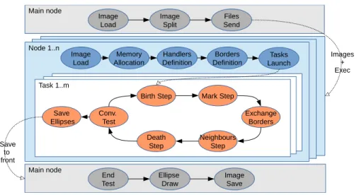
3.2.6 The Distributed PBD algorithm Figure 4 shows a workflow of the algorithm.
5http://cimg.eu/
Resource-Centered Distributed Processing of Large Histopathology Images 10 Main node Image Load Image Split Files Send Node 1..n Image Load Memory Allocation Handlers Definition Borders
Definition LaunchTasks Task 1..m
Save Ellipses
Birth Step Mark Step
Neighbours Step Death
Step Conv.
Test ExchangeBorders
Main node End Test Ellipse Draw Image Save Save to front Images + Exec
Figure 4: Workflow of PBD ORWL implementation
In an initial phase, a main node loads the entire image and splits it. A Ruby script using TAKTUK sends files to the computing node. Then each node loads its own image. Locations and handlers are defined. Several instances or orwl_tasks can be launched on a single node to parallelize the computation (see Listing 1). After the Mark Step, a new Exchange Border step is added. A first critical section enables a processor to write its border into a location. Then, a second critical section allows to retrieve the borders of the neighbors (Listing 5). Own borders are obtained by slicing the neighbors map. The interfaceStitchBand function adds neighbor borders to the processors neigh-bor map. Thereby, in the Neighneigh-bors Step the computation takes into account neighbors ellipses.
Once a Birth and Death process has converged, ellipse properties are sent to the main node. The final ellipse drawing can be done in a single loop.
4
Conclusion and future work
Thanks to the ORWL model, we presented a distributed version of the PBD algorithm that is ready to scale. It allows to properly consider all cell nuclei on the boundary between two partial images. An implementation of this algorithm is in progress and tests will be executed on a cluster of GPU and Xeon Phi accelerators. The final application is designed to map an entire slide in a few minutes. The goal is to let pathologists navigate through the slides, such that they may zoom in and out, just as they used to do with a microscope. Colored ellipses could be shown on a separate layer, helping to detect nuclei size atypia.
Resource-Centered Distributed Processing of Large Histopathology Images 11
References
[1] W. H. Organization, “GLOBALCAN 2012: Estimated Cancer Incidence, Mortality and Prevalence Worldwide in 2012,” 2012. [Online]. Available: http://globocan.iarc.fr/
[2] I. C. on Pattern Recognition, “MITOS-ATYPIA-14,” 2014. [Online]. Available: http://mitos-atypia-14.grand-challenge.org/
[3] C. Avenel, P. Fortin, and D. B´er´eziat, “Parallel birth and death process for cell nuclei extraction in histopathology images,” Jul. 2013, working paper or preprint. [Online]. Available: https://hal.archives-ouvertes.fr/hal-00844001 [4] K. E. Geltman, “The Simulated Annealing Algorithm,” 2014. [Online].
Available: http://katrinaeg.com/simulated-annealing.html
[5] F. J. Aherne, N. A. Thacker, and P. I. Rockett, “The bhattacharyya metric as an absolute similarity measure for frequency coded data,” Kybernetika, vol. 34, no. 4, pp. 363–368, 1998.
[6] C. Avenel, P. Fortin, and D. B´er´eziat, “Parallel birth and death process for cell nuclei extraction in histopathology images,” 2013.
[7] J. Gustedt and E. Jeanvoine, “Relaxed Synchronization with Ordered Read-Write Locks,” in Euro-Par 2011: Parallel Processing Workshops, ser. LNCS, M. Alexander et al., Eds., vol. 7155. Springer, May 2012, pp. 387–397. [Online]. Available: http://hal.inria.fr/hal-00639289
[8] B. Claudel, G. Huard, and O. Richard, “TakTuk, Adaptive Deployment of Remote Executions,” in Proceedings of the International Symposium on High Performance Distributed Computing (HPDC). Munich, Germany: ACM, 2009, pp. 91–100. [Online]. Available: https://hal.inria.fr/hal-00788923 [9] P.-N. Clauss and J. Gustedt, “Iterative Computations with Ordered
Read-Write Locks,” Journal of Parallel and Distributed Computing, vol. 70, no. 5, pp. 496–504, 2010. [Online]. Available: https://hal.inria.fr/inria-00330024
RESEARCH CENTRE NANCY – GRAND EST
615 rue du Jardin Botanique CS20101
54603 Villers-lès-Nancy Cedex
Publisher Inria
Domaine de Voluceau - Rocquencourt BP 105 - 78153 Le Chesnay Cedex inria.fr