HAL Id: hal-02668850
https://hal.inrae.fr/hal-02668850
Submitted on 31 May 2020
HAL is a multi-disciplinary open access
archive for the deposit and dissemination of
sci-entific research documents, whether they are
pub-lished or not. The documents may come from
teaching and research institutions in France or
abroad, or from public or private research centers.
L’archive ouverte pluridisciplinaire HAL, est
destinée au dépôt et à la diffusion de documents
scientifiques de niveau recherche, publiés ou non,
émanant des établissements d’enseignement et de
recherche français ou étrangers, des laboratoires
publics ou privés.
Copyright
A proteomic study of Methylobacterium extorquens
reveals a response regulator essential for epiphytic
growth
Benjamin Gourion, Michel Rossignol, Julia Vorholt
To cite this version:
Benjamin Gourion, Michel Rossignol, Julia Vorholt. A proteomic study of Methylobacterium
ex-torquens reveals a response regulator essential for epiphytic growth. Proceedings of the National
Academy of Sciences of the United States of America , National Academy of Sciences, 2006, 103 (35),
pp.13186-13191. �10.1073/pnas.0603530103�. �hal-02668850�
A proteomic study of
Methylobacterium extorquens
reveals a response regulator essential
for epiphytic growth
Benjamin Gourion*, Michel Rossignol†, and Julia A. Vorholt*‡
*Laboratoire des Interactions Plantes Micro-Organismes (LIPM), Institut National de la Recherche Agronomique兾Centre National de la Recherche Scientifique, BP52627, 31326 Castanet-Tolosan, France; and†Unite´ Mixte de Recherche 5546, Centre National de la Recherche兾Universite´ P. Sabatier, F-31326 Castanet-Tolosan, France
Edited by Steven E. Lindow, University of California, Berkeley, CA, and approved July 5, 2006 (received for review April 30, 2006) Aerial plant surfaces are colonized by diverse bacteria such as the
ubiquitous Methylobacterium spp. The specific physiological traits as well as the underlying regulatory mechanisms for bacterial plant colonization are largely unknown. The purpose of this study was to identify proteins produced specifically in the phyllosphere by comparing the proteome of Methylobacterium extorquens colo-nizing the leaves either with that of bacteria colocolo-nizing the roots or with that of bacteria growing on synthetic medium. We iden-tified 45 proteins that were more abundant in M. extorquens present on plant surfaces as compared with bacteria growing on synthetic medium, including 9 proteins that were more abundant on leaves compared with roots. Among the proteins induced during epiphytic growth, we found enzymes involved in methanol utilization, prominent stress proteins, and proteins of unknown function. In addition, we detected a previously undescribed type of two-domain response regulator, named PhyR, that consists of an N-terminal sigma factor (RpoE)-like domain and a C-terminal re-ceiver domain and is predicted to be present in essentially all
Alphaproteobacteria. The importance of PhyR was demonstrated
through phenotypic tests of a deletion mutant strain shown to be deficient in plant colonization. Among PhyR-regulated gene prod-ucts, we found a number of general stress proteins and, in partic-ular, proteins known to be involved in the oxidative stress re-sponse such as KatE, SodA, AhpC, Ohr, Trx, and Dps. The PhyR-regulated gene products partially overlap with the bacterial in
planta-induced proteome, suggesting that PhyR is a key regulator
for adaptation to epiphytic life of M. extorquens. fitness兩 sigma factor 兩 stress 兩 two-component system
M
olecular microbial ecology is often hampered by the difficulty of unraveling how the environment shapes bacterial physi-ology and allows microorganisms to multiply. One such habitat is the aerial parts of plants that are colonized by various microorgan-isms, mostly bacteria, which are often found in numbers averaging 106to 107cells per cm2. Epiphytes, defined as bacteria that are capable of multiplying on plant surfaces, encounter rather harsh conditions in the phyllosphere environment. This habitat is gener-ally considered to be poor in nutrients. In addition, residing microorganisms are exposed to the atmosphere and radiation and are subjected to rapid changes with respect to their physical environment (1). Many plant-colonizing bacteria do no apparent harm to their hosts and might even be beneficial to the plant, whereas others are plant pathogens and can, after establishment of an epiphytic population, ultimately destroy the tissue on which they are living.The chemical and physical features of leaf surfaces are not well known, and the same is true for the traits that allow these bacteria to multiply in the leaf habitat. Our current knowledge about bacterial physiology in the phyllosphere stems mainly from targeted approaches. Thus, phenotypes such as flagellar motility, UV-mediated mutagenic repair, and exopolysaccharide production contribute substantially to epiphytic fitness (2–4). In addition,
random mutagenesis has been performed to identify novel targets important for phyllosphere colonization (5). Gene expression pro-filing is a good strategy for providing information about adaptations to specific conditions or environments. One powerful strategy for targeting gene expression in the natural context is through pro-moter trap analysis [i.e., in vivo expression technology (IVET)] (6). Variants of this approach have been successfully applied to identify genes induced during phyllosphere colonization of bacterial patho-gens (7–9). However, by definition, the IVET strategy can only give an incomplete picture of the physiology of bacteria. Indeed, it is well known that changes at the protein level are not necessarily pre-dictable from transcript levels because of differences in translation efficiency, proteolysis, and posttranslational modifications. In this study, we have therefore chosen a more direct way to gain insights into the physiology of bacteria in the phyllosphere through the analysis of the proteome of a bacterium in this ecosystem.
For this work, we have used the Alphaproteobacterium
Methy-lobacterium extorquens AM1, a well studied model
pink-pigmented facultative methylotroph (PPFM) (10), whose draft genome sequence is available (see www.integratedgenomics.com兾 genomereleases.html#6). Methylobacterium spp. are common leaf epiphytes that represent an important bacterial population on leaves (11, 12) and have been found on all analyzed plants (13).
Methylobacterium spp. on plant surfaces benefit from methanol
produced by plants (14) by means of methylotrophy (10, 15). However, methanol is not the only carbon substrate that these bacteria are able to consume in the phyllosphere (14). The presence of Methylobacterium may be beneficial to plants through the pro-duction of plant hormones (13, 16). The ubiquitous presence of
Methylobacterium on plant surfaces makes them an interesting
model for discovering the particular traits that these bacteria have acquired as successful epipytes. This work provides the identifica-tion of previously undescribed candidate proteins of
Methylobacte-rium required for phyllosphere colonization and, in particular, the
identification of a key regulator controlling adaptation to this habitat.
Results and Discussion
Proteome Analysis ofM. extorquens During Phyllosphere Coloniza-tion.With the aim to identify proteins that are specifically induced when M. extorquens AM1 colonizes the phyllosphere of Arabidopsis
thaliana ColO plants, we performed a differential analysis of the
Conflict of interest statement: No conflicts declared. This paper was submitted directly (Track II) to the PNAS office.
Abbreviations: MM, minimal medium; ROS, reactive oxygen species; 2-DE, 2D gel electro-phoresis.
Data deposition: The sequence reported in this paper has been deposited in the GenBank database (accession no. DQ845291).
‡To whom correspondence should be sent at the present address: Institute of Microbiology,
Swiss Federal Institute of Technology Zurich, Eidgeno¨ssiche Technische Hochschule Ho¨ng-gerberg, CH-8092 Zurich, Switzerland. E-mail: vorholt@micro.biol.ethz.ch.
proteome of M. extorquens that had colonized plants under gno-tobiotic conditions after seed inoculation by comparison with the proteome of bacteria that were cultivated on the surface of synthetic minimal medium (MM) under the same conditions of light and temperature. We used succinate as a carbon source, because it enters directly into the tricarboxylic acid (TCA) cycle and allows us to observe the induction of methylotrophy markers (17). Proteins were separated by 2D gel electrophoresis (2-DE), and we identified those that were induced at least 3-fold based on image analysis (Fig. 1). In total, 40 proteins were identified as up-regulated during phyllosphere colonization (Table 1). To distinguish between pro-teins specific for phyllosphere colonization with respect to more general epiphytic adaptation, we compared the proteome of bac-teria from the aerial parts with that of rhizosphere-colonizing bacteria. We identified 9 proteins that were ⬎3-fold induced relative to the rhizosphere proteome (Table 1), out of which 5 were not identified in the earlier comparison. The high similarity be-tween the phyllosphere and rhizosphere proteomes suggests a similar adaptation to the epiphytic state in both plant environments. Relatively few down-regulated proteins were identified (see Table 3, which is published as supporting information on the PNAS web site), and these generally corresponded to housekeeping proteins, which could reflect a general down-regulation of metabolism during epiphytic growth compared with in vitro conditions.
Among the proteins induced during bacterial growth in the phytosphere (leaf and root surface environments), key markers of methylotrophic metabolism (17) were found to be up-regulated (e.g., MxaF and Fae) with respect to growth on synthetic medium containing succinate as a carbon source (Table 1). The induction of these enzymes upon epiphytic growth is in agreement with a previous study in which an advantage of wild-type (WT) M.
extorquens cells in competition with methylotrophy-minus mutants
was demonstrated, suggesting methanol utilization by the methyl-otroph (14). Another protein induced during phyllosphere coloni-zation was PhaA, which initiates synthesis of the reserve polyhy-droxy butyrate (PHB) (ref. 18; Table 1). It has been shown that PHB formation is stimulated by a deficiency of nutrients such as NH4⫹, SO42⫺, Mg2⫹, Fe2⫹, or Mn2⫹(19). The observed induction of
PhaA might thus represent part of a general adaptation to nutrient-limiting conditions as would be expected for phyllospheric growth where the carbon source might not be a growth-limiting factor (14). The nature of this limiting factor is possibly suggested by the phyllosphere-specific induction of two putative periplasmic ABC transporter components predicted to be involved in iron and sulfate uptake (Table 1). Interestingly, iron and sulfate have been sug-gested to be critical for phyllosphere colonization in other organ-isms (9).
We also found several putative dehydrogenases兾oxidoreductases to be induced during phytospheric growth (Table 1). Of these, RMQ03452 is phyllosphere-specific, sharing a high percentage of sequence identity with the AcoD of Ralstonia eutrophus, which is involved in the catabolism of acetoin and ethanol (20), and with AldB of Escherichia coli, which is thought to have a role in detoxifying alcohols and aldehydes (21). The RMQ03452 protein in
M. extorquens might contribute to carbon dissimilation or
detoxi-fication of alcohols or aldehydes that are produced by plants (22).
Methylobacterium spp. are specialists in dealing with toxic
com-pounds, as is already clear when considering that formaldehyde is a central intermediate of methylotrophic metabolism (10, 15). This question of detoxification vs. catabolism also arises for a putative lactoylglutathione lyase (GloA) (Table 1). In E. coli, GloA is required for detoxification of methylglyoxal, which is known to cause DNA damage (23). Because it has been reported that methylglyoxal is formed during catabolism of certain amino acids and other compounds such as acetone (24), methylglyoxal might therefore also be an intermediate produced upon breakdown of nutrients of the facultative methylotroph.
The analysis of the in planta proteome of M. extorquens AM1 clearly reflects an adaptation to survival under stress conditions (Table 1). The identified stress proteins fall into two classes: chaperones兾proteases and oxidative stress-related proteins. Among the former are two paralogues of the periplasmic DegP兾HtrA family (25), which suggests a response to extracytoplasmic stress and兾or the need for assistance in the maturation of components of the cell envelope required for epiphytic growth. In addition, there is a predicted protease of the DJ-1兾Pfp-1 superfamily that is a homologue of the general stress protein 18 (GSP18) of Bacillus
subtilis (26) and heat-shock proteins that are well known to be
induced by various types of environmental stress (27). The oxidative stress response is suggested by the up-regulation of superoxide dismutase, catalases, and the Dps protein. Dps is a nonspecific DNA-binding protein and a key component of the protection strategy against H2O2(28, 29), UV irradiation (30), and electro-philes such as methylglyoxal (23, 28) (see above). The formation of reactive oxygen species (ROS) is a normal event and a by-product of electron transport under aerobic conditions (31). Because the detoxification of ROS becomes particularly important under star-vation conditions (32), the observed induction of ROS-removing enzymes in M. extorquens AM1 might thus be a reaction to endogenously formed ROS. On the other hand, it is well known that plant cells challenge bacteria by means of an oxidative burst (33) and superoxide dismutase (SOD), and Kat and Dps have been shown to counteract the toxic effects of ROS produced by plants (34, 35). All of the stress proteins that we identified appeared to be epiphytic-specific rather than phyllosphere-specific. However, an-other protein, ClpP, was found to be induced in the phyllophere rather than in the rhizosphere. Clp proteases of E. coli are known to play an important role in cytoplasmic quality control and participate in numerous regulatory mechanisms that are important in nongrowing or slow-growing cells. ClpP interacts with ClpX or ClpA, which exhibit different substrate specificities (36).
A Response Regulator Common toAlphaproteobacteria Essential for Plant Colonization. The analysis of the in planta proteome of M.
extorquens AM1 revealed the induction of a putative
two-component system response regulator, RMQ08198, that we named
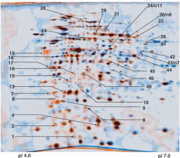
Fig. 1. Dual-channel image analysis of 2-DE protein pattern of M.
ex-torquens AM1 to reveal proteins induced during phyllosphere colonization.
Proteins from cells harvested from the phyllosphere of A. thaliana ColO are colored in orange, and those of cells harvested from the surface of solid MM containing succinate as the only carbon source are colored in blue. Spots of identified proteins are marked (see Table 1).
Gourion et al. PNAS 兩 August 29, 2006 兩 vol. 103 兩 no. 35 兩 13187
PhyR (for ‘‘phyllosphere-induced regulator’’). This protein is in-teresting in several respects as follows. (i) A National Center for Biotechnology Information (NCBI) CD search revealed that PhyR carries a RpoE (E)-like domain at the N terminus of the protein (Fig. 2A). A sigma-factor-like domain has not yet been described as part of a response regulator and might suggest that PhyR could possibly initiate transcription by itself. (ii) The sigma factor RpoE plays a major role in maintaining the integrity and function of the
envelope and provides resistance to environmental stresses includ-ing desiccation and oxidative stress in Pseudomonas spp. (4, 37, 38). (iii) The domain structure of PhyR shows that the predicted phosphorable receiver domain is located at the C terminus (Fig. 2 A), although this domain is usually located at the N terminus in described response regulators (39).
The identification of a previously undescribed type of response regulator in the proteome of in planta-grown M. extorquens AM1
Table 1. List of proteins from M. extorquens AM1 found to be induced during phyllosphere (P) colonization relative to MM and rhizosphere (R) colonization
Spot
no.* RMQ no. Gene product(s)† CD search Mr pl
Ratio,‡ P兾MM
Ratio,‡ P兾R
Metabolism
29 RMQ05966 MxaF, methanol dehydrogenase, large subunit (M31108) pfam01011 67.2 5.8 ⫹ 48 RMQ00044 MxaJ protein (M31108) pfam00497 27.4 6.0 ⬁ 4 RMQ09682 Fae, formaldehyde activating enzyme (L43136) 20.7 7.0 ⫹ 16 RMQ08765 PqqB, PQQ biosynthesis polypeptide (L25889) 30.6 5.4 ⬁
34 n11 RMQ03830 PhaA,-ketothiolase (AF287907) pfam00108 44.1 6.7 ⫹ ⫹ 42 RMQ09548 Crr, crotonyl-CoA reductase (L48340) pfam00107 47.5 6.3 ⬁
2 RMQ01365 Gap20 (AF442749) 19.0 5.8 ⬁
45 RMQ05381 Malyl-CoA lyase-like protein pfam03328 37.9 5.8 ⬁
24 n10 RMQ03452 Aldehyde dehydrogenase pfam00171 62.8 6.4 ⫹ ⬁ 28 RMQ07560 Xanthine oxidase-related aldehyde oxidoreductase pfam02738 80.6 5.3 ⫹
44 RMQ07805 Putative NADP-dependent oxidoreductase pfam00107 35.9 6.0 ⫹
P12 RMQ11717 Putative quinoprotein 36.0 6.9 ⫹
1 RMQ02894 GloA, lactoyglutathione lyase pfam00903 16.5 5.7 ⫹
n3 RMQ09259 Adenylate kinase pfam00406 21.3 5.1 ⫹
Transport
n4 RMQ08930 ABC-type Fe⫹ transport system, periplasmic component 36.6 6.6 ⫹ n6 RMQ06383 ABC-type sulfate transport system, periplasmic component 24.1 7.8 ⫹ 15 RMQ02495 Putative amino acid binding protein pfam00497 28.5 5.2 ⬁
31 RMQ05493 Putative oligopeptide binding protein pfam00496 70.2 6.7 ⫹ Stress proteins
P28 RMQ01248 DegP兾HrA, Trypsin-like serine proteases pfam00089 50.4 6.7 ⬁ 40 RMQ04833 DegP兾HrA, Trypsin-like serine proteases pfam00089 51.2 8.4 ⬁
n5 RMQ08088 ClpP, ATP-dependent Clp protease proteolytic subunit pfam00574 23.1 5.8 ⫹ 6 RMQ05519 Protease I (Serine protease) DJ-1兾Pfpl family (GSP18) pfam01965 20.7 4.9 ⬁
P22 RMQ06501 Hsp70, heat-shock protein 70 (DnaK) pfam00012 51.9 4.9 ⫹ P35 RMQ06982 Hsp70, heat-shock protein 70 (DnaK) pfam00012 68.5 5.2 ⫹ P4 RMQ02206 Hsp20, heat-shock protein 20 pfam00011 18.4 5.2 ⬁ P15 RMQ02531 SodA, Superoxide dismutase pfam02777 22.5 5.8 ⫹ 37a RMQ09549 KatE, catalase (L48340) pfam00199 63.5 7.1 ⬁ 37b RMQ11789 KatE, catalase pfam00199 59.9 5.8 ⬁ 9 RMQ05258 Dps, DNA protection protein pfam00210 19.9 5.0 ⫹
Proteins of unknown function
7, 12 RMQ09016 NfU-like兾thioredoxin-like protein pfam01106 20.3 4.8 ⬁
43 n7 RMQ10082 Major royal jelly protein§ pfam03022 41.4 6.3 ⬁ ⫹
P26 RMQ06718 Major royal jelly protein§ pfam03022 40.2 5.4 ⬁
n12 RMQ07439 Protein of unknown function 68.3 6.4 ⫹ 8 RMQ10020 Protein of unknown function pfam05974 18.5 5.4 ⬁
10 RMQ09099 Protein of unknown function 21.1 5.9 ⬁ 13 RMQ08861 Protein of unknown function 20.3 5.3 ⬁ 17 RMQ03063 Protein of unknown function 31.7 4.9 ⬁ 18 RMQ00428 Protein of unknown function§ 31.9 5.3 ⫹
P30 RMQ05730 Protein of unknown function§ 28.9 6.8 ⫹
19 RMQ01102 Protein of unknown function 43.5 5.0 ⬁ P25 RMQ00267 Protein of unknown function 39.1 5.2 ⬁ 39 RMQ03107 Protein of unknown function pfam00450 55.3 6.4 ⬁ 46 RMQ03170 Orf88, dioxygenase (AY034474) pfam00903 36.4 6.2 ⫹
36 n8 RMQ09688 Putative nucleoside binding protein 54.7 9.0 ⬁ ⬁ Regulator
P16 RMQ08198 Response regulator (PhyR, this work) pfam06182 29.1 4.8 ⬁
*Spot numbers that are preceded by ‘‘P’’ were identified on gels from independent experiments stained with silver nitrate rather than with SYPRO Ruby. Spot numbers preceded by ‘‘n’’ were detected to be induced in bacteria that were grown in the phyllosphere with respect to the rhizosphere. All other proteins were identified from the proteome of bacteria grown in the phyllosphere with respect to minimal medium supplemented with succinate.
†Accession nos. are in parentheses.
‡Spots indicated as ‘‘⬁’’ were only detectable in the proteome from bacteria grown in the phyllosphere and not in the references (MMand rhizosphere, respectively). Spots indicated as ‘‘⫹’’ were found to be at least 3-fold induced. Proteins were identified from 2D gels stained with SYPRO Ruby and found in two biological repetitions whereby the majority of the spots were in addition also found induced on gels that were stained with silver nitrate.
prompted us to evaluate the importance of PhyR for plant colo-nization by constructing a deletion strain. Growth rates of the mutant were found to be unaltered with respect to WT when plant colonization was mimicked in vitro under mixed growth conditions (i.e., in the presence of succinate and methanol) (14). However, in
planta colonization experiments revealed a severe growth defect of
the PhyR deletion mutant (Fig. 3). Cell numbers of the mutant were below the detection limit for 65% of 3-week-old plants. When we then cloned the PhyR gene in trans, we were able to restore the colonization capacity to the WT level (data not shown).
Interestingly, a BLAST search revealed that PhyR homologues are present in essentially all free-living Alphaproteobacteria for which a genome sequence is available, but not in any other bacteria. This finding clearly indicates a more general function for PhyR homologues than adaptation to the phyllosphere and an ancient origin within this proteobacterial subgroup based on phylogenetic analysis (Fig. 2B).
Identification of Proteins That Are Positively Regulated by PhyR.The phenotype of phyR mutants indicate that it is an important regulator and that one or more important physiological traits of phyllospheric growth are under PhyR control. To identify genes that are induced by PhyR, we performed proteome analysis with 2-DE so that we could readily recognize proteins that had been identified during the in planta proteome analysis. To this end, we cloned phyR in the expression vector pCM80 (40) and introduced the plasmid into M. extorquens AM1⌬phyR. The proteome of this overexpressing strain was compared with the phyR-deficient strain containing the empty vector as control. The 42 proteins that we identified as PhyR-regulated are in Table 2.
These results show that PhyR is partly responsible for the induction of some of the proteins that we had found to be induced during phyllospheric growth of the bacterium. However, we are unable to distinguish whether they are directly or indirectly regu-lated by PhyR. Among these proteins are KatE, SodA, Hsp20, Dps, GloA, and several uncharacterized proteins. As mentioned earlier, these proteins are known to be involved in coping with stress caused by electrophiles (GloA, Dps) and ROS (KatE, SodA, Dps). The latter group comprises proteins protective not only against super-oxide anions and hydrogen persuper-oxide but also alkyl hydropersuper-oxides (see induction of Ohr and AhpC; Table 2) that are all important components of the plant defense response against microbial infec-tion (33, 41) and by-products of aerobic metabolism (see above). Several dehydrogenases (pfam00107 and -00106) were found to be induced by PhyR. The substrate spectrum and role of these dehydrogenases is unknown. They might be involved in substrate utilization during starvation to furnish the additional energy supply associated with processes such as repair of oxidized proteins and lipids. Nevertheless, a possible role for one of the putative dehy-drogenases (RMQ06018) can be proposed. This protein represents a putative glutathione-dependent formaldehyde dehydrogenase (42) that might fulfill an auxiliary role coping with excess formal-dehyde alongside the well described H4MPT- and H4F-dependent pathways for formaldehyde oxidation in Methylobacterium (15).
In several model bacteria, stress responses have been well stud-ied. Whereas many regulators are specifically involved in one type of stress, other regulators control diverse functions.Sis a master regulator of the general stress response in bacteria that belong to
Gammaproteobacteria (43). Mutants deficient inSare less able to survive upon starvation and are more sensitive to oxidative and osmotic stress as well as UV and desiccation stress in both loga-rithmic- and stationary-phase cells in Enterobacteriaceae and pseudomonads (44–46). In Bacillus,Bhas been postulated to be the functional homologue ofS. The general stress regulon ofB provides the cells with a nonspecific, multiple, and preventive stress resistance in which the protection against oxidative stress is an essential part of the response (47).
PhyR has a central role in the adaptation of Methylobacterium to the plant environment. Our proteome analysis points to a rather large PhyR-dependent regulon within which the oxidative stress response is an important part (Table 2), reminiscent of the role of S兾Bin adaptation for surviving stress and starvation in nature (43, 47). So far, the regulatory elements representing the functional homologues ofS兾Bwith their corresponding activation mecha-nisms in Alphaproteobacteria are unknown. It is therefore tempting to speculate that PhyR is involved in a S兾B-like response. Biochemical analysis will be important to clarify whether PhyR represents a chimeric protein with a functional output domain that acts as a bona fide sigma factor, suggested by theE-like domain.
Conclusions
Little is known about traits important for phyllosphere coloniza-tion, and even less is known about the regulatory mechanisms that determine the adaptation of plant epiphytes in general and
Methy-lobacterium spp. in particular. Our proteome profiling approach for
bacteria that have colonized the phyllosphere is clearly
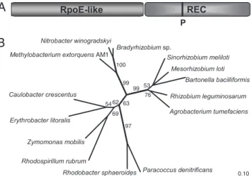
advanta-Fig. 2. Structure and phylogeny of PhyR. (A) Predicted two-domain structure of PhyR. A strictly conserved aspartate residue (corresponding to position 190 of PhyR) is predicted to be the phosphorylation site (P) of the receiver domain according to Prosite (www.expasy.ch). (B) Phylogenetic tree of PhyR homo-logues in various Alphaproteobacteria using the treepuzzle algorithm and the Jones–Taylor–Thornton evolutionary model and based on the amino acid sequences aligned with ClustalW (see Fig. 4, which is published as supporting information on the PNAS web site). Branches that were recovered in⬍50% of 1,000 reconstructed treepuzzle trees are shown as multifurcations; percent-age values for branches withⱖ50% recovery are given in the tree. Original tree construction included all available PhyR homologue sequences currently available in the GenBank database. A selection was made of 13 representative sequences plus PhyR of M. extorquens AM1.
Fig. 3. Plant colonization of M. extorquens WT and the phyR deletion strain. The detection limit was at 102cfu per plant. Three other independent exper-iments showed congruent results (data not shown).
Gourion et al. PNAS 兩 August 29, 2006 兩 vol. 103 兩 no. 35 兩 13189
geous in detecting the up-regulation of proteins that might have partially overlapping functions, as suggested by the identification of protein paralogues. This work provides a list of candidate proteins that need to be analyzed in more detail with respect to their importance for bacterial fitness. In addition, we have identified a previously undescribed type of two-domain regulator termed PhyR, which plays a key role in the adaptation of bacteria for plant colonization. The PhyR regulon suggests a role in dealing with the various stresses that the bacteria are likely to encounter in the phyllosphere. We assume that this regulator is also of importance in other Alphaproteobacteria.
Experimental Procedures
Bacterial Growth Underin Vitro and in Planta Conditions.For plant inoculation experiments, M. extorquens AM1 was grown in liquid MM (48) containing succinate (20 mM) to midexponential growth phase (OD600⫽ 1.2), centrifuged, washed, and resuspended in 10
mM MgCl2. The bacteria were adjusted to an OD600of 1.0 (108cfu per ml) and used for seed inoculation (4-h shaking at room temperature with slight moving) after sterilization of A. thaliana (ecotype Columbia) seeds. Sterilization of seeds was achieved through incubation in 2.4% hypochlorite for 5 min followed by eight washing steps. The plants were allowed to develop under controlled conditions on Murashige and Skoog medium in growth chambers under sterile conditions in Magenta boxes (1 week at 20°C, 16 h light兾8 h darkness; 2 weeks at 22°C, 9 h light兾15 h darkness). Preliminary experiments were performed to determine a suitable time point of harvest of M. extorquens AM1 from plants. It was chosen at 3 weeks to ensure that the overall bacterial population showed logarithmic development at this time point (average cell population: 106 cfu per aerial part of each plant). Sterility of uninoculated plants was verified by sonication of leaves in phos-phate buffer and plating on KingB medium. For each experiment, ⬇150 plants were grown. In addition, bacterial precultures (105
Table 2. List of proteins found to be positively regulated by PhyR Spot
no. RMQ no. Gene product* CD search Mr pl
Ratio†phy R⫹vs. phyR⫺
Found in phyllosphere proteome (see Table 1)
20, 21 RMQ08198 (PhyR, response regulator) pfam06182 29.1 4.8 ⬁ 1, 2 RMQ09549 KatE, catalase (L48340) pfam00199 63.5 7.1 ⫹ 12 RMQ02531 SodA, superoxide dismutase pfam02777 22.5 5.8 ⫹ 26, 28 RMQ02206 Hsp20, heat-shock protein 20 pfam00011 18.4 5.2 ⫹⫹ 29 RMQ05258 Dps, DNA protection protein pfam00210 19.9 5.0 ⬁ 4 RMQ11717 Putative quinoprotein (glucose dehydrogenase) 36.0 6.9 ⫹ 38 RMQ02894 Putative lactoylglutathione lyase pfam00903 16.5 5.7 ⬁ 8 RMQ03170 Orf88, dioxygenase (glyoxalase family protein) (AY034474) pfam00903 36.4 6.2 ⬁
43 RMQ01365 Gap20 (AF442748) 19.0 5.8 ⫹
25 RMQ09016 NifU-like兾thioredoxin-like protein pfam01106 20.3 4.8 ⬁ 24 RMQ08861 Protein of unknown function 20.3 5.3 ⬁ 15 RMQ00428 Protein of unknown function 31.9 5.3 ⫹
Additional proteins under PhyR control
3a RMQ06018 Glutathione-dependent formaldehyde dehydrogenase pfam00107 42.2 5.7 ⬁ 5 RMQ01240 ADH, alcohol dehydrogenase pfam00107 39.1 6.5 ⬁ 6 RMQ00842 ADH, alcohol dehydrogenase pfam00107 35.2 6.3 ⬁ 7a RMQ07799 Short-chain dehydrogenases兾reductases (GSP39) pfam00106 30.8 5.9 ⬁ 23 RMQ00500 Short-chain dehydrogenases兾reductases pfam00106 27.6 5.1 ⬁ 51 RMQ11711 PccA, propionyl-CoA carboxylase (AY181038) pfam02786 75.6 4.9 ⫹ 14 RMQ06958 MclA, malyl-CoA lyase (U72662) 38.0 5.6 ⫹ 18 RMQ06488 MDH, malate dehydrogenase (L33465) pfam02866 39.1 6.6 ⬁ 30, 31 RMQ02884 Phosphoglycerate mutase pfam00300 23.9 5.5 ⫹ 48 RMQ06654 NAD(P) transhydrogenase␣-subunit pfam01262 39.6 5.6 ⬁ 19 RMQ01528 FixB, Electron transfer flavoprotein,␣-subunit pfam00766 32.5 4.9 ⬁ 11 RMQ02643 WrbA, flavoprotein pfam00258 21.1 6.2 ⬁ 32 RMQ00895 Carbonic anhydrase pfam00484 28.4 9.1 ⫹ 36 RMQ06760 Ohr, organic hydroperoxide resistance protein-like protein (GSP17o) pfam02566 15.0 5.7 ⬁ 40 RMQ06032 AhpC (alkyl hydroperoxide reductase)兾TSA (thiol specific antioxidant) family protein pfam00578 18.0 5.9 ⫹
45 RMQ07144 Trx, thioredoxin pfam00085 17.4 9.7 ⫹
7b RMQ06181 Putative haloacetate dehalogenase兾non-heme chloroperoxidase pfam00561 30.6 5.8 ⬁ 33 RMQ07321 Putative phospholipid-binding proteins pfam01161 19.7 5.5 ⬁ 10 RMQ11238 Putative 3-hydroxyisobutyrate dehydrogenase 20.8 5.7 ⫹ 22 RMQ12169 CinA-like protein pfam00994 25.0 4.8 ⫹ 27 RMQ09145 Hbd,D--hydroxybutyrate dehydrogenase (AY391854) pfam00106 31.9 8.7 ⫹ 42 RMQ02665 GreA, transcription elongation factor pfam01272 22.1 6.9 ⫹ 13 RMQ06933 EF-Ts, elongation factor pfam00889 35.1 5.3 ⫹ 50 RMQ07718 CzcB, Cobalt-zinc-cadmium resistance protein pfam00529 46.3 6.2 ⫹ 3b RMQ03224 Hypothetical signaling protein 42.4 8.2 ⬁ 34 RMQ04652 Protein of unknown function (GSP26) pfam01243 19.0 5.7 ⬁ 9 RMQ05442 Protein of unknown function pfam01442 28.4 5.8 ⫹ 41 RMQ01392 Protein of unknown function pfam03928 14.8 5.0 ⫹ 35 RMQ12283 Protein of unknown function 14.9 5.6 ⬁ 37 RMQ12368 Protein of unknown function 12.7 6.6 ⬁ 44 RMQ07641 Protein of unknown function 16.8 5.4 ⬁
*Accession nos. are in parentheses.
†Spots indicated as ‘‘⬁’’ were only detectable in the proteome from M. extorquens AM1 ⌬phyR pBG11 (phyR overexpression) and not in the strain containing pCM80 (phyR minus). Spots indicated as ‘‘⫹’’ were found to be at least 3-fold induced.
cfu兾ml) were spread on the surface of agar-solidified MM com-plemented with 20 mM succinate for 5 days at 22°C, 9 h light兾15 h darkness.
Harvest of Bacteria and Preparation of Cell Extracts.The aerial parts of the plants were separated from the roots by cutting with a razor blade, and bacteria were harvested in aliquots of 15 plants in 50-ml plastic tubes filled with cooled TE buffer (10 mM Tris, pH 7.5兾1 mM EDTA) supplemented with PMSF (0.3 mg兾ml) and Percoll (GE Healthcare, Uppsala, Sweden; 20% final concentration) through alternating sonication and vortexing (45 s兾30 s, 3 times). The suspension was centrifuged (12,000 ⫻ g, 4°C for 10 min) whereby the addition of Percoll facilitated sedimentation of the bacteria, leaving small plant debris in the supernatant. Cells from one experiment were washed, pooled, and frozen until further use. Bacteria from roots and in vitro conditions were treated in parallel in a similar way. Total proteins were extracted by using a French pressure cell at 108Pa (two times, 4°C), and the cell extract was recovered after centrifugation (13,000⫻ g, 4°C for 30 min).
Proteome Analysis.2-DE was performed with 18-cm immobilized pH gradient strips (4.0–7.0; GE Healthcare) as described (17). Five independent experiments were performed, whereby the material (aerial parts, roots, in vitro) from two experiments was subjected to SYPRO Ruby staining (Molecular Probes, Leiden, The Nether-lands; using 350g of protein) and from three experiments to silver nitrate staining (using 120 g of protein). To identify proteins associated with epiphytic growth of M. extorquens, images were analyzed by using the Delta 2D software package (Decodon, Greifswald, Germany). Only proteins that were at least 3-fold induced in the independent experiments were identified. Protein identification was performed by peptide mass fingerprinting as described (17) and liquid chromotography兾tandem mass spectrom-etry (49). Identification of differentially expressed proteins was performed independently from the different gels that represent the different biological repetitions and that were stained with SYPRO
Ruby and silver nitrate, respectively, and had to give congruent results.
Mutant Generation and Construction of Complementation Strains.A
phyR mutant was generated by using the suicide vector pCM184
(50). Complementation of the phyR deletion mutant was achieved through cloning of phyR with its presumed promoter region by using the forward primer Prom-Phy-f-BamHI tggatcctgccgcgactacga-caaacgag (located 454 nt upstream of the predicted start codon) and the reverse primer Phy-r-KpnI catcggccggtaccttttcacgg into the XbaI and HindIII sites of the broad host range cloning vector pCM62 (40) resulting in pBG17. The plasmid was subsequently introduced in the ⌬phyR::kanR mutant. In addition, phyR was cloned downstream of the mxaF promoter into the PstI兾KpnI site of the expression vector pCM80 (40) resulting in pBG11 by using the primer Phy-PstI-f: catggctgcagcagcaacg and Phy-CM80-r-KpnI mentioned above.
Phenotypic Analysis ofM. extorquens AM1 Constructs and Analysis of the PhyR Regulon. Colonization of the phyR deletion strain was compared with M. extorquens AM1 WT and the complemented strain ⌬phyR::kanR pBG17. For this purpose, plant inoculation experiments were performed. For sampling, the aerial parts of the plants were placed individually in 1 ml of MM and sonicated for 5 min in an ultrasonication bath. Cell suspensions were then serially diluted and plated onto MM. To identify PhyR-regulated genes, we performed a differential proteome analysis by using M. extorquens AM1 ⌬phyR::kanR containing pBG11 and pCM80, respectively, grown to midexponential growth phase in the presence of succinate. Cells were harvested, washed in ice-cold TE buffer supplemented with PMSF, and cell extracts were prepared, and proteome analysis was performed as described above.
We thank Claudia Knief (Laboratoire des Interactions Plantes Micro-Organismes) for tree calculation. This work was supported by a grant from the Centre National de la Recherche Scientifique ‘‘Program Prote´omique et genie des prote´ines.’’ B.G. was supported by a fellowship of the French Ministry of Research. J.A.V. was supported by the Max-Planck-Gesellschaft.
1. Lindow, S. E. & Brandl, M. T. (2003) Appl. Environ. Microbiol. 69, 1875–1883. 2. Haefele, D. M. & Lindow, S. E. (1987) Appl. Environ. Microbiol. 53, 2528–2533. 3. Sundin, G. W. & Murillo, J. (1999) Environ. Microbiol. 1, 75–87.
4. Yu, J., Penaloza-Vazquez, A., Chakrabarty, A. M. & Bender, C. L. (1999) Mol. Microbiol. 33,712–720.
5. Lindow, S. E. (1993) Appl. Environ. Microbiol. 59, 1586–1592.
6. Rainey, P. B. & Preston, G. M. (2000) Curr. Opin. Biotechnol. 11, 440–444. 7. Boch, J., Joardar, V., Gao, L., Robertson, T. L., Lim, M. & Kunkel, B. N. (2002)
Mol. Microbiol. 44, 73–88.
8. Yang, S., Perna, N. T., Cooksey, D. A., Okinaka, Y., Lindow, S. E., Ibekwe, A. M., Keen, N. T. & Yang, C. H. (2004) Mol. Plant Microb. Interact. 17, 999–1008. 9. Marco, M. L., Legac, J. & Lindow, S. E. (2005) Environ. Microbiol. 7, 1379–1391. 10. Chistoserdova, L., Chen, S. W., Lapidus, A. & Lidstrom, M. E. (2003) J.
Bacteriol. 185, 2980–2987.
11. Hirano, S. S. & Upper, C. D. (1991) in Microbial Ecology of Leaves, eds. Andrews, J. H. & Hirano, S. S. (Springer, New York), pp. 271–294. 12. Corpe, W. A. & Rheem, S. (1989) FEMS Microbiol. Ecol. 62, 243–250. 13. Holland, M. A. (1997) Recent Res. Dev. Plant Physiol. 1, 207–213.
14. Sy, A., Timmers, A. C., Knief, C. & Vorholt, J. A. (2005) Appl. Environ.
Microbiol. 71, 7245–7252.
15. Vorholt, J. A. (2002) Arch. Microbiol. 178, 239–249.
16. Koenig, R. L., Morris, R. O. & Polacco, J. C. (2002) J. Bacteriol. 184, 1832–1842. 17. Laukel, M., Rossignol, M., Borderies, G., Vo¨lker, U. & Vorholt, J. A. (2004)
Proteomics 4, 1247–1264.
18. Korotkova, N. & Lidstrom, M. E. (2001) J. Bacteriol. 183, 1038–1046. 19. Bourque, D., Pomerleau, Y. & Groleau, D. (1995) Appl. Microbiol. Biotechnol.
44,367–376.
20. Priefert, H., Kru¨ger, N., Jendrossek, D., Schmidt, B. & Steinbu¨chel, A. (1992)
J. Bacteriol. 174, 899–907.
21. Xu, J. & Johnson, R. C. (1995) J. Bacteriol. 177, 3166–3175.
22. Graus, M., Schnitzler, J. P., Hansel, A., Cojocariu, C., Rennenberg, H., Wisthaler, A. & Kreuzwieser, J. (2004) Plant Physiol. 135, 1967–1975. 23. Booth, I. R., Ferguson, G. P., Miller, S., Li, C., Gunasekera, B. & Kinghorn, S.
(2003) Biochem. Soc. Trans. 31, 1406–1408.
24. Cooper, R. A. (1984) Annu. Rev. Microbiol. 38, 49–68. 25. Spiess, C., Beil, A. & Ehrmann, M. (1999) Cell 97, 339–347.
26. Antelmann, H., Bernhardt, J., Schmid, R., Mach, H., Volker, U. & Hecker, M. (1997) Electrophoresis 18, 1451–1463.
27. Weiner, L. & Model, P. (1994) Proc. Natl. Acad. Sci. USA 91, 2191–2195. 28. Martinez, A. & Kolter, R. (1997) J. Bacteriol. 179, 5188–5194.
29. Chen, L. & Helmann, J. D. (1995) Mol. Microbiol. 18, 295–300. 30. Nair, S. & Finkel, S. E. (2004) J. Bacteriol. 186, 4192–4198. 31. Gonzalez-Flecha, B. & Dimple, B. (1997) J. Bacteriol. 179, 382–388. 32. Nystrom, T. (2004) Annu. Rev. Microbiol. 58, 161–181.
33. Levine, A., Tenhaken, R., Dixon, R. & Lamb, C. (1994) Cell 79, 583–593. 34. Ceci, P., Ilari, A., Falvo, E. & Chiancone, E. (2003) J. Biol. Chem. 278,
20319–20326.
35. Kim, Y. C., Miller, C. D. & Anderson, A. J. (2000) Appl. Environ. Microbiol. 66, 1460–1467.
36. Weichart, D., Querfurth, N., Dreger, M. & Hengge-Aronis, R. (2003) J.
Bacteriol. 185, 115–125.
37. Schnider-Keel, U., Lejbolle, K. B., Baehler, E., Haas, D. & Keel, C. (2001) Appl.
Environ. Microbiol. 67, 5683–5693.
38. Keith, L. M. & Bender, C. L. (1999) J. Bacteriol. 181, 7176–7184. 39. West, A. H. & Stock, A. M. (2001) Trends Biochem. Sci. 26, 369–376. 40. Marx, C. J. & Lidstrom, M. E. (2001) Microbiology 147, 2065–2075. 41. Jalloul, A., Montillet, J. L., Assigbetse, K., Agnel, J. P., Delannoy, E.,
Tri-antaphylides, C., Daniel, J. F., Marmey, P., Geiger, J. P. & Nicole, M. (2002)
Plant J. 32, 1–12.
42. Gutheil, W. G., Kasimoglu, E. & Nicholson, P. C. (1997) Biochem. Biophys. Res.
Commun. 238, 693–696.
43. Hengge-Aronis, R. (2002) Microbiol. Mol. Biol. Rev. 66, 373–395.
44. Sarniguet, A., Kraus, J., Henkels, M. D., Muehlchen, A. M. & Loper, J. E. (1995)
Proc. Natl. Acad. Sci. USA 92, 12255–12259.
45. Miller, C. D., Kim, Y. C. & Anderson, A. J. (2001) Can. J. Microbiol. 47, 41–48. 46. Stockwell, V. O. & Loper, J. E. (2005) Microbiol. 151, 3001–3009.
47. Hecker, M. & Vo¨lker, U. (2001) Adv. Microb. Physiol. 44, 35–91.
48. Harder, W., Attwood, M. & Quayle, J. R. (1973) J. Gen. Microbiol. 78, 155–163. 49. Boudart, G., Jamet, E., Rossignol, M., Lafitte, C., Borderies, G., Jauneau, A.,
Esquerre-Tugaye, M. T. & Pont-Lezica, R. (2005) Proteomics 5, 212–221. 50. Marx, C. J. & Lidstrom, M. E. (2002) BioTechniques 33, 1062–1067.
Gourion et al. PNAS 兩 August 29, 2006 兩 vol. 103 兩 no. 35 兩 13191