HAL Id: tel-01803814
https://tel.archives-ouvertes.fr/tel-01803814
Submitted on 31 May 2018HAL is a multi-disciplinary open access archive for the deposit and dissemination of sci-entific research documents, whether they are pub-lished or not. The documents may come from teaching and research institutions in France or abroad, or from public or private research centers.
L’archive ouverte pluridisciplinaire HAL, est destinée au dépôt et à la diffusion de documents scientifiques de niveau recherche, publiés ou non, émanant des établissements d’enseignement et de recherche français ou étrangers, des laboratoires publics ou privés.
High-resolution structure of the nucleosome-H1 complex
and interaction with transcription factor Sox6
Ramachandran Boopathi
To cite this version:
Ramachandran Boopathi. High-resolution structure of the nucleosome-H1 complex and interaction with transcription factor Sox6. Cellular Biology. Université Grenoble Alpes, 2016. English. �NNT : 2016GREAV020�. �tel-01803814�
THÈSE
Pour obtenir le grade de
DOCTEUR DE LA COMMUNAUTÉ UNIVERSITÉ
GRENOBLE ALPES
Spécialité :Biologie cellulaire
Arrêté ministériel : 7 août 2006
Présentée par
Ramachandran BOOPATHI
Thèse dirigée par Dr. Stefan DIMITROV
préparée au sein duInstitut Albert Bonniot,INSERM U823
dans l'École Doctorale Chimie et Sciences du Vivant
Structure de haute résolution
du complexe nucleosome-H1
et son interaction avec le
facteur de transcription Sox6
Thèse soutenue publiquement le 30 Mai 2016, devant le jury composé de :
M. Hans GEISELMANN
Professeur, Université Grenoble Alpes (Président)
MME. Francesca PALLADINO
Directeur de Recherche CNRS (Rapporteur)
M. Christophe THIRIET
Chercheur CNRS (Rapporteur) M. Dimitar ANGELOV
Directeur de Recherche CNRS (Examinateur)
M. Stefan DIMITROV
2
Dedicated to
My Family
&
3
ACKNOWLEDGEMENTS
“ெசய்யாமல் ெசய்த உதவிக்கு ைவயகமும்
வானகமும் ஆற்றல் அrது”
A great Tamil poet Thiruvalluvar said that the heaven and earth is not an equivalent for a benefit which is conferred where none had been received. Same way during the journey of my doctoral studies many people have been helping me without expecting anything in return. Without them it would not have been possible for me to complete my thesis. I wish to take this opportunity to thank them for their unconditional support, guidance and assistance.
First and foremost, I wish to express my deepest gratitude to my mentor Dr. Stefan Dimitrov (University of Grenoble) for providing me an opportunity to work on this challenging project under his guidance. I would like to thank him for his support, encouragement and giving me freedom to think and to do things in my own way. I would like to thank our collaborator Dr. Dimitar Angelov (ENS de Lyon) for providing me an opportunity and space to work in his lab. I wish to thank him specially for teaching the chromatin biochemistry and for his support and guidance throughout my studies. I feel privileged to have been guided by two mentors during my PhD. It’s not just the scientific training which I have obtained under their guidance; they have nurtured me in every possible way such that I am confident to pursue anything independently.
I would like to thank Prof.Hans Geiselmann, Dr.Christophe Thiriet and Dr.Francesca Palladino for their time and efforts to evaluate the thesis.
I would like to take this opportunity to thank Prof.Tapas Kundu (JNCASR, India) for introducing me to field of molecular biology research. The 2 years spent in his lab prior to my graduate degree played an important role towards directing my interest in science and inculcating in me the curiosity to explore. I feel blessed to have the foundation of my scientific career laid by him.
I would like to extend my gratitude to all present and past lab members of Dr. Stefan’s and Dr. Dimitar’s team. Special thanks to Dr. Lama Soueidan (Lama Mama) for all the help from day one, especially for doing all French to English translation for me. Her presence made the lab environment lively and an enjoyable place to work. I would like thank Dr. Imtiaz
4
Nisar Lone for teaching me the footprinting techniques and providing valuable suggestions for my experiments. My thanks to Dr. Elsa, Dr. Herve and Ognyan for all the help and for the wonderful time we shared in Angelov’s team. I would like to thank all students and staff members of both ENS de Lyon and Institut Albert-Bonniot for their support and help.
Special thanks to Dr. Manish Grover (for me, Dr. Sheldon Cooper from the famous series “The Big Bang Theory”) for helping me with the editing of my thesis and supporting me during the course of thesis writing.
My special thanks to my friends especially the “Fish group members” Dr. Manju, Shwetha, Bharath, Vijitha, Uma Anivilla, Chaithanya, Sam and Suresh for those sweet memories we all shared together. I would like to thank my friends Babu anna, Chandru, Vivek, Balu, Madhu, Gopi, Sunil, Abhilash, Harsha, Akarsha and Madhura for making all my weekends in Lyon memorable.
I would like to thank all Dr. Tapas Kundu lab members especially Dr. Karthigeyan, Dr. Mohan Krishna and Dr. Selvi Bulusu for their valuable advice and guidance.
I wish to extend my gratitude to my friend Vinodhkumar for standing by me since school days sharing all my happiness and bearing all my stupidity.
Finally, I would like to thank my parents of their trust in me and letting me to do what I want to do in my life. My special thanks to my brother and sister for all those sacrifice they made for my education. I am glad that their sacrifice has paid off well and I have been able to successfully complete my Ph.D. With enormous love and gratitude I dedicate my thesis to my family.
5
INDEX
Thèse 1
Acknowledgements 3
Index 5
List of tables and figures 8
Resume de la these 10
Summary of the thesis 12
Information of the laboratories in which the thesis was prepared 14
List of abbreviations 15
Chapter I 16
I.1 Introduction 16
I.2 History of Chromatin 16
I.3 Chromatin Organization 18
I.4 Histone 19
I.4.1 Histone Octamer 19
I.5 Nucleosome core particle 21
I.5.1 Nucleosome positioning 22
I.6 Linker Histone H1 23
I.6.1 Domain architecture of linker histone H1 23
I.6.2 Location of Linker Histone H1 on Nucleosome 25
I.6.3 Histone H1 dynamics 26
I.6.4 H1 variants 28
I.6.4.1 H1.1- H1.5 30
I.6.4.2 H1.0 and H.1x 30
I.6.4.3 Germ line specific linker histone variants 31
I.6.5 Linker histone modifications 32
I.7 Structure of the 30-nm chromatin fiber 33
I.8 Chromatin and transcription 38
I.9 Structural Order Beyond the 30-nm fiber 39
I.10 Chromosome territories 40
I.10.1 Euchromatin 41
I.10.2 Heterochromatin 41
I.10.2.1 Facultative heterochromatin 42
I.10.2.2 Constitutive heterochromatin 43
I.11 Structural modification of chromatin fiber 43
I.11.1 Chromatin remodeling 43
I.11.1.1 Different Chromatin remodeling Families 44
I.11.1.1.1SWI/SNF family remodelers 44
I.11.1.1.2 ISWI family remodelers 45
I.11.1.1.3 CHD family remodelers 45
I.11.1.1.4 INO80 family remodelers 46
I.11.1.2 Remodeler mechanisms 48
I.11.1.2.1 Twist-diffusion model 48
I.11.1.2.2Bulge/Loop Propagation Model 49 I.11.2 Post translational modification of Histones 50
I.11.2.1 Histone acetylation 51
I.11.2.2 Histone phosphorylation 52
I.11.2.3 Histone methylation 52
6
I.11.2.5 Other modifications 54
I.11.3 Histone Variants 55
I.11.3.1 Histone H2A variants 55
I.11.3.1.1 Histone variant H2A.Z 55
I.11.3.1.2 Histone variant H2AX 56
I.11.3.1.3 Histone variant MacroH2A 56
I.11.3.1.4 Histone variant H2A.B 57
I.11.3.2 Histone H2B variants 58
I.11.3.3 Histone H3 variant 58
I.11.3.3.1 Histone variant CenH3 59
I.11.3.3.2 Histone Variant H3.3 60
I.12 Objective 61
I.13 Results: Manuscript 1: Structure and dynamics of the nucleosome in complex with linker histone H1
62
I.13.1 Abstract 63
I.13.2 Introduction 63
I.13.3 Results 64
I.13.3.1 Structure and dynamics of the linker DNA 64 I.13.3.2 Localization of H1 on the nucleosome 65 I.13.3.3 Crystal structure of the H1-bound
nucleosome 66
I.13.3.4 Nucleosome recognition by the GH1 domain 67 I.13.3.5 H1-nucleosome interactions mapped by
cross-linking and footprinting 68
I.13.4 Discussion 70
I.13.5 Methods summary 72
I.13.6 Acknowledgements 72
I.13.7 Methods 72
I.13.7.1 Cryo-electron microscopy and Image
Processing 72
I.13.7.1.1 Specimen preparation 72
I.13.7.1.2 Image Processing 73
I.13.7.1.3 Local resolution estimation 73 I.13.7.2 Crystal structure determination 74 I.13.7.2.1 Crystallization and Data collection 74 I.13.7.2.2 Structure Determination of the 197-bp Nucleosome (minus GH1)
74 I.13.7.2.3 Map scaling and noise estimation 74 I.13.7.2.4 Positioning of the GH1 domain 75
I.13.7.3 Molecular Docking 77
I.13.7.3.1Docking with HADDOCK 77
I.13.7.3.2 Docking with Autodock Vina 78
I.13.7.4 Biochemistry techniques 79
I.13.7.4.1 H1 modification 79
I.13.7.4.2 Nucleosome reconstitution for crosslinking experiments
79 I.13.7.4.3 Linker histone binding and
cross-linking
80
I.13.7.4.4 Cross-link Mapping 80
7
I.13.8 Supplemental Data 86
I.14 Perspectives 104
Chapter II 105
II.1 Introduction 105
II.2 Pioneer transcription factors 105
II.3 Sox proteins 110
II.3.1 High Mobility Group (HMG) DNA binding domain 111
II.4 Objective 112
II.5 Results 113
II.5.1 Binding of Sox6-HMG domain to the nucleosome 113 II.5.2 Effect of histone H1 on the Sox6 HMG domain specific
interaction with nucelosomal DNA
120
II.6 Discussion 122
II.7 Perspectives 123
II.8 Materials and methods 124
II.8.1 Sox6 HMG domain cloning and purification 124
II.8.2 Human core histone purification 124
II.8.3 Histone tetramers and dimers preparation 125
II.8.4 Preparation of DNA fragments 126
II.8.5 Nucleosome Reconstitution 127
II.8.6 Sox6 HMG domain binding reaction 127
II.8.7 NAP-1 mediated H1deposition 128
II.8.8 Hydroxyl radical footprinting 128
II.8.9 UV laser footprinting 129
8
LIST OF TABLES AND FIGURES
CHAPTER I
Figure 1 Diagram represent major discoveries in the history of chromatin
studies 17
Figure 2 Organization of eukaryotic chromatin fibers 19
Figure 3 Graphical representation of the domain architecture of all the core
histones 20
Figure 4 Histone octamer assembly 21
Figure 5 Nucleosome positioning sequences 22
Figure 6 Domain architecture of metazoan linker histone H1 24
Figure 7 Different models of H1 binding to nucleosome 26
Figure 8 A typical FRAP curve for H1 binding to chromatin 27 Figure 9 Different models for the reversible association of linker histone H1
with nucleosome
28
Table 1 The H1 histone family in human 29
Figure 10 Aminoacid sequence alignment of linker histone H1.5 and its
embryonic isoforms from different organisms 31
Figure 11 Linker histone H1.5 post-translational modifications 33
Figure 12 Models for the 30nm chromatin fiber structure 34
Figure 13 Comparison of chromatin models to raw images of folded chromatin 35
Figure 14 30nm chromatin fiber structure 36
Figure 15 Acidic patch of nucleosome and its interaction with H4 tail 38 Figure 16 Chromonema and radial-loop/protein scaffold models 40
Figure 17 Chromatin territories 41
Figure 18 Properties of euchromatic and heterochromatic regions 42
Figure 19 Domain architecture of all remodeler families 44
Table 2 Showing composition of remodeler complexes in different eukaryotes 46 Figure 20 The twist diffusion model of nucleosome remodeling 49 Figure 21 The loop/bulge propagation model of nucleosome remodeling 50 Table 3 A detailed list of known histone posttranslational modification
enzymes
54
MANUSCRIPT 1
Figure 1 Cryo-EM analysis of nucleosomal particles 81
Figure 2 Crystallographic analysis of the H1/nucleosome complex 82
Figure 3 Nucleosome recognition by GH1 83
Figure 4 Surface mapping of GH1 functional and phylogenetic data 84
Figure 5 Mapping of H1-nucleosomal DNA interactions 85
Table S1 Data collection and refinement statistics 86
Table S2 Interaction restraints used for docking with HADDOCK 87 Table S3 Summary of docking results obtained with HADDOCK 88 Table S4 Helix density in Omit 2Fo-Fc END maps compared to noise level in
corresponding RAPID maps 89
Figure S1 Cryo-electron microscopy and image analysis 90
Figure S2 Resolution curves for the nucleosomal particles 91
Figure S3 Local resolution measurements 91
Figure S4 Stereo omit maps of core histones 92
Figure S5 Structure of the linker histone globular domain 94 Figure S6 Sample of the 6-dimensional search space explored to position GH1
9
Figure S7 Rigid body refinement of GH1 configurations identified in the 6D
molecular replacement search 96
Figure S8 GH1 orientations related by the nucleosome dyad 97
Figure S9 Omit map showing density for GH1 helices 98
Figure S10 Stereo views of electron density covering the GH1
domain(orientation2) 99
Figure S11 Stereoview showing agreement between the crystal structure and cryo-EM map of the H1-nucleosome complex
100
Figure S12 Conserved surface residues of GH1 101
Figure S13 GH1 is able to specifically bind to nucleosomes bearing a single
10-bp linker 102
Figure S14 Summary of Haddock results 103
CHAPTER II
Figure 1 Initial targeting of pioneer factor and subsequent events 106 Table 1 Predicted/validated pioneer transcription factors 107
Figure 2 Sox HMG domain 111
Figure 3 Characterization of the reconstituted nucleosome 114 Figure 4 Binding of Sox6 HMG domain to 601 nucleosome containing single
binding site 115
Figure 5 Binding of Sox6 HMG domain to 601 nucleosome containing three
binding sites 118
Figure 6 Effect of histone H1 on the Sox6 HMG domain specific interaction with nucleosomal DNA
120
Figure 7 Human histone octamer assembly 126
Figure 8 Principal of UV laser footprinting 130
10
RESUME DE LA THESE
Structure de haute résolution du complexe nucleosome-H1 et son
interaction avec le facteur de transcription Sox6
Comprendre la structure et l’organisation de la chromatine est une question fondamentale dans le domaine de la régulation de l’expression des gènes. La cristallographie par rayons-X et d’autres techniques biophysiques on permit de comprendre la structure du nucléosome avec une précision quasi atomique. Malgré de nombreuses études, les données structurelles au delà de la particule de cœur nucléosomale (NCP) demeurent imprécises. Au cours des dernières décennies plusieurs tentatives ont été faites pour montrer comment l’histone de liaison H1 interagit avec les particules nucléosomales pour les condenser en fibre de chromatine. Ces études ont mené à différents modèle décrivant la position de l’histone de liaison H1 sur la chromatine. De récentes avancées sur l’histone de liaison H1 suggèrent que le domaine globulaire de H1 (GH1) et la partie C-terminale interagit avec la dyade du nucléosome et les 2 bouts d’ADN de liaison (modèle à 3 contacts) qui sont contraintes de former une structure en tige. Cependant, la conformation et la position précise de l’histone de liaison H1 reste inconnues et la controverse à ce sujet persiste.
Dans cette étude, nous avons déterminé la structure tridimensionnelle de nucléosomes contenant H1 par des techniques de cryo-microscopie électronique (cryo-EM) et de diffraction aux rayons-X dans des cristaux. Nous avons utilisé le chaperons d’histone, NAP1, pour déposer l’histone de liaison H1 sur les nucléosomes reconstitué à partir des histones de cœur recombinant et la séquence d’ADN positionnante 601 de 197 paires de bases (dite de Widom). Nos résultats de cryo-EM montrent que l’association de H1 compacte le nucléosome en réduisant la mobilité des ADNs et stabilisant ainsi les contacts entre les nucléotides précédant la sortie NCP et l’octamer d’histones. Nos résultats par diffusion de rayon-x dans des cristaux à une résolution de 7Ä montrent que la partie globulaire de H1 (GH1) est située sur la dyade et interagie simultanément avec les petits sillons de l’ADN à la dyade et les ADN de liaison à l’entrée et à la sortie du nucléosome. Les parties N- et C-terminales de H1 sont orientées vers l’extérieur du cœur du nucléosome à travers les différents ADN de liaison. Nous avons validé l’orientation de GH1 par des expériences de pontages ADN-proteine, après substitutions de cystéine par mutagénèse dirigée, empreinte par radicaux hydroxyles et « amarrage moléculaire ». Nos résultats révèlent l’effet de H1 sur la dynamique du nucléosome et apporte une vision détaillé de la conformation du « stem du nucléosome » lors de l’incorporation de H1.
Nous avons également étudié l’association spécifique du facteur de transcription Sox6 à ces de reconnaissance consensus présent à l’intérieur du nucléosome, associé ou non avec l’histone de liaison H1 par une empreinte biphotonique avec laser UV. Nos résultats montrent que le domaine HMG de Sox6 se fixe spécifiquement sur son motif consensus situé profondément à l’intérieur du nucléosome à l’exception sur la dyade. Cette association n’est pas influencée par la « fermeture » des ADN de liaison avec l’histone H1 démontrant l’existence d’un autre façon de reconnaissance que le modèle de Widom basés sur fluctuations thermodynamiques des ADN de liaison. Le résultat que Sox6 est capable de surmonter la
11
barrière nucléosomale (avec ou sans H1) suggère fortement que les facteurs de transcription de la famille Sox, de domaine de liaison de type HMG, jouent le rôle de facteurs « pionnier » dans la régulation de la transcription et en particulier dans l’initiation de la différentiation.
12
SUMMARY OF THE THESIS
High-resolution structure of the nucleosome-H1 complex and interaction
with transcription factor Sox6
Understanding the structural organization of chromatin is a fundamental issue in the field of gene regulation. X-ray crystallography and other biophysical techniques have enabled understanding of the nucleosome structure nearly at atomic precision. Despite numerous studies, the structural information beyond the nucleosome core particle (NCP) remains elusive. Over the last few decades several attempts have been made to reveal how the linker histone H1 interacts with the nucleosome particles and condenses them into a chromatin fiber. These studies have led to different models describing the position of linker histone H1 on chromatin. Recent advancements in linker histone H1 studies suggest that globular domain of histone H1 (GH1) interacts with the nucleosomal dyad and its C-terminal domain interacts with the linker DNA forming a stem like structure. However, the precise conformation of linker histone H1 and position of other domains still remains unknown.
In this study, we resolved the three-dimensional structure of H1-containing nucleosomes by using cryo-electron microscopy (cryo-EM) and X-ray crystallography. We have used the chaperone NAP-1 to deposit linker histone H1 onto nucleosomes reconstituted from recombinant core histones and 197 base-pair of 601 strong nucleosome positioning DNA sequence. Our cryo-EM results showed that association of H1 gives a more compact appearance of the nucleosome as it restricts the mobility of the two linker DNAs keeping them in close proximity and thereby stabilizing contacts between the histone core and nucleotides preceding NCP exit. Our X-ray crystallography results at 7 Ä resolution reveal that the globular domain of histone H1 (GH1) is positioned onto the nucleosome pseudodyad and recognizes the nucleosome core and both linker arms by contacting the DNA backbone in the minor groove. The N- and C-terminal domains of H1 are oriented away from the nucleosome core towards different DNA linkers. We further validated the orientation of GH1 by cross-linking experiments followed after cysteine substitutions mutagenesis, hydroxyl radical footprinting and by molecular docking. Our results reveal the effect of H1 on nucleosome dynamics and also provide a detailed view of the nucleosome stem conformation upon H1 incorporation.
13
We also studyed the nucleosome accessibility of transcription factor Sox6 and the impact of linker histone H1 incorporation to Sox6 binding on nucleosome by using UV laser biphotonic footprinting. Our results reveal that Sox6 HMG domain binds specifically to its consensus binding located deep inside of the nucleosomal DNA, but not at the nucleosomal dyad. Our in vitro footprinting results reveal that the “locking” of DNA linkers by incorporation of histone H1 on nucleosome does not show any impact on Sox6 HMG domain binding, evidencing an alternative to the Widom model based on thermal fluctuation “opening” of the nucleosome at the linkers.. The finding that Sox6 is able to overcome nucleosome (chromatosome) barrier in presence or absence of H1, strongly suggest that the HMG domain - based Sox family proteins it can act as a pioneer factor in transcription regulation, in particular in initiation of cell differentiation..
14
INFORMATION OF THE LABORATORIES IN WHICH THE THESIS
WAS PREPARED
a) Main Laboratory
Institut Albert Bonniot
CRI INSERM/UJF U823
Rond-point de la Chantourne
38706 La Tronche Cedex
France
b) Affiliated laboratory
Laboratoire de Biologie Moléculaire de la Cellule
École Normale Supérieure de Lyon- UMR5239
46, allée d'Italie
69364 LYON cedex 07
France
15
LISTS OF ABBREVIATIONS
2D: 2 Dimensions kDa: Kilo Dalton
3D: 3 Dimensions MNase: Micrococcal Nuclease
aa : Amino-Acid mRNA: Messenger RNA
ACF: ATP-utilizing chromatin assembly and remodeling factor
Nap1: Nucleosome Assembly Protein-1 AFM: Atomic Force Microscopy NASP: Nuclear Autoantigenic Sperm Protein ATP: Adenosine-5'-TriPhosphate NBP: Nucleosome Binding Protein
bp: base pair NCP: Nucleosome Core particle
CAF1: Chromatin Assembly Factor 1 nm: Nanometer
CBP: CREB Binding Protein NMR: Nuclear Magnetic Resonance
CENP-A: Centromere Protein A NP40: Nonyl phenoxylpolyethoxylethanol 40
CHD: Chromodomain NRL: Nucleosome Repeat Length
cryo-EM: cryo-electron microscopy NTD: N-Terminal Domain
CTD: C-Terminal Domain NURF: Nucleosome remodeling factor
DNA: DeoxyriboNucleic Acid PCR: Polymerase Chain Reaction
DSB: Double Strands Break PHD: Plant Homeo Domain
DTT: Dithiothreitol PTF: Pioneer transcription factor
EM: Electron Microscopy PTM: Post-Translational Modification
ES: Embryonic Stem R: Arginine
FRAP: Fluorescence Recovery After Photobleaching
RNA: Ribonucleic Acid
GD: Globular Domain RNAi: RNA Interference
GH1: Globular Domain H1 RNAPII: RNA polymerase II
H1: Histone H1 rRNA: ribosomal RNA
H2A.Bbd: Histone: H2A Barr body-deficient RSC: Chromatin structure remodeling
H2A: Histone H2A SDS-PAGE: Sodium dodecyl sulfate PAGE
H2B: Histone H2B SELEX: Systematic Evolution of Ligands by
Exponential Enrichment
H3: Histone H3 SNPs: Single nucleotide polymorphisms
H4: Histone H4 Sox6: SRY (Sex Determining Region
Y)-Box6
HATs: Histone Acetyltransferases SPR: Surface Plasmon Resonance
HDAC: Histone deacetylases SWI/SNF: Switch/Sucrose Non Fermentable HILS: histone H1-like Protein in Spermatids
1 TAD: Transactivation domain
HMG domain: High mobility group (HMG) box domain
TBE: Tris-borate-EDTA HMT: Histone Methyltransferase TF: Transcription factor HP1: Heterochromatin Protein 1 TSS: Transcription start site IN080 :Inositol requiring 8 Ub: ubiquitin
IPTG: isopropyl-beta-Dthiogalactopyranoside
UV: Ultra Violet
ISWI: Imitation switch WT: Wild Type
16
CHAPTER I
I.1 Introduction
The organization of genetic material inside a eukaryotic cell is a packaging wonder. You start unpacking the 6 µm wide nucleus and the DNA would stretch upto 2 metres. Such extraordinary level of compaction is achieved with the help of specialized proteins which enable the DNA to fold into multiple coils and loops. This complex of DNA with proteins is termed as “chromatin” and dynamic regulation of chromatin organization is essential for functionality of the genome. Understanding a small aspect of this massive packaging endeavor is the focus of this thesis. The following sections discuss in brief the history of chromatin discovery, the current state of knowledge about chromatin structure and how it led to the objectives of my study.
I.2 History of Chromatin
By late 19th century, cell biology had evolved with the development of light microscopes and availability of stains and fixatives[1]; however, the understanding of substance inside the nucleus still remained elusive. It all began in 1871 (Figure 1), when a student in E. Hoppe-Seyler lab called as F. Miescher, noticed a strong phosphorous-rich acid while developing a method to isolate nuclei from pus leukocytes[2]. He termed this as “nuclein” and ten years later in 1881, H. Zacharia described nuclein to be resistant to protease degradation through his microscopy studies on protease digested isolated nuclei. Around the same time, W. Flemming, who was studying cell division, described the presence of thread like structures inside the nucleus which had high affinity for dyes. He named this stained material as “chromatin” (the Greek word for colour) and correlating with Zacharia’s work suggested chromatin to be same as nuclein[3, 4]. Next major advancement again came from E. Hoppe-Seyler’s lab in 1884, when A. Kossel performed acid extraction of avian erythrocytic nuclei which resulted in a preparation of basic proteins. He called these proteins as “histon”[5].
In the beginning of the 20th century, the rush hour of chromatin studies came to a pause and the emerging field of genetics came to limelight. It commenced with the rediscovery of Mendelian principles by H. de Vries in 1900 followed by the development of gene theory and the concept of linkage in Drosophila melanogaster by T.H. Morgan in
17
1910[6]. Later studies on the “transforming principle” by F. Griffith in 1928 and experiments by Avery, MacLeod and McCarty in 1944 established DNA to be the genetic material [7, 8].
The dark ages of chromatin studies ended with the discovery of polytene chromosomes by Heitz and Bauer in 1933 and correlation established by Painter (1933) and Bridges (1935) between chromosome bands and gene localization. It was also the most awaited technical advancement for chromatin biologists as now they had access to a material to experiment with. Later in 1941, analysis by D. Mazia on polytene chromosome and plant chromosome using proteases and nucleases suggested that both polytene and plant chromosome are composed of a continuous framework which seems to be composed of histone like protein attached with nucleic acid through its phosphoric residues[9]. Back then in 1941 scientific investigators started to accept that chromosome and chromatin formed the structural basis of genes.
Figure 1: Diagram represent major discoveries in the history of chromatin studies. Image modified from[7].
Development in X-ray diffraction and biophysical techniques led to major discoveries in the field of chromatin structure towards the middle of the twentieth century. The discovery of the DNA double helix by Watson and Crick in 1953[10], preparation of soluble chromatin molecules by G. Zubay and P. Doty in 1959[11], fractionation of histones by E. Johns in 1960[12], single DNA molecule as the constituent of chromatid framework by J. Gall in 1963 and association between chromatin transcription and histone post translational modification
18
by V. Allfrey in 1964 are among the important milestones which led to the better understanding of the chromatin structure[13, 14].
In 1970s, H. Davies introduced electron microscopy in the field of chromatin biology and two independent studies by Olins & Olins and Woodcock described the “beads on string” structure for chromatin fibers[15-17]. A year later, R. Kornberg & J. Thomas proposed the model of chromatin structure based on nuclease digestion and histone crosslinking analysis which showed that approximately 200 base pairs of DNA forms a complex with pairs of four histones[18]. The chromatin subunit i.e. DNA wrapped over an octameric histone core, was termed as “nucleosome” in 1975 and multiple nucleosomes were found to be connected by another protein called as linker histone [19, 20]. The precise location of linker histone and its role in chromosome condensation is still an unresolved matter in the field.
The next most intriguing thing in the field was to elucidate the structure of the nucleosome. Using X-ray crystallography, three independent studies by T. J. Richmond (1984), K. Luger (1997) and C. A. Davey (2002) described structure of the nucleosome core particle to the resolution of 7 Ǻ, 2.8 Ǻ & 1.9 Ǻ respectively[21-23]. Later tetra nucleosome structure was solved by T. Schalch in 2005[24]. Following on the similar lines, this thesis work describes the crystal structure of linker histone positioned on a nucelosome core particle.
I.3 Chromatin Organization
In cell approximately 147bp of DNA wrapped around the octamer of core histones (pair of each H2A, H2B, H3 and H4) form the basic subunit of chromatin ‘nucleosome’ resulting five to ten fold compaction (~11nm)[18, 25]. Nucleosomes are linked to adjacent nucleosomes through ~10 to 80bp linker DNA form "beads-on-a-string" filament. The polynucleosome string folds into roughly 50 fold compact ~30 nm chromatin fiber and linker histone H1 stabilizes the chromatin fiber by binding to adjacent linker DNA of each nucleosome[25]. The long range of chromatin fiber interactions lead to proteinaceous scaffolds that organizes the chromatin into a highly condensed metaphase chromosome (Figure 2) [26].
19
Figure 2: Organization of eukaryotic chromatin fibers. The lowest level of organization is the nucleosome, in which 146 base pairs of left-handed superhelical DNA wrapped 1.65 turns around the histone octamer. Nucleosomes are connected to each other by short stretches of linker DNA. At the next level of organization, nucleosome is folded into ~30nm fiber and these fibers further folded into higher order structures. Condensations of chromatin fibers are mainly mediated through the internucleosomal interactions, linker histones and non-histone proteins. Figure adapted from [25].
I.4 Histone
Histones are highly abundant basic proteins found in the nuclei of all eukaryotic cells. Based on their amino acid composition they are divided into five types namely the linker histone H1 and the four core histones H2A, H2B, H3, and H4[27]. Histones are highly conserved in evolution from yeast to humans and genetic experiments using yeast showed that core histones are essential for cell viability[28]. In a cell, core histones are found in same equal molar stoichiometry whereas linker histone H1 is present in half the amount[29].
20
The core histones have a molecular mass ranging from 10-16 kDa and each one is structurally separated into three distinct domains based on the occurrence of various secondary structures (Figure 3). Approximately 70 amino acids in the central region of all core histones forms the histone fold domain comprising 3 to 4 turn α-helix (α1), a loop of 7-8 aa, an 8-turn α-helix (α2), a loop of 6 aa and a final 2 to 3 turn α-helix (α3). Histone fold extensions are less structured uniform elements that extend from histone fold domain: N-terminally in H3 and H2A (αN) and C-N-terminally in H2A and H2B (αC). Random coil elements of amino acid make the histone N-terminal ends which differ in length from histone to histone (16 aa for H2A to 44 aa for H3). N –terminal tails are flexible and unstructured at least in the context of a single nucleosome core[22]. Histone H2A also has C-terminal tails which differs in length between H2A variants[30]. Histones are subjected to many posttranslational modifications such as acetylation, methylation, phosphorylation, ubiquitination, sumoylation, biotinylation, glycosylation, parpylation and ADP-ribosylation in the tail region as well as in the core domains which regulates chromatin structure and functions[31].
Figure 3: Graphical representation of the domain architecture of all the core histones. The boxes indicate the helices of the histone fold domain and histone fold extensions. Figure is modified from [22].
I.4.1 Histone Octamer
Histone octamers are assembled in pairs of heterodimers (H2A-H2B and H3-H4). The α1-L1-α2-L2-α3 regions of histone in a heterodimer interact in antiparallel orientation through hydrophobic interactions. In detail, N-terminal α1 and C-terminal α3 helices of each histone fold back on center α2 generating left handed intervening loops. This folding gives a broad U shape to a single histone and causes terminal helices to interlink and central helices to overlap. This type of structure is stabilized by extensive hydrophobic interactions between the helices and formation of β bridges between the loops[22]. The α3 helices and the
C-21
terminal halves of the α2 helices of the two H3 within the H3-H4 dimers interact to form a four-helix bundle which assists tetramer formation (H3-H4)2. Two H2A-H2B dimers bind to
the two opposite sides of the horseshoe shaped tetramer and complete the octamer formation (Figure 4a & 4b). Histone oligomers exist as H2A-H2B dimers and H3-H4 tetramers under normal physiological conditions or in the absence of DNA and form histone octamer at high salt concentration or in the presence of DNA[22].
Figure 4: Histone octamer assembly. (a) Histone octamer consists of two copies of each of four core histone (H2A, H2B, H3 and H4) and it assembles through H3-H4 tetramer forming complex with H2A-H2B dimmers. (b) SDS electrophoresis gel of individual core histone, H3-H4 tetramer, H2A-H2A-H2B dimer and histone octamer.
I.5 Nucleosome core particle
Nucleosome core particle (NCP) is the crystalizable substrate of the canonical nucleosome[32]. In NCP, H3-H4 tetramers interacts with the central 60 base pairs and each H2A-H2B dimers bind to the adjacent 30 base pairs from the central region. H3 αN helices interact with the remaining 13 base pairs at the each end and keep them relatively straight. DNA bends around the histone octamer due to charge neutralization of the acidic DNA
22
phosphate groups, hydrophobic interactions and by hydrogen bond formation especially between main-chain amide groups and phosphate oxygen atoms[22]. N-terminal tails of both H3 and H2B pass through DNA gyres and are reported to interact with linker DNA [22, 33]. H4 tails are reported to interact with the acidic patch of adjacent nucleosomes suggesting their possible role in higher-order-structure formation [22, 23].
I.5.1 Nucleosome positioning
There are several factors which regulate the positioning of the nucleosome on the DNA. DNA sequence plays the most important role as it determines the intrinsic flexibility of the DNA strand, the orientation of the major and minor grooves relative to histone octamer and availability of DNA interaction sites[34]. Mostly in high affinity nucleosome binding sequence, A/T motifs are incorporated in the minor groove bending site and G/C motifs are incorporated in the major groove bending site. However, only rotational orientation of DNA sequence obeys this rule but not the translational position of DNA sequence [35]. Other factors which influence nucleosome positioning include DNA methylation, histone variants, PTMs, chromatin remodelers, DNA-binding proteins and higher-order structure.
Figure 5: Nucleosome positioning sequences. Different nucleosome core particle constructs are arranged in order of increasing salt stability measured by tyrosine fluorescence spectroscopy. Minor and major groove -inward- facing region on histone octamer are represented orange and black, respectively. DNA binding motifs of core histones are mentioned as L-loop and A-α helix, where the color represents histone proteins blue-H3, green-H4, yellow-H2A and red-H2B. Figure adapted from[36].
So far different high affinity nucleosome positioning sequences have been identified from naturally occurring sequences (5s rDNA) as well as from chemical synthesis (601 sequence). The latter has been used widely in many in vitro studies because it assembles strong, stable and single translational positioned nucleosomes [37]. Palindromic DNA derived from high affinity nucleosome positioning sequences increases nucleosome’s resolution in
X-23
ray crystallographic studies by providing perfect twofold symmetry[22, 38]. Recent studies on nucleosome positioning sequence showed that 601L palindromic sequence (derivative from 601 Widom sequence) has extraordinary salt stability compared to the human α-satellite NCP sequence and to the parental sequence (Figure 5) [36].
I.6 Linker Histone H1
Linker histones constitute the most diverse group of histone proteins. They are extremely rich in lysine and lack the histone folding domain unlike core histones. H1 binds to linker DNA in the nucleosome in the ratio of one molecule per NCP and helps to stabilize the wrapping of DNA around the nucleosome, consequently promoting folding and assembly of higher order structures of chromatin[39-41]. Canonical linker histone typically has a tripartite structure: a short unstructured N-terminal tail (20 aa), relatively conserved and stably folded central globular domain (80 aa) and a lysine rich unfolded C-terminal tail (100 aa) (Figure 6) [42]. In higher eukaryotes, the numbers of H1 variants are much larger within an organism and the differences are found mainly in the sequence composition of the C-terminal tail. There are 11 variants found in mammals out of which 5 are ubiquitously expressed in somatic cells in addition to other tissue and stage specific variants[43]. Knockout studies in mouse ES cells showed that lack of one or two H1 subtypes neither alters mice development or significantly changes chromatin structure suggesting H1 subtypes to have redundancy in their function[44]. In the case of lower eukaryotes, the numbers of H1 variants are much less and show an alternative domain architecture as compared to higher eukaryotes[39]. For example,
Saccharomyces cerevisiae has a single linker histone H1 like protein Hho1p, which contains
two globular domains lacking both N- and C- terminal tails whereas Tetrahymena
thermophile has a single linker histone H1 which lacks the entire globular domain[45-47].
Knockout studies on these proteins showed that they are not essential for cell survival. However, knocking out of Hho1p does affect chromatin compaction especially during the stationary phase in yeast[48]. Apart from serving the role of an architectural protein, linker histone H1 variants are also involved in gene regulation, triggering apoptosis in response to DNA damage and tissue specific chromatin repression[49-51].
I.6.1 Domain architecture of linker histone H1
Early micrococcal nuclease digestion studies on chicken erythrocyte chromatin showed that globular domain (GH1) of linker histone H1 is alone sufficient to protect ~20
24
base pair in addition to the NCP termed as “chromatosome”, however the remaining domains may be required for higher order structure formation[52]. The central domain belongs to the ‘winged helix’ family of DNA-binding proteins which is highly conserved, resistant to trypsin digestion and more hydrophobic in nature compared to other N- and C- terminal domains. X-ray crystal structure from linker Histone H5 (GH5) and 2D NMR studies on GH5 and chicken histone GH1 showed that globular domain adopts a mixed α/β fold, with three α-helices forming the core of the domain and β-strands. α1 and α2 are separated by β-strands, followed by α3 and 2 anti-parallel strands and a loop are formed by the extension of these 2 β-strands[53]. The N terminal domain (NTD) is less conserved as compared to globular domain (GH1). NTD has alanine and proline rich subdomains. Removal of NTD from H1 showed no alteration in the higher order structure of chromatin suggesting NTD is not required for chromatin condensation[54].
Figure 6: Domain architecture of metazoan linker histone H1. The unstructured N- terminal domain (NTD) consists of 25-35 amino acid residues, globular domain (G) consists of ~80 residues and the unstructured C-terminal domain (CTD) consist of 100-120 residues. Figure adapted from [55].
The long unstructured C-terminal domain (CTD) of linker histone is poorly conserved among subtypes and species. CTD is highly abundant with a repeat sequence of S/TPXK, lysine and alanine amino acids. Lysine residues alone constitute ~40% of total amino acid residues which are evenly distributed throughout the region whereas alanine represents 20-35% of total residues [56, 57]. It was predicted that the high basic nature of CTD neutralizes DNA charges and helps chromatin condensation by stabilizing high order structure. However, deletion of most part of CTD did not drastically alter the H1 induced chromatin fiber condensation. The ability to stabilize the chromatin fiber resides within the first 25 amino acids flanking next to the globular domain[58]. In solution, CTD is disordered and susceptible to protease cleavage but it can acquire α-helical structure in the presence of short DNA fragments or in the presence of secondary structure stabilizers (trifluorethanol and NaClO4)[59]. A recent finding suggests that CTD undergoes conformational changes from
25
random coil to more condensed form when binding happens between H1.0 to nucleosome and it is distinct from binding to DNA fragments[60]. However, the precise mechanistic properties of CTD in higher order structure formation are not clearly understood.
I.6.2 Location of Linker Histone H1 on Nucleosome
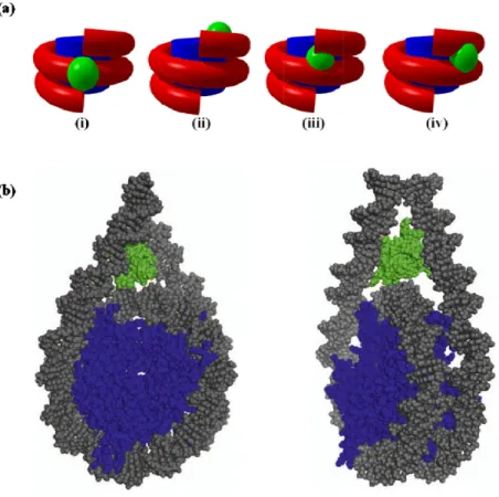
Over the last few decades numerous attempts have been made to understand the exact location of H1 globular domain on both native and reconstituted nucleosomal substrates. The very first symmetrical model was proposed in 1980 by Allan in which globular domain binds to the dyad of nucleosomes and interacts with ~10 base pairs linker DNA at both entry and exit points and stabilizes them[52]. Later, this model was validated by DNase I footprinting experiment[61]. Several attempts were made in the following years to improve the understanding about nucleosome-linker histone interaction. An W and colleagues performed MNase treatment on in vitro reconstituted nucleosomes using 5S RNA and suggested that H1 asymmetrically binds to DNA either at entry/exit site on nucleosome and protects ~20 base pairs[62]. Further, cross linking studies on 5S RNA nucleosome suggested that globular domain of H1 asymmetrically binds to the gyres of DNA near the dyad and protects 15 bp from one side and 5bp form the other side of nucleosome [63]. This model was opposed by bridging model proposed by Zhou and colleagues according to which site-specific protein-DNA photo-crosslinking on native chicken nucleosomes suggested that globular domain of linker histone H5 binds to the dyad and only one end of the linker DNA (either entry or exit side) on nucleosome[64]. However, FRAP studies on H1 globular domain and its mutants suggested the presence of two distinct DNA-binding sites one of which binds to major groove of DNA near the dyad and another binds to the minor groove either of linker DNA (Figure 7a) [65]. The recent cryo-electron microscopy (cryoEM) and hydroxyl radical footprinting studies on mono-, di- and tri-, nucleosomes supports the symmetrical model according to which globular domain binds to the dyad and CTD interacts with the linker DNA from both the ends of the nucleosome organizing them into a stem-like structure (Figure 7b) [66]. However, this binding requires “cavity” between linker DNA and nucleosome surface around the dyad. Any alteration in linker DNA orientation causes changes in the cavity which interferes with H1 binding on nucleosome. For example, depletion of docking domain from canonical H2A or replacing canonical H2A with variants H2A.Bbd or H2AL2 causes more opened nucleosome structure which leads to alteration in cavity and subsequently binding of H1 gets strongly compromised[67, 68]. None of these studies provide a complete understanding about the
molecular properties of linker histone H1, how it interacts and orients on the nucleosome, nucleosome array or the chromatin fiber.
Figure 7: Different models of H1 binding to nucleosome. (a) Simplified models showing the interaction of H1 globular domain with nucleosome, (i) Symmetrical model, (ii) asymmetrical model, (iii) asymmetrical DNA gyres model and (iv) bridging model. (b) Three-contact model for linker histone H1 globular domain binding within nucleosome, where globular domain (GH1), DNA and core histones are shown in green, grey and blue color respectively. Figures adapted from [55].
I.6.3 Histone H1 dynamics
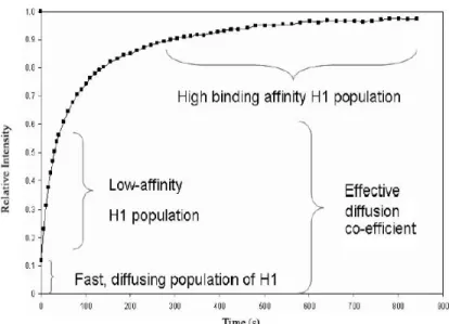
At any given point of time nuclear histone H1 is predominantly bound to the nucleosome; however, the binding is transient and its dynamic nature further varies between variants [69]. In vivo FRAP analysis showed that H1 binding to nucleosome is a reversible process and its residence time is higher (1-2 min) compared to the other chromatin binding proteins (HMG proteins -25 seconds) and lower when compared to core histones (30 min)[70, 71]. Histone H1 binding kinetics to chromatin suggests existence of three populations: a rapidly diffusing fraction, a weakly bound fraction and a strongly bound pool (Figure 8) [72].
Figure 8: A typical FRAP curve for H1 binding to chromatin showing multiple populations. Figure adapted from [73].
Three different models have been proposed to demonstrate histone H1 binding to the nucleosome (Figure 9). First model describes a two step process wherein unstructured C-terminal domain interacts with the linker DNA and directs the globular domain to bind at a specific position on the nucleosome. This model explains the secondary structure formation in CTD which is responsible for the low affinity of H1. If CTD fails to interact with linker DNA then binding will be compromised[65]. According to the second model, first, the globular domain non-specifically interacts with the nucleosome (low affinity H1 population) and induces conformational changes in CTD resulting in the formation of a stable complex (high affinity H1 population). However, FRAP analysis on CTD mutants showed that single mutation at either Thr152 or Ser183 into lysine increase histone H1 affinity to the nucleosome [74]. Remarkably, double lysine mutant at these residues failed to show higher binding affinity to nucleosomes. Also, this model failed to convince why globular domain alone initiates the non-specific binding to the nucleosome. This brings us to the third model according to which both globular domain and CTD simultaneously make an electrostatic clamp (low affinity population) leading to conformational changes in CTD necessary for high affinity binding.
28
A common theme which emerged from all these models is that binding of linker histone H1 on nucleosome is largely mediated through the globular domain and C-terminal domain. However, heterogeneity among H1 variants especially in the CTD results in different affinity towards the nucleosome. This hypothesis was further supported by FRAP experiments were histone H1 variants with shorter CTD length showed lowest residence time on the nucleosome [70]. In addition, with post-translational modifications especially phosphorylation and interaction with specific co-factors C-terminal domain alters the H1 binding affinity towards the chromatin fiber[75].
Figure 9: Different models for the reversible association of linker histone H1 with nucleosome. The low affinity binding of H1 with nucleosome is initiated through nonspecific electrostatic interaction between CTD of H1 and linker DNA (Model I) or by nonspecific interaction between GH1 and linker DNA (Model II) or by both the globular domain and CTD that makes electrostatic clamp with linker DNA simultaneously (Model III). This low affinity association triggers confirmation changes in CTD, resulting high affinity binding of H1 with nucleosome. Figure adapted from [69].
I.6.4 H1 variants
Heterogeneity is the characteristic feature of linker histone H1 family proteins and the numbers of their variants differ between species. Eleven subtypes of H1 have been identified in human and mouse genome and they are further classified into following subgroups based on their temporal and spatial expression patterns (Table 1) [76].
29
2. Genes with a variant mode of expression in somatic cells (histone H1.0and H1x) 3. Genes expressed specifically in germ cell (histone H1t, H1T2, H1LS1 and H1oo).
On the other hand linker histone genes can be classified as either cell cycle-dependent or replacement histone genes. H1.0 is the best example for a replacement histone gene which is expressed throughout the cell cycle and replaces other linker histones based on cellular signals[77]. Linker histone family genes exist either as clusters or solitary and their distribution patterns are conserved between human, mouse and rat genomes. The cell-cycle dependent H1genes are located as a cluster along with H1t gene and other core histone genes on human chromosome 6 (chromosome 13 in mouse) and the rest are located as solitary genes on other chromosomes (H1T2, H1LS1, H1oo, H1.0 and H1x) [78-83]. The following table describes in detail the H1 histone family in humans.
Table 1: The H1 histone family in human. Table adapted from[43] H1 protein (and synonyms) H1 gene (and synonyms) Gene locus (chromosome) Length (amino
acids)
Expression
(cell, tissue) Reference H1.1 (H1a) HIST1HA (H1F1) 6p21.3-22 214 Ubiquitous [84, 85] H1.2 (H1c) HIST1HC (H1F2) 6p21.3-22 212 Ubiquitous [85] H1.3 (H1d) HIST1HD (H1F3) 6p21.3-22 220 Ubiquitous [86] H1.4 (H1e) HIST1HE (H1F4) 6p21.3-22 218 Ubiquitous [86] H1.5 (H1b) HIST1HB (H1F5) 6p21.3-22 225 Ubiquitous [79] H1t HIST1H1T (H1FT) 6p21.3-22 206 Spermatocytes [87, 88] H1T2 H1FNT (HANP1) 12q13.11 233 Spermatids [89, 90] H1oo (H1foo) H1FOO 3q21.3f 345 Oocytes [91] HILS1
(Hils1) HILS1 17q21.33 230 Spermatids [92]
H1x (H1X, H1.X) H1FX 3q21.3 212 Ubiquitous [93, 94] H1.0 (H1°) H1FV (H1F0) 22q13.1 193 Differentiated cells [95]
30
I.6.4.1 H1.1- H1.5
Cell-cycle dependent histone H1 group genes share common characteristics features that they lack introns and have a short 5’ and 3’ non-coding sequences followed by hairpin loop at the end of mRNA which regulates their expression. In cell, expression of H1.1 is restricted to proliferation phase when cell undergoes successive division. It has the highest turnover rate with half-life of five days compared to other members in the group [96]. Studies on post-natal development of cortical neurons in rat brain showed that H1.1 expression reduces from 5% to 0.5% during this process and further H1.1 is replaced by H1.4[97]. Compared to other somatic variants histone H1.2 exhibits high level of mRNA expression with slower turnover rate. By interacting with YB1 and PURα, H1.2 forms a stable repressor complex which regulates p53-mediated trans-activation. This complex inhibits promoter specific p53-dependent and p300-mediated transcription by a direct interaction of H1.2 with p53, thereby blocking chromatin acetylation [98]. Lower level expression of H1.2 and H1.4 has been shown in actively transcribing chromatin as compared to facultative and heterochromatin stating that H1.2 is a ground state variant responsible for basal level chromatin compaction [99]. In addition to higher order formation H1.2 also regulates apoptosis in p53 dependent manner in response to DNA damage[51]. As like H1.2 H1.3 and H1.4 also expressed in non-dividing cells as well as in quiescent cells with low turnover rate and depleted from active chromatin region [99]. As compared to these variants H1.5 expressed in low level in quiescent cell and enriched in heterochromatin region located preferentially at nuclear periphery[100].
I.6.4.2 H1.0 and H.1x
These are replication independent H1 variants expressed all over the cell cycle. H1.0 expression is restricted to specific cell types and its expression is linked to cessation of DNA synthesis [77, 101]. Despite having the shortest CTD, H1.0 binds to the chromatin fiber with moderate affinity as compared to other H1 variants with long CTD [70]. H1.0 is most similar to avian replacement histone H5 and differences are found mainly in the distribution pattern of lysine and arginine residues. In the case of mammalian H1.0, lysine residues are present at more than ten positions in CTD whereas avian H5 predominantly carries arginine residues[95]. H1x is the least conserved linker histone H1 variant and is preferentially distributed into less accessible regions of the genome [94].
31
I.6.4.3 Germ line specific linker histone variants
Testis specific histone variant H1t is highly divergent in its primary sequence from other members of histone H1 family and its expression is restricted from pachytene spermatocytes to elongated spermatid stages. H1t binds to nucleosomes with less affinity compared to other variants and causes lower chromatin condensation[102]. This feature leaves chromatin relatively in open form which facilitates recombination events during meiosis [103]. The linker histone like H1LS protein is expressed exclusively in elongating and condensing spermatids and their expression pattern suggests that it could replace H1t in elongating spermatid and play a vital role in chromatin reorganization. H1LS also exhibits similar biochemical properties as like linker histones especially binding to mononucleosomes and condensing chromatin. H1.oo is the longest linker histone variant specifically expressed in oocytes. Primary sequence analysis of mammalian H1.oo and their homologs B4 (frog), cs-H1 (sea urchin) and dBigH1 (Drosophila) reveals enrichment of aspartic acid and glutamic acid residues in their CTD suggesting that sequence variation in CTD may affect the embryonic chromatin fiber condensation (Figure 10). This may lead to zygote genome activation and rapid cell division[104].
Figure 10: Aminoacid sequence alignment of linker histone H1.5 and its embryonic isoforms from different organisms. Primary sequence analysis reveals that C-terminal regions of H1 embryonic isoforms
32
are enriched with aspartic acid (Red-D) and glutamic acid (Red-E). Where identical residues are marked as “*”, substitutable residues as “.” and non-substitutable residues as “:”. Figure adapted from [104].
I.6.5 Linker histone modifications
As like core histones, linker histone H1 also undergoes several post translational modifications which alter its affinity towards the chromatin. Modifications found in H1 primarily include phosphorylation, acetylation, methylation, ubiquitination, formylation and citrullination (Figure 11) [105]. Phosphorylation is the most intensively studied modification of histone H1 and phosphorylated residues are found in all three domains ; however, their numbers and position may differ between variants. H1 phosphorylation has been suggested to play vital role in chromatin condensation, DNA replication, transcriptional regulation, DNA repair, apoptosis and ATP-dependent chromatin remodeling[106-109]. In cell, H1 phosphorylation is a reversible process and their levels increase to maximum level during G2 to mitosis phase and abruptly decrease soon after mitotic telophase[43]. Low level H1phosphorylation in S-phase drives H1 binding to DNA and causes chromatin de-condensation to promote DNA replication [110]. Hyperphosphorylation of H1 in mitotic phase induces chromatin condensation and promotes proper segregation of chromatids [110, 111]. Effects mediated by H1 phosphorylation are due to change in electrostatic charges and conformational changes in the structure especially in CTD. Phosphorylation of CTD reduces the proportion of α-helix and increases the β-sheet content resulting in alteration of the affinity towards DNA[112]. In addition, phosphorylation was also shown to affect transcription of mouse mammary tumor virus promoter (MMTV). In this particular case H1 phosphorylation increases transcription rate via improving the nucleosome positioning in vitro and promotes transcription through ATP dependent remodelers [113, 114].
H1 functional regulation through other PTMs like acetylation, methylation and citrullination are very little known as compared to the regulation by phosphorylation. Specific acetylation of K43in H1.4 found in promotor of active genes promotes H1 mobility and stimulates transcription through genera transcription factors recruitment [115]. Specific deacetylation and methylation of K26 in H1 by Polycomb repressive complex promotes the transcription repression by triggering H1binding with heterochromatin protein HP1 [116-118]. Citrullination of arginine 54 causes massive H1 dissociation from chromatin resulting drastic changes in the chromatin organization [119]. H1 formylation also have been
33
demonstrated during the DNA damage response however their precise role is not yet clear [120, 121].
Figure 11: Linker histone H1.5 post-translational modifications. Me—methylation, Ac—Acetylation, Ph— phosphorylation, Fo—formylation, Cr—crotonylation, Hib—2hydroxyisobutyrylation, Ub— ubiquitination, Cit—citrullination, Su—succinylation, Ar—ADPribosylation, OH—hydroxylation. The boxes indicate the globular domain. Figure adapted from [104].
I.7 Structure of the 30-nm chromatin fiber
The 30 nm-chromatin fiber is the next higher level of compaction achieved by genome after the “11 nm beads on string” confirmation. Existence of 30 nm fiber form the major fraction of interphase DNA in which most of the important biological process like transcription, DNA repair and replication take place. For the past 3 decades the structure of chromatin fiber has been studied using various methodologies including small angle X-ray scattering (SAXS), neutron scattering, X-ray crystallography, and direct imaging techniques (fluorescence, electron, cryo-electron and atomic force microscopy). Based on the experimental evidences, several models have been proposed to explain how nucleosomes folded into 30 nm chromatin fiber. They are classified into two major categories: first one is the one-start model in which nucleosome arrays are coiled with bent linker DNA and consecutive nucleosomes are arranged next to each other. Solenoid model and super coiled linker model are best examples for one start model [122, 123]. The second one is two-start model/zigzag model in which nucleosome arrays are assembled in a zigzag manner with straight linker DNA connecting two adjacent stacks of helically arranged nucleosome cores. Based on the twisting and coiling nucleosome stacks this model is further divided into 2 sub-classes : 1) Crossed-Linker Model and 2) Helical/ Twisted-Ribbon Model (Figure 12).
Figure 12: Models for the 30nm chromatin fiber structure. The image shows longitudinal views above and axial views below. (a) Solenoid model, (b) Crossed-linker model and (c) Helical ribbon model. Figure adapted from [124].
The solenoid model was first proposed by Finch and Klug in 1976 wherein one-start helical stack of nucleosomes were described to be linked together by bending linker DNA from adjacent nucleosomes. This model stated that dyads of all nucleosome faced towards the center of the helix and consequently linker DNA and histone H1 are located in the center of the chromatin fiber. This model also suggested that diameter of helix (30 nm) is independent of linker DNA length[122]. Another model for one start helix is the supercoiled linker model according to which linker DNA is supercoiled like DNA wrapped round histone octamers. This model suggested that dyad positioning of nucleosome is dependent on the linker DNA length[123]. The cross-linked fiber model is the most characterized among two start helix models. It describes nucleosomes to be stakced in two start left hand helix where the linker DNA is oriented approximately perpendicular to the fiber axis and passes through the center. In this model the diameter and mass per unit of fiber are dependent on the linker DNA length[125]. The twisted ribbon model suggests that chromatin fiber is formed by coiling of ribbon-like structure into a hollow cylinder where the linker DNA and histone H1 face the perimeter as like nucleosomes. In contrast to cross-linked model here the helical pitch increases and mass per unit decreases with increasing linker DNA length ; however, the diameter of pitch remains constant [126, 127]. Irregular straight linker model was postulated
through the observation of chromatin fiber by atomic force microscopy and electron tomographic in situ reconstruction. This model states that nucleosome arrays are loosely arranged in a zigzag manner without defined symmetry or geometry and histone H1 determines the linker DNA trajectory through its binding[128, 129].
Over the past three decades several attempts have been made to solve the structure of the 30 nm chromatin fiber ; however, there is no single model which is acceptable in all respects. The chemical synthesis of high affinity 601 sequence by Widom and studies based on this sequence with different linker lengths provided structural insights into nucleosome arrays. Chemical cross linking of folded nucleosomal arrays (by divalent cation Mg2+ or H1)
for stabilizing the inter-nucleosome interaction followed by nuclease digestion and EM imaging showed two parallel stacks of nucleosome supporting the two start model[130]. This model was further supported by the crystal structure of tetra-nucleosome at 9Ǻ resolution which was based on 167 NRL without linker histone H1 and showed that nucleosome array is folded in a two-start twisted ribbon manner with a diameter of 25 nm[24]. However, the presence of high salt, weak resolution and lack of histone H1 makes this result highly questionable.
Figure 13: Comparison of chromatin models to raw images of folded chromatin. (a) Cryo-EM images of folded chromatin arrays (22 nucleosomes with repeat length of 177bp). (b) View of interdigitated one-start helix model. (c) View of two-start crossed-linker model. Cryo-EM image analysis suggests that chromatin fibers are closely matched to the interdigitated one-start helix model, while they resemble the two-start crossed-linker model as well. The varying orientation of the coordinate axes describes the three dimensional orientation of the models in the corresponding rows. Figure adapted from [131].
Further, cryo-electron microscopy studies on series of 601 nucleosome arrays with different NRL (177, 187, 197,207, 217, 227 and 237 bp) in the presence of low divalent salt concentration (1.6 mM MgCl2) corresponding to physiological salt condition, showed two distinct structural classes with distinct fiber dimensions and ratio. Nucleosome arrays with shorter linker repeat length (NRLs 177 to 207) showed fibers of 33 to 34 nm diameter with a density of ~11 nucleosomes per 11 nm and in the case of longer linker DNA repeat (NRL 217 to 237) nucleosomes showed fibers of 43 nm with density of ~15 nucleosomes per 11 nm. In addition, cryo-EM image analysis showed that nucleosomes packing is achieved through the interdigitation of nucleosomes from adjacent helical gyres and modelling these images suggested that both fiber folding resembled into interdigitated one-start helices (solenoid model) (Figure13) [131].
Recent high resolution cryo-electron microcopy studies on H1 folded 601 nucleosome arrays with two different linker length (12 x 177 bp and 12 x 187 bp) supports two start model folding. In addition, mathematical analysis of 12 Å 3D cryo-EM images through mass-density showed two-start helix twisted by tetranucleosomal units (N1-N4, N5-N8 and N9-N12) and the structural properties are not affected by increase in the length of the linker (~10 bp). Within each tetranucleosomal unit nucleosomes are connected with a straight linker DNA whereas linker DNA is twisted between two tetranucleosomal units (Figure 14) [132]. However, these observations were made through the docking of tetranucleosomal crystal (4 x 167 bp) structure and failed to provide molecular details of H1 mediated compaction and the effect of longer linker DNA [24, 132].
Figure 14: 30nm chromatin fiber structure. (a) The crystallographic image of tetranucleosome with a twisted linker DNA segment. (b) 3D cryo-EM map of the 30-nm chromatin fibers reconstituted on 12x187bp nucleosome array with the three tetranucleosomal structural units highlighted by different colors. Figures adapted from [24, 132]
37
These experimental findings indicate that structure of 30 nm chromatin fiber is altered by various factors like the method of reconstitution done in in vitro, length of linker DNA, histone H1:nucleosome ratio and the inter-nucleosome interaction especially the one mediated through acidic patch of H2A-H2B dimer and H4 tails. Recent studies also showed that with increasing NRLs higher H1 per nucleosome is observed [133]. In addition, EM imaging in combination with sedimentation coefficient measurements suggested that folding of 30 nm chromatin fiber depends upon both NRL- and linker histone H1. Computational modelling studies on chromatin fiber having short, medium and long linker DNA using mesoscale modeling suggested that nucleosome having short and medium NRLs (173 to 209 bp) folds in an irregular zigzag manner ; however, the arrays with long linkers fold into solenoid form[134]. In the case of Monte Carlo simulations of oligonucleosome modelling, short and non-uniform NRL containing fiber formed a bent ladder rather than a compact chromatin fiber. Nevertheless, medium and long NRLs containing fibers fold in a regular zigzag manner. These experimental and computational modelling results suggested that polymorphism in 30 nm chromatin fiber structure is triggered by the variations in the length of the linker DNA. It has been suggested that condensed chromatin fiber structure is stabilized through electrostatic interaction between negatively charged acidic patch of histone dimers and H4 tails [135]. However, the mechanistic details of these interaction are still unclear. Indirect evidences from cross linking experiments, H4 tail deletion (residues 14-19) studies and specific modification at single residue (Lys16) showed these charge based specific internucleosomal interaction to be important for the formation of chromatin fiber[130, 136, 137]. Crystal structure of nucleosome provides evidence for the negatively charged acidic patch region formed by cluster of eight residues: Glu56, Glu61, Glu64, Glu90, Glu91, Glu92 of H2A and Glu102, Glu110 of H2B. In detail, α2-helix and the C-terminal extension of H2A forms the bottom of acidic patch and the edge is made through the α1- and αC-helices of H2B. Molecular dynamic simulation studies showed that residues 1-14 of H4 tail remain in unfolded form whereas residues 16-22 form α-helix and fit into to the acidic patch groove. In addition, residues K16, R19, K20 and R23 of H4 form non-covalent interaction extensively [138]. The residue K16 of H4 is the key residue which forms a salt bridge with H2A E61 and acetylation of K16 disrupts these non-covalent interactions (Figure 15) [139].
Figure 15: Acidic patch of nucleosome and its interaction with H4 tail. (a) Electrostatic potential view of the nucleosome surface. (b) Close-up view of acidic patch, where histone H2A and H2B shown in yellow and red respectively. The α1- and αC-helices of H2B, α2- and αC-helices of H2A and the eight residues that make up the acidic patch are labeled. (c) Close-up view of the histone H4 tail–acidic patch interaction observed in the crystal structure, where H4 tail, histones and acidic patch residues are shown in lime green, grey and pink respectively. Figures adapted from [138].
On the other hand, nucleosomes containing the variant H2A.Z, which has an acidic patch expanded with several residues, showed compaction more readily as compared to the canonical one indicating the involvement of acidic patch in higher order structure formation. However, mutations in the additional residues of H2A.Z acidic patch showed identical folding like canonical H2A [136]. Importance of acidic patch in chromatin fiber folding has also been demonstrated using another variant H2A.Bbd, which has areduced acidic patch and cannot fold the fiber in a regular form. Correcting H2A.Bbd acidic patch through replacing three residues (K61E, R91E and L92E) showed similar folding like regular H2A fibers [140].
As like polymorphism in NRLs, incorporation of histone variants, histone tail modifications and chromatin remodelers modulate the architecture of chromatin higher order structure. These factors and their role will be discussed in the later part of this chapter.
I.8 Chromatin and transcription
The highly compact nature of chromatin prevents the access of genome by the transcription machinery and the question arises how the initiation of transcription takes place. The answer lies with the subset of transcription factors commonly known as “Pioneering transcription factors”. The pioneering transcription factors have special features which allow them to invade the highly organized chromatin and bind to the specific binding site on the DNA wrapped around the histone octamers and thus initiate the transcription. One of the well studied factors is HNF-3 (FoxA) which was shown to bind the albumin gene enhancer elements in the reconstituted, H1 deposited chromatin fibers in the absence of chromatin
![Figure 1: Diagram represent major discoveries in the history of chromatin studies. Image modified from[7]](https://thumb-eu.123doks.com/thumbv2/123doknet/14429017.514816/19.918.141.778.479.762/figure-diagram-represent-discoveries-history-chromatin-studies-modified.webp)