HAL Id: inserm-00357408
https://www.hal.inserm.fr/inserm-00357408
Submitted on 26 Oct 2010
HAL is a multi-disciplinary open access
archive for the deposit and dissemination of
sci-entific research documents, whether they are
pub-lished or not. The documents may come from
teaching and research institutions in France or
abroad, or from public or private research centers.
L’archive ouverte pluridisciplinaire HAL, est
destinée au dépôt et à la diffusion de documents
scientifiques de niveau recherche, publiés ou non,
émanant des établissements d’enseignement et de
recherche français ou étrangers, des laboratoires
publics ou privés.
Cells lacking the fragile X mental retardation protein
(FMRP) have normal RISC activity but exhibit altered
stress granule assembly.
Marie-Cécile Didiot, Murugan Subramanian, Eric Flatter, Jean-Louis Mandel,
Hervé Moine
To cite this version:
Marie-Cécile Didiot, Murugan Subramanian, Eric Flatter, Jean-Louis Mandel, Hervé Moine. Cells
lacking the fragile X mental retardation protein (FMRP) have normal RISC activity but exhibit
altered stress granule assembly.. Molecular Biology of the Cell, American Society for Cell Biology,
2009, 20 (1), pp.428-37. �10.1091/mbc.E08-07-0737�. �inserm-00357408�
Molecular Biology of the Cell
Vol. 19, 428 – 437, January 1, 2009
Cells Lacking the Fragile X Mental Retardation Protein
(FMRP) have Normal RISC Activity but Exhibit Altered
Stress Granule Assembly
Marie-Ce´cile Didiot, Murugan Subramanian, Eric Flatter, Jean-Louis Mandel,
and Herve´ Moine
IGBMC (Institut de Ge´ne´tique et de Biologie Mole´culaire et Cellulaire); Inserm, U596; CNRS, UMR7104; and
Universite´ Louis Pasteur, Colle`ge de France, Chaire de Ge´ne´tique Humaine, Illkirch-Graffenstaden, F-67404
France
Submitted July 18, 2008; Revised October 22, 2008; Accepted October 30, 2008
Monitoring Editor: A. Gregory Matera
The fragile X mental retardation protein (FMRP) is an RNA-binding protein involved in the mRNA metabolism. The
absence of FMRP in neurons leads to alterations of the synaptic plasticity, probably as a result of translation regulation
defects. The exact molecular mechanisms by which FMRP plays a role in translation regulation have remained elusive.
The finding of an interaction between FMRP and the RNA interference silencing complex (RISC), a master of translation
regulation, has suggested that both regulators could be functionally linked. We investigated here this link, and we show
that FMRP exhibits little overlap both physically and functionally with the RISC machinery, excluding a direct impact of
FMRP on RISC function. Our data indicate that FMRP and RISC are associated to distinct pools of mRNAs. FMRP, unlike
RISC machinery, associates with the pool of mRNAs that eventually goes into stress granules upon cellular stress.
Furthermore, we show that FMRP plays a positive role in this process as the lack of FMRP or a point mutant causing a
severe fragile X alter stress granule formation. Our data support the proposal that FMRP plays a role in controlling the
fate of mRNAs after translation arrest.
INTRODUCTION
Fragile X syndrome, the most common form of inherited
mental retardation, is caused by the absence of the fragile X
mental retardation protein (FMRP). FMRP is an
RNA-bind-ing protein involved in the posttranscriptional control of
target mRNAs in particular at the level of synapses where
the absence of its function could alter synaptic plasticity and
the cognitive functions (Castets et al., 2005; Garber et al.,
2006; Huber, 2006). FMRP was found to be associated
spe-cifically with distinct classes of mRNAs: mRNAs harboring
G-quartet, poly (U) stretches, or a kissing complex motif
(Schaeffer et al., 2003; Jin et al., 2004a; Darnell et al., 2005). An
indirect interaction of FMRP with several mRNAs has also
been proposed through its interaction with the noncoding
BC1 RNA (Zalfa et al., 2003), but the biological importance of
this later type of interaction appears unclear (Iacoangeli et
al., 2008). Generally FMRP was found associated with
sev-eral aspects of the RNA metabolism, notably translation
repression (Laggerbauer et al., 2001; Castets et al., 2005),
transport (Dictenberg et al., 2008), and degradation (Zalfa et
al., 2007). These cellular events are generally related to
trans-lation arrest. Several of the mRNAs targeted by FMRP are
candidates for a translation repression (e.g., PP2A; Castets et
al., 2005) and MAP1B, Zhang et al., 2001), but the molecular
mechanism by which this repression occurs is still largely
unknown. The finding of the interaction of FMRP, and its
Drosophila ortholog dFXR, with the RNA Interference
Silenc-ing Complex (RISC; Caudy et al., 2002; Ishizuka et al., 2002;
Jin et al., 2004a,b) suggested that there could be a functional
link between FMRP and the RISC. Thus, it was proposed
that FMRP could be a modulator of the RISC function.
Conversely, the RISC could have been an effector of the
FMRP-mediated translational control or a combination of
both (Schaeffer et al., 2003). Up to now, however, a clear
demonstration of a direct role of FMRP in the RNA
interfer-ence (RNAi) and/or microRNA pathways controlled by the
RISC has been lacking. In fact it was proposed that
dFXR-deficient Drosophila S2 cells could have either no (Ishizuka et
al., 2002) or a moderate impact (Caudy et al., 2002) on RNAi
efficiency, and more recently FMRP was described to have a
positive impact on small interfering RNA (siRNA) efficiency
in mouse fibroblast cells (Plante et al., 2006).
To answer the question of the relationship between FMRP
and the RISC pathways and to examined the link between
FMRP and silent mRNAs in general, we analyzed the
local-ization of endogenous FMRP protein in cells with respect to
two known cellular structures where nontranslated mRNAs
have been shown to accumulate: the processing bodies (PBs)
and the stress granules (SGs) where RNA silencing and
integrated stress response are proposed to occur,
respec-tively. We also examined several aspects of the potential
physical and functional relationship between FMRP and the
RISC machinery in cells. In our model systems, FMRP was
found to have distinct localization properties than the
com-ponent of the RISC machinery and to have no impact on
RISC efficiency in various cell types. We identify however a
This article was published online ahead of print in MBC in Press
(http://www.molbiolcell.org/cgi/doi/10.1091/mbc.E08 – 07– 0737)
on November 12, 2008.
Address correspondence to: Herve´ Moine
(moine@igbmc.u-strasbg.fr).
new role of FMRP in mRNA metabolism as an effector of
stress granule assembly.
MATERIALS AND METHODS
Plasmids
pTL1-Iso7 and pTL1-Iso7-I304N encoding isoform 7 FMRP protein wild type and mutation I304N, respectively, were described previously (Sittler et al., 1996; Castets et al., 2005).
pRLTK-1x.The insertion of one miRNA23 target in the 3⬘ untranslated region
(UTR) of the Renilla luciferase (Rluc) gene in pRL-TK vector (Promega, Mad-ison, WI) was performed with QuickChange site-directed mutagenesis kit (Stratagene, La Jolla, CA) using oligonucleotides sense 5⬘-CATGTCTGCTCGAA-GCGGTCACTTTCCAGTGAGTTCCAGCCGCTCTAGAATTATTGT-3⬘ and antisense 5⬘-ACAATAATTCTAGAGCGGCTGGAACTCACTGGAAAGTG-ACCGCTTCGAGCAGACATG-3⬘ following the manufacturer’s instructions.
pRLTK-5x.The five target site-containing pRLTK was constructed with the two partially complementary DNA oligonucleotides: sense 5⬘-GC- TCTAGATGGAACTCACTGGAAAGTGACATATCTGGAACTCACTGGAAA-GTGACGAACTTGGAACTCACTGGAAAGTGACATGAC-3⬘ and antisense 5⬘-GCTCTAGAGTCACTTTCCAGTGAGTTCCAATATCGTCACTTTCCAG-TGAGTTCCAGTCATGTCACTTTCCAGTGAGTTCCAAGTTC-3⬘. The two oligonucleotides were annealed 5 min at 95°C, filled with Taq DNA polymer-ase 10 min at 72°C, cut with XbaI, and ligated into 3⬘UTR of pRL-TK XbaI site.
pRLTL5x-FBS.FBS sequence was produced by PCR using oligonucleotides. The original BamHI site was remove from pRL-TK by PCR-directed mu-tagenesis using oligonucleotide 5⬘-GTGCCACCTGGATACTTATCGATTT-TACC-3⬘. SalI and BamHI sites were added at the 3⬘ end of the five siRNA target sites by PCR-directed mutagenesis using oligonucleotide 5⬘-CC- AAACTCATCAATGTATCTTATCATGTGGATCCACTGCAGTCGACCT-GCTCGAAGCGGCCGCTCTAG-3⬘.
Short Duplex RNA Preparation
The oligonucleotide-directed synthesis of small RNA transcripts was per-formed with T7 RNA polymerase as previously described (Donze and Picard, 2002). One nanomole of each DNA oligonucleotide (P-sense, 5⬘-TGGAA-CTCACTGGAAAGTGACTATAGTGAGTCGTATTA; P-antisense, 5⬘-AA-GTCACTTTCCAGTGAGTTCTATAGTGAGTCGTATTA; B-sense 5⬘-T-GGAACTCACACCAAAGTGACTATAGTGAGTCGTATTA; B-antisense, 5⬘-AAGTCACTTTGGTGTGAGTTCTATAGTGAGTCGTATTA) was an-nealed with 1 nmol of T7 oligonucleotide (5⬘-TAATACGACTCACTATAC) in 50l of Tris-EDTA buffer, pH 8.0, for 3 min at 95°C followed by slow cooling to 20°C. Transcriptions were performed in 50l of mix (1⫻ T7 RNA polymerase buffer, 4 mM spermidine, 20 mM DTT, 0.1 mg/ml BSA, 4 mM each rNTP, 16 mM rGMP, 20 U RNAsin, and 5 U T7 RNA polymer-ase) on 400 pmol of annealed dsDNA template at 37°C for 2 h and treated with RNAse free DNAse. Sense and antisense 21-nt RNAs were then purified on 8 M urea, 25% polyacrylamide gel run at 30 mA in a Tris-borate-EDTA buffer. RNAs were visualized by UV shadowing, excised, and eluted in 200l of 0.5 M NH4acetate, 0.1 mM EDTA for 12 h at 4°C. RNAs were ethanol-precipitated and resuspended in water. Perfect and bulged sdRNAs were generated by mixing 1 nmol of each purified sense and antisense 21-nt RNA at 95°C for 5 min and for 1 h at 37°C.
Mouse Embryonic Fibroblasts Preparation
Pregnant female mice were killed by cervical dislocation at day 13 after coitus, and embryos were harvested. The head and the dark red organs were cut away from each embryo. Embryos were minced in 500l phosphate-buffered saline (PBS) and passed through an 18-gauge needle. Suspended cells were plated in 60-mm dishes in 2 ml of Dulbecco’s modified Eagle’s medium (DMEM) supplemented with 15% fetal calf serum and antibiotics at 37°C, 5% CO2. After 2 d, mouse embryonic fibroblasts (MEFs) were grown in DMEM supplemented with 10% fetal calf serum and 1 g/l glucose in the presence of antibiotics at 37°C in 5% CO2.
Cell Culture and Transfections
HeLa cells and all adherent mouse fibroblasts were grown in DMEM supple-mented with 10% fetal calf serum, 1 g/l glucose in the presence of antibiotics at 37°C in 5% CO2. Four hours before transfection, 4⫻ 104cells were plated into 24-well format plates in 500l of antibiotic-free medium. Transfections were performed in triplicate with Lipofectamine 2000 (Invitrogen, Carlsbad, CA) as directed by the manufacturer with 300 ng of pTL1-Iso7 or pTL1-Iso7-I304N. Twenty-four hours later, 100 ng of the reporter gene (pRLTK, pRLTK-1x, or pRLTK-5x, pRLTK-5x-FBS) were cotransfected by using Lipofectamine 2000 with 100 ng of the pFlashSV40 plasmid (Synapsys Solutions, Burgess Hill, West Sussex, United Kingdom), coding for the Firefly luciferase (Fluc) and used as normalizer, and 1–20 nM of sdRNA in a final volume of 600l
for each well. A nonspecific sdRNA scramble from Dharmacon Research (Boulder, CO; D-001205-01-05) was used as negative control at 5 nM. Plasmid pEGFP-hAgo2 expressing fusion GFP-human Ago2/eIF2C2 (W. Filipowicz, FMI, Basel, Switzerland) was transfected in HeLa cells in a 24-well format, using 0.8g DNA with Lipofectamine 2000 following the manufacturer’s instructions.
Luciferase Assays
Rluc and Fluc activities were determined using Luciferase Assay System (Promega) according to the manufacturer’s protocol. Assays were performed 24 h after transfection. Cell monolayers in 24-well cluster dishes were re-moved by scraping into 100l of reporter lysis buffer. Luciferase activities were measured using a Lumat LB 9501 luminometer (Berthold, Pforzheim, Germany). Rluc values were normalized with Fluc control values.
Real-Time Reverse Transcriptase PCR
To evaluate the expression of Renilla and Firefly mRNAs, total RNAs were extracted from transfected cells by using GenElute Mammalian Total RNA kit
Figure 1.
The majority of FMRP is not present in PBs in HeLa cells.
Confocal microscopy analysis of FMRP (red) and Dcp1 (green)
densities. Endogenous FMRP was stained with anti-FMRP antibody
(1C3) and an Alexa-fluor 594 –labeled mouse secondary
anti-body. Endogenous Dcp1 was stained with an anti-Dcp1a antibody
and an Alexa-fluor 488 –labeled anti-rabbit secondary antibody. (A
and B) Two different stacks taken 2
m apart from of one
represen-tative cell. (C and D) Two stacks from a cell showing a weak
colocalization of FMRP with PBs. These cells represent
⬍10% of cell
population. (E) Graph summarizing the colocalization of FMRP and
Dcp1 in HeLa cells. FMRP/Dcp1, indicates the percentage of FMRP
densities colocalizing with the Dcp1 densities (0.05
⫾ 0.02%). Dcp1/
FMRP, indicates the same for Dcp1 densities colocalizing with the
FMRP densities (8
⫾ 0.8%). The data are averages of 15 Z stacks
from 10 different representative cells (see Materials and Methods).
Error bars, SDs.
(Sigma, L’isle d’Abeau, France) as described by the manufacturer’s protocol. Reverse transcription reactions were performed on 0.5g of total RNA by using SuperScript III Reverse Transcriptase kit (Invitrogen) as described by the manufacturer. Real-time PCR was performed on 1:100 dilution of each reverse transcription sample in triplicate by using Platinum SybrGreen qPCR SuperMix (Invitrogen) in the MX4000 Thermocycler (Stratagene; 40 PCR cycles, denaturation at 95°C for 15 s, annealing at 55°C for 30 s, and extension at 72°C for 30 s). The real-time PCR reactions were carried out in the presence of 10 pmol of Renilla (forward 5⬘-TCTTCGTGGAAACCATGTTG-3⬘; reverse 5⬘-TGTTGGACGACGAACTTCAC-3⬘) and Firefly (forward 5⬘-TTCCATCT-TCCAGGGATACG-3⬘; reverse 5⬘-ATCCAGATCCACAACCTTCG-3⬘) gene-specific primers. The fluorescence was monitored at each annealing step. The Firefly reporter gene served as an internal control to monitor the transfection efficiency.
Polysomes Preparation
HeLa cells washed twice in PBS were lysed directly in the 10-cm plates in a lysis buffer containing 50 mM Tris-HCl, 100 mM KCl, 5 mM MgCl2, 1 mM DTT, 100g/ml cycloheximide, 40 U/ml RNasin (Sigma), Mini Complete antiprotease EDTA free (Roche, Indianapolis, IN) and 1% NP-40 or 1% so-dium deoxycholate as indicated in text. Total mouse embryonic day 16 (E16) brains were homogenized in 700l of lysis buffer without detergent by hand in a Kontes glass homogenizer (Vineland Glass Co., Vineland, NJ) fitted with
the loose-B pestle. The samples were transferred in microtubes, and 1% NP-40 was added to lyse the cells. The samples were centrifuged at 13,000 rpm at 4°C for 10 min. For fractionation, the clarified lysates were loaded on 15– 45% sucrose gradients and separated by ultracentrifugation with a SW41 rotor (Beckman) at 36,000 rpm at 4°C for 2 h. Linear sucrose gradients (15– 45%) were prepared with a Master Gradient 107ip (BioComp Instruments, Fred-ericton, NB, Canada) as indicated by the manufacturer in polyallomer tubes (Beckman Instruments, Fullerton, CA). Fractions of 500l were collected and A260was measured using a continuous flow cell UV detector (GE Healthcare Life Sciences, Piscataway, NJ). Polysomes of each fractions were precipitated with 2.5 vol of 100% ethanol with an incubation at⫺20°C overnight and by centrifugation at 13,000 rpm at 4°C for 20 min. Pellets were washed in 70% ethanol, briefly dried, and resuspended directly in SDS loading buffer.
Western Blotting
After denaturation in the loading buffer (100 mM Tris-HCl, pH 6.8, 4% SDS, 30% glycerol, 1.4 M-mercaptoethanol, and bromophenol blue) for 3 min at 95°C, proteins present in each polysome fraction were analyzed on a 6% SDS-polyacrylamide gel and immunoblotted onto nylon membrane (Schlei-cher & Schuell, Keene, NH) in Tris-glycine-SDS/20% ethanol buffer for 1 h at 200 mA. Membranes were incubated overnight at 4°C with mouse anti-FMRP 1C3 1:1000 (Devys et al., 1993), rabbit anti-hAgo2/GERp95 1:500 (Cikaluk et al., 1999), mouse anti-eIF2C2 1:1000 (ab57113, Abcam, Cambridge, MA), and
Figure 2.
FMRP and Ago2 are localized in
distinct subcellular compartments after
vari-ous cellular stresses in HeLa cells. (A) FMRP is
segregated in close but distinct subcellular
compartments than Dcp1 when translation
was arrested with 2 mM puromycin for 1 h
(Puro) or 500
M arsenite for 30 min (Ars).
Immunolabeling was as described in Figure 1.
(B) Ago2 (GFP-hAgo2 transiently transfected)
is localized in PBs like Dcp1 in nontreated cells
(NT) and remains there, unlike FMRP
(immu-nolabeled as in Figure 1), after 2 mM
puromy-cin for 1 h (Puro) or 500
M arsenite for 30 min
(Ars). The selected cells are representative of
the majority of cells expressing low or
moder-ate level of GFP-hAgo2.
M.-C. Didiot et al.
Molecular Biology of the Cell
430
anti-ribosomal protein L7 antisera (Ziemiecki et al., 1990) and then incubated at room temperature with peroxidase-conjugated goat anti-rabbit or goat anti-mouse antibodies (1/5000). Immunoreactive bands were visualized with the SuperSignal West Pico Chemiluminescent Substrate (Pierce, Rockford, IL).
Immunofluorescence and Fluorescent In Situ Hybridization
Cells for immunofluorescence (IF) and fluorescent in situ hybridization (FISH) were grown on glass coverslips, coating with poly-d-lysine, and ad-dition of fibronectin (1 pg/ml) was used for lymphoblast cultures. Cultures were incubated at 37°C in 5% CO2. Stress treatments were performed by incubating the cells at 43°C or with sodium arsenite (500 M final) for indicated time. After two washes with PBS, cells were fixed with PBS/4% paraformaldehyde (pH 7) at room temperature for 20 min. Cells were then washed for 15 min with 50 mM NH4Cl and permeabilized with PBS/0.2% Triton-X for 10 min at room temperature. Cells for IF were washed with PBS and blocked for 1 h at room temperature in PBS/0.1% Triton-X containing 5% BSA. Cells were incubated with primary antibodies mouse anti-FMRP 1C3 1:1000, rabbit anti-hDcp1a 1:1000 (J. Lykke-Andersen, University of Colo-rado), goat anti-TIA-1 1:1000 (C-20, Santa Cruz Biotechnology, Santa Cruz, CA), rabbit anti-MLN51 1:1000 (C. Tomasetto, IGBMC, Illkirch, France) or rabbit anti-eIF4E (FL-217, tebu-bio Laboratories, Le Perray-en-Yvelines, France), 1:1000 diluted in PBS/0,1% Triton-X overnight at 4°C. After three washes in PBS/0.1% Triton-X, cells were incubated with indicated fluorescent secondary antibodies (1:500) for 1 h at room temperature. Cells were washed three times at room temperature in PBS/0.1% Triton-X for 10 min and mounted in a Vectashield Mounting Medium with DAPI (1.5g/ml; Vector Laboratories, Burlingame, CA). Cells for FISH were rehydrated with 2⫻ SSC/50% formamide during 5 min at room temperature and then hybridized with 20 ng of oligo-dT probe in hybridization solution (2⫻ SSC, 50% form-amide, 30g E. coli tRNA, 0.02% RNAse-free BSA, 2 mM vanadyl-ribonucle-oside complexes, and 10% sulfate dextran) 12 h at 37°C in 40l. Oligo-dT probe was first heated 1 min at 90°C in 2⫻ SSC/50% formamide and tRNA. Cells were washed two times at room temperature in 2⫻ SSC/50% form-amide for 20 min and mounted as above. All image acquisitions and quanti-fication of fluorescent signal intensities were performed using standardized settings on a microscope (model DM4000 B, Leica, Deerfield, IL) equipped with CCD camera (CoolSnap CF, color) with 40⫻ or 63⫻ objectives and a confocal microscope (model SP1 and SP2-MP, Leica) with a 100⫻ objective. For colocalization analyses, JACoP localization analysis tool with ImageJ (http://rsb.info.nih.gov/ij/) was used on confocal images to determine Manders overlap coefficient (Bolte and Cordelieres, 2006). For SG quantifica-tion analysis, randomly selected images from each experiment were analyzed by counting SGs in at least 100 cells.
RESULTS
FMRP Localizes to Cytoplasmic Granules and SGs and
Has Little Overlap with PBs
We first compared by immunofluorescence the localization
of FMRP with the PB marker protein Dcp1a, which localizes
as bright foci punctuating the cytoplasm of nearly all cells. In
HeLa cells, FMRP was found distributed throughout the
cytoplasm in a granular manner with no overlap with PBs
(Figure 1, A and B). Occasionally, (i.e., in
⬍10% of the cells),
some FMRP granules were found to colocalize with PBs
(Figure 1, C and D). Applying JACoP localization analysis
tool with ImageJ (Bolte and Cordelieres, 2006) on confocal
stacks, the percentage of FMRP signal that overlapped with
PBs was estimated to be 0.05
⫾ 0.02% (Figure 1E). A similar
value was found when the fluorophores of the secondary
antibodies were inverted (data not shown). Thus, FMRP was
essentially excluded from PBs. This indicated that FMRP
and Dcp1 are present in distinct subcellular compartments
and only a very small fraction of total FMRP could
poten-tially colocalize with PBs. The round shape of cells showing
a partial colocalization of FMRP and PBs may indicate they
are in mitotic phase. These data suggest that if FMRP is
interacting with members of the RISC complex as previously
proposed, this interaction concerns only a very minor
frac-tion of FMRP pool and/or occurs in a narrow time frame.
Furthermore, this interaction should cease before the
mR-NAs submitted to RISC accumulate in PBs.
FMRP has been shown to be a component of SGs (Mazroui
et al., 2002), and PBs and SGs have been shown to be two
types of RNA granule structures in close relationship with
each other (Kedersha et al., 2005). We then examined the
relationship between FMRP, PBs, and SGs. We followed the
behavior of FMRP after different treatments that induce
the release of mRNAs from polyribosomes or their
stabili-zation and differently affect SGs and PBs. It has been shown
previously that puromycin, an aminoacyl-tRNA analog that
causes translation blocking by polysomes disassembly with
premature release of polypeptide chain and mRNA, leads to
an increase of the size and number of PBs (Sheth and Parker,
2003; Kedersha et al., 2005; Anderson and Kedersha, 2006).
After puromycin treatment, FMRP was found localized in a
few large granules close but distinct from PBs (Figure 2A). It
was previously shown that such treatment leads to SGs
assembly (Kedersha et al., 2000) as well as to PBs
enlarge-ment (Eulalio et al., 2007). Thus, when mRNAs are released
from polysomes by puromycin, FMRP localizes in SGs.
Ar-senite treatment had similar effect as puromycin (Figure 2B).
After 30 min of 500
M arsenite treatment or 15 min at 43°C
(see Figure 5, B and C, respectively), all immunodetectable
FMRP appeared in SGs as defined by the SG marker TIA-1.
Meanwhile, the PBs remained unaffected or slightly
in-creased in size. We showed next that this behavior is clearly
distinct from that of the core protein of the RISC, Ago2. In
nontreated cells Ago2 is localized in PBs like Dcp1 (Figure
2B, NT). On arsenite and puromycin treatment, Ago2
re-mained in PBs (Figure 2B) unlike FMRP that assembled into
SGs. Thus, FMRP, by being segregated away from the PBs in
conditions where the RISC members were found to
accumu-late (Pillai et al., 2005; Eulalio et al., 2007), exhibit a clear
distinct cellular behavior than the RISC components.
Cyclo-heximide treatment, which blocks translation at the
elonga-Figure 3.
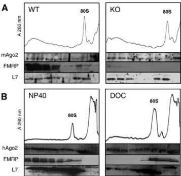
Fractionation of Ago2 and FMRP on sucrose gradients.
(A) Comparison of FMRP and mAgo2 mobility in 15– 45% sucrose
gradient prepared from wild-type or knockout littermate embryonic
mouse brain extracts. The densitograms (A
260) of gradients are
shown with corresponding pooled fractions tested for FMRP,
ribo-somal protein L7, and mAgo2 by Western blot analysis. (B)
Com-parison of FMRP and hAgo2 mobility in 15– 45% sucrose gradient
prepared with HeLa cell extracts submitted to NP-40 or sodium
deoxycholate (DOC) detergeants. The densitograms (A
260) are
shown with fractions tested for FMRP, ribosomal protein L7, and
hAgo2 by Western blot analysis.
tion level without disrupting polyribosomes, induced
in-stead both the granular distribution of FMRP to being more
disperse and PBs to decline (data not shown).
These data suggest that FMRP and RISC are associated to
distinct pools of mRNAs.
FMRP Exhibits Different Biochemical Behavior than Ago2
and Has No Impact on PBs Formation and RISC Activity
The apparent discrepancy between the previously reported
association of FMRP with members of the RISC complex,
namely Ago2 (argonaute 2 or eIF2C2), and the here above
reported differences in cellular localization, prompted us to
examine further the relationship between FMRP and RISC.
We first tested the possible impact of the absence of FMRP in
cells on PBs formation. For this, we analyzed the number
and intensity of PBs in MEFs from FMRP wild-type or
knockout (FMR1
⫺/⫺) littermates. In these cells, no significant
difference was observed either in PBs number or intensity as
labeled with an anti-Dcp1 antibody (data not shown). Thus,
the absence of FMRP has no impact on PBs formation in
MEFs.
We then analyzed the influence of FMRP on the
localiza-tion of Ago2, core of the RISC, in polyribosomes containing
fractions of embryonic (E18) mouse brain extracts. Using
Western blot analysis, Ago2 showed a ubiquitous
localiza-tion throughout the sucrose density gradient, whereas
FMRP was mostly concentrated in the heavy polyribosomes
fractions (Figure 3A). In the polyribosomes prepared from
littermate knockout mouse brain extracts, Ago2 had the
same sedimentation profile, indicating that the absence of
FMRP in cells had no detectable impact on Ago2 association
to mRNAs. Because FMRP sedimentation behavior on
su-crose density gradient is known to be sensitive to cationic
detergent (Khandjian et al., 2004), we then compared the
behavior of Ago2 and FMRP with respect to the type of
detergent used to extract polyribosomes. With the nonionic
detergent NP-40, FMRP was found enriched in the heavy
polyribosomes extracts of HeLa cells, whereas with the
cat-ionic sodium deoxycholate, FMRP was displaced from
heavy particles to the 80S fraction (Figure 3B) as already
reported (Khandjian et al., 2004). Interestingly, Ago2 was
insensitive to the nature of the detergent used. This
indi-cated that FMRP and Ago2 had distinct mRNA interaction
properties and likely belonged to different mRNP core
com-plexes. The fact that a treatment of cells with puromycin
induces the release of FMRP from heavy polysomes to free
mRNPs (Stefani et al., 2004), whereas the RISC members
remain associated to polyribosomes-like migrating particles
(Thermann and Hentze, 2007) further supports this idea.
We then tested the impact of FMRP on RISC activity using
an Rluc reporter gene bearing in its 3⬘ UTR one or five
targets of a microRNA (Figure 4A) as previously done to
evidence the miRNA-dependent RISC activity in cell
cul-tures (Doench et al., 2003; Zeng et al., 2003). The miRNA
miR23 was chosen based on evidence for a role in neural
specification (Kawasaki and Taira, 2003). The Rluc reporter
gene borne on a plasmid was transfected in various cell lines
together with a plasmid expressing the Fluc gene for
nor-malization purpose. First, we validated the system by using
short duplex RNAs (sdRNAs) that have been demonstrated
to work as miRNAs or siRNAs, depending upon their
complementarity with the target (Doench et al., 2003; Zeng et
al., 2003). We tested both perfect (sdRNA P) and imperfect/
bulged (sdRNA B) sdRNAs to mimic the siRNA or the
miRNA pathways, respectively. In HeLa cells, when no
target was present on the reporter gene or when a
nonspe-Figure 4.
FMRP has no impact on RISC
ac-tivity. (A) Scheme of the reporter gene Renilla
luciferase mRNAs (Rluc) bearing no (pRLTK),
one (pRLTK1x), five targets (pRLTK5x) of
miR23/short duplex RNAs (sdRNA), or one
target combined with a FMRP binding site
(pRLTK5x-Gq). (B) Controls of the RISC
ac-tivity assay. On the various Rluc mRNA
con-structs in HeLa cells extracts. C (f), P (u), and
B (䡺) are small duplex RNAs with no
comple-mentarity and perfect and imperfect matches
with Rluc mRNA, respectively. The
concen-trations of small duplex RNAs (5 or 10 nM)
used to treat cells are given below the graph.
The ratio of Rluc to Fluc activities (Rluc/Fluc)
set to 100 for the control is given. The relative
amount of Rluc-5X mRNA reporter (as
deter-mined by real-time reverse transcriptase PCR)
is indicated (E) with the different sdRNAs.
(C) Influence of FMRP on endogenous miR23
activity. Normalized Rluc activities of
report-ers bearing one (pRLTL1x) or five targets
(pRLTK5x) transfected in FMRP wild-type
(f) and knockout (䡺) MEF extracts are
shown. (D) Influence of FMRP on
sdRNA-dependent RISC activity. Rluc/Fluc values
measured in mouse FMR1
⫺/⫺fibroblasts
(Mazroui et al., 2002) transfected with empty
vector as control (f) or transfected with
pTL1-Iso7 expressing wild-type FMRP major
isoform 7 (u) or pTL1-Iso7-I304N expressing
mutant I304N FMRP isoform 7 (䡺). C, P, and
B are small duplex RNAs with no complementarity and perfect and imperfect matches with Rluc mRNA, respectively. The concentrations
of small duplex RNAs (5 or 20 nM) used to treat cells are given below the graph. The values in B, C and D are averages from at least three
independent transfections performed in triplicate. Error bars,
⫾ SD.
M.-C. Didiot et al.
Molecular Biology of the Cell
432
cific sdRNA (sdRNA C) was used, no effect was observed
(Figure 4B, pRLTK). When one or five targets of the sdRNA
was present in the 3⬘ UTR of the reporter gene, a specific and
very efficient inhibition was seen that reached more than
90% with 10 nM of sdRNA B and 5 nM of sdRNA P (Figure
4B, pRLTK 1X and pRLTK 5X, respectively), this without
apparent change in mRNA levels as measured by qRT-PCR
with pRLTK 5X. This indicates that the inhibitory effect of
both sdRNA P and B was at the translational level. Having
validated our reporter constructions, we then assessed the
action of endogenous miR23 in FMRP wild-type or
FMRP-deficient MEFs. Although miR23 could be detected in these
cells (Supplemental Figure S1), no impact of the FMRP
ab-sence on Rluc activity was detected whether one or five
miR23 targets were present in Rluc mRNA (Figure 4C). To
further trigger the siRNA or the miRNA pathways, we then
performed the same experiments with the different small
RNA duplexes described above. The efficiency of the sdRNAs
P and B in FMRP knockout fibroblasts was very similar to
the results obtained in HeLa cells (Figure 4D, f). When
FMRP major isoform 7 was expressed in these cells, the
efficiency of the RISC activity was unchanged (Figure 4D, u).
The same results were found independently of the
comple-mentarity of sdRNAs for its target (P or B), the number of
targets in Rluc mRNA (pRLTK 1X or 5X), and the dose of
sdRNAs (data not shown). The use of the loss-of-function
mutant FMRP bearing the point mutation I304N in the
sec-ond KH domain identified in a severe fragile X patient (De
Boulle et al., 1993) did not affect either these efficiencies
(Figure 4D,
䡺). Finally, we tested the contribution of an
FMRP-binding site located nearby the sdRNA sites. When
the guanine quartet RNA motif FBS previously
demon-strated to bind FMRP in vitro (Schaeffer et al., 2001) and in
vivo (Rackham and Brown, 2004) was inserted 29
nucleo-tides downstream of the five miR23 targets, again, no
spe-cific effect was detected (Figure 4D, pRLTK5x-Gq).
Alto-gether, these results indicate that the efficiency of the RISC
activity does not rely on the presence of FMRP in cells.
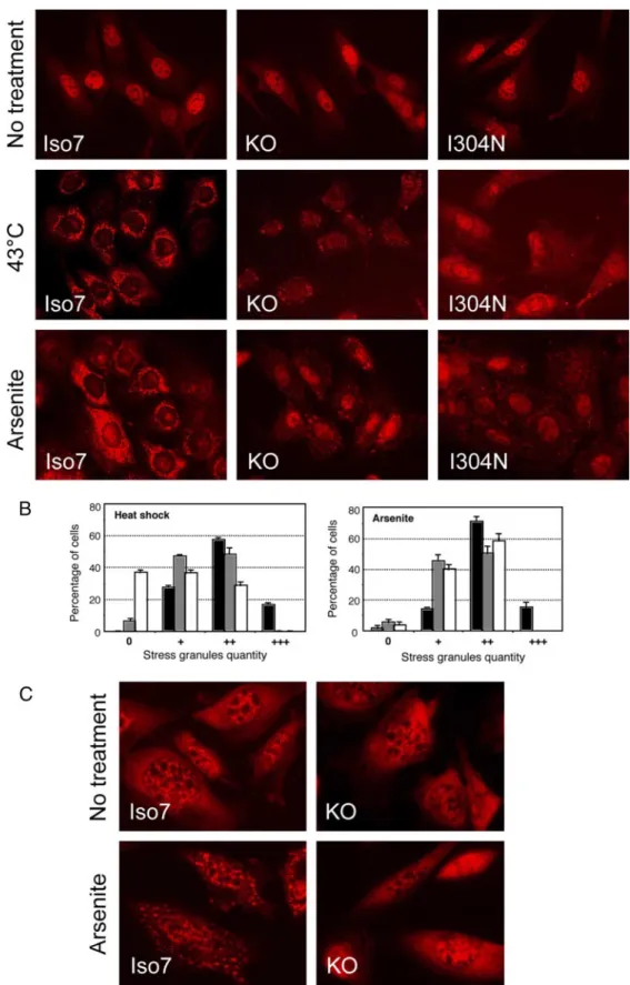
Absence of FMRP or a Mutation in Its Second KH
Domain Alters SGs Formation after Various Stresses
We then compared the assembly of FMRP into SGs with
other RNA-binding proteins known to localize in SGs, such
as TIA-1, a splicing factor proposed to be a core component
of SGs (Kedersha et al., 1999), eIF4E, the Cap-binding protein
of the initiation complex (Kedersha et al., 2005) and MLN51,
core protein of the exon junction complex (Baguet et al.,
2007). As expected, all four proteins that did not colocalize in
normal conditions (Figure 5A) steadily and similarly
local-ized into the SGs after a cellular stress such as arsenite
treatment (Figure 5B). Unexpectedly, however, we noticed
that these proteins had a different behavior after heat shock.
Thus, although FMRP fully colocalized with TIA-1 after 15
min at 43°C, eIF4E was in majority located in different foci
and MLN51 was absent from SGs (Figure 5C). These data
suggest that heat shock triggers a different stress response
than arsenite and that these mRNA-binding proteins have
distinct functions and/or associate with distinct pools of
mRNAs. Also, FMRP, like TIA-1, seemed to be an early SG
assembly protein. As TIA-1 had been demonstrated to be a
protein essential for SG formation (Kedersha et al., 1999), we
tested the possible involvement of FMRP in SGs formation.
We analyzed the ability of immortalized fibroblasts from
FMRP knockout mice to form SGs, whether they had been
stably transfected or not with the most frequent FMRP
iso-form 7 (Figure 6, A and B). Although the cells expressing
FMRP very efficiently formed SGs, as visualized by
immu-nofluorescence using anti-TIA-1 antibody and after
oxida-tive stress or heat shock, cells lacking FMRP comparaoxida-tively
showed reduced SG formation. This reduction mostly
af-fected the intensity rather than the number of SGs per cell.
Interestingly, SG formation was also similarly impaired in
cells stably expressing FMRP I304N (Figure 6A). Mutation
I304N caused a stronger impairment on SG formation than
Figure 5.
FMRP assembly in SGs. Fixed HeLa cells stained with an
anti-FMRP antibody and anti-mouse secondary antibody labeled
with Alexa-fluor 488 (green) or Alexa-fluor 594 (red), the anti-TIA-1
and anti-rabbit secondary antibody labeled with Alexa-fluor 594
(red), anti-eIF4E or anti-MLN51 and anti-rabbit secondary antibody
labeled with Alexa-fluor 488 (green), in the following conditions:
(A) nonstressed, (B) 500
M sodium arsenite for 30 min, (C) 15 min
at 43°C and with an anti-Dcp1a antibody.
Figure 6.
Absence of FMRP or point mutation I304N in its second KH domain impairs SG formation in mouse fibroblasts after various stresses. (A) SGs
were visualized by immunocytofluorescence using an anti-TIA-1 antibody and anti-rabbit secondary antibody labeled with Alexa-fluor 594 (red) in fixed
FMRP knockout fibroblasts stably transfected or not with FMRP isoform 7 (Iso7) or FMRP-I304N isoform 7 (I304N; Mazroui et al., 2002). The nature of
treatment is indicated for the various clones tested. (B) Histograms represent the percentages of cells (Y axis) for SGs quantity distributed in four categories:
no granules (0), weak (
⫹), moderate (⫹⫹), and strong (⫹⫹⫹) in wild-type (f), knockout (u) and FMRP-I304N (䡺) cells. Experiments shown reflect at
least three independent experiments with counting more than 100 cells in each case. (C) Fluorescent in situ hybridization with a poly-dT-Cy3 probe in fixed
wild-type or knockout fibroblasts.
M.-C. Didiot et al.
Molecular Biology of the Cell
434
the complete lack of FMRP after heat-shock treatment. SGs
are proposed sites of mRNA storage during stresses. Defect
in mRNA accumulation in SGs was visualized by using
fluorescent in situ hybridization with a poly(A)-specific
DNA probe (Figure 6C). Thus, mRNA storage in SGs after a
cellular stress was affected in cells lacking FMRP. To exclude
the possible influence of a clonal or immortalization effect on
the cell physiology, we analyzed the SG formation in
pri-mary cultures of MEFs from wild-type or knockout
litter-mates. A similar or stronger difference was observed
be-tween the wild-type and the knockout MEFs, with the
knockout cells showing SG assembly defect both at intensity
and number of SGs induced by oxidative stress (Figure 7A)
and heat shock (Figure 7B), confirming the results observed
in the immortalized cells. We then analyzed SG formation in
a lymphoblastoid cell line from a patient expressing an
FMRP protein bearing a point mutation I304N (De Boulle et
al., 1993). We compared the behavior of this cell line
sub-mitted to stress with the one from normal individuals
(Fig-ure 7C). Again, a marked defect in SG formation in response
to stress was observed with the cells expressing the mutant
FMRP protein in comparison with normal individuals.
Al-together these data provide strong evidence that the absence
of FMRP affects SG formation efficiency.
DISCUSSION
In this study, we examined the relationship between FMRP,
the protein absent in the fragile X syndrome, and two types
of cytoplasmic granules: the PBs and the SGs where silent
mRNAs are localized. PBs, also referred to as GW or Dcp
bodies, are site where decapping and mRNA decay occur
(Sheth and Parker, 2003; Cougot et al., 2004). Recently, it was
shown that mRNAs submitted to the RISC also accumulated
in PBs (Liu et al., 2005; Pillai et al., 2005). SGs are foci where
most mRNAs accumulate when cells are submitted to a
stress and when general translation is arrested (Anderson
and Kedersha, 2006). Both PB and SG RNA granules share
many components and establish constant dynamic
interac-tions (Anderson and Kedersha, 2002). We showed here that
FMRP does not colocalize with PBs in HeLa cells as only
0.05% of immunoreactive FMRP colocalized with PBs. This
quasi-absence of localization of FMRP within PBs
contra-dicts the previously reported association of FMRP into the
RISC seen in Drosophila (Caudy et al., 2002; Ishizuka et al.,
2002) and HeLa cells (Jin et al., 2004b). These results
contra-dict also the previously reported colocalization of dFMRP
within PBs in Drosophila neurons (Barbee et al., 2006).
The absence of FMRP in cells has also no impact on PB
formation contrarily to Ago2 and Dicer-2, which are
re-quired for PB integrity (Eulalio et al., 2007). The absence of
FMRP did not affect either the sedimentation profile of Ago2
on sucrose density gradient with mouse brain extracts,
in-dicating that the association of Ago2 RISC to mRNAs was
independent of FMRP. This together with the fact that the
absence of FMRP has no impact on the RISC activity in
immortalized or primary fibroblasts clearly indicates that
FMRP is not a regulatory element of the RISC nor of PB
related mRNA metabolism. Furthermore, FMRP and Ago2
have distinct behavior on sucrose density gradient: 1) the
use of cationic detergent releases FMRP from heavy
sedi-menting particles, whereas Ago2 remains unchanged (this
work) and 2) the puromycin treatment releases FMRP from
polyribosomes (Stefani et al., 2004), whereas mRNAs
sub-mitted to action of miRNAs (miRNPs) still migrates like
“pseudopolysomes” (Thermann and Hentze, 2007).
Alto-gether, these data suggest that FMRP and Ago2 belong to
distinct complexes. This is further suggested by the fact that
puromycin treatment, which releases mRNAs from
trans-lation pool, leads to accumutrans-lation of the RISC-silenced
mRNAs into PBs (Sheth and Parker, 2003; Eulalio et al.,
2007), whereas it causes the segregation of FMRP (and
some mRNAs) into the SGs. Thus, FMRP and Ago2 are
likely associated with distinct pools of mRNAs.
The processes controlling the fate of an mRNA submitted
to the RISC are still not well understood. In particular, it is
not clear what causes a specific mRNA to be degraded or
translationally repressed independently of its
complemen-Figure 7.
The lack of FMRP or the
FMRP-I304N mutation results in SG assembly defects
in various cells. (A) Arsenite induced SGs in
MEFs from wild-type or knockout littermates,
visualized with an anti-TIA-1 antibody and
anti-rabbit secondary antibody labeled with
Alexa-fluor 594 (red). (B) Heat-shock–induced
SGs visualized as in A. (C) Arsenite induced
SGs in human lymphoblasts from control
in-dividual (wild type) or from a fragile-X patient
bearing the I304N mutation visualized with an
anti-FMRP antibody and anti-rabbit
second-ary antibody labeled with Alexa-fluor 488
(green). Histograms represent the percentages
of cells (Y axis) for SGs quantity distributed in
two or three categories: no granules (0), weak
(
⫹), and moderate (⫹⫹) in wild-type (f) and
knockout or FMRP-I304N (䡺) cells.
tarity to a miRNA (Nilsen, 2007); for instance, in our hands
both perfect and nonperfect sdRNA caused translation
re-pression. As it was shown previously that FMRP has
intrin-sic RNA-binding properties, in particular for G-quartet RNA
motifs (Darnell et al., 2001; Schaeffer et al., 2001), a possibility
remained that FMRP could modulate the “susceptibility” of
an mRNA to the RISC when already bound to this mRNA,
for instance, through its nucleic acid chaperone properties
that could enhance miRNAs hybridization (Gabus et al.,
2004) or by preventing an mRNA from going into the PB
compartments. In fact, it is possible that mRNA silencing
could be initiated in the diffuse cytoplasm before its final
destination to PBs (Pillai et al., 2005), and the prior binding
of FMRP on such mRNAs could modulate their RISC
sus-ceptibility and thus affect the fate of the mRNAs. However,
we could not see any impact on RISC activity of the presence
of a G-quartet motif in the viscinity of miRNA targets, the
main consensus motif known to be bound by FMRP
(Schaef-fer et al., 2003). Thus, our data are in agreement with the
proposal that FMRP is mostly associated with translated
mRNAs (Corbin et al., 1997) and whether these mRNAs
should be submitted to silencing then they should dissociate
from FMRP prior their final destination to PBs.
On the contrary, FMRP does not dissociate from the
mRNAs submitted to a premature translation arrest (such as
during cellular stresses) and accompany them into the SG
storage compartments. Recruitment within SGs appears as a
hallmark of the RNA-binding proteins. FMRP had been
shown to follow this rule (Mazroui et al., 2002). Furthermore,
we showed that FMRP is likely to play a positive role in the
SG formation. We evidenced here a defect in the stress
granule formation process in absence of FMRP or in
pres-ence of a point mutation within its second KH motif, both
causing the fragile X syndrome. Thus, FMRP adds to the list
of RNA-binding proteins involved in SG formation after
TIA-1, Pumilio2, and MLN51 (Gilks et al., 2004; Vessey et al.,
2006; Baguet et al., 2007). The exact contribution of SG
pro-teins to SG formation during stress response appears
un-clear. SGs are consisting in stalled 48S preinitiation
com-plexes proposed to participate to the reprogramming of
mRNA metabolism for adaptation of cells to stress
condi-tions (Anderson and Kedersha, 2006). In this process TIA-1
is proposed to play a structural role in the SG assembly
process via the self-aggregation of its glutamine-rich motif
in a prion-like manner (Gilks et al., 2004). Meanwhile, these
interactions are very dynamic as TIA-1 shuttles rapidly in
and out the SGs (Kedersha et al., 2000). Self-aggregation
seems to be a characteristic of several of the SG proteins like
G3BP (Tourriere et al., 2003) and Pumilio2 (Vessey et al.,
2006). Whether FMRP has also some aggregation properties
remains to be established. Alternatively, the defect in SG
formation observed in the cells lacking FMRP could be due
to the loss of an active role of FMRP in the process of
translation inhibition. Our measurement of unaltered eIF2a
phosphorylation levels in cells lacking FMRP upon stress
(data not shown) do not favor this hypothesis however.
Altogether our data provide strong lines of evidence for an
involvement of FMRP in mRNA storage process during
stress conditions. Also, because SGs and the neuronal RNA
granules that are involved in the transport of silent mRNAs
share many components (Anderson and Kedersha, 2006;
Kiebler and Bassell, 2006), the defects reported for RNA
granule formation in neurons lacking FMRP (Aschrafi et al.,
2005) could be related to the SG defects seen here. It will be
important to determine now whether this defect in RNA
granule formation is mostly affecting those mRNAs
previ-ously reported to be targeted by FMRP.
ACKNOWLEDGMENTS
We thank T. Hobman (University of Alberta, Edmonton, Canada) for provid-ing the anti-Ago2/Gerp95, J. Lykke-Andersen for anti-hDcp1a, C. Tomasetto (IGBMC, Illkirch, France) for anti-MLN51, and W. Filipowicz (FMI, Basel, Switzerland) for pEGFP-hAgo2. We are grateful to B. Bardoni, E. Khandjian, E. Bertrand for material and helpful discussions, S. Pannetier for care to Xfra mice, and M. Boeglin and P. Kessler for help with microscopy analyzes. MCD was supported by a PhD fellowship from the Ministe`reDe´le´gue´ a` l’Enseignement Supe´rieur et a` la Recherche and from Association pour la Recherche sur le Cancer. This project benefited from grants from National Institutes of Health (R01 HD40612-01), from Agence Nationale de la Re-cheche, and from the Fondation pour la Recherche Me´dicale and the Fondation Jeroˆme Lejeune to H.M.
REFERENCES
Anderson, P., and Kedersha, N. (2002). Stressful initiations. J. Cell Sci. 115, 3227–3234.
Anderson, P., and Kedersha, N. (2006). RNA granules. J. Cell Biol. 172, 803– 808.
Aschrafi, A., Cunningham, B. A., Edelman, G. M., and Vanderklish, P. W. (2005). The fragile X mental retardation protein and group I metabotropic glutamate receptors regulate levels of mRNA granules in brain. Proc. Natl. Acad. Sci. USA 102, 2180 –2185.
Baguet, A., Degot, S., Cougot, N., Bertrand, E., Chenard, M. P., Wendling, C., Kessler, P., Le Hir, H., Rio, M. C., and Tomasetto, C. (2007). The exon-junction-complex-component metastatic lymph node 51 functions in stress-granule assembly. J. Cell Sci. 120, 2774 –2784.
Barbee, S. A., et al. (2006). Staufen- and FMRP-containing neuronal RNPs are structurally and functionally related to somatic P bodies. Neuron 52, 997– 1009.
Bolte, S., and Cordelieres, F. P. (2006). A guided tour into subcellular colo-calization analysis in light microscopy. J. Microsc. 224, 213–232.
Castets, M., Schaeffer, C., Bechara, E., Schenck, A., Khandjian, E. W., Luche, S., Moine, H., Rabilloud, T., Mandel, J. L., and Bardoni, B. (2005). FMRP inter-feres with the Rac1 pathway and controls actin cytoskeleton dynamics in murine fibroblasts. Hum. Mol. Genet. 14, 835– 844.
Caudy, A. A., Myers, M., Hannon, G. J., and Hammond, S. M. (2002). Fragile X-related protein and VIG associate with the RNA interference machinery. Genes Dev. 16, 2491–2496.
Cikaluk, D. E., Tahbaz, N., Hendricks, L. C., DiMattia, G. E., Hansen, D., Pilgrim, D., and Hobman, T. C. (1999). GERp95, a membrane-associated protein that belongs to a family of proteins involved in stem cell differentia-tion. Mol. Biol. Cell 10, 3357–3372.
Corbin, F., Bouillon, M., Fortin, A., Morin, S., Rousseau, F., and Khandjian, E. W. (1997). The fragile X mental retardation protein is associated with poly(A)⫹ mRNA in actively translating polyribosomes. Hum. Mol. Genet. 6, 1465–1472.
Cougot, N., Babajko, S., and Seraphin, B. (2004). Cytoplasmic foci are sites of mRNA decay in human cells. J. Cell Biol. 165, 31– 40.
Darnell, J. C., Jensen, K. B., Jin, P., Brown, V., Warren, S. T., and Darnell, R. B. (2001). Fragile X mental retardation protein targets G quartet mRNAs impor-tant for neuronal function. Cell 107, 489 – 499.
Darnell, J. C., Mostovetsky, O., and Darnell, R. B. (2005). FMRP RNA targets: identification and validation. Genes Brain Behav 4, 341–349.
De Boulle, K., Verkerk, A. J., Reyniers, E., Vits, L., Hendrickx, J., Van Roy, B., Van den Bos, F., de Graaff, E., Oostra, B. A., and Willems, P. J. (1993). A point mutation in the FMR-1 gene associated with fragile X mental retardation. Nat. Genet. 3, 31–35.
Devys, D., Lutz, Y., Rouyer, N., Bellocq, J. P., and Mandel, J. L. (1993). The FMR-1 protein is cytoplasmic, most abundant in neurons and appears normal in carriers of a fragile X premutation. Nat. Genet. 4, 335–340.
Dictenberg, J. B., Swanger, S. A., Antar, L. N., Singer, R. H., and Bassell, G. J. (2008). A direct role for FMRP in activity-dependent dendritic mRNA trans-port links filopodial-spine morphogenesis to fragile X syndrome. Dev. Cell 14, 926 –939.
Doench, J. G., Petersen, C. P., and Sharp, P. A. (2003). siRNAs can function as miRNAs. Genes Dev. 17, 438 – 442.
Donze, O., and Picard, D. (2002). RNA interference in mammalian cells using siRNAs synthesized with T7 RNA polymerase. Nucleic Acids Res. 30, e46.
M.-C. Didiot et al.
Molecular Biology of the Cell
436
Eulalio, A., Behm-Ansmant, I., Schweizer, D., and Izaurralde, E. (2007). P-body formation is a consequence, not the cause, of RNA-mediated gene silencing. Mol. Cell. Biol. 27, 3970 –3981.
Gabus, C., Mazroui, R., Tremblay, S., Khandjian, E. W., and Darlix, J. L. (2004). The fragile X mental retardation protein has nucleic acid chaperone proper-ties. Nucleic Acids Res. 32, 2129 –2137.
Garber, K., Smith, K. T., Reines, D., and Warren, S. T. (2006). Transcription, translation and fragile X syndrome. Curr. Opin. Genet. Dev. 16, 270 –275. Gilks, N., Kedersha, N., Ayodele, M., Shen, L., Stoecklin, G., Dember, L. M., and Anderson, P. (2004). Stress granule assembly is mediated by prion-like aggregation of TIA-1. Mol. Biol. Cell 15, 5383–5398.
Huber, K. M. (2006). The fragile X-cerebellum connection. Trends Neurosci. 29, 183–185.
Iacoangeli, A., Rozhdestvensky, T. S., Dolzhanskaya, N., Tournier, B., Schutt, J., Brosius, J., Denman, R. B., Khandjian, E. W., Kindler, S., and Tiedge, H. (2008). On BC1 RNA and the fragile X mental retardation protein. Proc. Natl. Acad. Sci. USA 105, 734 –739.
Ishizuka, A., Siomi, M. C., and Siomi, H. (2002). A Drosophila fragile X protein interacts with components of RNAi and ribosomal proteins. Genes Dev. 16, 2497–2508.
Jin, P., Alisch, R. S., and Warren, S. T. (2004a). RNA and microRNAs in fragile X mental retardation. Nat. Cell Biol. 6, 1048 –1053.
Jin, P., Zarnescu, D. C., Ceman, S., Nakamoto, M., Mowrey, J., Jongens, T. A., Nelson, D. L., Moses, K., and Warren, S. T. (2004b). Biochemical and genetic interaction between the fragile X mental retardation protein and the mi-croRNA pathway. Nat. Neurosci. 7, 113–117.
Kawasaki, H., and Taira, K. (2003). Functional analysis of microRNAs during the retinoic acid-induced neuronal differentiation of human NT2 cells. Nu-cleic Acids Res. Suppl. 3, 243–244.
Kedersha, N., Cho, M. R., Li, W., Yacono, P. W., Chen, S., Gilks, N., Golan, D. E., and Anderson, P. (2000). Dynamic shuttling of TIA-1 accompanies the recruitment of mRNA to mammalian stress granules. J. Cell Biol. 151, 1257– 1268.
Kedersha, N., Stoecklin, G., Ayodele, M., Yacono, P., Lykke-Andersen, J., Fritzler, M. J., Scheuner, D., Kaufman, R. J., Golan, D. E., and Anderson, P. (2005). Stress granules and processing bodies are dynamically linked sites of mRNP remodeling. J. Cell Biol. 169, 871– 884.
Kedersha, N. L., Gupta, M., Li, W., Miller, I., and Anderson, P. (1999). RNA-binding proteins TIA-1 and TIAR link the phosphorylation of eIF-2 alpha to the assembly of mammalian stress granules. J. Cell Biol. 147, 1431– 1442.
Khandjian, E. W., Huot, M. E., Tremblay, S., Davidovic, L., Mazroui, R., and Bardoni, B. (2004). Biochemical evidence for the association of fragile X mental retardation protein with brain polyribosomal ribonucleoparticles. Proc. Natl. Acad. Sci. USA 101, 13357–13362.
Kiebler, M. A., and Bassell, G. J. (2006). Neuronal RNA granules: movers and makers. Neuron 51, 685– 690.
Laggerbauer, B., Ostareck, D., Keidel, E. M., Ostareck-Lederer, A., and Fischer, U. (2001). Evidence that fragile X mental retardation protein is a negative regulator of translation. Hum. Mol. Genet. 10, 329 –338.
Liu, J., Valencia-Sanchez, M. A., Hannon, G. J., and Parker, R. (2005). MicroRNA-dependent localization of targeted mRNAs to mammalian P-bodies. Nat. Cell Biol 7, 719 –723.
Mazroui, R., Huot, M. E., Tremblay, S., Filion, C., Labelle, Y., and Khandjian, E. W. (2002). Trapping of messenger RNA by Fragile X Mental Retardation
protein into cytoplasmic granules induces translation repression. Hum. Mol. Genet. 11, 3007–3017.
Nilsen, T. W. (2007). Mechanisms of microRNA-mediated gene regulation in animal cells. Trends Genet. 23, 243–249.
Pillai, R. S., Bhattacharyya, S. N., Artus, C. G., Zoller, T., Cougot, N., Basyuk, E., Bertrand, E., and Filipowicz, W. (2005). Inhibition of translational initiation by Let-7 microRNA in human cells. Science 309, 1573–1576.
Plante, I., Davidovic, L., Ouellet, D. L., Gobeil, L. A., Tremblay, S., Khandjian, E. W., and Provost, P. (2006). Dicer-derived microRNAs are utilized by the Fragile X Mental Retardation Protein for assembly on target RNAs. J. Biomed. Biotechnol. 4, 1–12.
Rackham, O., and Brown, C. M. (2004). Visualization of RNA-protein inter-actions in living cells: FMRP and IMP1 interact on mRNAs. EMBO J. 23, 3346 –3355.
Schaeffer, C., Bardoni, B., Mandel, J. L., Ehresmann, B., Ehresmann, C., and Moine, H. (2001). The fragile X mental retardation protein binds specifically to its mRNA via a purine quartet motif. EMBO J. 20, 4803– 4813.
Schaeffer, C., Beaulande, M., Ehresmann, C., Ehresmann, B., and Moine, H. (2003). The RNA binding protein FMRP: new connections and missing links. Biol. Cell 95, 221–228.
Sheth, U., and Parker, R. (2003). Decapping and decay of messenger RNA occur in cytoplasmic processing bodies. Science 300, 805– 808.
Sittler, A., Devys, D., Weber, C., and Mandel, J. L. (1996). Alternative splicing of exon 14 determines nuclear or cytoplasmic localisation of fmr1 protein isoforms. Hum. Mol. Genet. 5, 95–102.
Stefani, G., Fraser, C. E., Darnell, J. C., and Darnell, R. B. (2004). Fragile X mental retardation protein is associated with translating polyribosomes in neuronal cells. J. Neurosci. 24, 7272–7276.
Thermann, R., and Hentze, M. W. (2007). Drosophila miR2 induces pseudo-polysomes and inhibits translation initiation. Nature 447, 875– 878. Tourriere, H., Chebli, K., Zekri, L., Courselaud, B., Blanchard, J. M., Bertrand, E., and Tazi, J. (2003). The RasGAP-associated endoribonuclease G3BP assem-bles stress granules. J. Cell Biol. 160, 823– 831.
Vessey, J. P., Vaccani, A., Xie, Y., Dahm, R., Karra, D., Kiebler, M. A., and Macchi, P. (2006). Dendritic localization of the translational repressor Pumilio 2 and its contribution to dendritic stress granules. J. Neurosci. 26, 6496 – 6508. Zalfa, F., Eleuteri, B., Dickson, K. S., Mercaldo, V., De Rubeis, S., di Penta, A., Tabolacci, E., Chiurazzi, P., Neri, G., Grant, S. G., and Bagni, C. (2007). A new function for the fragile X mental retardation protein in regulation of PSD-95 mRNA stability. Nat. Neurosci. 10, 578 –587.
Zalfa, F., Giorgi, M., Primerano, B., Moro, A., Di Penta, A., Reis, S., Oostra, B., and Bagni, C. (2003). The Fragile X syndrome protein FMRP associates with BC1 RNA and regulates the translation of specific mRNAs at synapses. Cell 112, 317–327.
Zeng, Y., Yi, R., and Cullen, B. R. (2003). MicroRNAs and small interfering RNAs can inhibit mRNA expression by similar mechanisms. Proc. Natl. Acad. Sci. USA 100, 9779 –9784.
Zhang, Y. Q., Bailey, A. M., Matthies, H. J., Renden, R. B., Smith, M. A., Speese, S. D., Rubin, G. M., and Broadie, K. (2001). Drosophila fragile X-related gene regulates the MAP1B homolog Futsch to control synaptic structure and function. Cell 107, 591– 603.
Ziemiecki, A., Muller, R. G., Fu, X. C., Hynes, N. E., and Kozma, S. (1990). Oncogenic activation of the human trk proto-oncogene by recombination with the ribosomal large subunit protein L7a. EMBO J. 9, 191–196.


![[PDF] Langage XML cours de base sur les concepts et pratiques pour reviser et s'entrainer | Cours xml](data:image/gif;base64,R0lGODlhAQABAIAAAP///wAAACH5BAEAAAAALAAAAAABAAEAAAICRAEAOw==)