HAL Id: hal-01927928
https://hal.archives-ouvertes.fr/hal-01927928
Submitted on 31 Jul 2019HAL is a multi-disciplinary open access
archive for the deposit and dissemination of sci-entific research documents, whether they are pub-lished or not. The documents may come from teaching and research institutions in France or abroad, or from public or private research centers.
L’archive ouverte pluridisciplinaire HAL, est destinée au dépôt et à la diffusion de documents scientifiques de niveau recherche, publiés ou non, émanant des établissements d’enseignement et de recherche français ou étrangers, des laboratoires publics ou privés.
Epstein-Barr Virus Nuclear Antigen 1 (EBNA1)
interacts with Regulator of Chromosome Condensation
(RCC1) dynamically throughout the cell cycle
Thibaut Deschamps, Quentin Bazot, Derek Leske, Ruth Macleod, Dimitri
Mompelat, Lionel Tafforeau, Vincent Lotteau, Vincent Maréchal, George
Baillie, Henri Gruffat, et al.
To cite this version:
Thibaut Deschamps, Quentin Bazot, Derek Leske, Ruth Macleod, Dimitri Mompelat, et al.. Epstein-Barr Virus Nuclear Antigen 1 (EBNA1) interacts with Regulator of Chromosome Condensation (RCC1) dynamically throughout the cell cycle. Journal of General Virology, Microbiology Society, 2017, 98 (2), pp.251-265. �10.1099/jgv.0.000681�. �hal-01927928�
Journal of General Virology
Epstein-Barr Virus Nuclear Antigen 1 (EBNA1) interacts with Regulator of
Chromosome Condensation (RCC1) dynamically throughout the cell cycle
--Manuscript
Draft--Manuscript Number: JGV-D-16-00550R2
Full Title: Epstein-Barr Virus Nuclear Antigen 1 (EBNA1) interacts with Regulator of Chromosome Condensation (RCC1) dynamically throughout the cell cycle Article Type: Standard
Section/Category: Animal - Large DNA Viruses Corresponding Author: Evelyne Manet
CIRI-International Center for Infectiology research;INSERM U1111; CNRS UMR5308; Université de Lyon; ENS de Lyon
Lyon, FRANCE First Author: Thibaut Deschamps Order of Authors: Thibaut Deschamps
Bazot Quentin Derek M Leske Ruth MacLeod Dimitri Mompelat Lionel Tafforeau Vincent Lotteau Vincent Maréchal George S Baillie Henri Gruffat Joanna B Wilson Evelyne Manet
Abstract: The Epstein-Barr virus (EBV) nuclear antigen 1 (EBNA1) is a sequence-specific DNA binding protein which plays an essential role in viral episome replication and
segregation, by recruiting the cellular complex of DNA replication onto the origin (oriP) and by tethering the viral DNA onto the mitotic chromosomes. Whereas the
mechanisms of viral DNA replication are well documented, those involved in tethering EBNA1 to the cellular chromatin are far from being understood. Here, we have identified Regulator of Chromosome Condensation 1 (RCC1) as a novel cellular partner for EBNA1. RCC1 is the major nuclear guanine nucleotide exchange factor (RanGEF) for the small GTPase Ran enzyme. RCC1, associated with chromatin, is involved in the formation of RanGTP gradients critical for nucleo-cytoplasmic transport, mitotic spindle formation, and nuclear envelope reassembly following mitosis. Using several approaches, we have demonstrated a direct interaction between these two proteins and found that the EBNA1 domains responsible for EBNA1 tethering to the mitotic chromosomes are also involved in the interaction with RCC1. The use of an EBNA1 peptide array confirmed the interaction of RCC1 with these regions and also the importance of the N-terminal region of RCC1 in this interaction. Finally, using confocal microscopy and FRET analysis to follow the dynamics of interaction between the two proteins throughout the cell cycle, we have demonstrated that EBNA1 and RCC1 closely associate on the chromosomes during metaphase, suggesting an essential role for the interaction during this phase, perhaps in tethering EBNA1 to mitotic chromosomes.
Epstein-Barr Virus Nuclear Antigen 1 (EBNA1) interacts with Regulator of
1
Chromosome Condensation (RCC1) dynamically throughout the cell cycle
2
3
Running title: EBNA1 localizes with RCC1 to chromatin during mitosis 4
5
Thibaut Deschamps1,3,4,5,6, Quentin Bazot1,3,4,5,6¶, Derek M. Leske7, Ruth MacLeod7, Dimitri
6
Mompelat1,3,4,5,6 ♯, Lionel Tafforeau2,3,6°, Vincent Lotteau2,3,4,5,6, Vincent Maréchal8, George
7
S. Baillie7, Henri Gruffat1,3,4,5,6, Joanna B. Wilson7 and Evelyne Manet1,3,4,5,6 *
8 9
1 CIRI, International Center for Infectiology Research, Oncogenic Herpesviruses team,
10
Université de Lyon, Lyon, 69364, France
11
2 CIRI, International Center for Infectiology Research, Cell Biology of Viral Infections team,
12
Université de Lyon, Lyon, 69364, France
13
3 INSERM, U1111, Lyon, 69364, France
14
4 CNRS, UMR5308, Lyon, 69364, France
15
5 Ecole Normale Supérieure de Lyon, Lyon, 69364, France
16
6 Université Lyon 1, Centre International de Recherche en Infectiologie, Lyon, 69364, France
17
7 College of Medical, Veterinary and Life Sciences, University of Glasgow, Glasgow G12
18
8QQ, UK
19
8 UPMC Univ Paris 6, Inserm, Centre d’Immunologie et des Maladies Infectieuses
(Cimi-20
Paris), UMR 1135, ERL CNRS 8255, F-75013 Paris, France
21 22
° Present address : Lionel Tafforeau, Cell Biology lab, University of Mons, Mons, Belgium
23
¶ Present address: Quentin Bazot, Section of Virology, Department of Medicine, Imperial
24
College London, St Mary’s Campus, London, UK
25
Manuscript Including References (Word document) Click here to download Manuscript Including References
♯ Present address: University Joseph Fourier, Pathogenesis and Lentiviral Vaccination
26
laboratory, Grenoble, France.
27
University of Oxford, Ludwig Institute for Cancer Research, Oxford, United Kingdom
28 29
* corresponding author: Email: evelyne.manet@ens-lyon.fr ; Tel: +33 (0) 472 72 89 54
30 31
Keywords: Epstein-Barr virus, EBV, EBNA1, Regulator of Chromosome Condensation,
32
RCC1
33 34
Subject category: animal/DNA viruses
35 36
Word count: 6496
37 38
ABSTRACT 39
The Epstein-Barr virus (EBV) nuclear antigen 1 (EBNA1) is a sequence-specific 40
DNA binding protein which plays an essential role in viral episome replication and 41
segregation, by recruiting the cellular complex of DNA replication onto the origin (oriP) 42
and by tethering the viral DNA onto the mitotic chromosomes. Whereas the mechanisms 43
of viral DNA replication are well documented, those involved in tethering EBNA1 to the 44
cellular chromatin are far from being understood. Here, we have identified Regulator of 45
Chromosome Condensation 1 (RCC1) as a novel cellular partner for EBNA1. RCC1 is 46
the major nuclear guanine nucleotide exchange factor (RanGEF) for the small GTPase 47
Ran enzyme. RCC1, associated with chromatin, is involved in the formation of RanGTP 48
gradients critical for nucleo-cytoplasmic transport, mitotic spindle formation, and 49
nuclear envelope reassembly following mitosis. Using several approaches, we have 50
demonstrated a direct interaction between these two proteins and found that the EBNA1 51
domains responsible for EBNA1 tethering to the mitotic chromosomes are also involved 52
in the interaction with RCC1. The use of an EBNA1 peptide array confirmed the 53
interaction of RCC1 with these regions and also the importance of the N-terminal region 54
of RCC1 in this interaction. Finally, using confocal microscopy and FRET analysis to 55
follow the dynamics of interaction between the two proteins throughout the cell cycle, we 56
have demonstrated that EBNA1 and RCC1 closely associate on the chromosomes during 57
metaphase, suggesting an essential role for the interaction during this phase, perhaps in 58
tethering EBNA1 to mitotic chromosomes. 59
60
INTRODUCTION 61
Epstein-Barr virus (EBV) is a ubiquitous herpesvirus associated with several human cancers
62
(Crawford, 2001). Following primary infection, the virus persists in a life-long, latent state in
memory B cells, with intermittent viral production occurring in the oropharynx. Ex vivo, EBV
64
has the capacity to induce growth transformation of resting primary human B-lymphocytes,
65
leading to the establishment of lymphoblastoid cell lines (LCLs). In such cell lines, only a
66
small number of viral genes are expressed which act in concert to induce and maintain
67
continuous cell proliferation and survival (Kieff & Rickinson, 2007).
68
In latently infected cells, the EBV genome persists as a multicopy, covalently closed,
69
double-stranded, nuclear episome. When cells proliferate, these episomes replicate once per
70
cell-cycle during S phase, using the cellular DNA-replication machinery and are subsequently
71
equally segregated to the daughter cells, such that a constant copy number of EBV genomes is
72
maintained through cell divisions (Adams, 1987; Nanbo et al., 2007; Yates & Guan, 1991).
73
Both replication and segregation depend on the presence of two viral elements, the EBV
cis-74
acting origin of plasmid replication (oriP) and the viral protein EBNA1 (Yates et al., 1984,
75
1985). oriP is composed of two functional elements: the dyad symmetry (DS) element and the
76
family of repeats (FR) (Reisman et al., 1985). Both contain recognition sites for the EBNA1
77
protein. The DS element, which comprises four EBNA1 binding sites arranged in pairs, is
78
required for DNA replication initiation (Rawlins et al., 1985; Reisman et al., 1985;
79
Wysokenski & Yates, 1989; Chaudhuri et al., 2001; Ritzi et al., 2003; Schepers et al., 2001).
80
The FR element consists of an array of twenty imperfect 30 bp repeats, each one containing
81
an 18 bp EBNA1 binding site (Rawlins et al., 1985; Reisman et al., 1985). FR functions by
82
tethering the viral episomes to human metaphase chromosomes via EBNA1 (Sears et al.,
83
2003, 2004; Wu et al., 2000, 2002) and this ensures the stable retention of oriP-episomes
84
within the cells (Kirchmaier & Sugden, 1995; Little & Schildkraut, 1995; Nanbo et al., 2007).
85
FR is also required for EBNA1-dependent tethering of EBV genomes to specific
86
perichromatic regions of host chromosomes during interphase (Deutsch et al, 2010) which
87
appears to be essential for efficient replication of the episome (Hodin et al., 2013).
EBNA1 is a homo-dimeric DNA-binding protein that recognises an 18 bp palindromic
89
sequence via its C-terminal domain (residues 459-607 have been co-crystallised with DNA)
90
(Ambinder et al., 1990, 1991; Bochkarev et al., 1996; Frappier & O’Donnell, 1991; Jones et
91
al., 1989; Rawlins et al., 1985; Shah et al., 1992). ChIPseq analyses have shown that, in
92
addition to binding the DS and FR regions of oriP, EBNA1 binds multiple sites in the host
93
genome (Lu et al., 2010; Tempera et al., 2015). Independently of its C-terminal-specific
94
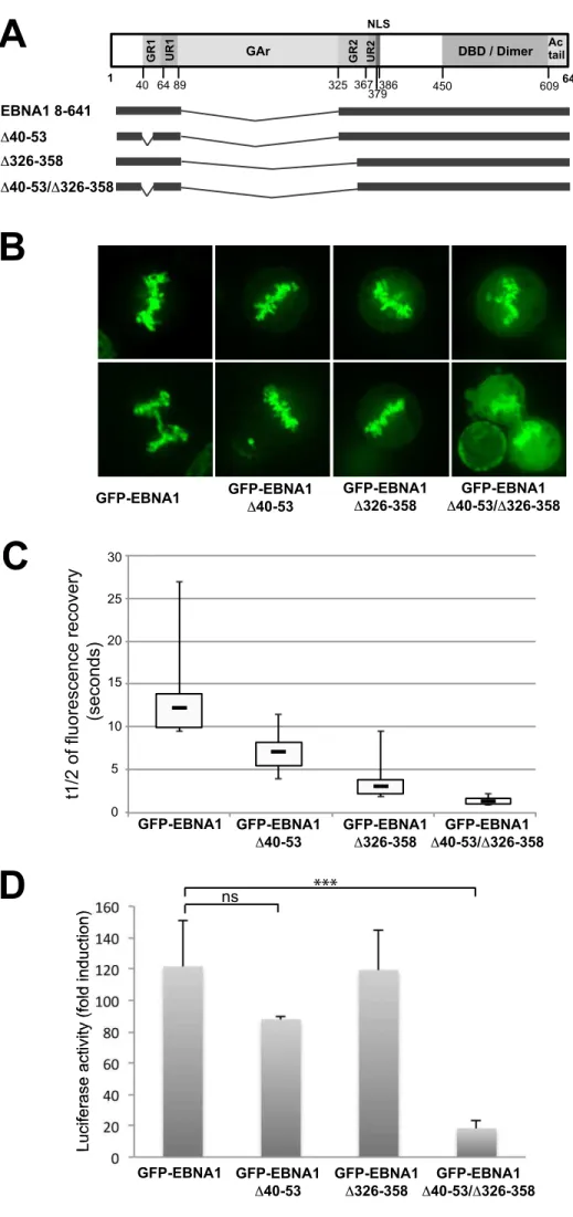
DNA-binding domain, EBNA1 can associate with chromatin throughout the cell cycle via its
95
N-terminal half. This N-terminal region carries two domains, called Linking Regions 1 (LR1:
96
aa 40-89) and 2 (LR2: aa 325-379) which confer intramolecular “linking” between
EBNA1-97
DNA complexes as revealed by electrophoretic mobility shift assays (Mackey et al., 1995).
98
Each of these domains consists of a region rich in arginine and glycine (RGG-rich region:
99
GR1 and GR2 respectively) and a unique region (UR1 and UR2 respectively). The RGG-rich
100
regions possess intrinsic AT-hook activity allowing binding to AT-rich DNA (Sears et al.,
101
2003, 2004). These regions have been found to be important for EBNA1 replication and
102
transcription activity (Mackey & Sugden, 1999) and to play an essential role in tethering
103
EBNA1 to cellular DNA during interphase (Coppotelli et al., 2011; Coppotelli et al., 2013).
104
EBNA1 attachment to metaphase chromosomes has been mapped to three independent
105
chromosome binding sites (CBS) - CBS-1 (aa 72 to 84), CBS-2 (aa 328 to 365) and CBS-3
106
(aa 8 to 54) - that correlate well with the ability of EBNA1 to confer plasmid maintenance
107
(Kanda et al., 2001; Maréchal et al., 1999; Wu et al., 2002). However, the mechanisms
108
responsible for EBNA1 interaction with mitotic chromosomes are still unclear. It has been
109
proposed that the AT-hook structures within the LR1/LR2 regions could be directly
110
responsible for EBNA1 attachment to the chromosomes (Kanda et al., 2013; Sears et al.,
111
2004). Interestingly, HMGA1a a cellular chromatin-binding protein which associates with
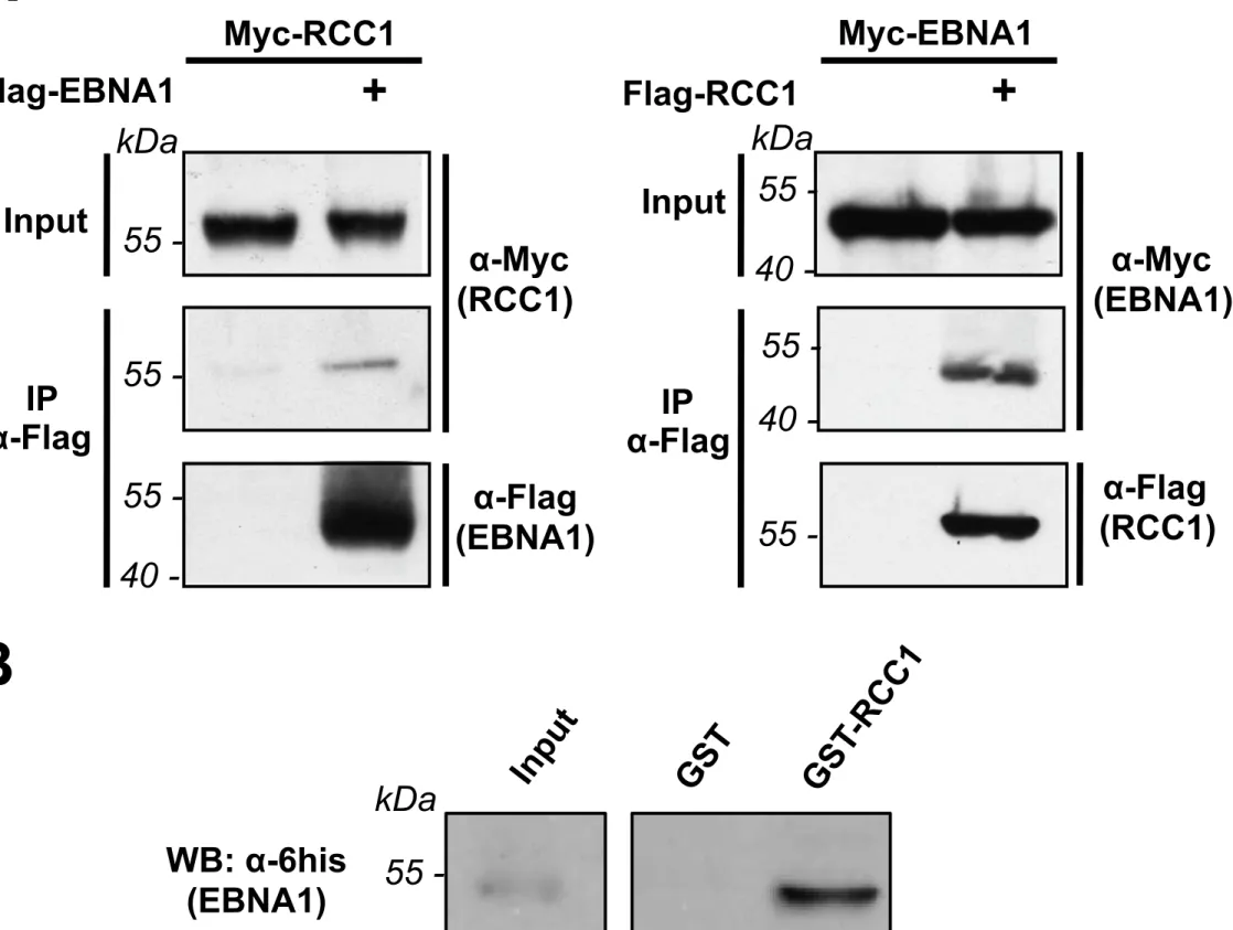
112
chromatin through its AT-hook domains, or histone H1, can functionally replace the amino
terminus of EBNA1 both in oriP plasmid replication and partitioning of the viral episome
114
(Hung et al., 2001; Sears et al., 2003; Thomae et al., 2008). EBNA1 may also interact with
115
chromatin through protein-protein interactions with one or several cellular partners. hEBP2
116
(human Epstein-Barr Binding Protein 2) was the first of the sort identified. (Kapoor et al.,
117
2005; Nayyar et al., 2009; Wu et al., 2000). hEBP2 binds to the LR2 region of EBNA1 (Shire
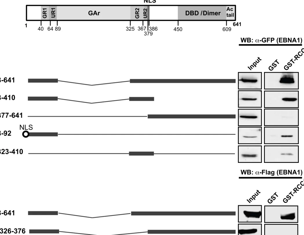
118
et al., 1999), which also corresponds to the CBS-2 region. In a yeast model, hEBP2 was
119
required (in the presence of EBNA1) for the maintenance of a plasmid carrying the EBV FR
120
sequence (Kapoor et al., 2001, 2003). However, a recent study demonstrated that hEBP2 and
121
EBNA1 do not interact during mitosis in living mitotic cells, suggesting that the involvement
122
of hEPB2 might not be direct (Jourdan et al., 2012; Frappier, 2012), or that it might have
123
another role. More recently, HMGB2 (high-mobility group box 2), a well known chromatin
124
component, has been identified as a new partner for EBNA1 (Jourdan et al., 2012). EBNA1
125
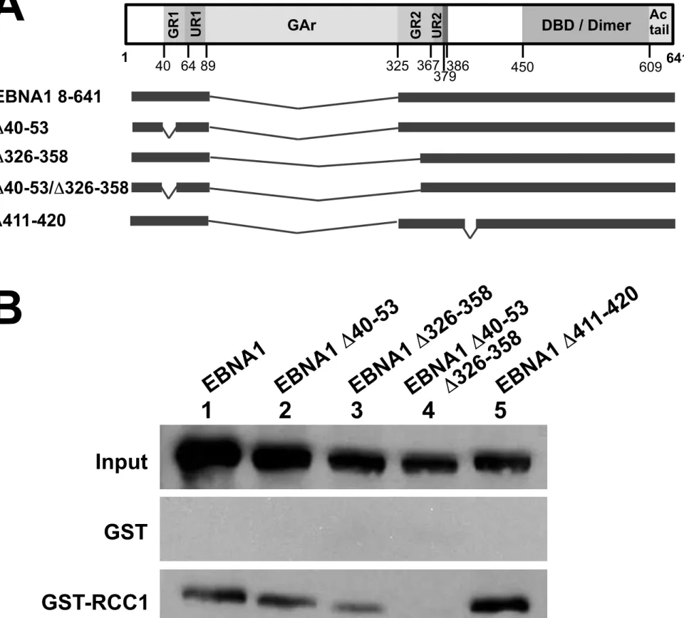
interacts with HMGB2 on chromatin during interphase and mitosis, and its depletion partially
126
alters EBNA1 association with the chromosomes. However, HMGB2 depletion is not
127
sufficient to alter EBV episome maintenance in Raji cells (Jourdan et al., 2012). Taken
128
together, these results suggest that several mechanisms cooperate to promote EBNA1
129
association with the chromosomes throughout mitosis and maintenance of the EBV genome
130
within proliferating cells.
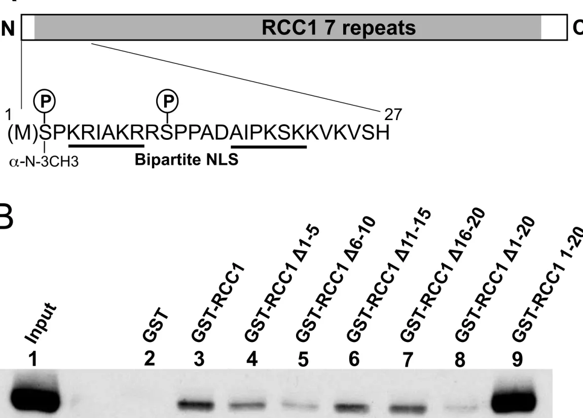
131
In order to identify novel proteins that could play a role in EBNA1 chromosomal
132
binding, we performed a yeast two-hybrid screen. From this screen, we identified Regulator
133
of Chromosome Condensation 1 (RCC1), a major nuclear guanine nucleotide exchange factor
134
(RanGEF) for the small GTPase Ran enzyme. In its association with chromatin, RCC1 is
135
involved in the formation of RanGTP gradients critical for nucleo-cytoplasmic transport
136
(Riddick & Macara, 2005), mitotic spindle formation and nuclear envelope reassembly after
137
mitosis (Askjaer et al., 2002; Bamba et al., 2002). RCC1 is a ubiquitous nuclear protein
structured as a seven bladed propeller with unstructured small N- and C-terminal tails
139
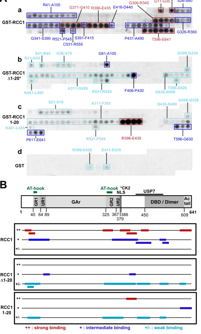
(Renault et al., 1998). RCC1 directly interacts with histones H2A/H2B (Nemergut et al.,
140
2001) and its structure (bound to Ran and the nucleosomes) has been solved: one face of the
141
protein binds to Ran (Renault et al., 2001) whereas binding to chromatin involves the
N-142
terminal tail of the protein as well as loop region in the fourth blade of its β-propeller
143
(England et al., 2010; Makde et al., 2011). RCC1 is modified in cells by removal of the initial
144
N-terminal methionine and mono-, di- or tri-methylation of the new N-terminal residue
145
(serine 2 in human). This modification is present throughout the cell cycle and is necessary
146
for stable chromatin association and normal mitosis (Chen et al., 2007). The association of
147
RCC1 with chromatin in interphase nuclei and mitotic chromosomes is highly dynamic
148
(Cushman et al., 2004; Li et al., 2003) and regulated by its interaction with Ran (Hao &
149
Macara, 2008; Zhang et al., 2002). It is also regulated in a cell cycle dependent manner by
150
various mechanisms including interaction with Ran-GTP-binding protein 1 (RanBP1) (Zhang
151
et al., 2014) and phosphoinositide 3-kinase beta (PI3Kβ) (Redondo-Muñoz et al., 2015). A
152
role of phosphorylation of serine 2 at the N-terminus of RCC1 has also been suggested but
153
remains controversial (Bierbaum & Bastiaens, 2013; Hutchins et al., 2004; Li & Zheng,
154
2004).
155
Due to its ability to interact with chromatin, especially through mitosis during which the
156
interaction is stabilized, RCC1 appears to be a good candidate to promote the association of
157
EBNA1 with chromatin. We have now confirmed the interaction between EBNA1 and RCC1
158
using various in vitro and ex vivo assays. We have demonstrated that this interaction is direct
159
and characterized the domains involved. Finally, we found that although the proteins
160
colocalized throughout the cell cycle, they only closely interact during metaphase, strongly
161
suggesting a role for RCC1 in stabilizing the interaction between EBNA1 and the chromatin
162
at this phase of the cell cycle.
164
RESULTS 165
Deletion of EBNA1 AT-hook motifs only partially modify its localization to the 166
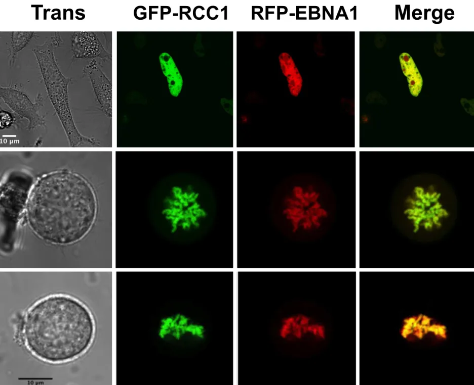
metaphasic chromosomes. 167
EBNA1 has been suggested to directly bind to AT-rich regions of the chromosomal DNA via
168
AT-hook motifs located within the LR1/GR1 and LR2/GR2 regions (Sears et al., 2004). In
169
particular, fusion proteins between mCherry and various combinations of the EBNA1 regions
170
containing AT-hook motifs, efficiently associated with chromosomes (Kanda et al., 2013).
171
However, the effect of specific deletion of these AT-hook motifs - in the context of the whole
172
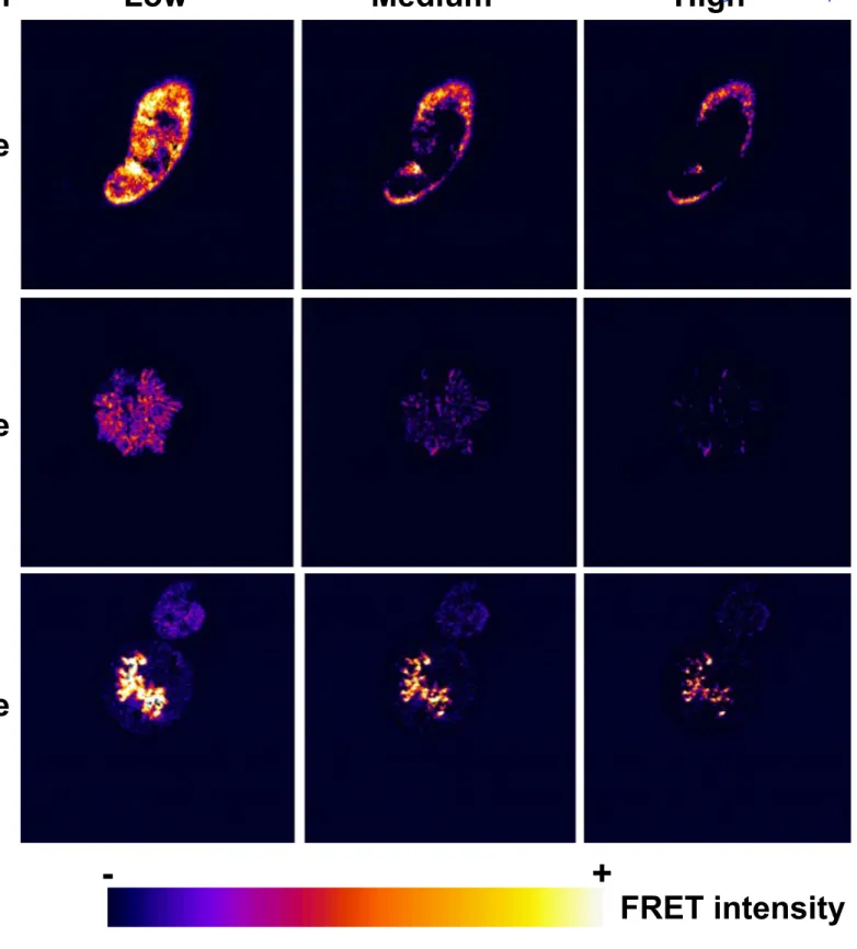
protein - on its association with cellular chromosomes has never been tested. We therefore
173
generated derivatives of GFP-EBNA1 with either aa 40 to 53 (deleting most of GR1) or 326
174
to 358 (deleting two thirds of GR2) or both regions deleted (Fig. 1A) and tested their capacity
175
to bind mitotic chromosomes and activate transcription. Association with chromosomes was
176
first analysed by confocal microscopy, following transfection of HeLa cells with expression
177
plasmids for GFP-EBNA1 or the mutated derivatives. Deletion of the GR regions led to the
178
appearance of a faint diffuse staining of the cell nuclei which was more accentuated in the
179
double mutant (Fig. 1B). However, even in the double mutant, a large proportion of the
180
protein remained localized to the metaphasic chromosomes. This suggests that the AT-hook
181
motifs are not the sole domains responsible for EBNA1 attachment to the chromosomes
182
during mitosis. Second, we performed a FRAP analysis during cell interphase to
183
comparatively evaluate the mobility of each protein. The half-time of fluorescence recovery
184
of the EBNA1 GR-deletion mutants, especially that of the double mutant, was strongly
185
diminished as compared with GR-wild type EBNA1, indicating a higher mobility of proteins
186
lacking the AT-hook motifs (Fig. 1C). Finally, to complete the characterization of these
187
mutants, we tested their transactivation ability since LR1 and LR2 regions were previously
reported to be important for transcriptional activation (Mackey & Sugden, 1999). The single
189
GR deletion mutants appear to activate LUC expression from the pGL2-FR-TK-LUC reporter
190
construct to similar levels as GR-wild type (Fig. 1D). By contrast the double mutant shows
191
significant reduction in transcriptional activation through FR. Taken together, these results
192
support a role for these two regions in transcriptional activation, but demonstrate that
193
although these regions appear to play an important role in chromatin association during
194
interphase, as could be deduced from the FRAP experiments, they are not absolutely essential
195
for tethering EBNA1 to the mitotic chromosomes.
196
EBNA1 interacts directly with RCC1 197
Since the AT-hook motifs, appear not to be essential for EBNA1 association with mitotic
198
chromatin, it is likely that one or more cellular partners are involved in mediating the linking
199
of EBNA1 with the chromosomes. The two cellular proteins which have been previously
200
found to play a role in this process - hEBP2 and HMGB2 - do not appear to be sufficient to
201
account for all the properties of EBNA1 during EBV replication and segregation. In order to
202
identify novel cellular partners of EBNA1, a yeast two-hybrid screen using EBNA1 as bait
203
was performed. From this screen, Regulator of Chromosome Condensation 1 (RCC1) (gene
204
ID: 1104), a guanine-nucleotide releasing factor that promotes exchange of Ran-bound GDP
205
with GTP, was identified. RCC1 plays a key role both in nucleo-cytoplasmic transport and in
206
the regulation of onset of chromosome condensation in S phase (Hadjebi et al., 2008).
207
The interaction between EBNA1 and RCC1 was first examined by
co-208
immunoprecipitation from transfected HeLa cells (Fig. 2A). Myc-tagged RCC1 specifically
209
co-immunoprecipitated with Flag-tagged EBNA1. Consistently, in a reverse experiment,
210
Myc-tagged EBNA1 specifically co-immunoprecipitated with Flag-tagged RCC1.
211
To assess if the interaction is direct, an in vitro GST-pulldown assay was performed
212
using both GST-RCC1 and 6xhis-EBNA1 produced in bacteria and purified. 6xhis-EBNA1
was incubated with similar amounts of GST or GST-RCC1 proteins bound to glutathione
214
sepharose beads. EBNA1 was efficiently retained on GST-RCC1 beads but not on GST alone,
215
which strongly suggests that a direct interaction occurs between EBNA1 and RCC1 (Fig. 2B).
216
EBNA1 binds RCC1 via domains previously reported to be essential for chromosome 217
binding of the protein 218
EBNA1 interaction with mitotic chromosomes has been reported to be dependent on three
219
regions: CBS-1 (aa 72 to 84), CBS-2 (aa 328 to 365) and CBS-3 (aa 8 to 54) (Fig. 3A) (Kanda
220
et al., 2001; Maréchal et al., 1999; Wu et al., 2002). We thus investigated the involvement of
221
these regions in the interaction with RCC1. For this, a series of GFP-tagged EBNA1 deletion
222
mutants were expressed in HeLa cells and the lysates incubated with GST-RCC1-bound
223
beads. Deletion mutants containing either CBS-1/CBS-3 (EBNA1 8-92), CBS-2 (EBNA1
224
323-410) or both (EBNA1 8-410) were all able to interact with RCC1, whereas EBNA1
377-225
641 with both regions deleted, showed considerably reduced interaction (Fig. 3B). This
226
preferential interaction of RCC1 with the CBS domains of EBNA1 supports a putative role
227
for RCC1 in EBNA1’s targeting to metaphase chromosomes. Surprisingly however, deletion
228
of region 326 to 376 completely abrogated the interaction with RCC1 (Fig. 3C), even though
229
the CBS-1/-3 domains still present in this mutant were sufficient for interaction with RCC1 in
230
mutant EBNA1 8-92 (Fig. 3B). This suggests that EBNA1 Δ326-376 protein’s general
231
topology may be altered such that the CBS-1/-3 domains are no longer accessible to interact
232
with RCC1.
233
We also tested the capacity of the GR-deleted mutants used in Figure 1, to interact with
234
RCC1 (Fig. 4). Although deletion of each region individually did not prohibit interaction with
235
RCC1, deletion of both motifs had a dramatic effect. This suggests that at least one intact GR
236
sub-region is required for the interaction with RCC1.
237
Taken together, these results indicate that the EBNA1 interaction domains with RCC1
overlap closely with the previously characterized EBNA1 chromosome binding regions.
239
However, it is to be noted that mutant Δ40-53/Δ326-358 with GR1 and most of GR2 deleted,
240
was still - at least partially - associated with the mitotic chromosome, whereas no interaction
241
with RCC1 could be detected in the GST-pulldown assay. Thus, although RCC1 is likely to
242
contribute to EBNA1 association with metaphasic chromosomes, it is probably not the only
243
factor involved.
244
The RCC1 N-terminal tail is essential for RCC1 interaction with EBNA1 245
RCC1 is composed of small N-terminal and C-terminal unstructured tails surrounding a
246
seven-bladed propeller structure (Makde et al., 2011) (Fig. 5A). Due to the highly structured
247
central domain of the protein, introducing large mutations into this region would likely
248
disorder the entire structure. Therefore, a single RCC1 mutant, RCC1 Δ1-20, with the
N-249
terminal tail deleted, was generated. Further, the N-terminal tail was cloned in fusion with
250
GST. GST-pulldown using transfected HeLa cell extracts were performed with these proteins
251
(Fig. 5B). Interestingly, we found that EBNA1 interacted strongly with the N-terminal tail of
252
RCC1 and very inefficiently with the rest of the protein. To further map the interaction region,
253
smaller deletions within the N-terminal extremity of RCC1 were introduced. Deletion of aa
254
11-15 or 16-20 did not significantly modify the interaction with EBNA1, in contrast to
255
deletions of aa 1-5 and 6-10 which both impaired the interaction. These results suggest that
256
the 10 first amino acids of the RCC1 N-terminal tail are important for the interaction of RCC1
257
with EBNA1.
258
Peptide array analysis confirms the RCC1 interaction with the CBS-3/-1 and CBS-2 259
domains and also defines potential supplementary RCC1 interaction regions in the C-260
terminal moiety of the protein. 261
With the aim to delimit the domains of EBNA1 involved in the interaction with RCC1 more
262
precisely, a peptide array analysis was undertaken. A library of overlapping peptides
mers), each shifted by 5 amino acids across the entire sequence of EBNA1 (including the
264
GAr) was immobilised onto membranes and probed with recombinant GST-RCC1 full length
265
(FL) and mutants (Fig. 6). Probing the EBNA1 peptide array with FL GST-RCC1 revealed
266
intermediate to strong binding to peptides covering regions that encompass both GR repeats
267
of EBNA1 as well as CBS-1 (aa 72 to 84) and CBS-3 (aa 8 to 54), which is consistent with
268
our GST-pulldown mapping. It is interesting to note that the strongest interacting regions
269
overlap with the AT-hook domains whose deletion in mutant EBNA1Δ40-53/Δ326-358,
270
completely abrogates binding in our GST-pull down assay (Fig. 4). Thus, there is good
271
agreement between the GST-pull down assay and the peptide array analysis. Moreover, the
272
peptide analysis revealed the presence of unexpected binding regions in the central and
C-273
terminal moiety of EBNA1: a first region between aa G371-E435 lies between the known
274
binding sites for CK2 and USP7 and is well conserved in EBV EBNA1 isolates (Hussain et
275
al., 2014); a second region incorporates the C-terminal tail of the protein, rich in negatively
276
charged residues. Reprobing the array with GST, indicated that the binding observed for
GST-277
RCC1 was specific to RCC1.
278
When a second array was probed with GST-RCC1Δ1-20, binding was observed for
279
largely the same set of peptides as for full length RCC1, but much weaker, with the notable
280
exception of the C-terminal tail peptides, which showed no binding. Therefore, consistent
281
with our GST-pulldown mapping, RCC1 with its N-terminal 20 aa deleted, only weakly
282
interacts with EBNA1. This suggests that either the N-terminal region of RCC1 is the primary
283
mediator of binding or that it is required for correct folding of full-length RCC1 to enable
284
binding to EBNA1, or possibly both.
285
To distinguish between these possibilities, the second array was stripped and re-probed
286
with just the N-terminal region of RCC1 fused to GST. GST-RCC1 1-20 showed intermediary
287
binding to the C-terminal tail peptides and strong binding to peptides covering region R396 to
E435. However, strong binding was not observed with peptides localised in the N-terminal
289
half of EBNA1 although these were strongly bound by full-length RCC1. This suggests that
290
residues 1-20 of RCC1 contribute to the conformation of RCC1, or otherwise promote the full
291
interaction, but may not completely comprise the binding site.
292
Regarding the strongest binding region identified for RCC1 1-20, analysis of the
293
EBNA1 amino acid sequence reveals that the stretch of residues in common between EBNA1
294
peptides interacting strongly with GST-RCC1 1-20 lies between aa 411 to 420
295
(EADYFEYHQE). This region contains 4 negatively-charged residues. By contrast, the
N-296
terminal region of RCC1 (MSPKRIAKRRSPPADAIPKS) contains 6 positively charged
297
residues and one negatively charged residue. It is therefore possible that the interaction of
298
RCC1 with these EBNA1 peptides is largely charge-based and possibly an artefact of the
299
array approach, if these stretches are not normally accessible in the folded protein. To explore
300
this possibility, a new array was generated with a series of mutated peptides spanning residue
301
401 to 430 and probed with GST-RCC1 1-20 (Supplementary Figure 1). This revealed that
302
indeed charge is critical to the binding, such that replacement of the 4 charged residues (E411,
303
D413, E416, E420) completely abrogated binding, however F415 and Y414 were also found to be
304
key for the interaction. This region was thus a candidate for being a core binding site between
305
the N-terminal region of RCC1 and EBNA1. However, an EBNA1 mutant deleted for this
306
region (EBNA1Δ411-420) still interacted with RCC1 in a GST-pulldown assay (Fig. 4B, lane
307
5). Therefore, it appears that this region is not required for stabilizing the EBNA1-RCC1
308
interaction in vitro. However, it cannot be ruled out that it might play a role in the interaction
309
in vivo, in a context where RCC1 is associated with chromatin.
310
EBNA1 interacts with RCC1 localized to chromatin during mitosis 311
In order to determine the subcellular localization of the two proteins in living cells,
312
several fluorescent-tagged forms of EBNA1 and RCC1 were expressed in HeLa cells and
observed by live cell imaging during interphase and mitosis. The EGFP-RCC1 and
EBNA1-314
RFP proteins colocalize almost perfectly in living cells during interphase and throughout
315
mitosis (Fig. 7): during interphase the proteins colocalize throughout the nucleoplasm, with
316
the exception of the nucleolus from which RCC1 is completely excluded and where weak
317
staining is observed for EBNA1. During prophase and metaphase, both EBNA1 and RCC1
318
appear to be associated with the mitotic chromosomes. Similar observations were made in
319
cells coexpressing other pairs of EGFP- and RFP-tagged forms of the proteins (data not
320
shown).
321
To confirm that EBNA1 interacts with RCC1 in living cells we performed a Förster
322
resonance energy transfer (FRET) analysis. FRET is a nonradioactive energy transfer that can
323
occur when a donor and a compatible acceptor fluorophore are located at less than 10 nm
324
from each other. FRET efficiency relies on the relative position and distance of the donor and
325
acceptor fluorophores, which can be affected by the position of the fluorophore in a fusion
326
protein. Therefore, both the following pairs of fusion proteins: EGFP-RCC1/EBNA1-RFP and
327
EBNA1/RFP-RCC1 were tested. No significant FRET was observed with the
EGFP-328
RCC1/EBNA1-RFP pair. However, the EGFP-EBNA1/RFP-RCC1 pair revealed clear FRET
329
activity during both interphase and metaphase (Fig. 8). During interphase, it is interesting to
330
note that although the proteins colocalize throughout the nucleoplasm (Fig. 7), they were only
331
in close interaction at the periphery of the nucleus (Fig. 8). Only very weak interaction was
332
observed during prophase. By contrast, strong interaction was observed between the two
333
proteins on metaphasic chromosomes. Taken together, these results suggest that the
334
interaction between RCC1 and EBNA1 is highly dynamic through the cell cycle. The strong
335
FRET signal observed specifically on metaphasic chromosomes supports a role for RCC1 in
336
stabilizing the EBNA1 interaction with the chromosomes during mitosis.
337 338
DISCUSSION 339
The mechanisms by which EBNA1 tethers the EBV genome to mitotic chromosomes are far
340
from understood. The AT-hook regions of the protein have been proposed to play a major role
341
in EBNA1 chromosome binding activity and episomal maintenance (Sears et al., 2003, 2004).
342
The use of netropsin, a small molecule that binds to the minor groove of AT-rich DNA, leads
343
to the loss of EBV genomes from cells, supporting the role of the AT-hooks in episomal
344
maintenance (Chakravorty & Sugden, 2015). However, we have found that deletion of the
345
EBNA1 AT-hook regions does not abrogate EBNA1’s general targeting to metaphasic
346
chromosomes. This result is consistent with a previous analysis revealing three independent
347
CBS regions (Marechal et al., 1999). In effect, specific deletion of the AT-hook domains
348
leaves CBS-1 (aa 72 to 84) intact. Therefore these data reinforce the idea of alternative or
349
complementary mechanisms of recruitment of EBNA1 to the metaphasic chromosomes,
350
possibly via the interaction with cellular chromatin binding factors.
351
Here, we have identified RCC1 as a novel mediator of EBNA1 interaction with
352
metaphase chromosomes. We have characterized the interaction between EBNA1 and RCC1
353
by various methods. Importantly, by performing an in vitro assay using both proteins purified
354
from bacteria, we have demonstrated that the two proteins can interact directly. Up to now,
355
however, we have not been able to perform a successful co-immunoprecipitation with
356
endogenous proteins. One explanation, other than a possible interference of antibodies with
357
the interaction and the low level of expression of EBNA1, is the small amount of cells from
358
the total population undergoing mitosis - the phase of the cell cycle in which our FRET
359
experiments demonstrate a close interaction between the proteins.
360
Characterization of the interaction domains revealed that the RCC1 interaction domains
361
of EBNA1 closely overlap with the CBS regions of the protein, known to be important for
362
tethering EBNA1 to the chromosomes (Maréchal et al., 1999). Accordingly, we have found
that region 8-92 which includes both CBS-3 and CBS-1, and region 323-410 which includes
364
CBS-2, can interact independently with RCC1. Surprisingly, however, deletion of CBS-2 in
365
mutant Δ326-376 previously found to impair hEBP2 interaction as well as oriP plasmid
366
maintenance and mitotic localization (Shire et al., 1999; Wu et al., 2000), completely
367
abolished EBNA1 interaction with RCC1. This latter result is not consistent with the finding
368
that the CBS-3/-1 region (still present in this mutant) is sufficient alone to mediate the
369
interaction with RCC1. This suggests that the conformation or accessibility of the CBS-3/-1
370
region is compromised in this mutant, affecting various functions of the protein without
371
necessarily reflecting the direct involvement of the deleted region.
372
Since the EBNA1 mutant with the two AT-hook domains deleted, appears to localize to
373
chromatin both during interphase and through mitosis (which had not previously been tested),
374
mechanisms other than the interaction of EBNA1 with AT-rich regions of DNA are likely to
375
be required. However, deletion of the two AT-hook domains also affected the interaction with
376
RCC1, suggesting that still other factors are involved. Another chromatin binding protein,
377
HMGB2, was previously identified as an EBNA1 interacting factor (Jourdan et al., 2012).
378
HMGB2 could thus be responsible for targeting EBNA1 to the chromatin, in the absence of
379
both direct interaction with DNA (via the AT-hooks) and interaction with RCC1. To
380
corroborate such an hypothesis it would be interesting to know more precisely where HMGB2
381
binds within EBNA1. Alternatively, another as yet unidentified partner could be involved in
382
the process. These possibilities are not mutually exclusive, indeed EBNA1 may employ
383
multiple mechanisms to tether the viral genome to chromatin and to associate with the
384
chromatin independently of the viral genome, through the different stages of the cell cycle and
385
under various conditions.
386
Regarding the domains of RCC1 involved in the interaction with EBNA1, the
N-387
terminal flexible region of RCC1 was identified as an essential domain. Interestingly, this
terminal tail, and in particular the serine at position 2, is the site of post-transcriptional
389
modifications (both α-N-methylation and phosphorylation) that are important for stable
390
chromatin association and regulation of RCC1’s NLS interaction with importins α and β
391
(Chen et al., 2007; Li & Zheng, 2004). Such modifications of RCC1 N-terminal tail within
392
mammalian cells could modulate or even prevent the interaction between the two proteins.
393
Conversely, since these modifications have been suggested to play an important role in the
394
mobility of RCC1 during metaphase and in its stabilization on the chromatin (Hutchins et al.,
395
2004; Li & Zheng, 2004), EBNA1’s interaction with these regions could affect the dynamics
396
of RCC1’s interaction with chromatin.
397
Use of EBNA1 peptide arrays permitted a more detailed mapping of the EBNA1
398
interaction regions with respect to full length RCC1 as well as the N-terminal tail. Interaction
399
of full-length RCC1 (and to lesser extent the N-terminal tail of RCC1 alone) with the
CBS-1/-400
3 and CBS-2 regions of EBNA1 was confirmed. In addition, two other regions of EBNA1
401
were identified that might be involved in the interaction: the C-terminal tail, and a negatively
402
charged region located between the previously characterized CK2 and USP7 binding sites.
403
Interestingly, these two regions (particularly the latter) are recognised by RCC1’s N-terminal
404
tail. In particular, the N-terminal tail of RCC1 critical for the interaction specifically contacts
405
a region of EBNA1 - DYFEYHQE - located between aa 413 and 420. When set in the context
406
of an in silico structural model of full-length EBNA1 (Hussain et al., 2014), this domain
407
appears to be located in a region that resembles a small pocket. This could potentially
408
accommodate the N-terminal region of RCC1, facilitating further interactions between the
409
CBS domains of EBNA1 and the seven-propeller helix of RCC1, and hence stabilising the
410
interaction between the two proteins. However, deletion of this domain does not preclude
411
binding of EBNA1 and RCC1 in the in vitro assays used here and we cannot exclude that it
412
may reflect artifactual binding to a site that is not normally accessible to RCC1.
With regard to the dynamics of interaction between the two proteins in live cells, the
414
combination of colocalization experiments in live cells and FRET analysis reveals that the
415
two proteins colocalize with the chromatin throughout the cell cycle. However, their
416
proximity varies according to the location within the cell nucleus as well as the phase of the
417
cell cycle: during interphase, although the two proteins appear to be colocalizing throughout
418
the cell nucleus, FRET could only be observed at the periphery of the nucleus, suggesting that
419
close interaction between EBNA1 and RCC1 could be linked to the latter being in a different
420
conformation when actively involved in nucleo-cytoplasmic transport. This result opens up
421
the possibility that the interaction of EBNA1 with RCC1 could play a role in functions other
422
than segregation of the viral episome.
423
Importantly, during mitosis, FRET was mainly observed in metaphase, indicating a
424
more specific role for the RCC1-EBNA1 interaction at this particular stage of mitosis that
425
precedes segregation of sister chromatids. This observation, together with the correlation
426
between EBNA1 regions interacting with RCC1 and EBNA1 domains previously
427
characterized for their role in chromosome binding and episome maintenance, argues for an
428
important role of RCC1 in EBV episome tethering to the chromosomes and subsequent
429
episome maintenance. However, this hypothesis will be difficult to prove directly since RCC1
430
is an essential protein whose downregulation leads to premature chromosome condensation or
431
arrest in the G1 phase of the cell cycle (Uchida et al., 1990). The observation that deletion of
432
the AT-hook domains still permits association of EBNA1 with metaphase chromosomes while
433
it appears to abrogate interaction with RCC1 in vitro, does not refute the hypothesis. First, the
434
assay conditions used to detect the interaction in vitro may not be optimum and a weak
435
interaction between EBNA1 and RCC1 might nevertheless occur in vivo in the absence of the
436
AT-hook domains. Alternatively, in the absence of an interaction between RCC1 and EBNA1,
437
alternative mechanisms tethering EBNA1 to the chromatin may act. Of note, depletion of
HMGB2 was found to affect the stability but not to prevent EBNA1 association with
439
chromatin, nor did it impact viral genome maintenance, despite the observed interaction
440
between EBNA1 and HMGB2 on chromatin through mitosis (Jourdan et al., 2012). It is thus
441
likely that several mechanisms are involved in EBNA1 tethering to the chromatin,
442
orchestrated to play a role at different stages of the cell cycle to both bring EBNA1 to the
443
chromatin and stabilize it once there: During interphase, the EBV genomes are distributed to
444
perichromatic regions of the nucleus in a manner dependent on the FR element and EBNA1
445
(Deutsch et al., 2010). It has been suggested that the AT-hook domains of EBNA1 could play
446
an important role in this tethering of the EBV genomes to the chromatin. This hypothesis is
447
strengthened by the demonstration that HMG1Aa, an AT-hook binding protein can
448
functionally replace the N-terminal domain of EBNA1 (Sears et al., 2003; Thomae et al.,
449
2008) and by the results of our FRAP analysis showing a higher mobility of EBNA1 with its
450
AT-hook domains deleted; HMGB2 is associated with EBNA1 on the chromatin during
451
interphase and more so during mitosis (Jourdan et al., 2012); RCC1 colocalizes with EBNA1
452
throughout the cell cycle but the interaction appears to be specifically stabilized during
453
metaphase. Moreover, interaction between EBNA1 and the chromatin could be facilitated by
454
a direct interaction through the AT-hook domains with nucleosomal DNA.
455
Finally, it is interesting to note that the ortholog of EBNA1 in KSHV (Kaposi Sarcoma
456
Herpes Virus), LANA (Latency-Associated Nuclear antigen), directly interacts with
H2A-457
H2B dimers to enable its binding to chromosomes (Piolot et al., 2001). The resolution of the
458
crystal structure of the nucleosome complexed with the first 23 amino acids of LANA
459
revealed that the LANA peptide forms a hairpin that interacts with an acidic H2A-H2B region
460
implicated in the formation of higher order chromatin structure (Barbera et al., 2006).
461
Interestingly, RCC1 targets the same region of the nucleosomal H2A-H2B dimer as LANA,
462
and the two proteins have been shown to compete for nucleosome interaction (England et al.,
2010). Thus, whereas LANA directly contacts the H2A-H2B dimer to enable its binding to
464
the chromosomes, EBNA1 may interact indirectly with the same H2A-H2B dimer through
465
RCC1. Similar to EBNA1, LANA also interacts with several cellular proteins that appear to
466
play a role in the tethering of LANA to chromosomes and/or episomal segregation (Krithivas
467
et al., 2002; Xiao et al., 2010). Thus EBNA1 and LANA have evolved similar but not
468
identical mechanisms to insure anchorage of the viral episomes onto the chromatin at different
469
stages of the cell cycle, allowing efficient replication and segregation of the respective viral
470 genomes. 471 472 METHODS 473
Cell culture and transfections 474
HeLa and HEK293T cells were grown at 37°C in DMEM, 10% FCS. Plasmid transfection
475
was performed using the PEI transfection reagent (Polysciences).
476
Plasmids 477
pEGFP-N1-EBNA1ΔGA (aa 8 to 641) has been described previously (Jourdan et al., 2012).
478
Unless otherwise indicated, all EBNA1 plasmids used were derived from this plasmid and
479
thus contain EBNA1 deleted for GAr as well as the first 7 N-terminal aa. EBNA1 and RCC1
480
N-terminal tail deletion mutants were generated by site-directed mutagenesis (QuickChange
481
Site-Directed Mutagenesis kit, Stratagene). For the two-hybrid screen, ORFs for EBNA1,
482
EBNA1 8-410 and EBNA1-381Cter were PCR-amplified (KOD Hot Start DNA
483
Polymerase®, EMD Millipore) cloned first in pDONR207 then into pGBKT7, using the
484
Gateway recombinational cloning system (Invitrogen). EBNA1 and deletion mutants cloned
485
into pDEST-Myc, pCI-3xFlag, pDEST15 or pDEST53 were also generated using the Gateway
486
system. A codon-optimised version of EBNA1-ΔGA was cloned into pET22b (Merck
487
Millipore) to generate pET22b-EBNA1. The ORF for full-length RCC1 (alpha isoform) was
transferred from pDONR223-RCC1 (obtained from a human ORFeome library) into
pDEST-489
Myc, pCI-3xFlag or pDESTTM15 (Invitrogen) using the Gateway system. pEGFP-C1-EBNA1
490
has been described previously (Jourdan et al., 2012). pRFP and pEGFP fusion proteins were
491
generated by cloning the relevant PCR-amplified ORFs in pRFP-N1, pRFP-C1, pEGFP-N1 or
492
pEGFP-C1, using the In-Fusion®HD cloning kit (Clontech). All oligonucleotides used are
493
listed in Supplementary Table 1.
494
Luciferase Assays 495
Renilla or Firefly luciferase activities were measured in a VeritasTM Luminometer (Turner
496
Biosystems) using the Renilla or Firefly Luciferase Assay system (Promega Madison Co).
497
Yeast two-hybrid screens 498
The screens were performed as previously described (Bazot et al., 2014) using
pGBKT7-499
EBNA1/-EBNA1-8-410 or -EBNA1-381Cter as bait vectors and a human LCL AD-cDNA
500
library (Invitrogen). Positive clones were sequenced and identified by automatic BLAST
501
(Pellet et al., 2010).
502
Co-immunoprecipitation and western blotting 503
Cells were lysed in 50 mM Tris-HCl pH 7.5, 150-300 mM NaCl, 1 mM Dithiotreitol (DTT)
504
and 0.5% Nonidet P-40 plus protease inhibitors. For immunoprecipitation of transiently
505
expressed Flag-tagged proteins, extracts were incubated with 20 μl of anti-Flag M2 affinity
506
gel (Sigma) for 4 h at 4°C. After washing, bound proteins were analysed by western blotting,
507
visualised using ECL (Thermo Fisher Scientific). Antibodies: anti-Flag rabbit polyclonal
508
antibody (Sigma), anti-His6 mouse monoclonal antibody (Roche Molecular Biochemicals),
509
anti-c-Myc (9E10) HRP-conjugated antibody (Santa Cruz Biotechnology, Inc). Anti-rabbit
510
and anti-mouse (HRP)-conjugated antibodies (GE Healthcare) were used as secondary
511
antibodies.
512
Production and purification of the 6xhis-EBNA1 protein 513
6xhis-EBNA1 was purified from Escherichia coli Rosetta (pLysS) strain transformed with
514
pET22b-EBNA1. Cells were lysed in 50 mM NaH2PO4, 1 M NaCl, 10 mM imidazole pH 8,
515
protease inhibitors and 1 mg/ml lysozyme. After sonication, the protein was purified by
516
gravity-flow chromatography using Ni-NTA agarose beads. Beads were washed with lysis
517
buffer plus 50 mM imidazole and the proteins eluted in lysis buffer containing 150 mM
518
imidazole.
519
In vitro GST-Pulldowns 520
Glutathione S-Transferase (GST) and GST-fusion proteins were purified from Escherichia
521
coli BL21 (DE3) codon plus strain extracts, with glutathione-Sepharose 4B beads (GE
522
Healthcare). Beads carrying the GST or the GST-fusion proteins were equilibrated in MTPBS
523
(150 mM NaCl, 16 mM Na2HPO4, 4 mM NaH2PO4, 100 mM EDTA, 1% Triton) and
524
incubated with either purified 6xhis-EBNA1 or transfected cell extracts for 4h in MTPBS.
525
Beads were washed 5 times in MTPBS and bound proteins analysed by western blotting.
526
EBNA1 peptides arrays 527
25mer peptides comprising the entire sequence of EBNA1 (B95.8 strain), with 5 residue shifts
528
(ie. initiating at residues 1, 6, 11, 16 etc) were synthesized by automatic SPOT synthesis
529
(Kramer and Schneider-Mergener, 1998) directly onto cellulose membranes using Fmoc
(9-530
fluorel methoxycarbonyl) chemistry and Autospot Robot ASS222 peptide synthetizer (Invatis
531
Bioanalytical Instruments AG). Arrays were bathed in ethanol and washed for 10 mins in
532
TBST (50 mM Tris.HCl pH7.5, 150 mM NaCl, 0,05% Tween-20), followed by blocking in
533
TBST, 5% non-fat milk powder (NFM) for 2 hours at RT and washed again with TBST.
534
Arrays were probed with purified GST (as control) or GST fusion proteins, at 2 to 5 ug/mL in
535
TBST, 1% NFM, shaking overnight at 4°C. After washing in TBST, membranes were
536
incubated with rabbit anti-GST-HRP and the array revealed using ECL (Pierce #32106). To
537
strip the array membranes for re-probing, they were covered in 60 mM Tris-HCl pH6.8, 20
mM DTT, 70 mM SDS, at 70°C for 30 min.
539
Confocal microscopy 540
HeLa cells were plated onto glass-bottomed dishes for confocal microscopy (Ibidi) and
541
transfected with expression vectors coding for EBNA1 and RCC1 fused to either EGFP or
542
RFP. Live cells were analyzed with a Zeiss LSM710 confocal microscope with ZEN software.
543
GFP and RFP signals were acquired using respectively an argon laser at 488nm and a laser
544
diode (DPSS) at 561nm. Z-stack series were also acquired for mitotic cells: the most
545
representative stacks are presented. All analyses were conducted with ImageJ Software.
546
Fluorescence recovery after photobleaching (FRAP) analysis 547
Cells used for FRAP acquisition were prepared as for classical microscopy and data collected
548
using a confocal spinning disk microscope. Same parameters were used to acquire all images.
549
Regions of interest were photobleached using a 494-nm laser during 510ms at full power.
550
Images were acquired with an EM gain of 30, 200ms exposure time and a 488-nm laser at
551
9.5% full power. 5 images were acquired before bleaching then 1 image every 0.5s for 5
552
seconds, 1 image per second for 1 minute and 1 image every 5 seconds for 30 seconds.
553
Analysis was performed using ImageJ and EasyFrap software.
554
Förster resonance energy transfer (FRET) analysis 555
Cells used for FRET acquisition were prepared as for confocal microscopy and data collected
556
with an LSM-710 confocal microscope. FRET analysis was performed using the FRET
557
Analyzer plugin (http://rsb.info.nih.gov/ij/plugins/fret-analyzer/fret-analyzer.htm). Three
558
tracks were used for the acquisition: EGFP (excitation: GFP, reception: GFP range), RFP
559
(excitation: RFP, reception: RFP range) and FRET (excitation: GFP, reception: RFP range).
560
Argon laser and DPSS were used at 2% and 7% power respectively. Gain level was 540 for
561
the GFP signal and 640 or 690 for the RFP signal. Spectral leakage was measured by
562
acquisition of 5 images for each track with EGFP or RFP fusions expressed alone. Double
transfected cells were used for FRET acquisition data. In each case EGFP, RFP and FRET
564
fluorescent signals were acquired for each track.
565 566
ACKNOWLEDGEMENTS 567
This work was supported by the ‘Institut National de la Santé et de la Recherche Médicale’
568
(INSERM); ‘the Cluster de Recherche Rhône-Alpes en Infectiologie’; ‘the Ligue Contre le
569
Cancer, comité du Rhône’; the ‘Association pour la Recherche contre le Cancer (ARC grant
570
n° R11176CC)’. T. D. and Q. B. have been recipient of a fellowship from the ‘Ministère de
571
l’enseignement et de la Recherche scientifique (MENRS), T. B. from the “Ligue Nationale
572
Contre le Cancer”, Q. B. from the ‘Association pour la Recherche contre le Cancer’ and D. M.
573
L. by a Medical Research Council (MRC) scholarship. We acknowledge the AniRA Genetic
574
Analysis and cytometry platforms and the “Platim” microscope facilities of the SFR
575
Biosciences Gerland-Lyon Sud (US8/UMS3444).
576 577
REFERENCES 578
Adams, A. (1987). Replication of latent Epstein-Barr virus genomes in Raji cells. J Virol 61, 579
1743–1746.
580
Ambinder, R. F., Shah, W. A., Rawlins, D. R., Hayward, G. S. & Hayward, S. D. (1990). 581
Definition of the sequence requirements for binding of the EBNA-1 protein to its palindromic
582
target sites in Epstein-Barr virus DNA. J Virol 64, 2369–2379.
583
Ambinder, R. F., Mullen, M. A., Chang, Y. N., Hayward, G. S. & Hayward, S. D. (1991). 584
Functional domains of Epstein-Barr virus nuclear antigen EBNA-1. J Virol 65, 1466–1478.
585
Askjaer, P., Galy, V., Hannak, E. & Mattaj, I. W. (2002). Ran GTPase cycle and importins 586
alpha and beta are essential for spindle formation and nuclear envelope assembly in living
587
Caenorhabditis elegans embryos. Mol Biol Cell 13, 4355–4370.
Bamba, C., Bobinnec, Y., Fukuda, M. & Nishida, E. (2002). The GTPase Ran regulates 589
chromosome positioning and nuclear envelope assembly in vivo. Curr Biol CB 12, 503–507.
590
Barbera, A. J., Chodaparambil, J. V., Kelley-Clarke, B., Joukov, V., Walter, J. C., 591
Luger, K. & Kaye, K. M. (2006). The nucleosomal surface as a docking station for Kaposi’s 592
sarcoma herpesvirus LANA. Science 311, 856–861.
593
Bazot, Q., Deschamps, T., Tafforeau, L., Siouda, M., Leblanc, P., Harth-Hertle, M. L., 594
Rabourdin-Combe, C., Lotteau, V., Kempkes, B. & other authors. (2014). Epstein-Barr 595
virus nuclear antigen 3A protein regulates CDKN2B transcription via interaction with MIZ-1.
596
Nucleic Acids Res 42, 9700–9716.
597
Bierbaum, M. & Bastiaens, P. I. H. (2013). Cell cycle-dependent binding modes of the ran 598
exchange factor RCC1 to chromatin. Biophys J 104, 1642–1651.
599
Bochkarev, A., Barwell, J. A., Pfuetzner, R. A., Bochkareva, E., Frappier, L. & 600
Edwards, A. M. (1996). Crystal structure of the DNA-binding domain of the Epstein-Barr 601
virus origin-binding protein, EBNA1, bound to DNA. Cell 84, 791–800.
602
Cai, X., Schafer, A., Lu, S., Bilello, J. P., Desrosiers, R. C., Edwards, R., Raab-Traub, N. 603
& Cullen, B. R. (2006). Epstein-Barr virus microRNAs are evolutionarily conserved and 604
differentially expressed. PLoS Pathog 2, e23.
605
Chakravorty, A. & Sugden, B. (2015). The AT-hook DNA binding ability of the Epstein 606
Barr virus EBNA1 protein is necessary for the maintenance of viral genomes in latently
607
infected cells. Virology 484, 251–258.
608
Chaudhuri, B., Xu, H., Todorov, I., Dutta, A. & Yates, J. L. (2001). Human DNA 609
replication initiation factors, ORC and MCM, associate with oriP of Epstein-Barr virus. Proc
610
Natl Acad Sci U S A 98, 10085–10089.
611
Chen, T., Muratore, T. L., Schaner-Tooley, C. E., Shabanowitz, J., Hunt, D. F. & 612
Macara, I. G. (2007). N-terminal alpha-methylation of RCC1 is necessary for stable 613
chromatin association and normal mitosis. Nat Cell Biol 9, 596–603.
614
Coppotelli, G., Mughal, N., Marescotti, D. & Masucci, M. G. (2011). High avidity binding 615
to DNA protects ubiquitylated substrates from proteasomal degradation. J Biol Chem 286,
616
19565–19575.
617
Coppotelli, G., Mughal, N. & Masucci, M. G. (2013). The Gly-Ala repeat modulates the 618
interaction of Epstein-Barr virus nuclear antigen-1 with cellular chromatin. Biochem Biophys
619
Res Commun 431, 706–711.
620
Crawford, D. H. (2001). Biology and disease associations of Epstein-Barr virus. Philos 621
Trans R Soc Lond B Biol Sci 356, 461–73.
622
Cushman, I., Stenoien, D. & Moore, M. S. (2004). The dynamic association of RCC1 with 623
chromatin is modulated by Ran-dependent nuclear transport. Mol Biol Cell 15, 245–255.
624
Deutsch, M. J., Ott, E. & Schepers, A. (2010). The latent origin of replication of Epstein-625
Barr virus directs viral genomes to active regions of the nucleus. J Virol 84, 2533-2546.
626
England, J. R., Huang, J., Jennings, M. J., Makde, R. D. & Tan, S. (2010). RCC1 uses a 627
conformationally diverse loop region to interact with the nucleosome: a model for the
RCC1-628
nucleosome complex. J Mol Biol 398, 518–29.
629
Frappier, L. & O’Donnell, M. (1991). Overproduction, purification, and characterization of 630
EBNA1, the origin binding protein of Epstein-Barr virus. J Biol Chem 266, 7819–7826.
631
Hadjebi, O., Casas-Terradellas, E., Garcia-Gonzalo, F. R. & Rosa, J. L. (2008). The 632
RCC1 superfamily: from genes, to function, to disease. Biochim Biophys Acta 1783, 1467–79.
633
Hao, Y. & Macara, I. G. (2008). Regulation of chromatin binding by a conformational 634
switch in the tail of the Ran exchange factor RCC1. J Cell Biol 182, 827–36.
635
Hodin, T. L., Najrana, T. & Yates, J. L. (2013). Efficient replication of Epstein-Barr virus-636
derived plasmids requires tethering by EBNA1 to host chromosomes. J Virol 87, 13020–
637
13028.
Hung, S. C., Kang, M. S. & Kieff, E. (2001). Maintenance of Epstein-Barr virus (EBV) 639
oriP-based episomes requires EBV-encoded nuclear antigen-1 chromosome-binding domains,
640
which can be replaced by high-mobility group-I or histone H1. Proc Natl Acad Sci U S A 98,
641
1865–1870.
642
Hussain, M., Gatherer, D. & Wilson, J. B. (2014). Modelling the structure of full-length 643
Epstein-Barr virus nuclear antigen 1. Virus Genes 49, 358–372.
644
Hutchins, J. R. A., Moore, W. J., Hood, F. E., Wilson, J. S. J., Andrews, P. D., Swedlow, 645
J. R. & Clarke, P. R. (2004). Phosphorylation regulates the dynamic interaction of RCC1 646
with chromosomes during mitosis. Curr Biol CB 14, 1099–1104.
647
Jones, C. H., Hayward, S. D. & Rawlins, D. R. (1989). Interaction of the lymphocyte-648
derived Epstein-Barr virus nuclear antigen EBNA-1 with its DNA-binding sites. J Virol 63,
649
101–110.
650
Jourdan, N., Jobart-Malfait, A., Dos Reis, G., Quignon, F., Piolot, T., Klein, C., Tramier, 651
M., Coppey-Moisan, M. & Maréchal, V. (2012). Live-cell imaging reveals multiple 652
interactions between Epstein-Barr virus nuclear antigen 1 and cellular chromatin during
653
interphase and mitosis. J Virol 86, 5314–5329.
654
Kanda, T., Otter, M. & Wahl, G. M. (2001). Coupling of mitotic chromosome tethering and 655
replication competence in epstein-barr virus-based plasmids. Mol Cell Biol 21, 3576–3588.
656
Kanda, T., Horikoshi, N., Murata, T., Kawashima, D., Sugimoto, A., Narita, Y., 657
Kurumizaka, H. & Tsurumi, T. (2013). Interaction between basic residues of Epstein-Barr 658
virus EBNA1 protein and cellular chromatin mediates viral plasmid maintenance. J Biol
659
Chem 288, 24189–24199.
660
Kapoor, P. & Frappier, L. (2003). EBNA1 partitions Epstein-Barr virus plasmids in yeast 661
cells by attaching to human EBNA1-binding protein 2 on mitotic chromosomes. J Virol 77,
662
6946–56.