Supplementary materials for
Surprising lack of liposome-induced complement activation by artificial 1,3-diamidophospholipids in vitro
Simon Bugna, MSca, Marzia Buscemaa, MSc, Sofiya Matviykiva, MSc,
Rudolf Urbanics, M.D., PhDb, Andreas Weinberger, PhDc, Tamas Meszarosb , MSc, Janos Szebeni, M.D., PhD, DScb, Andreas Zumbuehl, PhD*c, Till Saxer, M.D.*d, Bert Müller, PhD*a
a
Biomaterials Science Center, University of Basel, Allschwil, Switzerland
b
Nanomedicine Research and Education Center, Semmelweis University, Budapest, Hungary
c
Department of Chemistry, University of Fribourg, Fribourg, Switzerland
d
Cardiology Division, University Hospitals of Geneva, Geneva, Switzerland
*corresponding author: phone +41 61 265 9660, [email protected], Biomaterials Science Center, University of Basel, Gewerbestrasse 14, 4123 Allschwil, Switzerland
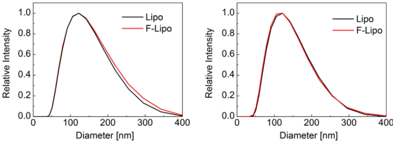
Appendix A. Size distribution of Pad-PC-Pad liposomes
The DLS measurements were carried out using the Zetasizer Nano S instrument (Malvern Instruments Ltd, Malvern, UK).
Figure S1 shows the results of the liposomes used to perform ELISA test on human sera (left diagram) and pig plasma (right diagram). Before the ELISA incubation, an aliquot of the liposomal suspension was filtered through a 100 nm tracked-edged polycarbonate membrane in order to dissipate any liposome aggregation (labelled F-Lipo). The remaining liposomal suspension was kept unfiltered (labelled Lipo). The experimentally derived size distributions are equivalent. They are characterized by a mean diameter of 122 nm with a full-width-at-half maximum (FWHM) of 152 nm. From these experiments, one can conclude that the liposomes do not excessively aggregate over a period of six days.
Figure S1. The size distributions of Pad-PC-Pad liposomes, which were applied for the
in vitro tests in human sera (left diagram) and pig plasma (right diagram), were determined
using DLS measurements.
Published in "Nanomedicine: Nanotechnology, Biology and Medicine 12(3): 845–849, 2016" which should be cited to refer to this work.
For the in vivo test, liposomes with a significantly narrower size distribution were used. The extrusion through 100 nm filters was repeated until the size distribution of liposomes became mono-disperse at (95.8 ± 1.4) nm. As these liposomes aggregated over night, a filtration-step before bolus injection was necessary.
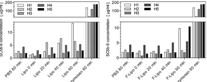
Appendix B. Supplementary data of the in vitro experiments
Figure S2. The ELISA results of complement activation in human sera demonstrate the variability between five donors.
Phosphate buffered saline (PBS) and zymosan served as negative and positive control, respectively. Their values determined after an incubation time of 60 minutes are compared with the results of the unfiltered liposomes (left diagram) and filtered liposomes (right diagram) using the incubation times indicated, i.e. starting condition and after 20, 40 and 60 minutes. One finds variability among sera donors H1 to H5. In all cases, zymosan caused a more than 20 times higher complement activation compared to liposomes. There is no complement activation by liposomes, as the values are not significantly higher than the ones measured for PBS. To determine the SC5b-9 concentration a standard curve was obtained using the blank subtracted absorbance-at-450-nm values and the assigned concentration for each standard. The slope, intercept, and correlation coefficient of the derived best-fit line for the SC5b-9 standards were within the standard ranges with a correlation coefficient of 0.99 (> 0.95), a slope of 0.00752 ± 0.00007 (0.0039 to 0.0123), and an intercept of -0.0182 ± 0.0066 (-0.189 to 0.201).
Figure S3. The ELISA results of complement activation in pig plasma show that the variability between the five donors is rather small.
The MicroVue Pan-Specific C3 Reagent Kit (Quidel, San Diego, USA) was used to measure complement activation in hirudinized pig plasma in vitro. The reagent kit converts the activity of C3 in the pig plasma to human 9 that is detectable with the MicroVue SC5b-9 Plus EIA kit (Quidel, San Diego, USA). The relative C3 consumption is inversely proportional to SC5b-9. The graphs in Figure S3 show direct proportionality with the SC5b-9 values. Limited variability between the plasma donors P1 to P5 occurs. Zymosan caused the two-fold complement activation in comparison to liposomes and negative control.
Appendix C. Supplementary information to the animal experiment
The three pigs were rested two days before experiment start. The in vivo experiments were approved by local Animal Subject Review Committees and performed at the big animal OP facility of Experimental Surgical Department of Semmelweis University, Budapest, Hungary. The pigs were sedated by an intramuscular Calypsol/Xilazine injection (ketamine, 200 to 300 mg). Anaesthesia was maintained during the experiment using isofluorane inhalation method at concentrations from 1.5 to 2.5 % in O2. Fluid supply with PBS was provided by
venous catheter to maintain stable circulation.
A tracheotomy was provided. The pigs were intubated with a 6.5 to 7.0 Fr tracheal tube to maintain free airways and to enable controlled breathing if necessary. A CAP10 Medlab (Medlab Medizinische Diagnosegeräte GmbH, Karlsruhe, Germany) capnograph was connected to the tracheal tube to monitor end tidal CO2 and respiratory rates, cf. Figure 1.
Oxygenation and heart rate were monitored by pulsoximeter. A thermometer was rectally inserted to monitor the temperature. A Swan-Ganz catheter was introduced through the right external jugular vein about 2 cm from the heart in order to supply NaCl, act as liposome injection site to the pulmonary artery, and to measure the pulmonary arterial pressure (PAP). A cannula with a pressure transducer was introduced into the left femoral artery to record the systemic arterial pressure (SAP). Blood samples were accessible through a catheter inserted
into the femoral vein. ECG was monitored at the standard Einthoven's 3-lead detection points. The hemodynamic parameters, i.e. PAP and SAP, heart rate, and ECG were continuously acquired with an AD Instruments PowerLab System with LabChart Pro v6 software at 1 kHz sampling rate.
The baseline blood samples and physiological parameters were recorded ten minutes before the first injection. The filtered liposomes were diluted in 5 mL PBS. The suspension was administered as a bolus injection into the left external jugular vein at time-point zero. Boluses were flushed into the circulation using 5 mL PBS. The complete CARPA test involved quantification of the cardiopulmonary, hemodynamic, haematological, skin colour and blood cell count changes. Skin changes were visually assessed. Blood samples for white blood cell (WBC), red blood cells, and platelets were collected before and after the injection of the liposomes at the selected time-points, i.e. 1, 3, 5, 10, 20, and 30 minutes. Blood cell counting was carried out using a DIATRON haematology analyser. Blood samples for clinicochemistry were taken before the test material injection and at the end of the experiment.
The pig with a weight of 18.4 kg was injected with a bolus of Pad-PC-Pad 0.5 mg/kg, calculated according the phospholipid content, followed by two injections each with a bolus of 5 mg/kg. The second pig with a weight of 22.8 kg was injected with a bolus of 0.5 and 5 mg/kg. The third pig with a weight of 20.2 kg was injected with non-filtrated, aggregates-containing liposome of 5 mg/kg. Immediately after these procedures, 0.1 mg/kg of zymosan was injected as positive control, before the animals were sacrificed under anesthesia with Eutasol/KCl overdose.