HAL Id: hal-03091738
https://hal.archives-ouvertes.fr/hal-03091738
Submitted on 31 Dec 2020HAL is a multi-disciplinary open access archive for the deposit and dissemination of sci-entific research documents, whether they are pub-lished or not. The documents may come from teaching and research institutions in France or abroad, or from public or private research centers.
L’archive ouverte pluridisciplinaire HAL, est destinée au dépôt et à la diffusion de documents scientifiques de niveau recherche, publiés ou non, émanant des établissements d’enseignement et de recherche français ou étrangers, des laboratoires publics ou privés.
Antimalarial activity of human group IIA secreted
phospholipase A2 in relation with enzymatic hydrolysis
of oxidized lipoproteins
Mélanie Dacheux, Véronique Sinou, Christine Payré, Louise Jeammet, Daniel
Parzy, Philippe Grellier, Christiane Deregnaucourt, Gerard Lambeau
To cite this version:
Mélanie Dacheux, Véronique Sinou, Christine Payré, Louise Jeammet, Daniel Parzy, et al.. Anti-malarial activity of human group IIA secreted phospholipase A2 in relation with enzymatic hydrolysis of oxidized lipoproteins. Infection and Immunity, American Society for Microbiology, 2019. �hal-03091738�
Antimalarial activity of human group IIA secreted phospholipase A2 in relation with 1
enzymatic hydrolysis of oxidized lipoproteins 2
3
Mélanie Dacheuxa, Véronique Sinoub, Christine Payréc, Louise Jeammetc, Daniel Parzyd, 4
Philippe Grelliera, Christiane Deregnaucourta#, and Gérard Lambeauc# 5
6 a
Unité Molécules de communication et adaptation des microorganismes (MCAM), Muséum 7
national d'Histoire naturelle, CNRS, CP52, 61 rue Buffon 75005, Paris, France. 8
b
UMR_MD1, Inserm U-1261, Faculté de Pharmacie, Université Aix-Marseille, Marseille, 9
France. 10
c Université Côte d’Azur, CNRS, IPMC, Valbonne Sophia Antipolis, France 11
d K-plan, Département de Biologie, Marseille, France 12
13
Running title: antimalarial activity of hGIIA sPLA2
14 15
KEYWORDS: Malaria, secreted phospholipase A2, lipoprotein, oxidation, Plasmodium 16
17
# Address correspondence to Christiane Deregnaucourt, christiane.deregnaucourt@mnhn.fr 18
or Gérard Lambeau, lambeau@ipmc.cnrs.fr 19
20 21
ABSTRACT (250 words) 22
The human group IIA secreted phospholipase A2 (hGIIA sPLA2) is increased in the 23
plasma of malaria patients but its role is unknown. In parasite culture with normal plasma, 24
hGIIA is inactive against Plasmodium falciparum, contrasting with hGIIF, hGV and hGX sPLA2s 25
that readily hydrolyze plasma lipoproteins, release non-esterified fatty acids (NEFAs) and 26
inhibit parasite growth. Here, we revisited the anti-Plasmodium activity of hGIIA in 27
conditions closer to malaria physiopathology where lipoproteins are oxidized. In parasite 28
culture containing oxidized lipoproteins, hGIIA sPLA2 was inhibitory with an IC50 value of 29
150.0 ± 40.8 nM, in accordance with its capacity to release NEFAs from oxidized particles. 30
With oxidized lipoproteins, hGIIF, hGV and hGX sPLA2s were also more potent, by 4.6-, 2.1- 31
and 1.9-fold, respectively. Using specific immunoassays, we found that hGIIA sPLA2 is 32
increased in plasma from 41 patients with malaria over healthy donors (median (IQR): 1.6 33
(0.7-3.4) nM, versus 0.0 (0.0-0.1) nM, respectively; P <0.0001). Other sPLA2s were not 34
detected. Malaria but not normal plasma contain oxidized lipoproteins and were inhibitory 35
to P. falciparum when spiked with hGIIA sPLA2. Injection of recombinant hGIIA to mice 36
infected with P. chabaudi reduced the peak of parasitaemia, and this was effective only 37
when the level of plasma peroxidation was increased during infection. 38
In conclusion, we propose that malaria-induced oxidation of lipoproteins converts 39
these latters into a preferential substrate for hGIIA sPLA2, promoting its parasite killing effect. 40
This mechanism may contribute to host defense against P. falciparum in malaria where high 41
levels of hGIIA are observed. 42
43 44
INTRODUCTION 45
Malaria is due to protozoan parasites of the genus Plasmodium that are transmitted to 46
vertebrates by mosquitoes. In mammalian hosts, Plasmodium spends most of its lifetime in 47
red blood cells (1). In humans, the intraerythrocytic parasite is responsible for the clinical 48
symptoms associated with malaria. The vast majority of clinical cases present as non-specific 49
febrile illnesses that are relatively easily terminated (uncomplicated malaria), but a minority 50
of cases progress to severe, life-threatening disease. According to the WHO World Malaria 51
Report 2015 (http://www.who.int/gho/malaria/en/) there were 214 million cases of malaria 52
globally in 2015, and 438,000 malaria deaths attributed to major complications. In this 53
context, a better knowledge of the actors of malaria physiopathology remains a key entry to 54
fight the disease. The work presented here focuses on the possible antimalarial role of a 55
family of secreted phospholipases A2 (sPLA2) released by mammalian host cells, with special 56
emphasis on human group IIA secreted PLA2 (hGIIA sPLA2). 57
sPLA2s are structurally-conserved enzymes with a small molecular mass (14-19 kDa) 58
which catalyze the hydrolysis of glycerophospholipids at the sn-2 position to release free 59
fatty acids and lysophospholipids (2-6). Mammalian sPLA2s exhibit unique tissue and cellular 60
distributions as well as different enzymatic properties (6-9), suggesting distinct physiological 61
and pathophysiological roles for each enzyme. Besides their role in the production of lipid 62
mediators such as eicosanoids and lysophospholipids, multiple evidence indicates that 63
sPLA2s participate in innate immunity, especially in the first line of host defense against 64
bacteria and other pathogens (10-25). 65
Among sPLA2s, human group IIA (hGIIA) sPLA2 is also known as the inflammatory-type 66
sPLA2. It is a strong bactericidal agent present in inflammatory fluids and in the plasma of 67
patients with sepsis (10, 11, 17, 20, 22, 26-28). In patients with malaria, hGIIA circulates at 68
abnormally high levels, an observation originally made by Vadas and colleagues in the early 69
90s' (29, 30). However, its role in malaria has remained unknown until nowadays. 70
When the studies by Vadas et al. were performed, only hGIB (pancreatic-type human 71
group IB sPLA2) and hGIIA were known in humans. Since then, additional genes coding for 72
sPLA2s have been identified in the human genome and up to 12 genes are now identified (IB, 73
IIA, IID, IIE, IIF, III, V, X, XIIA, XIIB, otoconin-95 and the pseudogene IIC) (2, 3, 7). Very recently, 74
a genome-wide association study of non-severe malaria has suggested the possible role for 75
one or more sPLA2s (31) present in a gene cluster containing 6 sPLA2 genes coding for hGIIA, 76
hGIID, hGIIE, hGIIF, and hGV (32), further strengthening our interest for the study of sPLA2s 77
in malaria. 78
In our previous studies, we demonstrated that various venom sPLA2s (33-36) as well as 79
several human sPLA2s including hGIIF, hGIII, hGV and hGX sPLA2s exert potent in vitro 80
antimalarial activity against Plasmodium falciparum, the most virulent species of human 81
parasites (33-37). However, in these typical in vitro infection assays of red blood cells by P. 82
falciparum where normal human serum is used, hGIIA sPLA2 was inactive (37). We depicted 83
a mechanism by which human sPLA2s exert their killing effect against P. falciparum indirectly, 84
by hydrolyzing phospholipids from human native lipoproteins present in the parasite culture 85
medium and generating lipid products such as non-esterified fatty acids (NEFAs) including 86
polyunsaturated fatty acids (PUFAs) which appeared as the key lipid products toxic to the 87
parasite and responsible for the sPLA2-dependent parasite death (37). 88
Interestingly, it has been shown that hGIIA sPLA2 hydrolyzes more efficiently oxidized 89
lipoproteins than their native counterparts (38-42). Oxidation of lipoproteins is observed in 90
malaria (13) and in other pathological situations including atherosclerosis, inflammatory 91
syndromes and infectious diseases (43-45). Since our above in vitro experimental conditions 92
using native human lipoproteins were likely not reflecting the in vivo physiopathological 93
conditions of malaria, we sought to re-investigate whether hGIIA and the other human 94
sPLA2s would be more effective against Plasmodium in the presence of oxidized lipoproteins. 95
We found that in vitro oxidation of human lipoproteins converts these latters into a 96
readily hydrolyzable substrate for hGIIA sPLA2, revealing its toxic effect towards the parasite. 97
Oxidation of lipoproteins also enhances the inhibitory effects of hGIIF, hGV and hGX sPLA2s. 98
To provide further in vivo relevance of these results, plasma from healthy and P. falciparum-99
infected people were analyzed for sPLA2 content, lipoprotein oxidation and capacity to 100
inhibit P. falciparum in vitro growth. hGIIA sPLA2 was increased in plasma from infected 101
patients, whereas the other sPLA2s were not detected. The level of lipoprotein oxidation was 102
higher in malaria plasma as compared to normal plasma, and only malaria plasma was able 103
to confer in vitro inhibitory activity of exogenously added hGIIA sPLA2 against P. falciparum. 104
The in vivo relevance of these observations was challenged by injection of recombinant 105
hGIIA sPLA2 to Plasmodium-infected mice. A significant decrease of parasitaemia was 106
observed in mice only when the sPLA2 was injected at the late timepoint in the course of 107
infection, at the time concomitant to increase in plasma peroxidation. 108
Together, our results suggest that the combined presence of high levels of oxidized 109
lipoproteins and hGIIA sPLA2 might synergize to help controlling parasite growth in human 110
malaria. 111
112 113
RESULTS 114
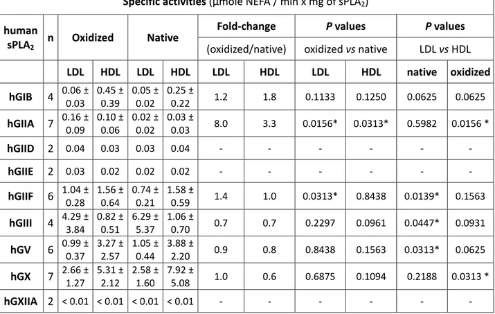
Oxidative modification of lipoproteins enhances hydrolysis by hGIIA and hGIIF but 115
not other sPLA2s — We previously reported the specific activities of different human sPLA2s 116
on native lipoproteins (Table 1 and (37)). To analyze the anti-Plasmodium activity of human 117
sPLA2s in a context more relevant to malaria where lipoproteins are oxidized (13), we 118
examined the capacity of various human sPLA2s to hydrolyze LDL and HDL particles after in 119
vitro oxidation.
120
Hydrolysis of lipoproteins was assessed by measuring the release of non-esterified 121
fatty acids (NEFAs). Seven of the 9 recombinant catalytically-active human sPLA2s hydrolyzed 122
oxidized and native LDL and HDL particles with the same specific activities (Table 1). In 123
contrast, hGIIA, and to a lower extent hGIIF, exhibited significantly higher activity on oxidized 124
lipoproteins. Oxidation of both LDL and HDL dramatically increased the activity of hGIIA 125
sPLA2 whereas oxidation of LDL but not HDL slightly increased the activity of hGIIF. A slight 126
fold-change was also observed for hGIB on LDL and HDL, but this change did not reach 127
significance. However, the specific activity of hGIIA on oxidized LDL and HDL remained lower 128
than that of hGIIF, hGIII, hGV and hGX sPLA2s, which are highly active on both native and 129
oxidized lipoproteins. 130
131
Oxidized LDL and HDL pretreated with human sPLA2s including hGIIA are active in
132
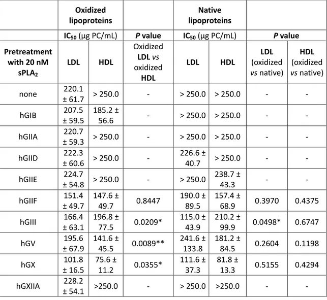
vitro against P. falciparum — We next compared the anti-Plasmodium activity of oxidized 133
lipoproteins before and after hydrolysis by recombinant human sPLA2s. Assays were carried 134
out with lipoproteins at concentrations close to the physiological ones (0.2 mg/mL of 135
phospholipids in the culture medium). Inhibitory concentrations of sPLA2-hydrolyzed LDL and 136
HDL against P. falciparum growth are presented in Table 2. Without sPLA2 pretreatment, 137
oxidized LDLs were barely inhibitory towards Plasmodium, whereas oxidized HDLs were not. 138
Pretreatment with 20 nM hGIIF, hGIII, hGV and hGX sPLA2s, but not other sPLA2s, increased 139
the toxicity of oxidized LDL and rendered oxidized HDL toxic to the parasite. However, 140
oxidized lipoproteins were not more potent than their native counterparts after hydrolysis. 141
Oxidized LDL were less potent than native LDL after hydrolysis by hGIII sPLA2. 142
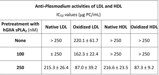
Since the circulating levels of hGIIA sPLA2 can be very high in severe cases of malaria 143
(29, 30) while pretreatment of oxidized lipoproteins with 20 nM hGIIA was ineffective against 144
P. falciparum, we evaluated the anti-Plasmodium potency of oxidized lipoproteins when
145
treated at higher concentrations. As shown in Table 3, pretreatment with 100 nM hGIIA 146
sPLA2 moderately increased the toxicity of oxidized LDL whereas 250 nM induced a marked 147
toxicity of both oxidized LDL and HDL and rendered native lipoproteins toxic. 148
149
The anti-Plasmodium activity of human sPLA2s including hGIIA is enhanced in culture
150
medium containing oxidized lipoproteins — To further analyze the effects of human sPLA2s 151
in conditions closer to physiology where all classes of lipoproteins are present, a total 152
lipoprotein fraction (oxidized or not) instead of purified LDL or HDL was used without 153
enzymatic pretreatment, and incubation was prolonged for 96 h to cover two parasite intra-154
erythrocytic cycles instead of one. As expected, hGIIF, hGV and hGX sPLA2s were inhibitory in 155
culture medium containing native lipoproteins (Table 4): hGIIF and hGX sPLA2s exhibited IC50 156
values of 19.8 and 1.5 nM, close to the IC50 values previously measured in human plasma 157
(10.7 nM and 2.9 nM, respectively (37)), whereas hGV sPLA2 was found to be more active 158
(IC50 = 21.9 nM) than in previous assays with plasma (IC50 = 94.2 nM, (37)). hGIIA sPLA2 was 159
not inhibitory in normal plasma (37) nor with native lipoproteins (Table 4). Remarkably, all 160
four sPLA2s had enhanced inhibitory activities in the presence of oxidized versus native 161
lipoproteins. hGV and hGX sPLA2s were 2-fold more active, and hGIIF sPLA2 was 4.6-fold 162
more active, consistent with its increased capacity to hydrolyze oxidized lipoproteins. hGIIA 163
sPLA2 was inhibitory in the presence of oxidized lipoproteins with an IC50 value of 150 nM 164
while it was completely inactive with native lipoproteins. 165
166
Oxidized PUFAs are not responsible for enhanced anti-Plasmodium activity of 167
sPLA2s ‒ To understand why sPLA2s exhibit greater anti-Plasmodium activities in assays with 168
oxidized lipoproteins, we tested whether oxidized PUFAs have greater toxicity than native 169
PUFAs. As far as we know, only one study by Kumaratilake et al. (46) reported the anti-170
Plasmodium activity of oxidized PUFAs, namely oxidized AA and DHA. The authors found that
171
these PUFAs were more active after oxidation. Assuming that oxidized phospholipids present 172
in lipoproteins can be hydrolyzed by sPLA2s such as hGIIA and hGX to release the 173
corresponding oxidized PUFAs (39, 47), we compared the effects of native and self-oxidized 174
AA, EPA and DHA on parasite growth in dose-response assays. When added to culture 175
medium containing human plasma, these PUFAs were inhibitory to P. falciparum in both 176
native and oxidized states (Table 5). The IC50 values for the three PUFAs in their native state 177
were in good accordance with those reported by Kumaratilake et al. (46). However, once 178
oxidized, they were not more efficient, exhibiting similar (AA) or even higher (EPA, DHA) IC50 179
values (Table 5). These results are thus different from those by Kumaratilake et al. who 180
reported a ≈ 1.5-fold (AA) and ≈ 2.0-fold (DHA) better capacity of these PUFAs to inhibit 181
Plasmodium after oxidation (46). The discrepancy might come from differences in the
182
amount and molecular identity of oxidation products as well as differences between 183
experimental settings for oxidation. Anyway, this indicates that there is no clear-cut 184
difference between the capacity of oxidized versus native PUFAs to inhibit Plasmodium 185
growth, and suggests that the enhancement of the anti-Plasmodium activity of sPLA2s in the 186
presence of oxidized lipoproteins might rather come from an increased activity of sPLA2s at 187
hydrolyzing oxidized particles and from prolonged incubation during the inhibition assay, 188
resulting in a greater production of various toxic lipids, beyond the oxidized ones. 189
190
Circulating levels of hGIIA sPLA2, but not hGIIF, hGV or hGX sPLA2s, are increased in
191
uncomplicated P. falciparum malaria — To provide a pathophysiological relevance of our in 192
vitro results, we tested the sPLA2 enzymatic activity and the presence of hGIIA, hGIIF, hGV 193
and hGX sPLA2s in plasma from 41 P. falciparum-infected Vietnamese adults versus 28 non-194
parasitized, healthy donors from the same geographic area. None of the parasitized 195
individuals presented signs of complicated malaria at the time of blood sampling. As shown 196
in figure 1A, sPLA2 enzymatic activity was dramatically increased in the plasma of P. 197
falciparum-infected patients relative to healthy donors (median (interquartile range: IQR):
198
260.0 (148.5-367.1) versus 16.20 (10.46-29.06) cpm/min x µL respectively, P value < 0.0001). 199
No correlation was found between the level of enzymatic activity and blood parasitaemia (P 200
value = 0.8079, Spearman’s r = 0.03969, not shown). 201
Plasma samples were next analyzed for the presence of hGIIA, hGIIF, hGV and hGX 202
sPLA2s by a sandwich ELISA-like method (Time-resolved fluoroimmunoassay, TR-FIA) using 203
specific antibodies and assays for each sPLA2 (27). Protein levels of hGIIA sPLA2 were 204
significantly increased in the plasma from parasitized patients (median (IQR): 1.6 (0.7-3.4) 205
nM, with a maximum value of 9.1 nM, as compared to the plasma from healthy donors 206
(median (IQR): 0.0 (0.0-0.1) nM) (P value <0.0001) (figure 1B). In contrast, hGIIF, hGV and 207
hGX sPLA2s were not detected in the parasitized plasma (not shown), indicating that these 208
enzymes do not circulate, at least at detectable levels, in uncomplicated malaria. In the 209
parasitized plasma, sPLA2 enzymatic activity and hGIIA protein concentration were highly 210
correlated (Figure 1C; P value <0.0001, Spearman’s r = 0.9542), indicating that hGIIA sPLA2 is 211
the predominant sPLA2 responsible for the circulating enzymatic activity. No correlation was 212
found between the plasma concentration of hGIIA and the parasitaemia from infected 213
donors (Figure 1D; P value = 0.6684, Spearman r = 0.0689). 214
215
Malaria but not normal plasma inhibits in vitro parasite growth when spiked with 216
recombinant hGIIA sPLA2.
217
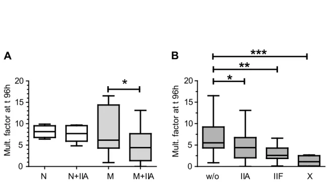
The capacity of hGIIA sPLA2 endogenously present in the plasma from malaria patients 218
at inhibiting Plasmodium development was evaluated in conventional in vitro culture 219
conditions. Ten plasma samples containing various levels of endogenous hGIIA sPLA2 up to 9 220
nM were tested (Figure 2A). It is important to note that in our culture conditions where 221
RPMI was supplemented with 8% human plasma, the final hGIIA concentrations were only in 222
the range of 0.1 to 1 nM. The growth rates of P. falciparum measured in the presence of 223
plasma from the different malaria patients was variable but not significantly lower than that 224
measured in the presence of plasma from different healthy donors (median growth rate 225
(IQR): 6.1 (4.3-14.4) in malaria plasma versus 8.1 (6.7-9.5) in normal plasma, P = 0.4278; 226
Figure 2A). We then evaluated the possible role of hGIIA sPLA2 present in the diluted plasma 227
by adding to the culture medium LY311727, a specific active site inhibitor of hGIIA sPLA2 (8). 228
Addition of LY311727 did not modify the parasite growth rate in the plasma samples 229
(multiplication factor after 96 h of cultivation: median (IQR): 7.08 (2.87-17.5) without 230
LY311727 versus 6.65 (4.14-10.7) with LY311727, P = 0.1550) (not shown). The absence of 231
effect of LY311727 i) indicates that the variable capacity of the different malaria plasma to 232
support parasite growth is not due to hGIIA, and ii) might be explained by too low 233
concentrations of endogenous hGIIA in our assay conditions to be effective at inhibiting the 234
growth of Plasmodium. To test this possibility, P. falciparum was grown for 96 h in plasma 235
from malaria patients versus healthy donors spiked with 100 nM recombinant hGIIA sPLA2. 236
This concentration was chosen based on hGIIA plasma concentrations reported in severe 237
cases of malaria (29, 30). As shown in Figure 2A, hGIIA spiked in malaria but not normal 238
plasma inhibited parasite development, demonstrating that 1) the concentration of 239
endogenous hGIIA in our assay conditions was not sufficient to inhibit parasite growth but 240
that higher concentrations were clearly effective, and 2) malaria-induced modifications of 241
plasma (possibly including oxidation of lipoproteins) is required to reveal the anti-242
Plasmodium activity of hGIIA. We also tested the spike of malaria plasma with 10 nM hGIIF
243
or 0.5 nM hGX recombinant sPLA2s for comparison. Both enzymes were inhibitory for P. 244
falciparum in malaria plasma and found more active than hGIIA sPLA2 (Figure 2B), as 245
anticipated from the in vitro results with human native plasma. 246
247
Lipoproteins from P. falciparum-infected plasma are oxidized — To add further 248
support to our hypothesis that the anti-Plasmodium activity of hGIIA sPLA2, and may be of 249
other sPLA2s,is promoted by the presence of oxidized lipoproteins in the plasma from 250
infected patients, we evaluated the level of lipoproteins and their oxidative status in malaria 251
plasma versus controls. In accordance with previous observations reporting a drop in HDL 252
content in patients' blood during malaria (48, 49), the level of lipoproteins, as measured by 253
total PC concentration was found to be lower in plasma from malaria patients as compared 254
to healthy donors (median (IQR): 1.20 (1.00-1.46) mg/mL versus 1.36 (1.19-1.68) mg/mL, P = 255
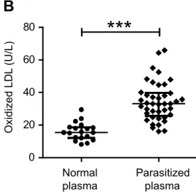
0.0158) (Figure 3A). Oxidation of lipoproteins in plasma was estimated using the specific 256
Mercodia oxidized LDL immunoassay kit (see methods). LDL oxidation was clearly increased 257
in the plasma from malaria patients as compared to healthy donors (median (IQR): 33.13 258
(25.65-39.79) vs 15.41 (12.13-18.69) U/L, P < 0.001) (Figure 3B). 259
Together, our results demonstrate that malaria-induced modifications of plasma 260
include oxidation of lipoproteins that most likely promotes the anti-Plasmodium activity of 261
hGIIA sPLA2 and enhances the inhibitory capacity of hGIIF and hGX sPLA2s, when spiked in 262
malaria plasma. 263
264
The anti-Plasmodium activity of hGIIA sPLA2 in malaria plasma requires its catalytic
265
activity — To demonstrate that the anti-Plasmodium activity of hGIIA sPLA2 in malaria 266
plasma requires enzymatic hydrolysis of oxidized lipoproteins, we first assessed the ability of 267
the enzyme to hydrolyze phospholipids from malaria plasma. Plasma samples were 268
incubated with hGIIA sPLA2 as well as hGX sPLA2, since this latter actively hydrolyzes both 269
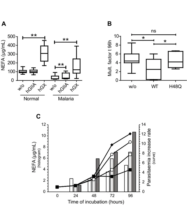
native and oxidized lipoproteins. hGIIA sPLA2 did not release NEFAs from healthy plasma, 270
whereas small but significant release occurred from malaria plasma (P = 0.0093) (Figure 4A). 271
As expected, hGX sPLA2 induced a net increase in NEFAs from both malaria (P = 0.0020) and 272
healthy donor (P = 0.0019) plasma (Figure 4A). 273
Second, we tested whether the enzymatic activity is essential for the anti-Plasmodium 274
effect of hGIIA sPLA2. Parasite growth inhibition assays were performed using the H48Q 275
catalytically-inactive mutant of hGIIA (<0.5% of WT enzymatic activity (50)). The parasite 276
development was not affected in malaria plasma spiked with the H48Q mutant, indicating 277
that hydrolysis of plasma phospholipids is required for parasite inhibition (Figure 4B). 278
Last, we analyzed the relationship between Plasmodium inhibition in malaria plasma 279
supplemented with hGIIA sPLA2 and the amount of NEFAs released by the enzyme in the 280
culture supernatant. We found that both parasite inhibition and NEFA amount in the culture 281
medium increase with the time of incubation and concentration of hGIIA sPLA2 (Figure 4C). 282
Interestingly, a small but noticeable inhibition of parasite development was observed in the 283
presence of 20 nM hGIIA beyond 48 h of incubation, suggesting a possible long-term effect 284
induced by relatively low concentrations of the enzyme. 285
Together, these results indicate that the anti-Plasmodium activity of hGIIA sPLA2 286
observed in the plasma from malaria patients is mediated by hydrolysis of oxidized 287
lipoproteins. 288
289
Recombinant hGIIA sPLA2 inhibits parasite development in Plasmodium-infected
290
mice only when injected at the time of plasma peroxidation. 291
To challenge our hypothesis that hGIIA sPLA2 might help controlling parasite 292
development in an in vivo situation where lipoprotein oxidation does occur, we injected 293
recombinant hGIIA sPLA2 to C57BL/6J mice infected with P. chabaudi at different time points 294
post-inoculation. The recombinant enzyme was injected twice daily for 3 consecutive days, 295
either at the onset of the patent phase (when the first parasites are observed on blood 296
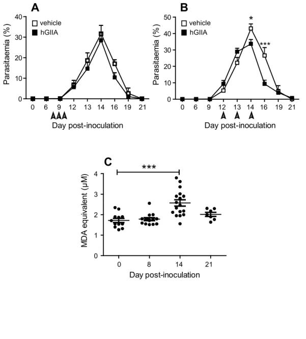
smears) or later, just prior to the parasitaemia peak. Interestingly, a significant decrease by 297
about 20% of the parasitaemia peak was observed after the late injection of hGIIA (Figure 298
5B), but not when hGIIA was injected at early time points (Figure 5A). 299
To assess the potential involvement of oxidized lipoproteins in the in vivo anti-300
Plasmodium effect of hGIIA, the plasma from infected mice was analyzed for lipid
301
peroxidation. The levels of TBARS were increased at day-14 following parasite inoculation 302
but not before (Figure 5C), i.e. at the time coincident with the one when injection of hGIIA 303
was found to be effective against Plasmodium infection. 304
DISCUSSION 306
Since the discovery in the early 90’s of an increase of hGIIA sPLA2 in the plasma from 307
malaria patients (29, 30), no comprehensive study on the potential role of this enzyme has 308
been made in the context of malaria physiopathology. Interestingly, an association of plasma 309
PLA2 enzymatic activity and related phospholipid products with brain swelling in pediatric 310
cerebral malaria has been reported in 2015 (51), suggesting a role of hGIIA sPLA2 in the 311
inflammatory events operating in cerebral malaria. More recently, a possible role for the 312
gene cluster encoding multiple sPLA2 (genes for hGIIA, hGIID, hGIIE, hGIIF and hGV sPLA2s) 313
has been suggested from results of a genome-wide association study performed on non-314
complicated malaria susceptibility (31), highlighting the possible role for one or more human 315
sPLA2s in malaria. 316
In line with the above findings, we provide here the first evidence that hGIIA sPLA2 and 317
maybe other sPLA2s might contribute as host defense factors to control Plasmodium 318
development. Our results indicate that hGIIA, as well as some other sPLA2s, might operate 319
through hydrolysis of lipoproteins, and more precisely oxidized lipoproteins that are 320
produced during the course of Plasmodium infection in malaria (13). This was demonstrated 321
by combining a series of in vitro studies on parasite infection, analysis of plasma from 322
malaria patients and finally by in vivo analysis of the anti-Plasmodium effect of hGIIA sPLA2 in 323
a mouse model of chronic malaria. 324
We previously demonstrated that hGIIF, hGIII, hGV and hGX but not hGIIA sPLA2s 325
inhibit the in vitro growth of P. falciparum through hydrolysis of native lipoproteins present 326
in human plasma (37). We now show that oxidized lipoproteins are also substrates for these 327
enzymes and participate to the inhibition of parasite growth, at least in vitro. Focusing on 328
hGIIA sPLA2 because of its presence in the plasma from malaria patients, we also show that i) 329
hGIIA hydrolyzes much more potently oxidized LDL and HDL than their native counterparts 330
(where no detectable hydrolysis was observed), a finding in accordance with previous results 331
(38, 39, 42), and ii) hGIIA exhibits anti-Plasmodium activity only in the presence of oxidized 332
lipoproteins. 333
In normal culture conditions, hGIIF, hGIII, hGV and hGX sPLA2s hydrolyze lipoprotein 334
phospholipids, producing toxic free fatty acids, mainly long chain PUFAs (37). It is known that 335
hGIIA sPLA2 has little activity on native lipoproteins but higher activity on oxidized 336
counterparts ((38, 39, 42) and this study). Identification and quantification of the 337
lysophospholipids and fatty acids produced upon hydrolysis of native lipoproteins by hGIIA 338
and other sPLA2s have been reported by us and others (37-39). It is known also that hGIIA 339
sPLA2 hydrolyzes oxidized lipoproteins to generate non-oxidized lipids and likely oxidized 340
products (38, 39, 41, 42). In vitro and in vivo antimalarial properties of native as well as 341
oxidized lipids, especially PUFAs, has been reported elsewhere (46, 52). Kumaratilake et al. 342
have shown that oxidized PUFAs are more active than native PUFAs against P. falciparum in 343
vitro (46). However, we did not find evidence that oxidized PUFAs are more toxic than native
344
PUFAs, yet PUFAs were indeed toxic. These differences might be due to experimental 345
variations between the two studies, and obviously, additional investigations will be 346
necessary to solve the question of the role of oxidized lipids in the anti-Plasmodium activities 347
of sPLA2s. Considering the other sPLA2s, it is interesting to note that hGIIF, hGV and hGX 348
sPLA2s displayed increased capacities to inhibit Plasmodium when incubated with oxidized 349
lipoproteins from the whole lipoprotein fraction and for a long incubation time (96 h, i.e. 350
two parasite cycles), whereas LDL or HDL first purified, and then oxidized and prehydrolyzed 351
with 20 nM of those sPLA2s did not exhibit greater toxicity than the native ones in the 352
classical 48 h inhibitory assay. This indicates that experimental conditions are crucial to 353
reveal the anti-Plasmodium properties of sPLA2s, and suggests that a specific 354
pathophysiological environment is a key factor in promoting their enzymatic and biological 355
activities. In line with our findings, many studies have now shown that hGIIA sPLA2 is only 356
active when acting on specific "non-cellular" and cellular phospholipid substrates, including 357
microparticles, exosomes or free mitochondria released by activated human platelets or 358
other immune cells, or damaged and apoptotic cells after oxidation or scramblase-mediated 359
phosphatidylserine externalization (5, 53-64). 360
We measured concentrations of hGIIA sPLA2 up to 9 nM in the plasma from a 361
Vietnamese cohort of 41 patients with uncomplicated malaria. These concentrations are 362
consistent with a previous study by Vadas et al in a small cohort of 14 Canadian adult 363
patients with uncomplicated malaria (29), where the mean level of hGIIA sPLA2 was 12.1 ± 364
4.8 nM (169.7 ± 67.3 ng/mL). We found that P. falciparum develops normally in such malaria 365
plasma (diluted to 8%, i.e. at a relatively low concentration of hGIIA in the nM range) 366
although it contains oxidized LDL. In a subsequent study by Vadas et al on Malawian children 367
with severe malaria, the mean level of hGIIA sPLA2 was estimated to be around 70 nM, with 368
the most severe cases exhibiting levels up to 200 nM (30). Of note, in these studies including 369
ours, plasma were likely collected at different times during the course of infection, and it is 370
not known how varies the concentration of hGIIA during the infection and according to levels 371
of parasitaemia. In line with such high concentrations, we show that addition of 100 nM 372
hGIIA sPLA2 to uncomplicated malaria plasma induces significant parasite inhibition, whereas 373
the same addition of hGIIA to normal plasma is ineffective. Furthermore, it should be 374
considered that hGIIA sPLA2 has high affinity for various heparan sulfate proteoglycans lining 375
the vascular endothelium and various cells, which likely acts as a reservoir of hGIIA in the 376
circulation, and may lead to an underestimation of the actual concentration of hGIIA in 377
blood (65-68). This is supported by the study by Nakamura et al. where the hGIIA sPLA2 378
activity measured in plasma was increased by about 3-fold between heparinized and non-379
heparinized patients (69). This indicates that conventional collection of plasma or serum 380
without heparin pretreatment leaves behind numbers of hGIIA molecules bound to the 381
heparan sulfates in the vasculature or circulating immune cells. With these data in mind, the 382
100 nM concentration of hGIIA sPLA2 used in our in vitro experiments may not be that far 383
from the concentration in the vascular compartment of malaria patients, especially in severe 384
cases of malaria. A last point to consider when comparing our in vitro data with what might 385
occur in vivo is the fact that hGIIA needs an appropriate substrate to be effective, in our case 386
oxidized lipoproteins. Besides the various levels of hGIIA circulating in patients, the quality 387
and quantity of this particular substrate might also vary among individuals, leading to 388
variable anti-Plasmodium host response. 389
By investigating the effects of injection of recombinant hGIIA sPLA2 to parasitized mice, 390
we show that Plasmodium development is reduced in those mice. Interestingly, the enzyme 391
is effective only when injected in the late course of infection (at days 12-14 p.i.) and not at 392
earlier time points (at days 8-10 p.i.). Consistent with the in vitro results, plasma 393
peroxidation was increased at day 14 but not at day 8, in line with the time window at which 394
the injected hGIIA sPLA2 is effective against Plasmodium in parasitized mice. 395
It must be noticed that the drop in parasitaemia induced by injection of hGIIA is rather 396
modest, i.e. about 20%. This may reflect a transient effect of the injected enzyme due to its 397
rapid in vivo clearance or capture by the vascular wall components, and hence lower 398
bioavailability of the soluble enzyme at hydrolyzing plasma oxidized lipoproteins. This 399
situation may differ from that in infected patients where the enzyme is continuously 400
produced by the sustained inflammation and circulates in plasma at steady state high levels. 401
The use of transgenic mice continuously overexpressing the human enzyme may be more 402
relevant in this context, as they were previously used to demonstrate the antibacterial effect 403
of hGIIA sPLA2 in vivo (16-18, 70-72) and have been shown to have altered lipoprotein 404
profiles (73, 74). 405
One may also consider that the in vivo antimalarial activity of hGIIA sPLA2 results from 406
multiple mechanisms of action, with the release of PUFAs directly toxic to the parasite being 407
only one among several mechanisms. Indeed, there is considerable evidence that hGIIA 408
sPLA2 is an important effector of the innate immune response and may activate platelets or 409
neutrophils among different immune cells involved in malaria (22, 62, 63, 75, 76). The 410
production of lipid mediators by sPLA2s, as an integral component of the inflammatory 411
reaction, plays a major role in protecting the host against invading pathogens (3, 5, 22). 412
However, lipid products might also induce deleterious effects to the host, as exemplified by 413
recent metabolomics analyses showing a positive association between lipid products of the 414
PLA2 pathway and brain swelling in pediatric cerebral malaria (51). 415
Concerning the possible in vivo role of other human sPLA2s, we showed that 416
exogenous hGIIF and hGX sPLA2s are active against Plasmodium in plasma from malaria 417
patients as well as healthy donors, in accordance with their ability to hydrolyze lipoproteins 418
under native or oxidized states. These sPLA2s were however not detected in the plasma of P. 419
falciparum-infected or healthy subjects. It is tempting to speculate that they may act locally
420
at sites relevant to Plasmodium infection. Indeed, while there is now considerable evidence 421
that hGIIA sPLA2 is the only sPLA2 highly present in the circulation under pathological 422
conditions associated with inflammation, tissue injury or infection, there is little evidence for 423
the presence of other sPLA2 isoforms in serum, except for hGIB sPLA2 in some disease 424
situations (27, 77). However, several sPLA2s other than hGIIA are also involved in various 425
types of infection, suggesting that these sPLA2s exert their effects in diverse pathologies 426
locally, within the microenvironment of the disease, and either in an autocrine or paracrine 427
manner (3, 5, 6). Examples include studies on the role of GIII, GV and GX sPLA2s after 428
infection with various pathogens (19, 21, 78-81), GIII and GX sPLA2s in cancer (6, 82, 83), or 429
GIIF, GIII, GV and GX sPLA2s in cardiovascular or skin diseases, and allergy or asthma (84-89). 430
It is also interesting to note that hGX sPLA2 has been found in atherosclerotic plaques but 431
not in plasma from patients with atherosclerosis (90), suggesting that the enzyme is 432
produced locally but not systemically. All together, it is possible that sPLA2s other than hGIIA 433
also contribute to malaria within the vascular wall or via local secretion from immune cells, 434
even though these enzymes are not detected in plasma. 435
In summary, we have shown that the concomitant presence of high concentrations of 436
hGIIA sPLA2 and oxidation of lipoproteins, as found in the serum of malaria patients, might 437
participate to the host defense mechanism against P. falciparum. The mechanism of action 438
would be based on a synergistic effect between the enzyme and its capacity to hydrolyze 439
oxidized lipoproteins serving as one of its preferred substrates to release toxic lipids for 440
Plasmodium, yet other indirect mechanisms may occur. By extrapolation, this mechanism of
441
action of hGIIA on oxidized lipoproteins might also be involved in other pathological 442
conditions where both hGIIA sPLA2 and oxidized lipoproteins are present, including 443
atherosclerosis or other infectious diseases. It also remains to determine if other sPLA2s such 444
as hGIIF, hGIII, hGV and hGX which are active in vitro, may also be active in an in vivo 445
situation, even though these enzymes are not found in plasma from malaria patients. 446
MATERIALS AND METHODS 447
Materials — The FcB1 strain of the human Plasmodium: P. falciparum (Columbia) and the 448
864VD strain of the murine Plasmodium: P. chabaudi chabaudi used in this work was from 449
the Unicellular Eukaryotes Collection from the National Museum of Natural History (MNHN-450
CEU-224-PfFcB1). The 864VD strain of the murine Plasmodium P. chabaudi chabaudi was 451
from the MCAM Research Unit’s Plasmodium collection. C57BL/6JOlaHsd mice for the in vivo 452
assays were from Envigo RMS SARL (Gannat, 03800 France). Plasma and red blood cells for 453
the in vitro studies were from the O– or A+ blood groups and were supplied by the French 454
National Agency for Blood (Etablissement Français du Sang (EFS)), convention reference: C 455
CPSL UNT 13/EFS/126. RPMI 1640 and Albumax II® were from Life Technologies (Cergy 456
Pontoise, France). Diff-Quick staining reagents were from Medion Diagnostics AG. 457
The NEFA-C and the Phospholipid B kits, respectively used for quantitative 458
determination of non-esterified fatty acids (NEFAs) and phospholipids, were from WAKO 459
Chemicals (Oxoid S.A., Dardilly, France). The oxidized LDL ELISA kit was from Mercodia SAS 460
sales in France. Purified recombinant human sPLA2s and the hGIII sPLA2 domain were 461
prepared as described (91). The sPLA2 inhibitor LY311727 (3-{[3-(2-Amino-2-oxoethyl)-2-462
ethyl-1-(phenylmethyl)-1H-indol-5-yl]oxy}propyl]-phosphonic acid (164083-84-5)) that 463
targets hGIIA sPLA2 was from Sigma. 464
The polyunsaturated fatty acids (PUFAs): arachidonic acid (AA, C20:4, n-6), 465
eicosapentaenoic acid (EPA, C20:5, n-3) and docosahexaenoic acid (DHA, C22:6, n-3) were 466
from Cayman Chemicals (Interchim). 3H-hypoxanthine monohydrochloride (370 GBq-1.11 467
TBq/mmol) was from Perkin-Elmer. Other high quality grade biochemical reagents were 468
from Sigma. 469
Methods 471
P. falciparum cultivation — In routine culture conditions, P. falciparum was grown in 472
red blood cells from the A+ group at 2% haematocrit and 2-4% parasitaemia, in RPMI 473
supplemented with 11 mM glucose, 27.5 mM NaHCO3, 100 UI/mL penicillin, 100 µg/mL 474
streptomycin, adjusted to pH 7.4 (basic medium), supplemented with 8% heat-inactivated 475
human A+ plasma (complete medium), according to the procedure of Trager and Jensen (92). 476
When specified, the serum substitute Albumax II® (0.5% w/v final) was used in culture 477
medium instead of heat-inactivated human plasma. Culture flasks were gassed with 91% N2, 478
6% O2 and 3% CO2 before being incubated at 37°C. In those conditions, the intra-erythrocytic 479
cycle of the FcB1 strain was 48-h long. Parasitaemia (%) was established by optical 480
examination of Diff-Quik-stained smears from the formula = [infected erythrocytes number / 481
total erythrocytes number] x 100. 482
483
Purification of lipoproteins and oxidation — Non-fasted human plasma was split into 484
aliquots and frozen at -20°C. One aliquot was thawed to prepare LDL and HDL by differential 485
centrifugation, according to Havel et al. (93). Briefly, chylomicrons and VLDL were removed 486
by a first round of centrifugation at 1.006 g/mL density, then LDL and HDL were purified 487
separately by successive centrifugations at 1.053 g/mL and 1.210 g/mL densities, 488
respectively. When specified, total lipoprotein fraction was purified by a single run of plasma 489
at 1.210 g/mL density. Lipoproteins were dialyzed at 4°C against NaCl 9 g/L, then against 490
RPMI, and sterilized by 0.2 µm filtration. Oxidation was achieved by storing lipoproteins in a 491
transparent flask at room temperature under sterile air exchange for 14 days. Native 492
lipoproteins were prepared from another aliquot of the plasma just prior to assays. 493
Experiments were performed within 1 week of lipoprotein storage at 4°C under N2 in the 494
dark. Phosphatidylcholine (PC) content of lipoproteins was measured by using the 495
Phospholipid B dosage kit, according to the manufacturer's instructions. 496
497
Oxidation of PUFAs — Concentrated solutions of commercial AA (76.5 mM), EPA (60 498
mM) and DHA (100 mM) in ethanol were stored frozen under N2. An aliquot of 20 µL of each 499
PUFA was allowed to dry in a glass tube and then exposed to air and light for 1 week under 500
sterile conditions. Dried PUFAs were re-suspended into 100 µL CHCl3, and then diluted 1/20 501
in RPMI containing 8% heat-inactivated human plasma. 502
Prior to dose-response assays, non-oxidized PUFAs were prepared from another 20 µL 503
aliquot of each concentrated solution, mixed to 80 µL of CHCl3 and diluted into culture 504
medium as above. 505
506
Measurement of lipid oxidation in lipoproteins and plasma — The level of TBARs 507
(thiobarbituric acid reactive substances) was determined as a marker of lipid peroxidation in 508
purified human lipoproteins (94, 95). Briefly, 150 µL of diluted sample containing 0.2% 509
butylated hydroxytoluene was mixed with an equal volume of 0.25 N HCl containing 15% 510
trichloroacetic acid and 0.4% thiobarbituric acid. The mixture was heated at 85°C for 15 511
minutes and then cooled on ice. 150 µL of butanol was added, the sample was vortexed, and 512
then let stand on ice for phase separation to occur. The butanol phase was taken for TBARs 513
determination at 515 nm excitation and 550 nm emission wavelengths on a 514
spectrofluorometer (AMINCO-Bowman series 2). 1,1,3,3-tetraethoxypropane was used as an 515
external standard. Three independent measurements with plasma lipoproteins from 516
different donors were performed. Before oxidation, TBARS ranged between 0.30-0.50 and 517
0.05-0.15 nmoles of malondialdehyde (MDA)/mg of protein in LDL and HDL, respectively. 518
After two weeks of air-light oxidation, TBARS concentration had raised to 1.50-2.50 (LDL) and 519
0.30-0.50 (HDL) nmoles of MDA/mg of protein. For comparison, TBARS in LDL and HDL 520
purified from the serum of malaria cases were reported to be around 1.0 and 0.2 nmol/mg 521
of protein, respectively (13). 522
Due to the presence in plasma of substances other than MDA and susceptible to react 523
with thiobarbituric acid (96), the level of lipoprotein oxidation in crude human plasma was 524
determined by quantifying oxidized LDL using the Ox-LDL ELISA kit from Mercodia, a 525
sandwich ELISA based on the mouse monoclonal antibody 4E6, which is directed against a 526
conformational epitope in oxidized ApoB-100. The ELISA assay was performed according to 527
the manufacturer’s instructions. Oxidation of mouse plasma was measured by quantifying 528
TBARS because the Mercodia kit is not appropriate for mouse plasma. 529
530
Hydrolysis of lipoproteins and plasma by sPLA2 — Native and oxidized LDL and HDL
531
purified from the same batch of human plasma were adjusted to 1 mg phospholipid/mL in 532
RPMI supplemented with 1 mM CaCl2. They were incubated with and without recombinant 533
sPLA2 for various times at 37°C. sPLA2s were used at different concentrations. hGIB was used 534
at 100 nM (number of independent experiments, n = 4), hGIIA was used at 50, 100 and 250 535
nM (n = 7), hGIID was used at 100 and 200 nM (n = 2), hGIIE was used at 200 and 250 nM (n 536
= 2), hGIIF was used at 30 and 40 nM (n = 6), hGIII was used at 50 and 100 nM (n = 4), hGV 537
was used at 10, 40 and 50 nM (n = 6), hGX was used at 5, 10 and 20 nM (n = 7), hGXIIA was 538
used at 100 and 200 nM (n = 2). NEFAs were measured at different time points using the 539
NEFA-C kit (WAKO) following manufacturer's instructions. Values were normalized by 540
subtracting NEFAs measured from lipoproteins incubated without sPLA2. Specific activity was 541
deduced from the linear part of the curve [NEFA] = f (t). 542
To assess the ability of hGIIA and hGX sPLA2s to hydrolyze phospholipids in plasma 543
from P. falciparum-infected patients versus healthy donors, 1 mM final CaCl2 was added to 544
each plasma sample and then enzymes were added at 400 nM (hGIIA) and 8 nM (hGX) final 545
concentration. Samples were incubated overnight at 37°C. Control for endogenous release of 546
NEFAs was without added sPLA2. NEFA content before and after incubation was determined 547
using the NEFA-C kit, according to manufacturer’s instructions. 548
549
Anti-Plasmodium activity assays with sPLA2-hydrolyzed lipoproteins — The ability of
550
human sPLA2s to promote the toxicity of lipoproteins was tested in dose-response assays as 551
described (37). Briefly, LDL and HDL (0.6 mg phospholipid/mL in RPMI) were incubated 552
overnight at 37°C with 20 nM sPLA2 or alone, then tested for parasite inhibition at a LDL and 553
HDL final concentration of 0.2 mg/mL (ie close to physiological concentrations) in RPMI 0.5% 554
Albumax II® for 48h in normal culture conditions. Albumax II® instead of human plasma was 555
used throughout the test to avoid any contribution of lipoproteins from human plasma. 556
Incubations with each sPLA2 added alone were also performed to check for lipoprotein-557
independent toxicity of sPLA2s in these conditions. Assays analyzing the anti-Plasmodium 558
effect of lipoproteins pre-treated with higher concentrations of hGIIA sPLA2 (100 nM and 559
250 nM) were performed similarly. Assays analyzing the anti-Plasmodium effect of sPLA2s co-560
incubated with total lipoprotein fraction but without pretreatment were carried out on two 561
parasite cycles (i.e. 96 h). 562
563
Anti-Plasmodium activity assays with oxidized PUFAs ‒ Dose-response assays with 564
oxidized and non-oxidized AA, EPA and DHA were performed as described in (37) with some 565
modifications. Decreasing concentrations of each PUFA were established by 3-fold dilution 566
steps in culture medium, distributed in a 96-well plate (50 µL/well) and mixed with a culture 567
of P. falciparum (0.25% parasitaemia, 4% haematocrit, 50 µL/well). Parasites were allowed to 568
grow for 48 h in normal culture conditions, then 9.25 kBq 3H-hypoxanthine were added per 569
well. After an additional 48 h incubation period, plates were frozen and the experiment 570
proceeded as in (37). 571
572
Human plasma collection in Vietnam — Venous whole blood samples were collected 573
in Acid Citrate Dextrose (ACD) (BD, India) vacutainers from patients attending commune 574
health centers in the Binh Phuoc province, VietNam. Malaria positivity was evaluated by 575
using the rapid diagnostic test OptiMAL-IT (DiaMed AG, Switzerland). All infected patients
576
had P. falciparum malaria. The final study population included 41 individuals with malaria 577
and 28 healthy donor controls from the same area. From the malaria-infected cohort, there 578
were 33 men and 8 women, with an average age of 27.1 years (range 16-58 years). 579
Blood samples were centrifuged at 2,400 g for 5 min at room temperature and the 580
plasma was immediately stored at -80°C. Plasma samples were then shipped to the Museum 581
(Paris, France) where they were thawed on ice, aliquoted and stored at -80°C until use. 582
The study was approved by the VietNam People’s Army Department of Military 583
Medicine. The purpose of the study was explained to participants in their own language, 584
and oral consent was obtained. Positive patients were treated with dihydroartemisinin-585
piperaquine combination in accordance with the national drug policy of VietNam. 586
587
Time-resolved fluoroimmunoassays (TR-FIA) — TR-FIA assays for hGIIA, hGIIF, hGV 588
and hGX sPLA2s in plasma were performed as described earlier (27). The assays use rabbit 589
polyclonal anti-sPLA2 antibodies to set up ELISA-like sandwiches, which were shown to be 590
highly specific for each sPLA2 isoform (97). hGIII sPLA2 could not be measured, due to the 591
lack of sensitivity of the corresponding immune serum. 592
593
Enzymatic assays on E. coli membranes — sPLA2 activity in plasma was measured by 594
hydrolysis of E. coli membranes radiolabeled with [3H]-oleic acid and autoclaved (98). Briefly, 595
40 µL of radiolabeled E. coli membranes (100,000 dpm in activity buffer consisting of 0.1 M 596
Tris-HCl pH 8.0, 10 mM CaCl2, 0.1% BSA) were incubated with plasma (20 µL, diluted 1:150 in 597
activity buffer) at 37°C for 30 minutes. Enzymatic assays were stopped by adding 80 µL of 0.1 598
M EDTA/0.2% fatty acid-free BSA. Reactions were spun down for 5 min at 10,000 g, and the 599
supernatant was collected and counted in a 1450 Microbeta counter (Wallac, Perkin Elmer). 600
601
P. falciparum cultivation in malaria plasma and related assays — To analyze the anti-602
Plasmodium effect of endogenous sPLA2 in plasma from infected people, the FcB1 strain was 603
grown in red blood cells of the O- group, brought to 0.1-0.2% parasitaemia, washed and then 604
transferred at 2% haematocrit into basic RPMI supplemented with 2.5 µM hypoxanthine and 605
1 mM CaCl2 (RPMI-calcium). The cell suspension was distributed into wells of a 96-well 606
microplate (100 µL/well) and then 8 µL of plasma from an infected donor or control normal 607
plasma was added per well. To avoid thermal denaturation of the endogenous sPLA2 (23, 99), 608
plasma samples were not heat-inactivated prior to parasite cultivation. The microplate was 609
incubated in a candle jar for 96 h, then parasitaemia in each well was determined from Diff-610
Quik-stained smears. 611
To assess for parasite inhibition by exogenously added hGIIA sPLA2, experiment was 612
carried out as above, with recombinant hGIIA added at the specified concentration at the 613
start of incubation. To evaluate the contribution of hGIIA catalytic activity, recombinant 614
hGIIA sPLA2 in its native (WT) or catalytically inactive form (H48Q mutant)(50), and/or the 615
specific hGIIA inhibitor LY311727, were added to the culture at 100 nM (hGIIA WT and 616
H48Q) and 10 µM (LY311727) at the start of incubation. 617
618
In vivo assays with Plasmodium chabaudi-infected mice 619
Mice were housed in the National Museum of Natural History (MNHN) animal facilities 620
accredited by the French Ministry of Agriculture for performing experiments on live rodents. 621
Work on animals was performed in compliance with French and European regulations on 622
care and protection of laboratory animals (EC Directive 2010/63/UE, French Law 2013-118, 623
February 1st, 2013). All experiments were approved by the MNHN’s Ethics Committee and 624
registered under the deposit n° APAFIS 201802281454598. 625
C57BL/6J mice (males, 8 to 12-week-old) were inoculated intraperitoneally (IP) with 626
1x10⁶ P. c. chabaudi 864VD-infected mouse red blood cells (RBC) in Alsever’s solution. Tail 627
blood was taken every 2-3 days within 21 days following inoculation. Parasitaemia was 628
established by optical examination of Diff-Quik-stained blood smears and counting of 2,000 629
RBCs. 630
Parasitized mice were IP-injected with 100 µL of recombinant hGIIA (0.125 mg/kg) in 631
PBS containing 1% mouse serum. Injections were performed twice daily for 3 days, either at 632
the beginning of the patent phase (early injection) or later, i.e. prior to the parasitaemia 633
peak (late injection). Blood parasitaemia was determined every 2-3 days within 22 days 634
following P. chabaudi inoculation. Three independent experiments were performed: one 635
early injection experiment with 5 control mice and 6 hGIIA-injected mice, and two late 636
injection experiments, with, respectively, 5 (control) and 7 (injected) mice, and 5 (control) 637
and 6 (injected) mice. 638
To measure plasma peroxidation, mice were sacrificed by exsanguination at day 0 639
before parasite inoculation and at days: 8, 14 and 21 post-inoculation (p.i.). Blood was 640
recovered into EDTA-coated tubes and centrifuged at 800 g for 15 minutes at room 641
temperature. Plasma was frozen at -80°C before TBARS measurement. 642
643
Statistical analysis — Data were analyzed using the GraphPad Prism software (San 644
Diego, CA). Normality of groups with n numbers superior to 6 was tested using the Shapiro-645
Wilk test. When sampling distribution was normal, the parametric unpaired or paired t-test 646
with a two-tailed P value was applied. When sampling distribution was not normal and/or 647
the n number was too small (≤ 6), the non-parametric Mann-Whitney U test for independent 648
samples or the Wilcoxon matched-pair test for dependent samples were used. The effect of 649
hGIIA sPLA2 injection to P. chabaudi-infected C57BL/6 mice was analyzed by using the two-650
way ANOVA test with Bonferroni post-test. Statistical analysis of plasma peroxidation level in 651
the course of infection was performed using the one-way ANOVA with Dunn’s multiple 652
comparison test. Differences with p values <0.05 (*), <0.01 (**) and <0.001 (***) were 653
considered as statistically significant. 654
655
ACKNOWLEDGMENTS 656
Funding. This work was supported by the ATM blanche 2016-2017 to C. Deregnaucourt 657
from the National Museum of Natural History (Paris, France) and by grants to GL from CNRS, 658
the National Research Agency (MNaims ANR-17- CE17-0012-01) and the Fondation de la 659
Recherche Médicale (DEQ20180339193). 660
REFERENCES 662
1. Cowman AF, Healer J, Marapana D, Marsh K. 2016. Malaria: Biology and Disease. Cell 663
167:610-624. 664
2. Lambeau G, Gelb MH. 2008. Biochemistry and Physiology of Mammalian Secreted 665
Phospholipases A2. Annu Rev Biochem 77:495-520. 666
3. Murakami M, Taketomi Y, Girard C, Yamamoto K, Lambeau G. 2010. Emerging roles of 667
secreted phospholipase A2 enzymes: Lessons from transgenic and knockout mice. 668
Biochimie 92:561-82. 669
4. Dennis EA, Cao J, Hsu YH, Magrioti V, Kokotos G. 2011. Phospholipase A2 enzymes: 670
physical structure, biological function, disease implication, chemical inhibition, and 671
therapeutic intervention. Chem Rev 111:6130-85. 672
5. Murakami M, Taketomi Y, Miki Y, Sato H, Yamamoto K, Lambeau G. 2014. Emerging 673
roles of secreted phospholipase A enzymes: The 3rd edition. Biochimie 107PA:105-674
113. 675
6. Murakami M, Sato H, Miki Y, Yamamoto K, Taketomi Y. 2015. A new era of secreted 676
phospholipase A(2). J Lipid Res 56:1248-61. 677
7. Valentin E, Lambeau G. 2000. Increasing molecular diversity of secreted 678
phospholipases A2 and their receptors and binding proteins. Biochim Biophys Acta 679
1488:59-70. 680
8. Singer AG, Ghomashchi F, Le Calvez C, Bollinger J, Bezzine S, Rouault M, Sadilek M, 681
Nguyen E, Lazdunski M, Lambeau G, Gelb MH. 2002. Interfacial kinetic and binding 682
properties of the complete set of human and mouse groups I, II, V, X, and XII secreted 683
phospholipases A2. J Biol Chem 277:48535-48549. 684
9. Masuda S, Murakami M, Ishikawa Y, Ishii T, Kudo I. 2005. Diverse cellular localizations 685
of secretory phospholipase A2 enzymes in several human tissues. Biochim Biophys 686
Acta 1736:200-210. 687
10. Vadas P, Pruzanski W. 1993. Induction of group II phospholipase A2 expression and 688
pathogenesis of the sepsis syndrome. Circ Shock 39:160-7. 689
11. Weinrauch Y, Abad C, Liang NS, Lowry SF, Weiss J. 1998. Mobilization of potent 690
plasma bactericidal activity during systemic bacterial challenge. Role of group IIA 691
phospholipase A2. J Clin Invest 102:633-638. 692
12. Koprivnjak T, Weidenmaier C, Peschel A, Weiss JP. 2008. Wall Teichoic Acid Deficiency 693
in Staphylococcus aureus Confers Selective Resistance to Mammalian Group IIA 694
Phospholipase A2 and Human alpha-Defensin 3. Infection and Immunity 76:2169-695
2176. 696
13. Sibmooh N, Yamanont P, Krudsood S, Leowattana W, Brittenham G, Looareesuwan S, 697
Udomsangpetch R. 2004. Increased fluidity and oxidation of malarial lipoproteins: 698
relation with severity and induction of endothelial expression of adhesion molecules. 699
Lipids Health Dis 3:15. 700
14. Fenard D, Lambeau G, Valentin E, Lefebvre JC, Lazdunski M, Doglio A. 1999. Secreted 701
phospholipases A2, a new class of HIV inhibitors that block virus entry into host cells. 702
J Clin Invest 104:611-618. 703
15. Koduri RS, Gronroos JO, Laine VJ, Le Calvez C, Lambeau G, Nevalainen TJ, Gelb MH. 704
2002. Bactericidal properties of human and murine groups I, II, V, X, and XII secreted 705
phospholipases A2. J Biol Chem 277:5849-5857. 706
16. Piris-Gimenez A, Paya M, Lambeau G, Chignard M, Mock M, Touqui L, Goossens PL. 707
2005. In vivo protective role of human group IIA phospholipase A2 against 708
experimental anthrax. J Immunol 175:6786-6791. 709
17. Movert E, Wu Y, Lambeau G, Kahn F, Touqui L, Areschoug T. 2013. Secreted Group IIA 710
Phospholipase A2 Protects Humans Against the Group B Streptococcus: Experimental 711
and Clinical Evidence. J Infect Dis 208:2025-35. 712
18. Pernet E, Guillemot L, Burgel PR, Martin C, Lambeau G, Sermet-Gaudelus I, Sands D, 713
Leduc D, Morand PC, Jeammet L, Chignard M, Wu Y, Touqui L. 2014. Pseudomonas 714
aeruginosa eradicates Staphylococcus aureus by manipulating the host immunity. Nat 715
Commun 5:5105. 716
19. Kim JO, Chakrabarti BK, Guha-Niyogi A, Louder MK, Mascola JR, Ganesh L, Nabel GJ. 717
2007. Lysis of human immunodeficiency virus type 1 by a specific secreted human 718
phospholipase A2. J Virol 81:1444-1450. 719
20. Nevalainen TJ, Graham GG, Scott KF. 2008. Antibacterial actions of secreted 720
phospholipases A2. Review. Biochim Biophys Acta 1781:1-9. 721
21. Balestrieri B, Maekawa A, Xing W, Gelb MH, Katz HR, Arm JP. 2009. Group V secretory 722
phospholipase A2 modulates phagosome maturation and regulates the innate 723
immune response against Candida albicans. J Immunol 182:4891-4898. 724
22. Dore E, Boilard E. 2019. Roles of secreted phospholipase A2 group IIA in 725
inflammation and host defense. Biochim Biophys Acta Mol Cell Biol Lipids 1864:789-726
802. 727
23. Paganelli FL, Leavis HL, He S, van Sorge NM, Payre C, Lambeau G, Willems RJL, 728
Rooijakkers SHM. 2018. Group IIA-Secreted Phospholipase A2 in Human Serum Kills 729
Commensal but Not Clinical Enterococcus faecium Isolates. Infect Immun 86. 730
24. van Hensbergen VP, Movert E, de Maat V, Luchtenborg C, Le Breton Y, Lambeau G, 731
Payre C, Henningham A, Nizet V, van Strijp JAG, Brugger B, Carlsson F, McIver KS, van 732
Sorge NM. 2018. Streptococcal Lancefield polysaccharides are critical cell wall 733
determinants for human Group IIA secreted phospholipase A2 to exert its bactericidal 734
effects. PLoS Pathog 14:e1007348. 735
25. Edgar RJ, van Hensbergen VP, Ruda A, Turner AG, Deng P, Le Breton Y, El-Sayed NM, 736
Belew AT, McIver KS, McEwan AG, Morris AJ, Lambeau G, Walker MJ, Rush JS, 737
Korotkov KV, Widmalm G, van Sorge NM, Korotkova N. 2019. Discovery of glycerol 738
phosphate modification on streptococcal rhamnose polysaccharides. Nat Chem Biol 739
15:463-471. 740
26. Gronroos JO, Laine VJ, Nevalainen TJ. 2002. Bactericidal group IIA phospholipase A2 741
in serum of patients with bacterial infections. J Infect Dis 185:1767-1772. 742
27. Nevalainen TJ, Eerola LI, Rintala E, Laine VJ, Lambeau G, Gelb MH. 2005. Time-743
resolved fluoroimmunoassays of the complete set of secreted phospholipases A2 in 744
human serum. Biochim Biophys Acta 1733:210-223. 745
28. Tan TL, Goh YY. 2017. The role of group IIA secretory phospholipase A2 (sPLA2-IIA) as 746
a biomarker for the diagnosis of sepsis and bacterial infection in adults-A systematic 747
review. PLoS One 12:e0180554. 748
29. Vadas P, Keystone J, Stefanski E, Scott K, Pruzanski W. 1992. Induction of circulating 749
group II phospholipase A2 expression in adults with malaria. Infection and Immunity 750
60:3928-3931. 751
30. Vadas P, Taylor TE, Chimsuku L, Goldring D, Stefanski E, Pruzanski W, Molyneux ME. 752
1993. Increased serum phospholipase A2 activity in Malawian children with 753
falciparum malaria. Am J Trop Med Hyg 49:455-459. 754