Biophysical and Structural Characterization of Components
From The Nuclear Pore Complex and the Ubiquitin Pathway
by
James R. Partridge
B.S. University of California at DavisDavis, CA 2004
SUBMITTED TO THE DEPARTMENT OF BIOLOGY
IN PARTIAL FULFILLMENT OF THE REQUIREMENTS FOR THE DEGREE OF DOCTOR OF PHILOSOPHY
AT THE
MASSACHUSETTS INSTITUTE OF TECHNOLOGY MAY 2010
© 2010 Massachusetts Institute of Technology. All rights reserved.
(K~2III7
/
Signature of Author... James R. Partridge Department of Biology May 4, 2010 Certified by... . ... . Thomas U. Schwartz Associate Professor of Biology Thesis SupervisorAccepted by...
Stephen P. Bell Professor of Biology Chair, Biology Graduate Committee
ARCHIVES
sA0u~sc NST~UTE OF TECHNOLOGYJUN
0
2
2010
LBRL
LIBRARIES
Lu7-From The Nuclear Pore Complex and the Ubiquitin Pathway
byJames R. Partridge
Abstract
Formation of an endomembrane system in the eukaryotic cell is a hallmark of biological evolution. One such system is the nuclear envelope (NE), composed of an inner and outer membrane, used to form a nucleus and enclose the cell's genome. Access to the nucleus from the cytoplasm is mediated by a massive macromolecular machine called the nuclear pore complex (NPC). The NPC resides as a circular opening embedded in the NE and is composed of only -30 proteins that assemble with octagonal symmetry as biochemically defined
subcomplexes to form the NPC. One such subcomplex is the Nspl / Nup62 complex, composed of three proteins and stabilized by coiled-coil interactions. Here we reconstitute a tetrameric assembly between the Nspl-complex and a fourth nucleoporin (Nup) Nic96. Nic96 harbors a 20 kDa coiled-coil domain at the N-terminus followed by a 65 kDa stacked helical domain. The coiled-coil domain of the Nspl -complex and the N-terminus of Nic96 combine to form a tetrameric assembly, integrated into the NPC lattice scaffold via the stacked helical domain of
Nic96. We characterized the coiled-coil assembly with size exclusion chromatography and analytical ultracentrifugation. Deletion experiments and point mutations, directed by hydrophobic cluster analysis, were used to map connecting helices between members of the protein
assembly.
Although the core of the NPC is a rigid scaffold built for structural integrity, the NPC as a whole is a dynamic macromolecular machine. Protein transport is regulated by the small G
protein Ran. Ran interacts with the NPC of metazoa via two asymmetrically localized components, Nupl53 at the nuclear face and Nup358 at the cytoplasmic face. Both Nups contain distinct RANBP2 type zinc finger (ZnF) domains. We present crystallographic data detailing the interaction between Nup1 53-ZnFs and RanGDP. A crystal-engineering approach led to well-diffracting crystals so that all ZnF-Ran complex structures are refined to high resolution. Each of the four zinc finger modules of Nup1 53 binds one Ran molecule in largely independent fashion. Nupl53-ZnFs bind RanGDP with higher affinity than RanGTP, however the modest difference suggests that this may not be physiologically meaningful. ZnFs may be used to concentrate Ran at the NPC to facilitate nucleocytoplasmic transport.
In a separate study we present a structural analysis of the HECT domain from the E3 ubiquitin ligase HUWE1 and with biophysical data we show that an N-terminal helix stabilizes the HECT domain. This element modulates activity, as measured by self-ubiquitination induced in the absence of this helix, distinct from its effects on Ub conjugation of substrate Mcl-1. Such subtle structural elements in this domain potentially regulate the variable substrate specificity displayed by all HECT domain type, E3 ubiquitin ligases.
Thesis Supervisor: Thomas U. Schwartz Title: Associate Professor of Biology
Table of Contents
Abstract... 2
Figure and Table Legend ... 5
Chapter 1: Introduction ... 8
Introduction to the Nuclear Pore Com plex... 9
Overall structure... 9
M odularity...11
Protein com position...12
Dynam ics...12
Dom ain architec eture ... 13
FG-repeats ... 13
Coiled-coils...14
p-Propellers ... 15
a-Heli cal dom ains ... 16
ACE1 dom ains... 17
Structural characterization of NPC subcom plexes ... 18
NPC subcom plexes...18
In vitro nucleoporin subcom plex reassem bly ... 20
Purification of NPC subcomplexes for crystallographic and biophysical studies ... 20
The Nspl / Nup62 subcom plex...21
The interaction between Nup153 and the small GTPase Ran ... 23
Nup153 is a versatile nucleoporin...23
RanBP2-type zinc fingers in the eukaryotic cell ... 24
Structural and biophysical analysis of Nup153 zinc fingers and RanGDP... 26
Figures -Chapter 1...28
Chapter 2: Crystallographic and Biochemical Analysis of the Ran-Binding Zinc Finger D om ain ... ... ... ... ... 34
Introduction... 35
Results and Discussion ... 37
Crystallographic analysis of Nup153-ZnFeRanGDP com plexes... 37
Overall structure of the ZnF*RanGDP com plex... 38
Engineering im proved crystal contacts... 40
Com parison of the four Nup153 zinc fingers bound to RanGDP ... 41
Isotherm al titration calorim etry...43
Conservation of RanBP2-type ZnF cassettes in nucleoporins ... 45
Com parison of binding interactions am ong RanBP2-type zinc fingers... 46
Discussion... 47
M aterials and M ethods ... 47
Protein purification ... 48
Crystallization...49
Structure determ ination...50
Isotherm al titration calorim etry...50
Sequence analysis...51
Protein Data Bank accession num bers...51
Acknow ledgem ents ... 51
Trimeric Nspl/Nup57/Nup49 Complex ... 69
Introduction ... 70
Results... 75
Reassem bly of the trim eric yNspl / rNup62 subcom plex ... 75
M apping interactions w ithin the Nspl com plex... 77
Biophysical characterization of the Nspl com plex... 79
Assembly of a tetrameric assembly between the Nsp1 complex and Nic96 ... 81
Discussion ... 82
M ethods ... 83
Protein purification ... 84
Hydrophobic cluster analysis...85
M apping helical interactions ... 85
Analytical ultracentrifugation...86
Figures - Chapter 3 ... 87
Chapter 4: A Structural Element Within The HUWE1 HECT Domain Modulates Self-and Substrate U biquitination A ctivities... 95
Introduction ... 96
M echanism of ubiquitination m ediated by E3 HECT dom ains ... 97
Structural characteristics of the E3 HECT dom ain ... 98
Results...100
Structure of the HUW E1 HECT dom ain ... 100
Catalytic activity of the HECT dom ain ... 103
Thioester form ation in the HECT dom ain... 105
Substrate ubiquitination catalyzed by the HECT dom ain... 106
Catalytic activity of the C4341A m utants... . 107
Discussion ... 107
M ethods ... 110
Plasm ids ... ... ... ... .. ... 110
Bacterial protein expression and purification... 111
Circular dichroism ... 111
Biochem ical assays ... 111
M cl-1 ubiquitination assay... 112
Thioester assay... 112
Single-turnover assay...113
Crystallization of HUW E1 HECT dom ain... 113
Data collection and processing... 114
Acknow ledgem ents ... 114
Figures - Chapter 4 ... 116
Chapter 5: Conclusion ... 127
Sum m ary ... 128
Future Directions ... 129
A role for Nup153 zinc fingers in nucleocytoplasm ic transport...129
The tetrameric assembly between Nic96 and the Nspl subcomplex... 131
The HUW E1 HECT dom ain... 132
A balance between conformational flexibility and enzyme promiscuity...132
Figure and Table Legend
Figure 1. 1 - Overall structure of the Nuclear Pore Complex ... 28
Figure 1. 2 - Cryo-electron tomography of the NPC at -60
A
resolution ... 29Figure 1. 3 -Schematic representation of the modular NPC assembly...30
Figure 1. 4 -Inventory of the N PC ... 31
Figure 1. 5 - Structures of nucleoporins...32
Figure 1. 6 -Ran regulates nucleocytoplasmic transport... 33
Figure 2. 1 - Domain architecture of Nup153...52
Figure 2. 2 -Alignment of the four Ran-binding zinc fingers of Nupl53 from R. norvegicus... 52
Figure 2. 3 -Ribbon diagram representation of the Nupl53-ZnF2.RanGDP complex...53
Figure 2.4 - Crystallographic lattice formed in the crystallization of a Nupl53-ZnFRanGDP co m p lex ... 5 4 Figure 2. 5 -Comparison of the four individual zinc fingers with RanGDP ... 55
Figure 2. 6 -Conserved water network and non-conserved hydrogen bond ... 57
Figure 2. 7 -Surface representation of RanGDP / RanGTP with or without NuplS3-ZnF...58
Figure 2. 8 -Isothermal titration calorimetry (ITC) for each individual ZnF binding RanGDP ... 60
Figure 2. 9 -ITC illustrating low micro-molar affinity between RanGDP and two tandem ZnF pairs from the Nup153- and Nup3S8-ZnF domain ... 62
Figure 2. 10 -Weblogos from members of the RanBP2 class of zinc fingers known to have unique b in d in g p artn ers ... 6 3 Figure 2. 11 -Sequence alignment of the human Nup358/RanBP2 and Nupl53 ZnFs, including the respective C-term inal linker regions...64
Figure 2. 12 - Nup153ZnF12.Ran crystal contains Ran and intact Nup153ZnF12... 65
Table 2.1 -Crystallographic Data and Refinement Statistics...66
Table 2.2 -Thermodynamic parameters for ZnF-Ran binding in ITC experiments...67
Table 2.3 -Experimental constructs, abbreviations, and descriptions... 68
Figure 3. 1 -Domain architecture of both the Nspl and Nup62 trimeric complex...87
Figure 3. 2 -Hydrophobic cluster analysis of the Nup62 trimeric assembly...88
Figure 3. 3 - Systematic deletion of helices from Nup57 map an interaction between NupS7 and N sp l ... 9 0 Figure 3. 4 - Analytical ultracentrifugation of Nsp1 / Nup62 assemblies ... 91
Figure 3. 5 -Purification of a tetrameric assembly between full-length Nic96 and the trimeric Nspl co m p lex ... 9 3 Table 3.1 - AUC data analysis... 94
Figure 4. 1 -The al helix stabilizes the HUWE1 HECT domain ... 116
Figure 4.2 -Structure of the HUWE1 HECT domain ... 117
Figure 4. 3 -E3 Ubiquitin ligase activity of HUEW1 HECT domain...119
Figure 4. 4 -Detection of Ub-thioesters in the HUWE1 HECT domains ... 120
Figure 4. 5 -Substrate ubiquitination activity of the HUWE1 HECT domains ... 121
Figure 4. 6 -Ubiquitination activity of the HUWE1 HECT domains at 37*C... 122
Figure 4. 7 -Both HUWE1 Aal and +al HECT domains maintain the _L conformation ... 123
Figure 4. 8 -Mutation of the catalytic cysteine (C4341) to alanine abolishes activity of the HECT d o m a in ... 1 2 4 Table 4.1 - Crystallographic data and refinement statistics... 125
The work presented in this dissertation would not possible without the efforts and dedication of several people and institutions. First and foremost I want to thank my advisor Thomas Schwartz. Thomas has provided a seemingly limitless amount of guidance and
encouragement during the past five years. With clarity and passion Thomas has trained me as a crystallographer and I am thankful for his direction. Prior to entering the graduate program at MIT there were several other scientists that guided my research and instructed me over the years. As an undergraduate at UC Davis, lacking any research experience, Clark Lagarias was
brave enough to take me into his lab and give me an independent research project. The
experience in his lab intensified my interest in biological research and for this I am grateful. Also at UC Davis, my work with Jodi Nunnari finally convinced me to pursue a PhD and I am thankful for her sustained guidance over the years. My short three months of work with Frank Slack at Yale University convinced me to leave the great state of California and come to the East Coast. Frank's patience in training me how to work with C. elegans and think about developmental biology was remarkable. He is a fantastic teacher and research scientist and I am grateful for his instruction.
Four months after Thomas started his lab at MIT he met three naive graduate students, Steve Brohawn, James Whittle, and myself. Together the three of us have formed the core of Thomas' lab and worked day after day as both friends and colleagues. In the presence of some very gifted undergraduates, fellow grad students, and postdocs we have remained close friends. I want to thank Steve and James for being great friends, as well as a source of sanity, insanity, and fresh ideas. Thank you to Brian, Nina, and Silvija for being great work colleagues. Big thanks to Sandra and T.B. for continuing to be great friends and helping during my formative years in the Schwartz lab.
The members of my thesis committee, Amy Keating and Mike Yaffe have been a constant source of guidance of encouragement throughout the years. I appreciate their work, not only as thesis advisors, but also as teachers inside the classroom. I had the pleasure of having both Amy and Mike as instructors and I want to thank them for their work and dedication.
None of this work would be possible without my family. I want to say thank you to my brothers and sister, Tom, Vince, and Roz. Living so far away it is sometimes hard to express how often I think about you guys. Your visits throughout the years have made me so much happier than you can imagine. To my dad, I love you more than you will ever know. To my mom, I want to say thank you for all of your love and support throughout the years. You are always a source of inspiration for me and I would not have been able to finish this journey without you. To Greg, I appreciate all that you have done in our lives and especially for showing me how to live life to the fullest.
To my best friend and wife, Teresa. There is really no way I can adequately thank you. You have remained my biggest supporter during our time here at MIT. You have always
believed in me, but most importantly you believed in me when I did not. Your unconditional love has been a constant source of happiness for me and I am forever grateful.
Chapter 1: Introduction
A portion of the material presented in this chapter was adapted, with permission, from the following publication:
Brohawn, S.G., Partridge, J.R., Whittle, J.R., and Schwartz, T.U. (2009). The nuclear pore complex has entered the atomic age. Structure 17, 1156-1168.
Chapter 1: Introduction
Introduction to the Nuclear Pore Complex
The hallmark of eukaryotic cells is an elaborate endomembrane system that creates membrane-enclosed organelles. The nucleus is a prominent organelle, as it harbors the genetic material of the cell. The Nuclear Pore Complex (NPC) is the sole gateway into the nucleus and
perforates the nuclear envelope where the inner nuclear membrane (INM) and outer nuclear membrane (ONM) of the nuclear envelope (NE) are fused. NPCs are among the largest multiprotein assemblies in the quiescent cell and were first described 50 years ago by electron microscopy (Watson, 1959). For general reviews the reader is also referred to (D'Angelo and Hetzer, 2008; Lim et al., 2008b; Tran and Wente, 2006) and for the mechanism of NPC assembly to (Antonin et al., 2008) and for nucleocytoplasmic transport of proteins and RNA-based molecules to (Carmody and Wente, 2009; Cook et al., 2007; Pemberton and Paschal, 2005; Stewart, 2007; Weis, 2003). The emerging role of the NPC in gene regulation and nuclear organization is addressed in (Akhtar and Gasser, 2007; Heessen and Fornerod, 2007).
Overall structure
The first electron micrographs of the NPC showed that it forms an octagonal ring whose central channel is less electron dense then the eight lobes that surround it. Considering overall shape, scanning electron microscopy experiments (SEM) have recorded some of the most stunning NPC images (Fig. 1.1). While the architectural core is grossly symmetric about the plane of the membrane, the peripheral components on the nuclear and cytoplasmic faces are distinct. These peripheral components recapitulate the eightfold symmetry about the transport axis exhibited by the architectural core. On the cytoplasmic side, eight knobs, thought to be attachment sides for fibrous extensions, are visible in NPCs from multicellular species (Kiseleva et al., 2000). In yeast, these features are less pronounced, but are likely still present (Kiseleva et al., 2004). On the nucleoplasmic side, a ring termed the nuclear basket is suspended from eight filaments that join the NPC. Concerning size, the diameter of the NPC appears to be
similar in all eukaryotes, about 90-120nm (Akey and Radermacher, 1993; Beck et al., 2007; Fahrenkrog, 2000; Hinshaw et al., 1992; Stoffler et al., 2003). However, there is still
considerable uncertainty about the height, determined to be -30-50nm (Alber et al., 2007b; Elad et al., 2009).
Further work has led to progressively more detailed reconstructions of the NPC. Cryo-electron microscopy that relies on averaging images from many NPCs has been employed to study the core NPC structure, the part that spans the distance between the faces of INM and ONM (Akey and Radermacher, 1993; Hinshaw et al., 1992). These studies have shown that the scaffold ring structure, the electron dense material near the nuclear membrane, has alternating thicker and thinner regions, hence it is often called the spoke ring (Akey and Radermacher,
1993; Hinshaw et al., 1992). The scaffold structure appears to penetrate the pore membrane to form a perinuclear ring structure. Using cryo-electron tomography, the best pictures of complete
NPCs have been achieved, extending even to a resolution of ~ 6 nm (Beck et al., 2007; Elad et al., 2009). With this technique, details of the ring structures become apparent. The scaffold can be divided into three main ring elements: a central spoke ring is sandwiched between a
cytoplasmic ring and a nucleoplasmic ring. The rings appear to float on top of one another, indicating that material connecting them is less electron-dense than the rings themselves. Alternatively, this may be due to technical difficulties, such as the 'missing cone' problem or
poor resolution in the Z-direction.
The central transport cavity of the nuclear pore complex shows no distinct structural features, consistent with the perception that it is filled by an aqueous meshwork formed by
natively unfolded FG-domains, which are long polypeptide sequences found in several nucleoporins that contain phenylalanine-glycine (FG) repeats but are otherwise hydrophilic. These extensions are thought to form a distinct, semi-permeable environment that prevents the
Chapter 1: Introduction
diffusion of large molecules, unless they are bound to nuclear transport factors that facilitate entry into this central cavity.
In addition to the central channel, the scaffold itself likely harbors additional, peripheral channels. The spoke ring appears porous in cryo-EM/-ET structures, with gaps of ~ 9 nm diameter close to the NE membrane (Hinshaw et al., 1992; Stoffler et al., 2003). Peripheral channels have been discussed in several studies, and postulated to transport small proteins and
ions (Kramer et al., 2007). It also has been suggested that the peripheral channels transport membrane proteins destined for the INM. These are inserted into the ER membrane following translation and stay membrane-anchored until they reach their final destination (the ER, ONM, and INM are all contiguous). Perhaps these membrane proteins pass the NPC into the nucleus via these peripheral channels (Powell and Burke, 1990; Zuleger et al., 2008). The
nucleoplasmic domains of INM proteins are limited in size to ~ 40 kDa, about as large as cavities of the observed channels.
While the general NPC architecture is well established, the cryo-EM/-ET structures do not permit the assignment of individual proteins, since their boundaries are not visible at this resolution. For this, higher resolution methods are required.
Modularity
A characteristic of the NPC is its high degree of modularity, which manifests itself at several levels. First, the NPC is organized around a central eightfold rotational symmetry. Second, only -30 nucleoporins, composed of a limited set of domain topologies, come together to construct the NPC. Third, nucleoporins have various dwell times at the NPC, with only a fraction being stably attached at all times. Finally, the stably attached nucleoporins are arranged into subcomplexes, each of which assembles in multiple copies to build the entire NPC (Fig. 1.2 and Fig. 1.3). This modularity is the basis for approaching structural determination of the assembly at atomic resolution (Schwartz, 2005).
Protein composition
Two studies, using S. cerevisiae (Rout et al., 2000) and rat hepatocytes (Cronshaw et al., 2002) as starting material, determined an inventory of nucleoporins. In both studies, cell extracts were enriched for NPCs by fractionation and analyzed by mass-spectrometry to identify the proteins purified thereby. The set of proteins found in both organisms is largely identical. The nucleoporins can be broadly classified into three categories (Fig. 1.4). -10 contain disordered N- and/or C-terminal regions that are rich in phenylalanine-glycine (FG) repeats. These FG-repeat regions emanate into and form the transport barrier in the channel of the NPC. -15 nucleoporins have distinct architectural functions and form the NPC scaffold structure. Three nucleoporins have transmembrane domains and anchor the NPC in the circular openings in the NE. Immunogold-labeling of all nucleoporins shows that the majority of the Nups, notably scaffold nucleoporins, are symmetrically localized around a two-fold symmetry axis in the plane of the NE, perpendicular to the eightfold rotational symmetry about the main transport channel
(Rout et al., 2000). Based on simple hydrodynamic and volumetric calculations the size of the NPC was estimated to range from 66 MDa in S. cerevisiae (Rout and Blobel, 1993) to 125 MDa in vertebrates (Reichelt et al., 1990). Calculations based on the stoichiometry of nucleoporins obtained in the proteomic studies, however, indicate that the NPC size is only 44 MDa in S.
cerevisiae and ~ 60 MDa in rat. The discrepancy supports the conclusion that the NPC is a porous, lattice-like assembly, rather than a solid entity (Brohawn et al., 2008; Hinshaw et al., 1992), which accounts for the overestimate of mass based on volumetric analysis.
Dynamics
An important aspect of the NPC is that it is not a rigidly defined machine, but a rather dynamic entity. Inverse fluorescence recovery after photobleaching experiments using GFP-tagged nucleoporins showed that different parts of the NPC have drastically different residence times (Rabut et al., 2004). Some mobile components detach from the NPC within seconds,
Chapter 1: Introduction
while other components are stable throughout the entire cell cycle. Notably, the components of the structural scaffold - the Y-complex and the Nic96 complex - are stably attached, while
FG-nucleoporins are more dynamic. These studies on nucleoporin dynamics are consistent with the very slow protein turnover of scaffold nucleoporins (D'Angelo et al., 2009; Daigle et al., 2001).
The scaffold structure of the NPC can be viewed as a docking site for more mobile
nucleoporins, which often have functional roles at sites away from the NPC (Kalverda and Fornerod, 2007).
Domain architecture
Until about five years ago, very little high-resolution structural information on nucleoporins was available. This was largely due to the technical difficulties of obtaining nucleoporins of sufficient quantity and quality for structural studies, a challenge particularly severe in the case of scaffold nucleoporins. Despite the scarcity of experimental evidence, structural predictions grouped nucleoporins into a small set of fold classes (Berke et al., 2004; Devos et al., 2004; Devos et al., 2006; Schwartz, 2005). First, FG-domains, the primary transport factor interaction sites, are present in about one third of all nucleoporins. Second, coiled-coil domains are present in a number of nucleoporins. Third, scaffold nucleoporins are largely composed of p-propellers, a-helical domains, or a tandem combination of both. Using this simple classification, about 76 % of the mass of the yeast NPC was accounted for.
FG-repeats
A total of 13 % of the NPC mass is made up of FG-repeat domains. The repeats are found at the terminal ends of -10 nucleoporins and make up the NPC physical transport barrier. NTRs specifically interact with the FG-regions, which allow them to enter the central transport channel. How FG-repeat regions exactly form the transport barrier is vigorously investigated, and hotly debated (Frey and Gorlich, 2007; Lim et al., 2007; Peters, 2009; Rout et al., 2003).
Systematic deletion of FG-regions from different Nups has shown that the total mass of these filaments is more important than any one individual FG-filament, arguing for substantial redundancy in the meshwork (Terry and Wente, 2007). The intrinsic disorder of the
FG-filaments stretches is well documented in a series of crystal structures (Bayliss et al., 2000; Fribourg et al., 2001; Grant et al., 2003; Liu and Stewart, 2005). Only short peptide stretches are orderly bound to the convex outer surface of the HEAT-repeats that build NTRs, with the phenylalanine side chains inserting between neighboring helices. Otherwise, the filaments remain without structure. Little is known about the intervening, non-FG sequences. They are poorly conserved, but are rich in polar and charged residues, important for the biophysical properties of the transport barrier.
Coiled-coils
Coiled-coils in the NPC fulfill significant structural roles. The nuclear basket of the NPC is mainly constructed from the large coiled-coil proteins Mlp-1/2 in yeast and Tpr in vertebrates. Coiled-coils are often used for protein-protein interactions, thus the nuclear basket may serve as a general recruitment platform to bring accessory factors close to the NPC. The desumoylating enzyme Ulpl, for example, is stably associated with the nuclear basket (Li and Hochstrasser, 2000). Lining the central NPC channel are six nucleoporins containing coiled-coil regions. The FG-Nup Nspl is part of two distinct entities, the Nspl-Nup57-Nup49 complex (Grandi et al., 1993) and the Nspl-Nup82-Nupl59 complex (Bailer et al., 2001). In both, the proteins are held together by coiled-coil interactions (Bailer et al., 2001) and the Nspl-Nup57-Nup49 complex is, in addition, tethered to the NPC scaffold via the N-terminal coiled-coil region of Nic96 (Grandi et al., 1995). So far, only a homodimerized 10 kDa fragment of Nup58 (the vertebrate orthologue to Nup57) has been structurally characterized (Melc k et al.). Biochemical analysis suggests that the network involves specific rather than promiscuous interactions, arguing for a specific tethering function for the coiled-coil segments. It will be interesting to see these coiled-coil
Chapter 1: Introduction
interactions in atomic detail in order to manipulate them and potentially swap the attached
FG-domains within the NPC. Such experiments could provide important insight into the organization of the FG-network.
fl-Propellers
A large portion of the NPC scaffold is built from p-propellers, one of the most abundant classes of proteins, especially in eukaryotes, and with diverse functions (Chaudhuri et al., 2008; Paoli, 2001). Sets of Nups were initially identified as p-propellers based on sequence analysis. In yeast, only Sec13 and Seh1 contain the signature WD-40 repeat motif and were among the very first p-propellers to be recognized (Pryer et al., 1993). More Nups have since been
recognized as p-propellers despite the lack of signature sequence motifs. The N-terminal domain of Nup1 33 was the first experimentally determined p-propeller of the NPC and after this
structure was solved, the additional non-canonical p-propeller domains in the NPC were
identified (Berke et al., 2004). To date, five of the eight universally conserved p-propellers in the NPC are structurally characterized (Fig. 1.5). In Nup133, Nup120, and Nup159 (hNup214) the p-propellers are N-terminal and seven-bladed. While forming a distinct entity in Nup133 and Nupl59 (Weirich et al., 2004), physically tethered but otherwise not interacting strongly with the C-terminal part of the protein, the
p-propeller
in Nupl20 is fully integrated with an adjacent helical domain to build one continuous oblong domain (Leaks et al., 2009). Seh1 and Secl3 are so far unique variations of p-propellers in that they are open and 6-bladed (Brohawn et al., 2008; Debler et al., 2008; Fath et al., 2007; Hsia et al., 2007). Their partner proteins insert a seventh blade into the p-propeller to complete the domain in trans. The function of thep-propellers is architectural and it is widely assumed that they serve as protein-protein interaction sites. Peripheral p-propellers can recruit accessory proteins, like the mRNA export factor Dbp5
(von Moeller et al., 2009), whereas those more centrally located likely are used to connect subcomplexes.
a-Helical domains
a-Helical domains make up more than half of the mass of the NPC scaffold. Structural predictions have classified the non-coiled-coil a-helical domains into a strongly related group of a-helical solenoids (Devos et al., 2006). a-solenoids are characterized by a two or three helix unit that is repeatedly stacked to form an elongated, often superhelical domain with N and C terminus at opposite ends of the molecule (Kobe and Kajava, 2000). Such regular, a-helical repeat structures are, often in combination with
p-propellers,
common scaffolds in large protein assemblies such as the clathrin vesicle coat (Edeling et al., 2006), the protein phosphatase 2A holoenzyme (Xu et al., 2006) and the anaphase promoting complex (Herzog et al., 2009), to name a few. Surprisingly, structural characterization of a-helical domain containing Nups has revealed three different a-helical folds, each distinct from a regular a-solenoid arrangement (Boehmer et al., 2008; Jeudy and Schwartz, 2007; Leksa et al., 2009; Schrader et al., 2008b). Nic96 was the first experimentally determined a-helical structure of a scaffold nucleoporin and it showed an unexpected, atypical a-helical topology (Jeudy and Schwartz, 2007; Schrader et al., 2008). The 30 helices of the -65 kDa domain, excluding the -200 N-terminal coiled-coil domain, are arranged in a J-like topology, forming an oblong domain. The chain starts in the middle of the elongated domain, traverses up on one side of the molecule, folds back over a stretch of 7 helices and then continues past the N terminus to the other end of the molecule (Fig. 1.5). Three other a-helical scaffold nucleoporins (Nup84, Nup85 and Nup145C) have since been structurally characterized and shown to adopt the same fold as Nic96, pointing to a common ancestor (Brohawn et al., 2008; see below). A second, distinct a-helical fold has been identified in structures of Nupl33 and Nup1 70, which are more distantly related, but share an extendedChapter 1: Introduction
and stretched a-helical stack (Boehmer et al., 2008; Whittle and Schwartz, 2009), substantially different from the first group. The third was revealed in the structure of Nup120, which forms a domain that fully integrates a p-propeller with an helical domain (Leksa et al., 2009). The a-helical segment is built around a central stalk of two long helices wrapped with 9 additional helices in an unprecedented fashion. In summary, the a-helical domains that occur in the NPC fall in unique classes that provide a significant challenge for structure prediction methods. One obvious challenge is the exceedingly low sequence conservation, even between orthologs, apparent in the inconsistent nucleoporin nomenclature. Poor sequence conservation is likely due to some degree of malleability of the scaffold structure and the construction from common sequence elements (Aravind et al., 2006). Whether poor sequence conservation is further the result of adaptive evolution, linking several architectural nucleoporins to speciation, is an intriguing possibility that should be explored in more detail (Presgraves et al., 2003; Tang and
Presgraves, 2009).
ACE1 domains
As mentioned above, the four a-helical scaffold nucleoporins Nic96, Nup145C, Nup85, and Nup84 are constructed around a common -65 kDa domain composed of 28 helices (there are some non-canonical additions in each of these nucleoporins). Notably, this domain has to date only been identified outside the NPC in Sec3l, one of the main building blocks of the COPIl vesicle coat. The commonality was surprising. Sequence conservation between the five members is so low that no specific structural relationship was inferred previously (Alber et al., 2007, Hsia et al., 2007). This domain, which we termed Ancestral Coatomer Element 1 (ACE1), is a structural manifestation of the likely common origin of the NPC and the COPIl vesicle coat (Devos et al., 2004). ACE1 is constructed from three modules, crown, trunk and tail, that
together form an elongated molecule of ~ 140 A x 45 A x 45 A. Structural superposition of ACE1
proteins shows that individual modules are closely aligned, while differences in linkers between 17
modules results in significant differences in their relative orientations. These differences, as well as proteolytic susceptibility data, suggest at least modestly flexible hinges connect the modules, especially the trunk and the tail. This likely explains why all crystal constructs except for Nic96 contain either the trunk and crown (Brohawn and Schwartz, 2009; Brohawn et al., 2008; Debler et al., 2008; Hsia et al., 2007) or the tail (Boehmer et al., 2008). Even with the structural
information in hand, it is difficult to find additional ACE1 proteins. Beyond a few residues conserved between orthologs, ACE1 is not characterized by a distinct sequence motif. The reason for this amazing degeneracy of sequence is that for folding the ACE1 domain only some general sequence profiles need to be satisfied. For example, helices a5, a7, al 5 and al 7 are typically hydrophobic, because they are incased by surrounding helices and are largely buried and solvent inaccessible. Thus, a combination of sequence profile evaluation, a-helical
prediction, and overall length are currently the only indicators for the ACE1 domain. The two remaining a-helical scaffold nucleoporins without any crystallographic structural information are
Nupl88 and Nup1 92. Whether they also belong to the ACE1 class, remains to be determined, but it appears unlikely.
Structural characterization of NPC subcomplexes
NPC subcomplexesMost nucleoporins are organized into discrete subcomplexes each present in multiple copies that arrange according to the symmetry elements of the NPC to form the complete structure. The subcomplexes are biochemically defined and reflect the stable interaction of subsets of nucleoporins. Interestingly, these subcomplexes are also found as entities in mitotic extracts of higher eukaryotes, when the nuclear envelope breaks down during open mitosis (Matsuoka et al., 1999). At the end of mitosis, NPCs reassemble from these subcomplexes in a defined order (Dultz et al., 2008). The eight spokes are arranged around the central rotational axis and composed of 5 subcomplexes (Fig. 1.3). Nup82/Nupl59/Nspl form a subcomplex
Chapter 1: Introduction
localized at the cytoplasmic side of the NPC (Fig. 1.3) (Belgareh et al., 1998). A second pool of Nsp1 complexes with Nup57 and Nup49 and resides in the center of the NPC, forming the bulk of the central transport barrier (Grandi et al., 1993). The scaffold ring is constructed from two major subcomplexes: the heptameric Y- or Nup84-complex and the heteromeric Nic96 complex. The Y-complex is the best-characterized subcomplex of the NPC and is essential for its
assembly, as shown in several organisms (Boehmer et al., 2003; Fabre and Hurt, 1997; Galy et al., 2003; Harel et al., 2003; Walther et al., 2003a). It has 7 universally conserved components
-Nup84, Nup85, Nup1 20, Nup1 33, Nupl45C, Secl3 and Seh1 - that assemble stoichiometrically and exhibit the eponymous Y-shape in electron micrographs (Kampmann and Blobel, 2009; Lutzmann et al., 2002; Siniossoglou et al., 2000). In many eukaryotes, notably excluding S.
cerevisiae, three additional proteins, Nup37, Nup43, and ELYS/MEL-28, are considered
members of the Y-complex, but their architectural role is unclear (Cronshaw et al., 2002; Franz et al., 2007; Rasala et al., 2006). In most models, the Y-complex is thought to symmetrically
localize to the cytoplasmic and the nucleoplasmic face of the NPC sandwiching the Nic96 complex. The Nic96 complex is not as well defined as the Y-complex, likely reflecting the fact that it associates less stably. However, Nic96 interacts directly with Nup53/59 (Hawryluk-Gara et al., 2005), and co-immunoprecipitation with Nupl88 (Nehrbass et al., 1996) and with Nupl92 have been reported (Kosova et al., 1999). Further, the Nic96 complex is the tether to the Nspl complex in the center of the NPC. The newest defined subcomplex contains the transmembrane Nup Ndcl, considered an anchor for the NPC in the pore membrane. This complex contains Nup1 57/170 and Nup53/59, which connect the Ndcl complex to the Nic96 complex (Makio et al., 2009; Onischenko et al., 2009). The other two transmembrane Nups, Pom34 and Pom1 52, are reported to interact with Ndcl as well, albeit less strongly. Mlp1/2 are attached to the NPC ring via Nup60 (Feuerbach et al., 2002) and likely form the nuclear basket structure (Strambio-de-Castillia et al., 1999).
In vitro nucleoporin subcomplex reassembly
The NPC exhibits an extraordinarily high level of symmetry, such that the study of modular and biochemically distinct NPC subcomplexes offers a feasible inroad for studying the entire NPC (Schwartz, 2005). The research described in this dissertation offers a glimpse at our progress towards reassembly of large protein subcomplexes in vitro, followed by biophysical and crystallographic analysis that allows for the description of binary nucleoporin interactions at atomic resolution. Assembly of large NPC subcomplexes in vitro requires significant knowledge of the domain architecture and secondary structural elements for every nucleoporin in question. This information allows for the identification of minimal structural elements responsible for binary protein interactions between nucleoporins. Our research has benefited greatly from increasingly powerful secondary structure prediction methods that allow us to identify and methodically probe minimal binding domains prior to recombinant protein expression and purification studies.
Purification of NPC subcomplexes for crystallographic and biophysical studies
Nucleoporins (Nups) are characteristically large proteins that are typically insoluble when recombinantly expressed in vitro. Although large and difficult to work with on an individual basis, Nups interact via minimal binding domains that can be isolated and expressed recombinantly in
E. coli to yield milligram amounts of protein from 1 liter of liquid bacteria culture. The ability to produce milligram amounts of soluble protein is critical for many of the biophysical and
crystallographic studies presented in this dissertation and solubility is typically improved when working with smaller minimal binding domains rather then the entire nucleoporin. In addition, many of these binding domains are largely insoluble unless coexpressed from a polycistronic vector, or coexpressed from two individual plasmids. Polycistronic vectors have become a critical tool for expression studies of large complexes and are especially useful when
Chapter 1: Introduction
that many of these large nucleoporin complexes are unstable or improperly folded in the absence of appropriate subcomplex binding partners. For instance, the Nup62 / Nspl complex described herein, solubility of the trimeric complex Nup62 / Nspl complex is significantly improved with coexpression of all three components. Following the expression and subsequent purification of a stable trimeric complex, biophysical and crystallographic studies have provided a powerful means for further characterization of NPC subcomplex assembly.
The Nsp1 / Nup62 subcomplex
As with several other Nups, the Nspl complex was originally identified in extracts of fractionated rat liver nuclei as a biochemically stable subcomplex composed primarily of three proteins (Fig. 1.3) (Davis and Blobel, 1986; Rout and Blobel, 1993; Snow et al., 1987). The nomenclature of Nups varies between species and the Nspl subcomplex is not spared from this confusion. The yeast nomenclature for the three members of the Nspl complex is as follows: Nspl, Nup57, and Nup49. In mammals these same proteins are named: Nup62, Nup54, and Nup58 respectively. In several instances we will compare the two trimeric complexes and we will invoke the yeast nomenclature for generalizations made between the two complexes. The size and sequence of Nspl, Nup57, and Nup49, differ among eukaryotes, however the overall structural characteristics remain concordant. Nspl, Nup57, and Nup49 are symmetrically localized within the NPC as judged by immunogold electron microscopy (Rout et al., 2000). Each member of the Nspl complex serves as an essential component for both function and structure of the NPC, as determined with deletions and point-mutants (Grandi et al., 1995; Hurt, 1988; Wente et al., 1992), as well as being essential for the formation of a functional minimal NPC structure (Strawn et al., 2004). Additional in vitro experiments, such as the
immunodepletion of the Nspl complex during reconstitution of the NPC with rat nuclei and
as revealed by scanning EM (Finlay et al., 1991; Mutvei et al., 1992). In addition to the three major components of the Nsp1 complex a fourth protein, Nic96 (yeast) / Nup93 (metazoan), is present as a partially stable component of the Nspl complex (Grandi et al., 1993; Grandi et al., 1995; Guan et al., 1995). It is believed that Nic96 serves as a bridging molecule, linking together the heterotrimeric Nspl complex with the more structurally significant five-membered Nic96 subcomplex.
The Nspl complex occupies the central channel of the NPC and plays a key functional role in the transport of cargo across the nuclear membrane (Fahrenkrog et al., 1998). Nsp1 is also known to interact with Nup82 and Nup1 59 on the cytoplasmic face of the NPC as a separate subcomplex (Belgareh et al., 1998), an interaction that was not part of this thesis. Domain architecture of each component from the Nspl complex maintains a conserved arrangement of secondary structure consisting of a coiled-coiled domain, approximately 200 residues in length, flanked by fiber-like extensions of unstructured amino acid sequence
containing Phe-Gly-rich (FG) domains (Hu et al., 1996; Schwarz-Herion et al., 2007). FG-repeat domains are found in a total of -11 different Nups and remarkably account for almost 13% of NPC's total mass to fill the central channel of the NPC (Brohawn et al., 2009). It is well established that FG-repeats are the primary interaction sites between the NPC and nuclear transport receptors (NTRs) that shuttle cargo during nucleocytoplasmic transport (Denning et
al., 2003; Isgro and Schulten, 2007a, b; Macara, 2001; Peters, 2005). The coiled-coil domains from each member of the Nspl complex interact to form a trimeric assembly that lines the inner-channel of the NPC, while the FG-repeat regions are largely unstructured and as such are nonessential for formation of the Nspl complex (Fig. 1.4) (Bailer et al., 2001; Finlay and Forbes, 1990; Hu et al., 1996; Strawn et al., 2004). This large trimeric coiled-coil assembly serves as a scaffold to support the FG-repeat domains and fuse these functional elements to the core NPC scaffold.
Chapter 1: Introduction
Although the core of the NPC is a rigid scaffold built for structural integrity, the NPC is at least in part also a dynamic macromolecular machine. Evidence is emerging to suggest that the cytoplasmic ring, the nuclear basket, and the luminal spoke ring of NPC undergo significant conformational rearrangements (Beck et al., 2007). Aside from itself being a dynamic and flexible machine, some nucleoporins are dynamic and shuttle on and off the NPC (Rabut et al., 2004). Moreover, following breakdown of the nuclear envelope during cell division in metazoa, the NPC disassembles into the discrete subcomplexes mentioned above, and is believed to reassemble in a step-wise manner at the completion of mitosis (Dultz et al., 2008; Matsuoka et al., 1999). Little is known of the binary interactions between nucleoporins that facilitate
assembly between subcomplexes. Nic96 is a large helical nucleoporin that is believed to be the "linker" nucleoporin that connects the Nic96 complex with the Nspl complex (Fig. 1.3) (Grandi et al., 1993). Nic96 contains a coiled-coil domain at the N-terminus that facilitates a binary interaction with Nspl to bridge together the Nic96 and Nspl complexes to form a tetrameric coiled-coil assembly. The study of such a large coiled-coil assembly not only offers promise towards a better understanding of NPC subcomplex assembly, but offers a great deal of insight towards understanding how large coiled-coil interactions maintain specificity.
The interaction between Nup153 and the small GTPase Ran
Nup153 is a versatile nucleoporinAlthough many nucleoporins share common structural domains, Nup153 is one protein that defies many of these structural classifications. Nup1 53 is an essential protein (Galy et al., 2003; Harborth et al., 2001) that is found in higher order eukaryotes. Its domain topology is roughly conserved between species, but distinct differences are observed (Dimaano et al., 2001; Shah and Forbes, 1998; Sukegawa and Blobel, 1993). Curiously, Nup1 53 is absent in single cell eukaryotes, including Saccharomyces cerevisiae and Saccharomyces pombe,, arguing for a specific role in metazoa. Aside from a direct role in regulating transport Nupl53 is
important in coordinating disassembly and reformation of the NPC (Favreau et al., 1996; Walther et al., 2003b). Current data suggests that Nup1 53 is primarily localized to the
nucleoplasmic side of the NPC and that it does interact directly with the NPC scaffold (Boehmer et al., 2003; Fahrenkrog et al., 2002; Krull et al., 2004; Pante et al., 2000; Sukegawa and Blobel, 1993).
The unstructured N-terminal domain of Nup1 53 can be divided into 3 smaller sub-domains based on function and known interaction partners (Fig. 2.1). The extreme N-terminus of human Nup1 53, residues 1-144, is known as the nuclear envelope targeting domain (NETD) and contains a predicted amphipathic helix that directs Nup1 53 to the nuclear envelope
(Enarson et al., 1998). The nuclear pore association region, located between residues 39-339, is responsible for targeting Nupl53 to the NPC structural scaffold via the Nupl07 complex (Vasu et al., 2001; Walther et al., 2003a; Walther et al., 2001). Overlapping partially with the nuclear pore association region is the RNA binding domain between residues 250-400 (Ball et al., 2007; Bastos et al., 1996; Dimaano et al., 2001). Similar to roughly a third of all nups,, Nupl53 contains an FG-repeat domain at the C-terminus (residues 881-1475) and thus plays a direct role in the transport of cargo through the NPC.
RanBP2-type zinc fingers in the eukaryotic cell
Our primary focus for this study was the protein binding zinc finger domain located in the middle of Nup153 between residues 650-880,. The domain of the human protein consists of four C2-C2 zinc fingers that bind the small GTPase Ran (Sukegawa and Blobel, 1993). This protein binding zinc finger domain is found in only two Nups, Nup153 and Nup358, although Nup358 harbors eight zinc fingers instead of four (Wu et al., 1995; Yokoyama et al., 1995). The Ran binding zinc fingers found in Nupl53 and Nup358 (Nup358 is also known as RanBP2) have previously been referred to as the RanBP2-type of protein binding zinc fingers, and more generally fall within the family of Npl4 type zinc fingers (NZF) (Meyer et al., 2002; Plambeck et
Chapter 1: Introduction
al., 2003; Wang et al., 2003). RanBP2-type/NZF zinc fingers conform to a consensus sequence
pattern: W-X-C-X(2,4)-C-X(3)-N-X(6)-C-X(2)-C (Fig. 2.2). Structural studies have described the
overall fold of the RanBP2/NZF class of zinc fingers as two orthogonal
p-hairpin
loops with two cysteines each to coordinate a single zinc ion in the central zinc finger core. In addition to four highly conserved cysteine residues, an absolutely conserved tryptophan residue stabilizes a hydrophobic core while an absolutely conserved asparagine residue bridges the two hairpins together (Higa et al., 2007; Plambeck et al., 2003; Yu et al., 2006). Additional studies provide structural evidence for a diverse set of binding partners associated with the RanBP2 class of zinc fingers, detailing interactions made with ubiquitin, RanGDP, and RNA (Alam et al., 2004; Partridge and Schwartz, 2009; Schrader et al., 2008a). Specifically, the mammalian nuclear protein localization 4 (Np14) protein contains a single RanBP2-type zinc finger that binds ubiquitin (Meyer et al., 2002), and with ubiquitin fusion degradation 1 (Ufdl), Npl4 forms an adaptor complex for the AAA ATPase p97 (known as Cdc48 in yeast). Together the complex between Npl4 and Ufd1 performs several processes, including nuclear envelope closure, regulated ubiquitin dependent processing, endoplasmic reticulum-associated degradation, and mitotic spindle disassembly (Cao et al., 2003; Hetzer et al., 2001). Similar to Nup1 53, the yeast homologue of Npl4 does not contain a ubiquitin binding zinc finger (Meyer et al., 2002; Ye et al., 2003). Aside from Npl4 other NZF containing proteins bind ubiquitin, including: Vps36 (Alam et al., 2004), and Tab2/Tab3 (Kanayama et al., 2004). Crystallographic data demonstrates that ubiquitin and RanGDP share the same binding interface on NZF/RanBP2-type zinc finger molecules (PDB codes: 1Q5W, 3GJ3, 3CH5) from Npl4 and Nupl53 respectively (Alam et al., 2004; Schrader et al., 2008a), and it is clear that ubiquitin does not bind with Nup1 53/Nup358 zinc fingers (Higa et al., 2007). These studies clearly demonstrate that only two or three residues from each zinc finger module, a domain that is only twenty residues long, mediate ligand specificity. Another protein with a RanBP2-type zinc finger is ZRANB2 and was recentlyshown to bind single-stranded RNA in a manner unique from zinc finger interactions described in structures with Npl4 and Nup153 (Loughlin et al., 2009). Binding between ZRANB2 and single-stranded RNA (ssRNA) occurs on the side of the zinc finger module, away from the "knuckle" where both ubiquitin and RanGDP bind. A tryptophan stacking interaction exists whereby a highly conserved tryptophan side chain from ZRANB2 is stacked between two bases of target ssRNA sequence. The authors speculate that this particular zinc finger module may play some role in splicing, as the recognition motif strongly resembles a 5' splice site (Loughlin et al., 2009), although the details of ZRANB2's role in the splicing mechanism are yet to be determined. Together this data highlights the significant diversity of the RanBP2-type zinc finger motif and confirms the relevance of small, modular protein binding zinc fingers in multiple
processes inside the eukaryotic cell.
Structural and biophysical analysis of Nup153 zinc fingers and RanGDP
The master regulator of nucleocytoplasmic transport through the NPC is the small GTPase Ran. Ran, a member of the Ras superfamily, is a small protein, only 216 amino acids long and 24.4 kDa in weight, but is one-hundred percent conserved throughout eukaryotes and essential for regulation of mitosis and nucleocytoplasmic transport (Bischoff and Ponstingl,
1991 a, b; Drivas et al., 1990; Melchior et al., 1993; Moore and Blobel, 1993). Transport between the nucleus and the cytoplasm occurs through an energy dependent process and the NPC acts as a physical barrier to block cytoplasmic proteins from entering the nucleus.
However, following translation in the cytoplasm nuclear proteins are actively imported through the NPC and into the nucleus. This dynamic process is regulated by a concentration gradient of Ran between the nucleus and the cytoplasm, with a high concentration of RanGTP inside the nucleus and a high concentration of RanGDP in the cytoplasm. This gradient is regulated by the asymmetric localization of two regulator proteins, RanGAP and RCC1 (Fig. 1.6).
Chapter 1: Introduction
RanBP2-type zinc fingers from both Nup1 53 and Nup358 interact directly with Ran, however the details are uncertain. As a canonical small GTPase, Ran has two switch regions that adopt a significant conformational change when bound to either GDP or GTP (Stewart et al., 1998). RanGTP hydrolysis at the cytoplasmic face of the NPC releases export cargo from the ternary cargo-exportin-RanGTP complex, while RanGTP at the nuclear face of the NPC releases import cargo from the binary cargo-importin complex (Gorlich and Kutay, 1999). This well established gradient of RanGTP versus RanGDP across the nuclear envelope provides the energy for nucleocytplasmic protein transport. Since both Nup153 and Nup358 are each
preferentially positioned at either side of the NPC, we asked whether they might influence the Ran gradient by selectively binding to Ran in one or the other nucleotide-bound state. The existing literature on the selectivity of RanBP2-type zinc fingers is controversial (Higa et al., 2007; Nakielny et al., 1999; Yaseen and Blobel, 1999). In this dissertation we present a series of crystallographic and biophysical experiments detailing the interaction between Nup1 53, Nup358, and Ran.
Figures - Chapter
1
a
cytoplasmic side nucleoplasmic sideb
Xenopus laevis side view Drosophila Sachwomyces melanoaaster cerevisiae cytoplasmic viewFigure 1. 1 - Overall structure of the Nuclear Pore Complex
(a) Representative micrographs of NPCs from diverse eukaryotes and obtained by scanning electron microscopy. The distinct surface features that define cytoplasmic and nucleoplasmic face of the NPC are conserved, so are the overall dimensions in the plane of the nuclear envelope. Scale bar indicates 100nm.
(b) Cryo-electron tomographic (cryo-ET) reconstruction of the human NPC. The nuclear basket structure
and the cytoplasmic extensions are omitted for clarity. The central eightfold rotational symmetry is clearly visible. A comparison between cryo-ET reconstructions of NPCs from diverse species (not shown) reveals substantial differences in the overall height (Elad et al., 2009).
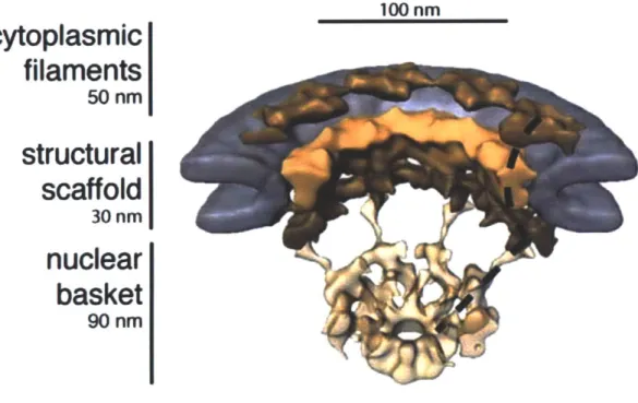
Chapter 1: Introduction 100 nm
cytoplasmic
filaments
50 nm
structural
scaffold
30 nmnuclear
basket
90 nmFigure 1. 2 - Cryo-electron tomography of the NPC at ~60 A resolution
Cut away view of the NPC (colored in brown/beige) embedded in the nuclear envelope (gray). The three major domains of the NPC, the cytoplasmic filaments, the structural scaffold, and the nuclear basket, are each clearly distinguishable. This view highlights the eight-fold rational symmetry exhibited by the NPC and four distinct sections are shown in this orientation. To better highlight this, one of the repeating sections has been marked with a black dotted line (Beck et al., 2007).
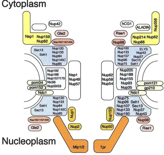
Cytoplasm
Nup42)~
Nsp1 Nup159 Nup2l4 Nup62
Nup82
Sec13 Nup120 Nuplee ELYS
Nup133Nu33
Nup84 NUplO7 NUP37
Nup145C Nup98 Sect3
Nup5 Nup75 5
Nup12(
Nup188 Nsp1 Nup62 Nup188
Nup157/170 Nup49 Nup58 Nup155
m34 NIc96 Nup57 Nup54 Nup93C poml2l
om152 Nup59/53 C 210 Ndcl )Ndc Nup120 Nup133 Seh1 Nup84 Sec13 Nup145C Nupu5 Nu85q~pl60Nup43Nu6
NNucleoplasm
Figure 1. 3 - Schematic representation of the modular NPC assembly
The NPC is built from -30 nucleoporins, organized in a small set of defined subeomplexes. This cartoon shows the major subcomplexes that make up the lattice-like scaffold (blue), the membrane-attachment (green), and the FG-network (gray) of the NPC. S. cerevisiae components on the left, metazoan with specific additional components on the right. A few peripheral Nups are left out for clarity. Simplified representation and connections are not to be taken literally.
Chapter 1: Introduction
Nucleoponn Metazoan Domain Architecture Abundance Mass per NPC Fraction of
homolog cytosasmc = 1 MDa uctured total NPC
ernosasmec m Mo unstucturaO Nup192 Nup205 0 0 Nup188 Nup188 0 0 Nic96 Nup93 0000 eW -35% Nupl57/170 Nupl55 000 a Nup53/5 Nup35 e 0 0 Nup133 Nup133 l 6 Nup120 Nup160 e e 0 Nup84 Nup107 0 0 -Nup85 Nup85 0 m 20% Nup145C Nup96 e e Seh1 Seh1 ' -: e Sec13 Sec13 40 u Nup37 Nup43 Mip1 Tpr e mm MIp2 Tpr 0
-Nup2 Nup50 (ZIDIEII 1 1 12%
Nup1 -Nup6O e
=0
Nup153 Pom152 0 0 Ndcl Ndc1 ( O-*= 0 0 9% Pom34 -C~)- e . gpero Pom121 Nsp1 Nup62 1 1 0000 mamm Nup49 Nup58 e * e 10% Nup57 Nup54 m * eNup159 Nup214/CAN IIIDUED III - -11 0
Glel Gle1 0 Nup42 NLP1IhCG1 (0-111111 D 7% Nup82 Nup88 e e0 a Nup358/RanBP2 ALADIN Rae1/Gle2 Rae1/Gle2 0 0 '
Nup145N Nup98 Ce11 HM
NuplOO Nup98 M I1
Nup116 Nup98 dillM 1llilli e
(hll(ACE1) Calculated Composition of the NPC from S cereisiae
C) p-rope ue J total
W cotd coO EM l ]
am 0 doman 11.3 6.5 6,3 5.3 2O 1.311 59 6 6 46 MDa
a doman
trans memrn Figure 1. 4 - Inventory of the NPC
Summary of the nucleoporins that make up the NPC. Domain architecture of nucleoporins from S.
cerevisiae as determined by x-ray crystallography or prediction (where structural information is still
lacking). Abundance and derived mass calculations are based on published Nup/NPC stoichiometries (Cronshaw et al., 2002; Rout et al., 2000). Nucleoporins specific to metazoa are italicized.
NIc96 Nup85-Sehl PDB Code: 2QX5, 2RFO 3EWE, 3F3F
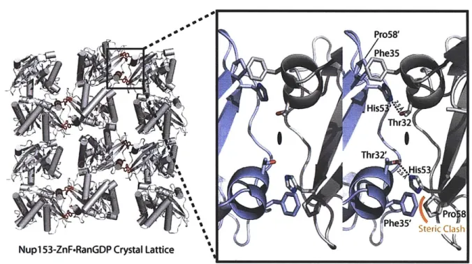
1 18 839 Nup85 1 744 Seht 1 I 349 hNup1O7-hNup133 314R, 3C0 hNup1O7 1 658925 658 hNup133 11 M 101156 517 Nup170 315P, 3150 1 79 502 yNup145C-hSec13 3BG1 yNup145 11 7 1317 741 1158 hSec13 1 K1322 316 rNup58 20SZ 1 585~ss 327-411 327-411 Nspl-FGS-Importin P 2BPT NsPl 11 1076 974-1012 IrmB 3 861 Nup120 3HXR 1 1037 757 mNup35 1WWH 1 [X1325 156-261 1 [1325 156-261 rNup153-ZnF-RanGDP 3CH5, 3GJ3-8 rNup153 1 11502 691-909 rRan 1 E216 ~y~I
L~rr
hNup133 1XKS 1 61156 67 514 Nupl59 (hNup214) 1XIP, 201T 1 37 11460 387Figure 1. 5 - Structures of nucleoporins
Comprehensive list of all representative nucleoporin structures published up until July 2009. PDB
accession codes are indicated. Structures are gradient-colored red- or blue-to-white from N to C terminus. Residue information for each crystallized fragment is given below the structure. Structures are shown in the assembly state that is supported by crystallographic and biochemical evidence. Structures are from S.
cerevisiae unless noted otherwise (h, human; m, mouse; r, rat). 2QX5(Jeudy and Schwartz, 2007);
hNup98 1K06, 2Q5X/Y 1 E 677 920 hNup358-RanBD.RanGDP 1RRP hNup358 11 /103224 han 216 1171-1304
ilk
Chapter 1: Introduction
2RFO(Schrader et al., 2008b); 3EWE(Brohawn et al., 2008); 3F3F(Debler et al., 2008); 3CQC(Boehmer et al., 2008); 314R, 315P, 315Q (Whittle and Schwartz, 2009), 3BG1(Hsia et al., 2007); 3HXR(Leksa et
al., 2009); 1XKS(Berke et al., 2004); 1XIP(Weirich et al., 2004); 20IT(Napetschnig et al., 2007); 20SZ(Melcdk et al.); 1WWH(Handa et al., 2006); 1K06(Hodel et al., 2002); 2Q5X/Y(Sun and Guo,
2008); 2BPT(Liu and Stewart, 2005); 3CH5(Schrader et al., 2008a); 3GJ3-8(Partridge and Schwartz, 2009); 1RRP(Vetter et al., 1999). Importin-a mRnportin-a CRM1 Cytopytoplasm NTF k"P~rtRanGAP1 GTPr Export Nucleus R D r importin- i CRMi Importin-a
Figure 1. 6 - Ran regulates nucleocytoplasmic transport
Ran is the master regulator of nucleocytoplasmic transport. RanGDP is recycled from the cytoplasm and into the nucleus by NTF2. In the nucleus RCC1I acts as a nucleotide exchange factor to reload Ran with GTP. In the nucleus RanGTP causes disruption of imported complexes between importins and cargo containing an NILS. RanGTP interacts directly with importin-P to stimulate this process. Binding of RanGTP with the exportin CRM1 promotes the assembly of an export complex formed between CRM1 and cargo containing a NES. In the cytoplasm, RanGTP interacts with the Ran GTPase activating protein (RanGAP) and RanBP2 or RanBP1 to stimulate hydrolysis of GTP and dissociate the exportin complex. RanGDP, importins, and exportins are all recycled through the NPC and the cycle begins again. Image taken from (Clarke and Zhang, 2008).
Chapter 2: Crystallographic and Biochemical Analysis of the
Ran-Binding Zinc Finger Domain
The material presented in this chapter was adapted, with permission, from the following publication:
Partridge JR, Schwartz TU. Crystallographic and biochemical analysis of the Ran-binding zinc finger domain. J. Mol. Biol. 2009, v391, 375-89.
Chapter 2: Crystallographic and Biochemical Analysis of the Ran-Binding Zinc Finger Domain
Introduction
Nucleocytoplasmic transport is controlled and facilitated by protein assemblies termed nuclear pore complexes (NPCs). NPCs reside in circular openings of the nuclear envelope where inner and outer nuclear membranes are fused. The NPC is a large macromolecular
assembly with a calculated mass of -50 MDa (Alber et al., 2007b). Based on electron-microscopy (EM) studies from X. laevis oocytes and S. cerevisiae, the general shape and structure of the pore is conserved across eukaryotes (Kiseleva et al., 2004; Kiseleva et al., 2001). These EM studies define the pore as a ring embedded in the nuclear envelope, exhibiting 8-fold rotational symmetry around a central axis and imperfect 2-fold symmetry between the cytoplasmic and nucleoplasmic faces. Despite its size, the NPC is only made up of -30 proteins, or nucleoporins (Nups), arranged in a few biochemically defined subcomplexes that assemble the entire structure in a modular fashion (Schwartz, 2005). As the single
constitutive barrier to regulate permeability, the NPC transports a wide range of substrates across the double membrane of the nuclear envelope (D'Angelo and Hetzer, 2008; Tran and Wente, 2006; Weis, 2003). Active transport through the NPC is mediated by nuclear transport receptors (NTRs), also called karyopherins or importins/exportins (Chook and Blobel, 2001; Cook et al., 2007).
The small G protein Ran is the master regulator of NTR-mediated, nucleocytoplasmic protein transport (Gorlich and Kutay, 1999). Ran selectively promotes binding or release of import or export cargos to NTRs by means of a chemical gradient. Ran binds mostly GDP in the cytoplasm, and mostly GTP in the nucleus. The GTPase-activating protein RanGAP, localized to the cytoplasmic face of the NPC, and the chromatin-bound GTP exchange factor, RCC1, together promote this asymmetry by modulating nucleotide hydrolysis and exchange
respectively. The established gradient provides directionality to protein transport. In the nucleus, RanGTP releases import-cargo from NTRs by competitive binding. RanGTP is recycled back to