HAL Id: inserm-00159119
https://www.hal.inserm.fr/inserm-00159119
Submitted on 2 Jul 2007
HAL is a multi-disciplinary open access
archive for the deposit and dissemination of
sci-entific research documents, whether they are
pub-lished or not. The documents may come from
teaching and research institutions in France or
abroad, or from public or private research centers.
L’archive ouverte pluridisciplinaire HAL, est
destinée au dépôt et à la diffusion de documents
scientifiques de niveau recherche, publiés ou non,
émanant des établissements d’enseignement et de
recherche français ou étrangers, des laboratoires
publics ou privés.
Cardiac Motion Estimation in Multislice Computed
Tomography Imaging Using a 4D Multiscale
Surface-Volume Matching Process
Antoine Simon, Mireille Garreau, Dominique Boulmier, Christine Toumoulin,
Hervé Breton
To cite this version:
Antoine Simon, Mireille Garreau, Dominique Boulmier, Christine Toumoulin, Hervé Breton. Cardiac
Motion Estimation in Multislice Computed Tomography Imaging Using a 4D Multiscale
Surface-Volume Matching Process. 2005, pp.219-222. �inserm-00159119�
Cardiac Motion Estimation in Multislice Computed Tomography Imaging
Using a 4D Multiscale Surface-Volume Matching Process
A Simon
1, M Garreau
1, D Boulmier
2, C Toumoulin
1, H Le Breton
1,21
LTSI, INSERM U642, Universit´e de Rennes 1, Campus de Beaulieu, 35042 Rennes, France
2Centre Cardio-Pneumologique, CHU Pontchaillou, 35033 Rennes, France
Abstract
A new generation of Multislice Computed Tomography (MSCT) scanners, which allow a complete heart coverage, is becoming widely available. With retrospective data re-construction, it offers new perspectives for cardiac kinetics evaluation in non invasive imaging with volume sequences of high spatial and temporal resolutions. A new method is proposed for cardiac motion extraction in MSCT. It is based on a 3D surface-volume matching process associ-ated to a hierarchical description of shapes. Needing the segmentation of only one volume, it provides, on a whole dynamic sequence, 3D velocity fields associated to the left ventricle inner surface and an estimation of the localiza-tion of this surface. The estimated molocaliza-tion can be used for global and local functional parameters quantification. Re-sults obtained on simulated and real data show the good behavior of this method.
1.
Introduction
Cardiovascular diseases represent the major cause of mortality in industrialized countries. Different cardiac functional parameters are used to guide diagnosis, treat-ment and follow-up of these diseases. The most used indi-cators provide information on the heart global function, but the detection and the treatment of some pathologies would need the precise quantification of cardiac motion and de-formation.
Because the heart is a nonrigid object that deforms in 3D space, only 3D representation based methods can provide accurate motion descriptors. Image processing methods, applied on 3D cardiac dynamic sequences, can be used to quantify heart motion.
Assessment of heart motion with minimally invasive modalities has been studied from four-dimensional (3D+T) data sets with magnetic resonance (MR) imaging [1] (and especially MR tagging [2, 3]), transthoracic ultra-sound images [4, 5] and the Dynamic Spatial Reconstructor (DSR) [6, 7, 8, 9]. See [10] for a more exhaustive review
of cardiac image functional analysis methods.
The recent significant advances of spiral computed tomography, with the introduction of ultra-fast rotating gantries, multi-rows detectors and retrospective ECG-gated reconstructions, provide higher contrast and spatio-temporal resolutions and allow a huge progress toward the imaging of moving organs. Some first reference studies have been conducted in MSCT for the automatic detection of coronary diseases [11, 12] and some first clinical studies have been realized for cardiac functional analysis [13], but very few works have been developed for the quantitative 3D cardiac motion automatic estimation [14].
Based on these 4D CT images, a new method is pro-posed to extract non-rigid motion of 3D structures and is applied to the left ventricle. This estimation method ex-tracts the motion between a 3D segmented surface which has been reconstructed at one time and the volume image corresponding to the next time. A surface estimation pro-cess, using the extracted motion, provides the localization of the surface corresponding to the second time. This way to proceed enables to apply the segmentation tool only to the first moment of the sequence, providing a method which avoids segmentation temporal coherence problems.
2.
Method
2.1.
Global process
The issue is to compute the motion, and to estimate the localization, of the endocardial surface represented in a dy-namic sequence of 3D volumes. The global process in-volves three main steps:
(a) a preprocessing step, including a segmentation, a
sur-face reconstruction and a sursur-face regularization processes;
(b) a hierarchical motion estimation step, applied between
the surface corresponding to the considered time t1and the volume corresponding to the following time t2;
(c) a surface estimation step resulting in the surface
cor-responding to time t2.
Step (a) is applied only to the first considered volume. Steps (b) and (c) are iteratively applied to the whole
se-HAL author manuscript inserm-00159119, version 1
HAL author manuscript
Computers in Cardiology (2005) 219-222
This material is presented to ensure timely dissemination of scholarly and technical work. Copyright and all rights therein are retained by authors or by other copyright holders.
All persons copying this information are expected to adhere to the terms and constraints invoked by each author's copyright. In most cases, these works may not be reposted without the explicit permission of the copyright holder.
quence in order to estimate both the cardiac motion and the endocardial surface during a whole cardiac cycle.
In order to obtain the mesh corresponding to the first considered time of the sequence, step (a) is decomposed in this way: a segmentation tool, based on a 3D region growing process bounded by a gradient information, is ap-plied [15]. The segmented surface is then reconstructed using the Marching Cubes algorithm. Finally, in order to prepare the matching process, the resulting surface mesh is regularized in such a manner that each node coordinates correspond to one volume voxel coordinates.
In order to obtain the surface corresponding to time t2, the surface estimation step (c) relies on the deformation of the surface corresponding to time t1 with the motion estimated between times t1and t2. A regularization step is then performed in order to fill mesh holes and to suppress redundant nodes.
2.2.
Hierarchical motion estimation
The 3D motion field estimation (step (b)) is performed by a matching process applied between the surface repre-senting the endocardium at time t1and the original volume corresponding to the next time t2. A hierarchical process is used to gain both in terms of result quality and of com-putational efficiency.
The matching process will firstly be described at one res-olution, then the multiresolution scheme will be detailed.
2.2.1. Matching process
The 3D motion field to compute between two suc-cessive instants is considered as a realization f = {fi/i = 1, ..., NS}of a 3D random field F (NSbeing the number of considered sites in the field). The set of sites Sof the field F is given by all the 3D mesh nodes at time t1. The labels assigned to these sites, expressed by the fi estimations, are given by the voxels found (at time t2) in best correspondence with the 3D nodes.
According to Bayes’ theory, the posterior probability of the realization f according to the observation d (the mesh nodes at time t1and the voxels at time t2) is given by:
P(f |d) = P(f ) P(d|f )
P(d) , (1)
with P(f) the prior probability, P(d|f) the conditional probability of the observation process d and P(d) the ob-servation probability which is considered independent of f. According to the maximum a posterior (MAP) esti-mator, the most probable realization f is provided by the maximisation of the posterior probability P(f|d) , with P(f |d) ∝ P(f ) P(d|f ).
The mechanical properties of the heart induce a spatio-temporal regularity of the motion field. To capture this
characteristic, the random field F is modeled by a Markov Random Field (MRF) in relation to a neighbourhood sys-tem µ: the local neighbourhood µiof each node i is given by all nodes j which have a common edge with i.
According to the Hammersley-Clifford theorem and as-suming that P(f) > 0, the random field F is a Gibbs Random Field in relation to the neighbourhood system µ. Therefore, the probability distribution function is given by:
P(f ) = 1 Z exp{−Ur(f )} = 1 Z exp{− X c∈C Vc(fc)} (2)
with Ur(f )the field internal energy function, defining the interactions between the field sites and having a regulari-zation effect, and Z a normaliregulari-zation constant. The inter-action potentials Vc (c ∈ C) are defined for each clique c belonging to the cliques set C. C is defined as all pairs of neighbouring nodes and fc= {fi, i ∈ c}. Vcis given by:
Vc(fc) = || − → fs−−→ft||
dist(s, t) ∀ c = {s, t} ∈ C (3) where−→fs(resp.−→ft) is the motion vector estimated at site s (resp. t).
The conditional probability of the observation process is provided by the data model:
P(d|f ) = exp{−Ud(f, d)} = exp{−X i∈S
Vi(ni, fi)}. (4) Ud(f, d)models the estimation error with:
Vi(ni, fi) = αp.Ep(ni, fi) + αt.Et(ni, fi) +αc.Ec(fi), (5) where Ep(ni, fi)is the Euclidean distance between node ni and voxel fi, and Ec(fi) corresponds to the belong-ing of voxel fito a contour (estimated by a Canny filter). Et(i, fi)expresses a topological correspondence between the node i and the voxel fi: the topology is described, for the surface, in terms of coordinates of the neighbouring nodes and compared, in the volume, to the corresponding voxel contour values.
Finally, the total energy to minimize, noted U(f, d), is thus defined by the linear combination of two terms:
U (f, d) = Ud(f, d) + α1Ur(f ). (6)
2.2.2. Hierarchical scheme
The previously described motion extraction process is applied according to a hierarchical scheme which allows to focus the correspondence research area and to reduce computing needs. This multiresolution scheme is consid-ered to preserve local Markov property according to the high spatial resolution provided by MSCT data.
The surface mesh at time t1and the volume at next time t2are defined with decreasing scales as follows: each data set from upper resolution Ri (i = 1, . . . , nl, nlbeing the number of used resolution levels) is restricted in space at a lower level scale Ri−1 by the application of a mean fil-tering (or Gaussian filfil-tering for the volume) and of a sub-sampling process in order to provide a regular mesh cor-responding to volume voxel coordinates at the same level. The matching process is first applied at the lowest resolu-tion (with an initializaresolu-tion to a null moresolu-tion) to guide the motion estimation with the coarsest details. The result of that first estimation is used as an initialization, after an in-terpolation step, for the correspondences computation at the next finer resolution. This extraction motion process is applied iteratively, with an adaptation of energy weighting coefficients, until the estimation is obtained at the desired resolution.
The global optimization of the correspondences is per-formed with a stochastic relaxation Metropolis algorithm combined with a simulated annealing process at the first lower resolution level and with an Iterated Conditional Mode (ICM) algorithm at the upper resolution levels.
3.
Results
3.1.
Tests on simulated data
Numerical simulations have been used to test the mo-tion extracmo-tion process between two successive instants. In order to obtain the simulated data, a segmented endocar-dial surface, corresponding to the first instant, is deformed using five kinds of motion (translation, twisting, rotation, global expansion/contraction and local deformations) re-sulting in the mesh corresponding to the second instant. Then, this deformed mesh is inserted into a volume pre-processed by a Canny filter followed by an endocardial suppression step. The hierarchical matching process is fi-nally applied between the surface before deformation and this volume at three increasing scales.
Using this process, the real correspondences are known. It enables to measure the error of matching of the proposed method and to study the evolution of the matching process along iterations and at each stage of the hierarchical pro-cess. The impact of the different parameters involved in the computation of the energies or in the optimization pro-cess, as well as meaningful information provided by scale refinement, can also be evaluated.
Figure 1 illustrates an example of results obtained at the two lowest resolution levels (R1with volume size 64
3and R2(1283)). In color is represented the applied (a,b) and estimated (c,d) motion amplitude. In red are represented the displacements directed inside the cavity. Considering the real voxel size (0.35 mm), with an initial mean match-ing error of 10.5 voxels at R1level, the process converges
(a) (b)
(c) (d)
Figure 1. Simulated (a,b) and estimated (c,d) motion am-plitude with a left oblique anterior view at two resolutions (levels 643 (a,c) and 1283 (b,d)) (colours: in blue (resp. red): motion directed outside (resp. inside) the cavity), measures in voxels.
to a final mean error of 1.0 voxel at R3 level. We have observed that this hierarchical process enables to gain in precision and in error deviation.
3.2.
Results on real data
The algorithm has been applied on real human heart data with a temporal database acquired by a Siemens SO-MATOM Sensation 16 with ten volume images represent-ing a whole cardiac cycle. Each volume contains about 300 slices of 512 × 512 pixels, giving a resolution for each voxel of 0.35 × 0.35 × 0.5 mm.
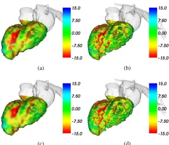
The proposed method has been applied to this dynamic sequence considering three resolution levels (from 643to 2563). Figure 2 illustrates results obtained at the two lo-west levels at end-diastolic (a,b) and end-systolic (c,d) ins-tants. The contraction movements during systole as well as expansion movements during diastole are identified and coherent with cardiac phases. We can see that the lowest level representation is meaningful and that the displace-ments obtained at upper levels provide the means to extract local clinical parameters at various scales.
4.
Discussion and conclusions
A new solution for motion extraction combined with surface estimation has been introduced and applied to the left ventricle in 4D cardiac MSCT imaging. This ap-proach is based on a hierarchical surface-volume
(a) (b)
(c) (d)
Figure 2. Estimated motion amplitude at end-diastolic (a,b) and end-systolic (c,d) instants, at two resolutions (lev-els 643(a,c) and 1283(b,d)) (colours: in blue (resp. red): motion directed outside (resp. inside) the cavity).
ing method formulated with a Markov Random Field and provides, with one unique process, left cavity surfaces and associated 3D motion vector fields. The algorithm has been tested with simulated and real MSCT dynamic data, highlighting encouraging results and confirming the great potential of MSCT imaging for quantitative clinical mea-sure assessment in cardiac applications. Further works will carry on algorithm optimization and on extensive evalua-tion.
Acknowledgements
This work is supported by Brittany region and the French Medical Research and Health National Institute (INSERM). The authors express their thanks to Siemens, Medical Division, France.
References
[1] Shi P, Sinusas AJ, Constable RT, Ritman E, Duncan JS. Point-tracked quantitative analysis of left ventricular sur-face motion from 3-D image sequences. IEEE Trans Medi-cal Imaging jan 2000;19:36–50.
[2] Park J, Metaxas D, Young AA, Axel L. Deformable models with parameter functions for cardiac motion analysis from tagged MRI data. IEEE Trans Medical Imaging jun 1996; 15:278–289.
[3] Huang J, Abendschein D, Davila-Roman V, Amini AA. Spatio-temporal tracking of myocardial deformations with a 4D B-spline model from tagged MRI. IEEE Trans Medi-cal Imaging oct 1999;18:957–972.
[4] Maehle J, Bjoernstad K, Aakhus S, Torp HG, Angelsen BA. Three-dimensional echocardiography for quantitative left ventricular wall motion analysis: a method for recon-struction of endocardial surface and evaluation of regional dysfunction. Echocardiography jul 1994;11:397–408. [5] Papademetris X, Sinusas AJ, Dione DP, Duncan J.
Estima-tion of 3D left ventricular deformaEstima-tion from echocardiogra-phy. Medical Image Analysis mar 2001;5:17–28.
[6] Amini AA, Duncan J. Bending and stretching models for LV wall motion analysis from curves and surfaces. Image and Vision Computing July/Aug. 1992;10:418–430. [7] Benayoun S, Ayache N. Dense non-rigid motion
estima-tion in sequences of medical images using differential con-straints. Int Journal of Computer Vision jan 1998;26:25–40. [8] Gorce JM, Friboulet D, Magnin IE. Estimation of three-dimensional cardiac velocity fields: assessment of a differ-ential method and application to three-dimensional CT data. Medical Image Analysis apr 1997;1:245–261.
[9] Eusemann CD, Ritman EL, Robb RA. Parametric visualiza-tion methods for the quantitative assessment of myocardial motion. Academic Radiology jan 2003;10:66–76. [10] Frangi AF, Niessen WJ, Viergever MA. Three-dimensional
modeling for functional analysis of cardiac images, a re-view. IEEE Trans Medical Imaging jan 2001;20:2–25. [11] Schroeder S, Kopp AF, et al. Accuracy and reliability of
quantitative measurements in coronary arteries by multi-slice computed tomography: experimental and initial clini-cal results. Cliniclini-cal Radiology jun 2001;56:466–474. [12] Larralde A, Boldak C, Garreau M, Toumoulin C,
Boul-mier D, Rolland Y. Evaluation of a 3D segmentation soft-ware for the coronary characterization in multi-slice com-puted tomography. In Lecture Notes in Computer Science, Functional Imaging and Modeling of the Heart (FIMH’03). Lyon, France, jun 2003; 39–51.
[13] Yamamuro M, Tadamura E, et al. Cardiac functional analysis with multi-detector row CT and segmental recon-struction algorithm: Comparison with echocardiography, SPECT, and MR imaging. Radiology feb 2005;234:381– 390.
[14] Garreau M, Simon A, Boulmier D, Guillaume H. Cardiac motion extraction in multislice computed tomography by using a 3D hierarchical surface matching process. In Proc. IEEE Computers in Cardiology (CinC’04). Chicago, USA, sep 2004; 549–552.
[15] Guillaume H, Garreau M. Segmentation de cavit´es car-diaques en imagerie scanner multi-barettes. In 12`eme Fo-rum des Jeunes Chercheurs en G´enie Biologique et M´edical (2003). Nantes, France, may 2003; 92–93.
Address for correspondence: Antoine Simon
LTSI, INSERM U642, Universit´e de Rennes 1, Campus de Beaulieu, 35042 Rennes, France [email protected]