The
Jour
nal
o
f
P
hysiology
Lack of muscle recovery after immobilization in old rats
does not result from a defect in normalization of the
ubiquitin–proteasome and the caspase-dependent
apoptotic pathways
Hugues Magne
1, Isabelle Savary-Auzeloux
1, Emilie Vazeille
1, Agn`es Claustre
1, Didier Attaix
1, Listrat Anne
2,
Sant´e-Lhoutellier V´eronique
3, Gatellier Philippe
3, Dominique Dardevet
1∗and Lydie Combaret
1∗1INRA, UMR 1019 Unit´e de Nutrition Humaine, 63122 Saint Gen`es Champanelle, France, and Clermont Universit´e, UFR M´edecine,
UMR 1019 Nutrition Humaine, 63000 Clermont-Ferrand, France
2INRA, UR 1213, Unit´e de Recherche sur les Herbivores, 63122 Saint Gen`es Champanelle, France 3INRA, UR 370, Qualit´e des Produits Animaux, 63122 Saint Gen`es Champanelle, France
Non-technical summary Immobilization periods increase with age because of decreased mobility and/or because of increased pathological episodes that require bed-rest. Then, sarcopaenia might be partially explained by an impaired recovery of skeletal muscle mass after a catabolic state due to an imbalance of muscle protein metabolism, apoptosis and cellular regeneration. Mechanisms involved during muscle recovery have been little studied and in elderly they remain almost unknown. We show, in rats, that a short immobilization period during ageing initiated muscle atrophy that was indeed not recovered after 40 days. Immobilization was associated with an activation of both the ubiquitin–proteasome and the mitochondria-associated apoptotic pathways and the inflammatory and redox processes, and a decrease of cellular regeneration. We show that the lack of muscle recovery during ageing is not due to a defect in proteolysis or apoptosis down-regulation. These observations lead us to hypothesize that muscle protein synthesis activation after immobilization was altered during ageing.
Abstract Immobilization periods increase with age because of decreased mobility and/or increased pathological episodes that require bed-rest. Sarcopaenia might be partially explained by an impaired recovery of skeletal muscle mass after a catabolic state due to an imbalance of muscle protein metabolism, apoptosis and cellular regeneration. Mechanisms involved in muscle recovery have been poorly investigated, and remain almost unknown in the elderly. This study aimed at studying the regulation of the capsase-dependent apoptotic and the ubiquitin–proteasome-dependent proteolytic pathways during immobilization and subsequent recovery during ageing. Old rats (22–24-months old) were subjected to unilateral hindlimb casting for 8 days (I8) and allowed to recover for 10 to 40 days (R10 to R40). Immobilized gastro-cnemius muscles atrophied by 21%, and did not recover even at R40. Apoptotic index, amount of polyubiquitinated conjugates, proteasome chymotrypsin- and trypsin-like, apoptosome-linked caspase-9, -3, and -8 activities increased at I8. Conversely, the amount of the myogenic factor myf-5 decreased at I8. These changes paralleled the increase of intramuscular inflammation and oxidative stress. All these parameters normalized as soon as R10. The XIAP/Smac-DIABLO protein ratio decreased by half in immobilized muscles and remained low during recovery. Surprisingly, the non-immobilized leg also atrophied from R20, concomitantly with a decreased XIAP/Smac-DIABLO protein ratio. Altogether, this suggests that the impaired recovery following immobilization in ageing does not result from a lack of normalization of the caspase-dependent
apoptotic and the ubiquitin–proteasome-dependent pathways, and also that immobilization could induce a general muscle loss and then contribute to the development of sarcopaenia in elderly.
(Resubmitted 28 October 2010; accepted after revision 26 November 2010; first published online 29 November 2010)
Corresponding author H. Magne: INRA, UMR 1019 Unit´e de Nutrition Humaine, 63122 Saint Gen`es Champanelle,
France. Email: hugues.magne@clermont.inra.fr
Abbreviations I, immobilized; MCP-1, monocyte chemotactic protein-1; NI, non-immobilized; PF, pair-fed; Smac/
DIABLO, second mitochondria-derived activator of caspase; TBARS, thiobarbituric acid reactive substances; Ub, ubiquitin; UPS, ubiquitin–by-proteasome-dependent proteolysis; XIAP, X-linked inhibitory apoptotic protein.
Introduction
Normal ageing is associated with a progressive loss of muscle mass and strength, a condition known as sarcopaenia (Rosenberg, 1989). This phenomenon is inevitable and has also been reported among healthy people and physically active elderly subjects (Hughes
et al. 2001). Skeletal muscle is the major reservoir of
body proteins and amino acids that can be used to cope with nutritional, infectious or traumatic stress. Therefore, sarcopaenia is a highly predictive factor of frailty, of limited mobility, of increased susceptibility to injury and of impaired recovery (Evans, 1997; Harris, 1997).
Many mechanisms have been proposed to explain sarcopaenia. Skeletal muscle is clearly resistant to anabolic stimuli such as food intake during ageing (for review see Balage & Dardevet, 2010), and impaired recovery of skeletal muscle mass has also been observed after an acute catabolic state (Dardevet et al. 1995; Mosoni
et al. 1999). The succession of catabolic periods followed
by incomplete recoveries results in a significant muscle mass loss over time and has been named recently ‘the catabolic crisis model’ (English & Paddon-Jones, 2010). An increase of prolonged periods of immobilization as a consequence of weakness, hospitalization or bed-rest is commonly associated with these catabolic states. However, the effect of disuse by itself on skeletal muscle in elderly individuals has not been extensively investigated and the subsequent recovery ability has been even less studied despite the fact that an impaired recovery prevailed in old rats (Chakravarthy et al. 2000) and elderly humans (Suetta et al. 2009) after immobilization-induced muscle atrophy. This impaired recovery has been linked only to decreased muscle strength and neuronal motor function (Suetta et al. 2009).
Knowledge of the cellular and molecular mechanisms underlying this lack of recovery is limited during ageing, but they have been related only to a decline in the pathways regulating the activation of muscle satellite cells during muscle regrowth (Zarzhevsky et al. 2001; Conboy et al. 2003). Beside this impaired regenerative
process, an increased apoptosis could also contribute to the loss of myocytes. This suggests an imbalance between regeneration and apoptotic processes following immobilization in old muscles. In addition, changes in skeletal muscle protein mass depend on the overall balance between rates of protein synthesis and break-down. Muscle recovery after immobilization results not only from a normalization of changes in muscle protein metabolism but also from increased protein synthesis, decreased proteolysis, or simultaneous changes in both processes. To our knowledge, and surprisingly, these aspects (i.e. apoptosis and muscle protein metabolism) have not been assessed during the recovery period following immobilization in ageing.
Ubiquitin–proteasome-dependent proteolysis (UPS) is responsible for the breakdown of the major contractile proteins (Attaix et al. 2005). In this pathway, substrates are tagged by covalent attachment of multiple ubiquitins and then recognized and degraded by the 26S proteasome. We and others have demonstrated that the ubiquitin pathway is highly activated during disuse atrophy in adult rats (Wing et al. 1995; Taillandier et al. 1996; Ikemoto et al. 2001; Vazeille et al. 2008), and rapidly normalizes during the recovery period as is the activation of apoptosis (Vazeille et al. 2008). However, during ageing, we have shown that muscle proteolysis becomes less sensitive to anabolic or catabolic stimuli (Dardevet et al. 1995; Mosoni et al. 1999; Combaret et al. 2005). This deregulation of the UPS may in part account for the lack of recovery of skeletal muscle mass following catabolic states.
Two main apoptotic pathways have been involved in caspase activation. The extrinsic pathway is triggered by the activation of a family of death receptors (e.g. TNFR) at the cell surface and controls the activation of caspase-8. In the intrinsic pathway (also called the mitochondria-associated apoptotic pathway), the mitochondria releases cytochrome c, which associates in the cytoplasm with Apaf-1 and caspase-9 to form the apoptosome (Adhihetty & Hood, 2003). This complex further activates caspase-3, a key cell death protease
that initiates DNA fragmentation (Kujawa et al. 2005; Dupont-Versteegden, 2005). Both apoptotic pathways are believed to be physiologically important in sarcopaenia (Dirks & Leeuwenburgh, 2004; Pistilli et al. 2006; Marzetti
et al. 2009), and in regulating disuse-induced muscle
atrophy both in younger adult (Andrianjafiniony et al. 2010) and in old (Alway et al. 2003; Plant et al. 2009) animals. The mitochondria-associated apoptotic pathway was concomitantly activated with the UPS during immobilization-induced atrophy (Vazeille et al. 2008) and was normalized during recovery (Vazeille et al. 2008; Andrianjafiniony et al. 2010), whereas the death receptor-mediated apoptotic pathway promoting the activation of caspase-8 remained elevated during recovery following unloading in adult rats (Andrianjafiniony
et al. 2010). This study tested whether this impaired
recovery during reloading in ageing results from a lack of normalization of the UPS and/or the caspase-dependent apoptotic pathways.
Methods
Animals and experimental design
All procedures were performed in accordance with institutional guidelines on animal experimentation in France and comply with the policies and regulations of The Journal of Physiology given by Drummond (2009). Male Wistar rats aged 22–24 months were housed individually under controlled environmental conditions (room temperature 22◦C; 12 h light–dark cycle, light period starting at 08.00 h), fed ad libitum a basal diet, and given free access to water. Basal diet was composed of (in g kg−1diet): 152.2 casein, 1.8L-cystine, 30 colza oil, 3 sunflower oil, 27 peanut oil, 35 cellulose, 35 AIN93 mineral mix, 10 AIN93 vitamin mix, 2.5 cholin, 100 saccharose, 134.5 lactose and 469 wheat flour.
After a 3 week adaptation period, 42 rats were anaesthetized with isoflurane inhalation and subjected to unilateral hindlimb cast immobilization with an Orfit-soft plaque (Gibaud, Saint Etienne, France) for 8 days (I8). The foot was positioned in plantar extension to induce maximal atrophy of the gastrocnemius muscles (Goldspink, 1977). Casted rats reduced their food intake during the immobilization period. Therefore, 35 control non-casted rats were pair-fed (PF) to the casted group. For muscle recovery studies, casts were removed and animals were allowed to recover for 10 (R10), 20 (R20) or 40 (R40) days. Eight rats were studied as control before the immobilization period (I0). At each time point animals were killed under pentobarbital sodium anaesthesia (50 mg kg−1 I.P.). Hindlimb gastrocnemius muscles were carefully dissected, weighed and frozen in liquid nitrogen. The central part of gastrocnemius samples
was frozen in isopentane chilled at –196◦C by liquid nitrogen for apoptotic nuclei measurements.
Apoptotic nuclei measurements
Frozen sections of gastrocnemius muscles were cut into 10μm-thick cross sections with a cryostat (Cryo-star HM560MV, Microm International, Walldorf, Germany). The DNA fragmentation of nuclei was monitored using a TUNEL (terminal deoxynucleotidyl transferase biotin-dUTP nick-end labelling) fluorescence detection kit according to the manufacturer’s instructions (Roche Applied Science, Indianapolis, IN, USA). This corresponds to global nuclear apoptosis measurements with evaluation of total nuclei contained in muscle.
Measurement of proteasome and apoptosome activities
Gastrocnemius muscle powder (150 mg) from pair-fed (PF), non-immobilized (NI) and immobilized (I) muscles at each time point were homogenized in 10 volumes of an ice-cold buffer containing 50 mM Tris-Cl (pH 7.5), 5 mMMgCl2, 250 mMsucrose, 1 mMdithiothreitol (DTT), 10 nMadenosine triphosphate (ATP) and protease inhibitors (10μg ml−1 antipain, 10μg ml−1 leupeptin, 10μg ml−1 aprotinin, 10μg ml−1 pepstatin A, 20μM phenylmethanesulphonyl fluoride (PMSF)). Apoptosome complexes co-sediment with proteasomes (Cain et al. 1999) and the apoptosome linked caspase-9 activity co-elutes with peptidase activities of the proteasome on 10–40% sucrose gradients (Cain et al. 1999; Vazeille et al. 2008). Apoptosome complexes and proteasomes were therefore isolated by three sequential centrifugations as described previously (Fang et al. 2000). Briefly, extracts were centrifuged at 10,000 g for 20 min at 4◦C. Supernatants were then centrifuged at 100,000 g for 1 h at 4◦C and resulting supernatants were finally centrifuged at 100,000 g for 5 h at 4◦C. The resulting protein pellets were resuspended in 150μl of a buffer containing 20% glycerol, 5 mMMgCl2and 50 mMTris-Cl (pH 7.5) (Buffer A). Protein concentration was determined on these resuspended pellets according to Lowry et al. (1951). The proteasome chymotrypsin- and trypsin-like activities and the apoptosome-linked caspase-9 activity were determined by measuring the hydrolysis of the fluoro-genic substrates succinyl-Leu-Leu-Val-Tyr-7-amino-4-methylcoumarin (LLVY-AMC) (Sigma, St. Louis, MO, USA), Boc-Leu-Arg-Arg-7-amino-4-methylcoumarin
(LRR-AMC) (Enzo Life Sciences, Farmingdale,
NY, USA) and N -acetyl-Leu-Glu-His-Asp-7-amino-4-methylcoumarin (LEHD-AMC) (Biomol, Hamburg, Germany), respectively.
To measure the proteasome chymotrypsin- and trypsin-like activities, 15μg of proteins from the resuspended pellets diluted in 15μl of Buffer A were
added to 60μl of reaction buffer (50 mM Tris-Cl (pH 7.5), 11.25 mM MgCl2, 1.25 mM DTT, 0.01 U apyrase) containing either 300μMLLVY-AMC or 800μM LRR-AMC. To measure the apoptosome-linked caspase-9 activity, 25μl of the resuspended pellets was diluted to 50μl in buffer A and then incubated with 50 μl of a reaction buffer (100 mM Pipes, pH 6.5, 0.2 mM EDTA, 10 mMDTT) containing 100μM LEHD-AMC (Biomol, Hamburg, Germany). Pilot experiments were performed with or without inhibitors of chymotrypsin-like (40μM MG132, AFFINITI Research Products Ltd, Exeter, UK), trypsin-like (100μM lactacystin, Sigma, St. Louis, MO, USA), or caspase-9 (LEHD-CHO, Biomol, Hamburg, Germany) activities to ensure that they were totally inhibited.
Activities were determined by measuring the accumulation of the fluorogenic cleavage product (amino-4-methylcoumarin, AMC) using a luminescence spectrometer (FLX800, Biotek, Winooski, VT, USA) during 45 min at 380 nm excitation wavelength and 440 nm emission wavelength. For the apoptosome-linked caspase-9 activity, final data were corrected by the amount of protein. The time course for the accumulation of AMC after hydrolysis of the substrate was analysed by linear regression to calculate activities, i.e. the slopes of best fit of accumulation AMC vs. time.
Caspase-3 and caspase-8 activity measurements Caspase-3 and caspase-8 activities were assessed in total cytosolic protein extracts. Powder (200 mg) of gastro-cnemius muscles was homogenized in 3.75 volumes of a buffer containing 10 mMNaCl, 1.5 mMMgCl2, 20 mM Hepes, 20% glycerol, 0.10% Triton X-100 and 1 mMDTT (Buffer B). Extracts were centrifuged at 1500 g for 5 min at 4◦C and the resulting supernatants were subjected to three further centrifugations at 3500 g for 5 min at 4◦C. The last supernatants were stored as total cytosolic protein extracts at−80◦C. A protease inhibitor cocktail containing 104 mM4-(2-aminoethyl)benzenesulfonylfluoride hydro-chloride (AEBSF), 0.08 mM aprotinin, 2 mM leupeptin, 4 mM bestatin, 1.5 mM pepstatin A and 1.4 mM E-64 (Sigma, St. Louis, MO, USA) was added to a small part of these supernatants, which were then stored at −80◦C for subsequent determination of protein levels for the X-linked inhibitory apoptotic protein (XIAP) and the second mitochondria-derived activator of caspase (Smac/DIABLO) (see below). Protein concentration was determined according to Lowry et al. (1951).
Caspase-3 and caspase-8 activities were assessed using the fluorogenic substrates, Ac-Asp-Glu-Val-Asp-AMC (DEVD-AMC, Calbiochem, Darmstadt, Germany) and
N -acetyl-Ile-Glu-Thr-Asp-AMC (Ac-IETD-AMC, Sigma,
St. Louis, MO, USA), respectively. Twenty-five microlitres of the total cytosolic protein extracts was diluted to 50μl in
Buffer B (see above) and incubated with 50μl of a medium containing 100 mM Pipes (pH 6.5), 0.2 mM EDTA, 10 mM DTT, 20% glycerol and 100μM DEVD-AMC. Pilot experiments were performed with or without inhibitor of caspase-3 (Ac-Asp-Met-Gln-Asp-CHO, DMQD-CHO, Calbiochem, Darmstadt, Germany) or caspase-8 (Ac-IETD-CHO, Sigma, St. Louis, MO, USA) to ensure that the activity was totally inhibited. Accumulation of the fluorogenic cleavage product AMC was followed using a luminescence spectrometer (FLX800, Biotek, Winooski, VT, USA) (as described above) and the final data were corrected by the amount of protein.
Muscle XIAP, Smac/DIABLO, ubiquitin(Ub)-conjugated and myf-5 protein contents
Sixty micrograms of total protein extract was separated
on 7.5% and 12% acrylamide gels for XIAP
and Smac/DIABLO, respectively, and then transferred on polyvinylidene fluoride (PVDF) membranes (GE Healthcare, Orsay, France). Antibodies against XIAP and Smac/DIABLO (Cell Signaling Technology, Inc., Danvers, MA, USA) were used at 1:1000 and at 1:4000 dilutions respectively.
The accumulation of poly(Ub)conjugates was assessed on myofibrillar protein extracts. Powder (150μg) of gastrocnemius muscles was homogenized in an ice-cold buffer containing 5 mMTris-HCl (pH 7.5), 5 mMEDTA (pH 8.0), 1 mMPMSF, 5 mMN -ethyl-maleimide (NEM), 5μg ml−1 leupeptin (Buffer C). Homogenates were centrifuged for 5 min (1500 g, 4◦C) to pellet myofibrillar proteins, which were then washed three times in Buffer C containing 1% Triton X-100. Myofibrillar proteins were then resuspended in 8M urea/5 mMTris-HCl (pH 7.5). Protein concentrations were determined as described above. Twenty-five micrograms of myofibrillar proteins was separated on 7.5% acrylamide gels and trans-ferred onto PVDF membranes (GE Healthcare, Orsay, France) to measure the accumulation of Ub-conjugates using the FK1 antibody, which recognizes poly-Ub chains (Millipore, Billerica, MA, USA) at 1:1,000 dilution.
The abundance of myf-5 was assessed on an aliquot of the supernatant obtained after the second 100,000 g centrifugation performed for proteasome and apoptosome extraction (see above). Protein concentration was determined as described previously. Fifty micro-grams of total protein extract was separated on a 12% acrylamide gel and transferred on PVDF membranes (GE Healthcare, Orsay, France). Antibody against myf-5 (Santa Cruz Biotechnology, Inc., Santa Cruz, CA, USA) was used at 1:1000 dilution. On each gel a reference sample (composed of a pool of samples) was systematically analysed and then used to compare the different gels.
Signals were detected using the ECL+ detection kit (GE Healthcare, Orsay, France) after exposition onto radiographic film (Hyperfilm ECL, GE Healthcare, Orsay, France), quantified by densitometry using the ImageJ software and normalized against the signal obtained with the reference sample.
Intramuscular oxidative stress and inflammatory marker measurements
All measurements described below were performed on gastrocnemius muscle powder. Oxidative stress was evaluated by measuring the content of intramuscular total glutathione and of oxidized proteins and lipids. Briefly, for total glutathione measurement, powder was homo-genized in a solution containing 20% perchloric acid and 5 mMEDTA. The homogenate was then centrifuged for 15 min at 14,000 g, and the supernatant was assayed for total glutathione content using a standard enzymatic assay described by Robinson et al. (1992). To assess protein oxidation, the carbonyl groups were detected by their reactivity with 2,4-dinitrophenylhydrazine (DNPH) to form hydrazone derivates, using the spectrophotometric method described by Oliver et al. (1987) and slightly modified by Mercier et al. (1998). The results were expressed as nanomoles of DNPH fixed per milli-gram of protein. Lipid oxidation was evaluated by the thiobarbituric acid reactive substances (TBARS) method according to Lynch & Frei (1993) and modified by Mercier
et al. (1998). This method involves the breakdown of lipid
peroxides into malondialdehyde (MDA) molecules. MDA reacts subsequently with the thiobarbituric acid (TBA), producing compounds suitable for spectrophotometric detection. The results were expressed as milligrams of MDA per kilogram of muscle.
Inflammatory status was evaluated by measuring muscle content of MCP-1, a cytokine recruiting inflammatory cells. MCP-1 was detected with an ELISA kit (RayBio Rat MCP-1 ELISA, RayBiotech, Inc., Norcross, GA, USA) which uses an antibody specific for rat MCP-1. This assay was performed according to the manufacturer’s instructions. The results were expressed as nanograms of MCP-1 per milligram of muscle.
Statistical analyses
All data are expressed as means±S.E.M. for n= 8 (I0),
n= 15 (I8 PF), n = 16 (I8), n = 5 (R10PF), n = 8 (R10), n= 7 (R20PF), n = 9 (R20) n = 8 (R40PF) and n = 9
(R40). Food intake and body weight comparisons were performed using repeated measures analysis of variance test (StatView statistical software package, version 5, SAS Institute, Cary, NC, USA). For other measurements, the effects of time (I8 to R40) and group (PF, NI or I) were analysed using a two-way ANOVA. When significant differences were detected, post hoc comparisons
between groups were made using the Fisher’s PLSD test. Significance was defined at the P< 0.05 level.
Results
Animal characteristics
Before the immobilization period, food intake was 21.3± 0.28 g day−1 (I0). Casted rats reduced their food intake by 62% (Fig. 1A) on the first day of immobilization (8.1± 0.47 g day−1). Then animals increased their food intake up to 13.4± 0.28 g day−1 at I8 and progressively reached initial values (20.3± 0.04 g day−1 at R40) after cast removal. Food intake of the pair-fed group perfectly matched the one of the casted group during immobilization (8.0± 0.22 g day−1at I8) and subsequent recovery.
Casted rats exhibited a slight decrease of body weight during immobilization (−9.6% at I8), which worsened during the recovery period and reached 13.4% and 16.9% at R10 and R20, respectively (Fig. 1B). During the same
A
Casted
Rats Pair-Fed Rats 0 5 10 15 20 25 30 0 5 10 15 20 25 30 35 40 45 50 R10 R20 R40 I8 FOOD INT AKE (g/day) I0 Days B % OF INITIAL BODY WEIGHT R10 R20 I8 I0 70 75 80 85 90 95 100 105 110 0 5 10 15 20 25 30 35 40 45 50 R40 * Days
Figure 1. Food intake (A) and body weight (B) of non casted pair-fed (PF) and casted rats during immobilization and subsequent recovery
Food intake is similar between casted and PF animals during the whole experiment, whereas body weight of casted rats significantly decreased compared to PF rats from R10. I0: before immobilization; I8: 8 days of casting; R10 to R40: 10 to 40 days of recovery. Food intake is expressed in g day−1and body weight is expressed as a percentage of initial body weight.∗P < 0.05 using repeated
measures analysis of variance test, casted ratsvs. PF. Data are
Table 1. Effect of casting on gastrocnemius muscle mass before and during immobilization and recovery
Time Non-casted rats
I0 2.643± 0.042a
Non-casted pair fed rats Casted rats
Immobilized leg Non immobilized leg I8 2.611± 0.049a 2.059± 0.081b 2.606± 0.084a R10 2.619± 0.045a 2.086± 0.094b 2.579± 0.050a R20 2.605± 0.048a 2.048± 0.052b 2.303± 0.079c R40 2.583± 0.032a 2.185± 0.078b 2.407± 0.062c I0: before casting; I8: 8 days of casting; R10 to R40: 10 to 40 days of recovery. Muscle mass is expressed in grams. The muscle mass change from I0 to R40 was assessed using a two-way ANOVA (time, group); values with different letters are significantly different from each other (P < 0.05). When significant differences
were detected,post hoc comparisons between groups were made using Fisher’s PLSD test. Data are means±S.E.M.
period, the body weight of pair-fed animals decreased only by 6.8% at I8 and remained constant during recovery (∼−8% at R10 and R20).
Skeletal muscle mass during immobilization and recovery
Gastrocnemius muscle masses from pair-fed rats were stable during the whole experiment (2.583± 0.032 g at R40 vs. 2.643± 0.042 g at I0) (Table 1). Immobilized gastrocnemius muscles atrophied at I8 by 21% (Table 1,
P< 0.0005; I leg vs. NI leg or vs. PF group). After cast
removal, gastrocnemius muscles from the I leg did not recover at all even at R40 (2.185± 0.078 g) (Table 1). More surprisingly, gastrocnemius muscle masses from the NI leg remained constant between I8 (2.606± 0.084 g) and R10 (2.579± 0.050 g), but then decreased down to 2.303± 0.079 g and 2.407 ± 0.062 g at R20 and R40, respectively (Table 1, P< 0.05; NI leg vs. PF group). Regulation of ubiquitin–proteasome-dependent pathway
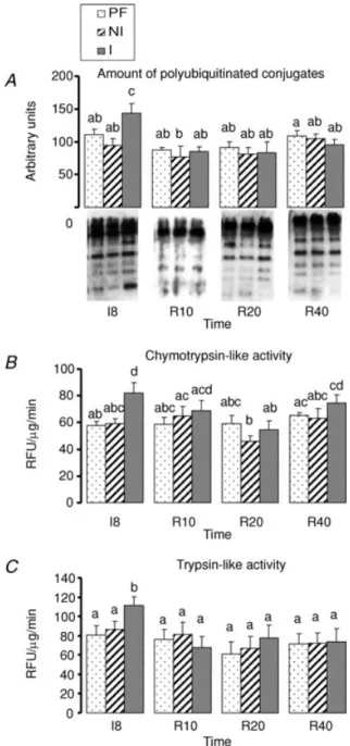
This pathway involves two distinct steps, i.e. the ubiquitination of proteins and their subsequent degradation by the 26S proteasome. We have investigated both steps by measuring (i) the accumulation of poly(Ub) conjugates, which are the final products of the ubiquitination step, and (ii) the chymotrypsin- and the trypsin-like activity of the proteasome.
Figure 2 shows that neither poly(Ub) conjugates nor chymotrypsin- and trypsin-like activities of the
proteasome changed in muscles from the PF group or the NI leg of the casted rats throughout the whole experiment. The amount of poly(Ub) conjugates increased at I8 in gastrocnemius muscle from the I leg of the casted rats compared to muscles from the NI leg or from the PF group of rats (+30% and +53%, respectively, P < 0.05) (Fig. 2A). Accordingly, the proteasome chymotrypsin-and trypsin-like activity also increased in I muscles at I8 by 29 or 33% (P< 0.05, vs. NI or PF group) and by 32% or 38% (P< 0.05, vs. NI or PF group) (Fig. 2B). Both increased poly(Ub) conjugates levels and chymotrypsin- and trypsin-like activities of the proteasome were normalized at R10 in muscles from the I leg compared to muscles from the NI leg or the PF group (Fig. 2A and B).
Regulation of the caspase-dependent apoptotic pathway
We and others have reported that disuse-induced muscle atrophy in adults was associated with increased Smac/DIABLO, decreased XIAP protein levels, and increased caspase-3 activity (Vazeille et al. 2008; Siu & Alway, 2005), suggesting the activation of the mitochondria-associated apoptotic pathway. Furthermore, this pathway is down-regulated in muscles during recovery in adults (Vazeille et al. 2008; Andrianjafiniony et al. 2010). In addition, these alterations in the ratio of XIAP to Smac/DIABLO protein levels also prevailed in unloaded muscles from old birds (Siu et al. 2005b). Finally, disuse-induced muscle atrophy was also associated with increased caspase-8 activity in younger adult (Ferreira et al. 2008; Andrianjafiniony et al. 2010),
and in old animals (Alway et al. 2003), suggesting that the death receptor-mediated apoptotic pathway is also activated. This pathway remained elevated during recovery in suspended rats (Andrianjafiniony et al. 2010). We therefore addressed the regulation of these two apoptotic pathways by measuring (i) the ratio between XIAP and Smac/DIABLO protein levels, (ii) the apoptosome-linked caspase-9 activity, and (iii) the caspase-3 and -8 activities.
Figure 3 shows the amount of the pro- and the anti-apoptotic proteins XIAP and Smac/DIABLO. Levels of both proteins remained unchanged in muscles from the PF group during immobilization and recovery. XIAP protein levels tend to increase in the NI leg of the casted rats at R10 (+22% vs. PF, NS), and returned to basal levels at R20 and R40 (Fig. 3A). Smac/DIABLO protein levels were stable between I8 and R10 in the NI leg, but then progressively tend to increase from R20 (+23% vs. PF, NS) to R40 (+48% vs. PF, NS) (Fig. 3B). Consequently, the ratio of XIAP to Smac/DIABLO progressively decreased in muscles from the NI leg compared to the PF value from R10 to R40 (Fig. 3C). XIAP protein levels decreased by ∼23% (Fig. 3A), while Smac/DIABLO protein levels increased by∼70% (Fig. 3B) in muscles from the I leg at I8 (P< 0.05 vs. NI leg or the PF group). Smac/DIABLO protein levels remained slightly elevated during the recovery period in muscles from the I leg compared to PF group or the NI leg, while XIAP protein levels were normalized to the PF values as soon as R10 (Fig. 3A and B). Thus, the XIAP to Smac/DIABLO protein ratio diminished by∼50% in muscles from the I leg at I8, and remained low during the recovery period (∼−20 to ∼−50% vs. PF group) (Fig. 3C).
This decreased XIAP to Smac/DIABLO protein ratio at I8 in gastrocnemius muscles from the I leg was correlated with increased apoptosome-linked caspase-9 (Fig. 4A,+60%, P < 0.05 I leg vs. NI leg), and caspase-3 (Fig. 4B, +48% P < 0.05 I leg vs. NI leg) activities. The caspase-8 activity also increased at I8 in the I leg (+26%
P< 0.05 I leg vs. NI leg) (Fig. 4C). All these activities
were normalized as soon as R10 in muscles from the I leg when compared to muscles from the NI leg or the PF group. These two last groups exhibited similar apoptosome-linked caspase-9, caspase-3 and caspase-8 activities, which remained constant throughout the whole experimentation (Fig. 4A–C).
The apoptotic index was calculated as the number of apoptotic nuclei measured for 1000 total nuclei. At I8, this index was similar between PF and NI values. Immobilization induced a large increase in the number of apoptotic nuclei resulting in increase of the apoptotic index in the I leg of the casted rats at I8 (+250% P < 0.05
vs. NI leg) (Fig. 5A).
Figure 2. Ubiquitin–proteasome-dependent pathway in gastrocnemius muscles during immobilization and recovery
A, accumulation of polyubiquitinated polyconjugates was assessed
on 25μg of myofibrillar proteins by imunoblotting using an antibody that recognizes polyubiquitin chains.B and C,
chymotrypsin-like activity (B) and trypsin-like activity (C) of the
proteasome was measured by using the fluorogenic substrate succinyl-LLVY-AMC and Boc-LRR-AMC as indicated in Methods. Data are expressed in relative fluorescence units (RFUμg−1min−1). I8: 8 days of casting; R10 to R40: 10 to 40 days of recovery; PF: Pair-fed rats; NI: Non-immobilized leg; I: Immobilized leg. The change in the considered parameter from I0 to R40 was assessed using a two-way ANOVA (time, group). When significant differences were detected,
post hoc comparisons between groups were made using Fisher’s
PLSD test. Values with a different letter are significantly different (P < 0.05). Data are means ±S.E.M.
Regulation of muscle regeneration
Figure 5B shows that the amount of the myf-5 protein was similar in gastrocnemius muscles from the PF group and the NI leg of the casted rats. In addition, these levels were unchanged throughout the whole experiment. The
Figure 3. Regulation of pro- and anti-apoptotic factors XIAP (A) and SMAC (B), respectively, and the ratio XIAP/SMAC (C) in gastrocnemius muscles during immobilization and recovery
Amount of the both factors was evaluated by immunoblotting on 60μg of total protein extracts. Signals were then quantified by using ImageJ software and normalized against the signal obtained with a reference sample systematically analysed on each gel. I8: 8 days of casting; R10 to R40: 10 to 40 days of recovery; PF: Pair-fed rats; NI: Non-immobilized leg; I: Immobilized leg. The change in the considered parameter from I0 to R40 was assessed using a two-way ANOVA (time, group). When significant differences were detected,
post hoc comparisons between groups were made using Fisher’s
PLSD test. Values with a different letter are significantly different (P < 0.05). Data are means ±S.E.M.
myf-5 protein levels decreased at I8 in muscles from the I leg (−48% P < 0.05 vs. NI leg), and returned to normal values at R10. Then, we observed an increase of myf-5 protein levels at R20 in the I leg (+58% P < 0.05 vs. NI leg) before a complete normalization at R40.
Figure 4. Caspase-dependent apoptotic pathways in gastrocnemius muscles during immobilization and recovery
A, apoptosome activity was evaluated by measuring
apoptosome-linked caspase-9 activity by addition of the caspase-9 fluorogenic substrate as described in Methods.B and C, caspase-3
(B) and caspase-8 (C) activities were assessed on 25 μl of cytosolic
protein extracts by using a specific substrate. All activities were obtained by calculating the accumulation of AMC after hydrolysis of the substratevs. time and are given in relative fluorescence units (RFUμg−1min−1). I8: 8 days of casting; R10 to R40: 10 to 40 days of recovery; PF: Pair-fed rats; NI: Non-immobilized leg; I: Immobilized leg. The change in the considered parameter from I0 to R40 was assessed using a two-way ANOVA (time, group). When significant differences were detected,post hoc comparisons between groups
were made using Fisher’s PLSD test. Values with a different letter are significantly different (P < 0.05). Data are means ±S.E.M.
Intramuscular inflammatory and redox status
Figure 6 shows that MCP-1 protein levels, total glutathione content, protein carbonyl content and TBARS did not change during the immobilization and recovery in muscles from the PF group and the NI leg. At I8, MCP-1 content increased in the I leg (+77% and +68% vs. PF group and NI leg respectively, P< 0.05) and was normalized thereafter (Fig. 6A).
Figure 6B shows an increase in the total glutathione content in the I leg at I8 (+41% and +37% vs. PF group and NI leg respectively, P< 0.05). The total glutathione content was elevated in muscles from the I leg at R20 (+16% vs. PF group, P < 0.05); it remained identical between the PF and the NI group during the recovery period. Protein carbonyl content also increased at I8 in muscles from the I leg (+14% vs. PF group or NI leg,
P< 0.05) and was normalized between the I leg and the
PF group during the recovery (Fig. 6C). No difference in TBARS contents was observed between I muscles compared either to the NI or the PF groups. However, they tend to increase progressively between I8 and R40 in the I leg (+25%, P < 0.05), and at R40 between the I and the PF groups (P= 0.098) (Fig. 6C).
Discussion
In the present study, we demonstrated that 8 days of immobilization in old rats resulted in skeletal muscle atrophy associated with a co-activation of the ubiquitin–proteasome-dependent proteolysis (UPS) and the caspase-dependent apoptotic pathways. During the subsequent recovery, these pathways were rapidly normalized, although skeletal muscle mass did not recover even 40 days after cast removal.
We reported that all processes measured in the mitochondria-associated apoptotic or the UPS pathways were concomitantly activated during immobilization. Firstly, we showed an imbalance between apoptotic and regeneration/differentiation processes in favour of apoptosis leading to an increase by 250% of the number of apoptotic nuclei in the immobilized limb. This DNA fragmentation has also been observed in adult rats during disuse (Allen et al. 1997), but to a lesser extent in the same model of cast immobilization (+96%) (Vazeille et al. 2008). Conversely, the apoptotic index increased much more in adult (∼+500%) than in old (∼+200%) rats subjected to hindlimb suspension (Jackson et al. 2010). Mechanisms responsible for these discrepancies in old rat muscles in response to disuse remain unknown. However, the increase in apoptotic nuclei may result specifically from apoptosis of cells of the stroma (e.g. satellite cells, smooth vascular cells). This would suggest for instance an impaired capacity of satellite cells to differentiate or alterations of the micro-vascularization of muscle cells,
leading to a reduced ability of myofibres to cope with mechanical stress. The ratio of XIAP to Smac/DIABLO proteins may play a key role in the activation of the mitochondria-associated apoptotic pathway. Indeed, XIAP binds to the apoptosome complex and inhibits the activation of pro-caspase-3 (Burke et al. 2010). This may ultimately modulate the degree of DNA fragmentation. In addition, the Smac/DIABLO proteins are pro-apoptotic factors secreted by the mitochondria, which can increase the caspase-processing activity of the purified apoptosome complex by 6- to 8-fold (Twiddy et al. 2004). Thus the ratio of XIAP to Smac/DIABLO proteins should reflect the entry into apoptosis through the apoptosome. Accordingly, this ratio decreased to the same extent in adult and old rats (∼−50%, Vazeille et al. (2008) and the present study). However, a lower increase of muscle apoptosome-linked
Figure 5. Apoptotic index and myogenic marker myf-5 as assessed as regenerative potential of the gastrocnemius muscles
A, the amount of the early myogenic marker myf-5 was assessed by
immunoblotting. Representative immunoblots are also shown. Signals were quantified by using ImageJ software and normalized against the signal obtained with a reference sample systematically analysed on each gel.B, apoptotic index is defined as the number of apoptotic nuclei per 1000 total nuclei and was measured at I8. I8: 8 days of casting; R10 to R40: 10 to 40 days of recovery; PF: Pair-fed rats; NI: Non-immobilized leg; I: Immobilized leg. The change in the considered parameter from I0 to R40 was assessed using a two-way ANOVA (time, group). When significant differences were detected,
post hoc comparisons between groups were made using Fisher’s
PLSD test. Values with a different letter are significantly different (P < 0.05). Data are means ±S.E.M.
caspase-9 and caspase-3 activities was observed in our study compared to the changes observed in adult rats. Upstream pro- and anti-apoptotic proteins (i.e. Bax and Bcl-2) modulate the mitochondrial integrity and the release of pro-apoptotic proteins such as cytochrome c into the cytosol. They are therefore key regulators of the intrinsic mitochondrial apoptotic pathway (Harrada & Grant, 2003). Alterations of Bax and Bcl-2 expression have been reported in aged animals (Alway et al. 2003) subjected to hindlimb suspension (Siu et al. 2005a,c). Altogether, this suggests that changes of the abundance of these upstream regulators should also prevail in immobilized muscles from old rats, leading to changes in the XIAP to
Smac/DIABLO protein ratio and to the activation of the apoptosome and caspase-3.
We also report an activation of caspase-8
during immobilization, suggesting that the death receptor-mediated apoptotic pathway may also be enhanced. This is in accordance with previous reports of disuse perturbations in youger adult (Ferreira et al. 2008; Andrianjafiniony et al. 2010) and old rats (Alway
et al. 2003). In addition, alterations of the death-receptor
apoptotic pathway prevailed in old muscles (Pistilli
et al. 2006; Marzetti et al. 2008). However, we cannot
exclude the role of other apoptotic pathways, such as the mitochondrial caspase-independent apoptotic
I PF NI Time NS Group P<0.01 Time x Group NS TBARS mg MDA/kg of muscle 1.0 1.2 1.4 1.6 1.8 2.0 2.2 D R40 R10 R20 I8 I0 Time P=0.008 Group P=0.06 Time x Group NS Time P<0.0001 Group P<0.0001 Time x Group NS MCP-1 content R40 R10 R20 I8 I0 ng/mg of muscle 0 20 40 60 80 100 A * Time P=NS Group P<0.0001 Time x Group P<0.0001 Total glutathione content
μ mol/g of muscle 0.5 0.6 0.7 0.8 0.9 1.0 B I8 I0 R10 R20 R40 * * nmol/mg protein
Protein carbonyl content
2.2 2.4 2.6 2.8 3.0 3.2 3.4 C R40 R10 R20 I8 I0 *
Figure 6. Inflammatory and oxidative stress markers induced by immobilization in gastrocnemius muscles
A, the MCP-1 protein content was elevated in I muscles at I8 and then normalized. MCP-1 protein content was
expressed in ng (mg of muscle)−1.B, the total glutathione increased in I muscles at I8 indicating an activation of
antioxidant defences. Total glutathione content is expressed inμmol (g of muscle)−1.C and D, protein and lipid oxidation were assessed by measuring protein carbonyl content (C) and TBARS (D), respectively. Protein oxidation
is generated in I muscles at I8 whereas lipid oxidation was generated at R40. Protein carbonyl content is expressed in nmol (mg proteins)−1and TBARS in mg MDA (malonedialdehydes) (kg of muscle)−1. I8: 8 days of casting; R10 to R40: 10 to 40 days of recovery; PF: Pair-fed rats; NI: Non-immobilized leg; I: Immobilized leg. The change in the considered parameter from I0 to R40 was assessed using a two-way ANOVA (time, group). When significant differences were detected,post hoc comparisons between groups were made using Fisher’s PLSD test.∗P < 0.05, Ivs. PF; †P < 0.05, I vs. NI. Data are means ±S.E.M.
pathway. Indeed, this pathway, mediated by AIF (Apoptosis-inducing factor) and endo G, increased with skeletal muscle mass loss during disuse (Siu & Alway, 2005) and ageing (Siu et al. 2005a), and may be differentially regulated upon disuse during ageing (Siu
et al. 2005a). Altogether, this suggests that activation
of both intrinsic and extrinsic pathways of apoptosis resulted in disuse-induced muscle atrophy in old rats, but also that the caspase-independent apoptotic pathway may be involved.
We also demonstrated that immobilization induced an activation of the UPS in old muscles, as previously reported in young rats during hindlimb suspension (Taillandier et al. 1996; Bodine et al. 2001; Berthon et al. 2007) or cast immobilization (Krawiec et al. 2005; Vazeille
et al. 2008). The UPS was also activated to a lesser extent
during ageing: the proteasome chymotrypsin-like activity increased only by 53% in old rats (present study), while a 138% increase was reported in adult rats (Vazeille
et al. 2008). However, the immobilization-induced muscle
atrophy remained similar at both ages with 21% and 23% in old and adult rats, respectively. This indicates that additional mechanisms leading to muscle loss during disuse may take place during ageing. As the size of the muscle protein compartment also depends on the rate of protein synthesis, it may be hypothesized that muscle protein synthesis was probably more affected in old animals than in younger adults to generate the same atrophy at both ages. Even if a decrease in protein synthesis cannot be excluded in the mechanisms leading to muscle atrophy during disuse in adults (Goldspink, 1977; Paddon-Jones et al. 2006; de Boer et al. 2007), this differential response of muscle proteolysis between adult and old animals has nevertheless been previously observed in other catabolic and/or anabolic situations. Loss of proteasome function has been reported in several old human and animal tissues and reviewed in Combaret
et al. (2009). For example, muscle proteolysis remained
less sensitive to the effect of glucocorticoids in old rats than in adults despite similar muscle atrophy (Dardevet
et al. 1995). In that situation, muscle wasting resulted from
depressed protein synthesis in old animals (not recorded in adults). Altogether, this suggest that these altered proteolytic and apoptotic responses to immobilization may, at least in part, account for the lower ability of the aged organism to adapt to external stresses.
In contrast with our previous work showing that muscle recovery was initiated as soon as 15 days after cast removal in adult rats (Vazeille et al. 2008), we reported here that old rats were unable to recover muscle mass even 40 days after cast removal. These observations confirm that ageing alters the muscle capacity to recover from any catabolic period, which may be pathological, nutritional or result from inactivity. Indeed, this age-related lack of muscle mass recovery has been already observed
following immobilization (Chakravarthy et al. 2000; Pattison et al. 2003), but also following other catabolic situations such as glucocorticoid treatment (Dardevet
et al. 1995) or starvation (Mosoni et al. 1999). This
absence of recovery leads to cumulative muscle mass loss during ageing that could worsen sarcopaenia, but also if repeated over time can be a mechanism by which sarcopaenia develops. The cause of this lack of muscle mass recovery in the elderly following immobilization remains mostly unknown. Chakravarthy et al. (2000) have nevertheless shown that the proliferative potential of gastrocnemius resident satellite cells decreased after a single bout of cast immobilization and this could partly explain the lack of recovery of muscle mass. In the pre-sent study, the decreased content of the myogenic factor myf-5, which is normally induced during satellite cell activation, was normalized during the recovery period and even increased after 20 days of recovery. In our study, the absence of muscle mass recovery cannot be totally explained by a diminished regenerative potential in old rat skeletal muscle. On the other hand, we observed a complete normalization of both the UPS and the caspase associated pathways of apoptosis, which occurred early in the recovery period, i.e. as soon as R10. This clearly indicates that the absence of recovery observed in our study did not result from a defect in the down-regulation of these two pathways. However, muscle mass recovery also depends in the generation of a positive nitrogen balance, which results from a muscle protein synthesis greater than proteolysis. Taken together, our data strongly suggest that old rat muscles were unable to enhanced protein synthesis after cast removal. It is not so surprising since some data already indicated that muscle protein synthesis was resistant to anabolic stimuli in the elderly. Indeed, it is now well established that the stimulating effect of food intake on muscle anabolism is altered in both animals and humans (reviewed in Balage & Dardevet, 2010). These alterations in muscle protein synthesis response have been proposed to be a consequence of a low grade inflammation and/or oxidative stress development (Marzani et al. 2008; Rieu et al. 2009). Interestingly, according to other studies (Kondo et al. 1991; Zarzhevsky
et al. 2001; Lawler et al. 2003; Biolo et al. 2008), we
reported here that immobilization leads to an increase of inflammatory and oxidative stress markers. We further showed that a chronic ‘low grade’ oxidative stress may also be present during the recovery period, as TBARS slightly increased from R10 to R40 in the recovering I leg. Indirectly, measurement of caspase-8 activity may also be related to the inflammatory status of the animals. The extrinsic apoptotic pathway, which controls the activation of caspase-8, is triggered by the activation of the death receptor family (e.g. proinflammatory cytokine TNFα receptors) (Pistilli et al. 2006). Muscle protein anabolism might have been challenged by the local maintenance
of chronic immobilization-induced oxidative damages and inflammation during the recovery period, and this may have contributed to the lack of recovery in old rats.
Surprisingly, a significant muscle mass loss prevailed in the non-immobilized leg 20 days after cast removal. To our knowledge this effect has never been described. We exclude the possibility of a general diminution of physical activity because this phenomenon occurred during the recovery period and not during the immobilization period. Moreover, this phenomenon is absent in the group of non-casted pair-fed animals, and therefore cannot be attributed to the decreased food intake during immobilization. As observed in the immobilized leg during the recovery period, the ratio of XIAP to Smac/DIABLO decreased also in the non-immobilized leg. This diminution is in favor of the entry into apoptosis and has been reported to be sensitive to minocycline, an anti-inflammatory compound (Scarabelli et al. 2004). Altogether, this suggested that immobilization by casting might have generated a systemic inflammatory effect in old rats, which induced muscle mass loss also in the non-immobilized leg or more generally at the whole body level. Our observation is consistent with the study of Chen
et al. (2007) who showed that immobilization by casting
of adult volunteers affected not only the immobilized limb but also the gene expression in the contralateral leg. The genes affected were involved in stress response, sarcomere structure, cell growth/death and interestingly in protein turnover regulation. However, we could not record any increase in the UPS or the caspase-dependent apoptotic pathways in the non-immobilized leg. Further studies are thus necessary to assess the mechanisms by which immobilization may affect whole body protein turnover and muscle mass.
Taken together, we showed that, if the primary effect of casting was the atrophy of the immobilized limb, the secondary one was not only the absence of muscle recovery, but also a muscle loss in the contralateral leg that may reflect a generalized whole body protein loss. The impaired muscle recovery during reloading in ageing did not result from a defect in the normalization of the UPS or the caspase-dependent apoptotic pathways, but presumably resulted from the inability of old muscles to develop an anabolic response following cast removal. Further investigations are clearly needed to identify the limiting factor for muscle accretion after an immobilization-induced atrophy and to explore more deeply the mechanism(s) responsible for the contralateral leg muscle atrophy.
References
Adhihetty PJ & Hood DA (2003). Mechanisms of apoptosis in skeletal muscle. Basic Appl Myol 13, 171–179.
Allen DL, Linderman JK, Roy RR, Bigbee AJ, Grindeland RE, Mukku V & Edgerton VR (1997). Apoptosis: a mechanism contributing to remodeling of skeletal muscle in response to hindlimb unweighting. Am J Physiol Cell Physiol 273, C579–587.
Alway SE, Degens H, Krishnamurthy G & Chaudhrai A (2003). Denervation stimulates apoptosis but not Id2 expression in hindlimb muscles of aged rats. J Gerontol 58, 687–697. Andrianjafiniony T, Dupre-Aucouturier S, Letexier D,
Couchoux H & Desplanches D (2010). Oxidative stress, apoptosis, and proteolysis in skeletal muscle repair after unloading. Am J Physiol Cell Physiol 299, C307–315. Attaix D, Ventadour S, Codran A, Bechet D, Taillandier D &
Combaret L (2005). The ubiquitin-proteasome system and skeletal muscle wasting. Essays Biochem 41, 173–186. Balage M & Dardevet D (2010). Long-term effects of leucine
supplementation on body composition. Curr Opin Clin Nutr
Metab Care 13, 265–270.
Berthon P, Duguez S, Favier FB, Amirouche A, Feasson L, Vico L, Denis C & Freyssenet D (2007). Regulation of
ubiquitin-proteasome system, caspase enzyme activities, and extracellular proteinases in rat soleus muscle in response to unloading. Pflugers Arch 454, 625–633.
Biolo G, Agostini F, Simunic B, Sturma M, Torelli L, Preiser JC, Deby-Dupont G, Magni P, Strollo F, di Prampero P, Guarnieri G, Mekjavic IB, Pisot R & Narici MV (2008). Positive energy balance is associated with accelerated muscle atrophy and increased erythrocyte glutathione turnover during 5 wk of bed rest. Am J Clin Nutr 88, 950–958. Bodine SC, Latres E, Baumhueter S, Lai VK, Nunez L, Clarke
BA, Poueymirou WT, Panaro FJ, Na E, Dharmarajan K, Pan ZQ, Valenzuela DM, DeChiara TM, Stitt TN, Yancopoulos GD & Glass DJ (2001). Identification of ubiquitin ligases required for skeletal muscle atrophy. Science 294, 1704–1708.
Burke SP, Smith L & Smith JB (2010). cIAP1 cooperatively inhibits procaspase-3 activation by the caspase-9 apoptosome. J Biol Chem 285, 30061–30068.
Cain K, Brown DG, Langlais C & Cohen GM (1999). Caspase activation involves the formation of the aposome, a large (approximately 700 kDa) caspase-activating complex. J Biol
Chem 274, 22686–22692.
Chakravarthy MV, Davis BS & Booth FW (2000). IGF-I restores satellite cell proliferative potential in immobilized old skeletal muscle. J Appl Physiol 89, 1365–1379.
Chen YW, Gregory CM, Scarborough MT, Shi R, Walter GA & Vandenborne K (2007). Transcriptional pathways associated with skeletal muscle disuse atrophy in humans. Physiol
Genomics 31, 510–520.
Combaret L, Dardevet D, Bechet D, Taillandier D, Mosoni L & Attaix D (2009). Skeletal muscle proteolysis in aging. Curr
Opin Clin Nutr Metab Care 12, 37–41.
Combaret L, Dardevet D, Rieu I, Pouch MN, Bechet D, Taillandier D, Grizard J & Attaix D (2005). A leucine-supplemented diet restores the defective
postprandial inhibition of proteasome-dependent proteolysis in aged rat skeletal muscle. J Physiol 569, 489–499.
Conboy IM, Conboy MJ, Smythe GM & Rando TA (2003). Notch-mediated restoration of regenerative potential to aged muscle. Science 302, 1575–1577.
Dardevet D, Sornet C, Taillandier D, Savary I, Attaix D & Grizard J (1995). Sensitivity and protein turnover response to glucocorticoids are different in skeletal muscle from adult and old rats. Lack of regulation of the ubiquitin-proteasome proteolytic pathway in aging. J Clin Invest 96, 2113–2119. de Boer M, Selby A, Atherton P, Smith K, Seynnes O,
Maganaris C, Maffulli N, Movin T, Narici M & Rennie M (2007). The temporal responses of protein synthesis, gene expression and cell signalling in human quadriceps muscle and patellar tendon to disuse. J Physiol 558, 241–251. Dirks AJ & Leeuwenburgh C (2004). Aging and lifelong calorie
restriction result in adaptations of skeletal muscle apoptosis repressor, apoptosis-inducing factor, X-linked inhibitor of apoptosis, caspase-3, and caspase-12. Free Radic Biol Med 36, 27–39.
Drummond GB (2009). Reporting ethical matters in The
Journal of Physiology: standards and advice. J Physiol 587,
713–719.
Dupont-Versteegden EE (2005). Apoptosis in muscle atrophy: relevance to sarcopenia. Exp Gerontol 40, 473–481.
English KL & Paddon-Jones D (2010). Protecting muscle mass and function in older adults during bed rest. Curr Opin Clin
Nutr Metab Care 13, 34–39.
Evans W (1997). Functional and metabolic consequences of sarcopenia. J Nutr 127, 998S–1003S.
Fang CH, Li BG, Fischer DR, Wang JJ, Runnels HA, Monaco JJ & Hasselgren PO (2000). Burn injury upregulates the activity and gene expression of the 20 S proteasome in rat skeletal muscle. Clin Sci (Lond) 99, 181–187.
Ferreira R, Neuparth MJ, Vitorino R, Appell HJ, Amado F & Duarte JA (2008). Evidences of apoptosis during the early phases of soleus muscle atrophy in hindlimb suspended mice. Physiol Res 57, 601–611.
Goldspink DF (1977). The influence of immobilization and stretch on protein turnover of rat skeletal muscle. J Physiol
264, 267–282.
Harrada H & Grant S (2003). Apoptosis regulators. Rev Clin
Exp Hematol 7, 117–138.
Harris T (1997). Muscle mass and strength: relation to function in population studies. J Nutr 127, 1004S–1006S.
Hughes VA, Frontera WR, Wood M, Evans WJ, Dallal GE, Roubenoff R & Fiatarone Singh MA (2001). Longitudinal muscle strength changes in older adults: influence of muscle mass, physical activity, and health. J Gerontol 56, B209–217.
Ikemoto M, Nikawa T, Takeda S, Watanabe C, Kitano T, Baldwin KM, Izumi R, Nonaka I, Towatari T, Teshima S, Rokutan K & Kishi K (2001). Space shuttle flight (STS-90) enhances degradation of rat myosin heavy chain in association with activation of ubiquitin-proteasome pathway. FASEB J 15, 1279–1281.
Jackson JR, Ryan MJ, Hao Y & Alway SE (2010). Mediation of endogenous antioxidant enzymes and apoptotic signaling by resveratrol following muscle disuse in the gastrocnemius muscles of young and old rats. Am J Physiol Regul Integr
Comp Physiol (in press).
Kondo H, Miura M & Itokawa Y (1991). Oxidative stress in skeletal muscle atrophied by immobilization. Acta Physiol
Scand 142, 527–528.
Krawiec BJ, Frost RA, Vary TC, Jefferson LS & Lang CH (2005). Hindlimb casting decreases muscle mass in part by
proteasome-dependent proteolysis but independent of protein synthesis. Am J Physiol Endocrinol Metab 289, E969–980.
Kujawa M, Baran W & Jankowska-Steifer E (2005).
Quantitative ultrastructural changes in satellite cells of rats immobilized after soleus muscle denervation. Exp Mol Pathol
78, 78–85.
Lawler JM, Song W & Demaree SR (2003). Hindlimb unloading increases oxidative stress and disrupts
antioxidant capacity in skeletal muscle. Free Radic Biol Med
35, 9–16.
Lowry OH, Rosebrough NJ, Farr AL & Randall RJ (1951). Protein measurement with the Folin phenol reagent. J Biol
Chem 193, 265–275.
Lynch SM & Frei B (1993). Mechanisms of copper- and iron-dependent oxidative modification of human low density lipoprotein. J Lipid Res 34, 1745–1753. Marzani B, Balage M, Venien A, Astruc T, Papet I,
Dardevet D & Mosoni L (2008). Antioxidant
supplementation restores defective leucine stimulation of protein synthesis in skeletal muscle from old rats. J Nutr 138, 2205–2211.
Marzetti E, Carter CS, Wohlgemuth SE, Lees HA, Giovannini S, Anderson B, Quinn LS & Leeuwenburgh C (2009). Changes in IL-15 expression and death-receptor apoptotic signaling in rat gastrocnemius muscle with aging and life-long calorie restriction. Mech Ageing Dev 130, 272–280.
Marzetti E, Wohlgemuth SE, Lees HA, Chung HY, Giovannini S & Leeuwenburgh C (2008). Age-related activation of mitochondrial caspase-independent apoptotic signaling in rat gastrocnemius muscle. Mech Ageing Dev 129,
542–549.
Mercier Y, Gatellier P, Viau M, Remignon H & Renerre M (1998). Effect of dietary fat and vitamin E on lipid and protein oxidation in turkey meat during storage. Meat Sci 48, 301–317.
Mosoni L, Malmezat T, Valluy MC, Houlier ML, Attaix D & Mirand PP (1999). Lower recovery of muscle protein lost during starvation in old rats despite a stimulation of protein synthesis. Am J Physiol Endocrinol Metab 277, E608–616.
Oliver CN, Ahn BW, Moerman EJ, Goldstein S & Stadtman ER (1987). Age-related changes in oxidized proteins. J Biol Chem
262, 5488–5491.
Paddon-Jones D, Sheffield-Moore M, Cree MG, Hewlings SJ, Aarsland A, Wolfe RR & Ferrando AA (2006). Atrophy and impaired muscle protein synthesis during prolonged inactivity and stress. J Clin Endocrinol Metab 91, 4836–4841.
Pattison JS, Folk LC, Madsen RW & Booth FW (2003). Selected Contribution: Identification of differentially expressed genes between young and old rat soleus muscle during recovery from immobilization-induced atrophy. J Appl Physiol 95, 2171–2179.
Pistilli EE, Jackson JR & Alway SE (2006). Death
receptor-associated pro-apoptotic signaling in aged skeletal muscle. Apoptosis 11, 2115–2126.
Plant PJ, Bain JR, Correa JE, Woo M & Batt J (2009). Absence of caspase-3 protects against denervation-induced skeletal muscle atrophy. J Appl Physiol 107, 224–234.
Rieu I, Magne H, Savary-Auzeloux I, Averous J, Bos C, Peyron MA, Combaret L & Dardevet D (2009). Reduction of low grade inflammation restores blunting of postprandial muscle anabolism and limits sarcopenia in old rats. J Physiol 587, 5483–5492.
Robinson MK, Rounds JD, Hong RW, Jacobs DO & Wilmore DW (1992). Glutathione deficiency increases organ
dysfunction after hemorrhagic shock. Surgery 112, 140–147; discussion 148–149.
Rosenberg IH (1989). Summary comments. Am J Clin Nutr 50, 1231–1233.
Scarabelli TM, Stephanou A, Pasini E, Gitti G, Townsend P, Lawrence K, Chen-Scarabelli C, Saravolatz L, Latchman D, Knight R & Gardin J (2004). Minocycline inhibits caspase activation and reactivation, increases the ratio of XIAP to smac/DIABLO, and reduces the mitochondrial leakage of cytochrome C and smac/DIABLO. J Am Coll Cardiol 43, 865–874.
Siu PM & Alway SE (2005). Mitochondria-associated apoptotic signalling in denervated rat skeletal muscle. J Physiol 565, 309–323.
Siu PM, Pistilli EE & Alway SE (2005a). Apoptotic responses to hindlimb suspension in gastrocnemius muscles from young adult and aged rats. Am J Physiol Regul Integr Comp Physiol
289, R1015–1026.
Siu PM, Pistilli EE, Ryan MJ & Alway SE (2005b). Aging sustains the hypertrophy-associated elevation of apoptotic suppressor X-linked inhibitor of apoptosis protein (XIAP) in skeletal muscle during unloading. J Gerontol 60, 976–983.
Siu PM, Pistilli EE, Butler DC & Alway SE (2005c). Aging influences cellular and molecular responses of apoptosis to skeletal muscle unloading. Am J Physiol Cell Physiol 288, C338–349.
Suetta C, Hvid LG, Justesen L, Christensen U, Neergaard K, Simonsen L, Ortenblad N, Magnusson SP, Kjaer M & Aagaard P (2009). Effects of aging on human skeletal muscle after immobilization and retraining. J Appl Physiol 107, 1172–1180.
Taillandier D, Aurousseau E, Meynial-Denis D, Bechet D, Ferrara M, Cottin P, Ducastaing A, Bigard X, Guezennec CY, Schmid HP, et al. (1996). Coordinate activation of
lysosomal, Ca2+-activated and ATP-ubiquitin-dependent
proteinases in the unweighted rat soleus muscle. Biochem J
316, 65–72.
Twiddy D, Brown DG, Adrain C, Jukes R, Martin SJ, Cohen GM, MacFarlane M & Cain K (2004). Pro-apoptotic proteins released from the mitochondria regulate the protein composition and caspase-processing activity of the native Apaf-1/caspase-9 apoptosome complex. J Biol Chem 279, 19665–19682.
Vazeille E, Codran A, Claustre A, Averous J, Listrat A, Bechet D, Taillandier D, Dardevet D, Attaix D & Combaret L (2008). The ubiquitin-proteasome and the mitochondria-associated apoptotic pathways are sequentially downregulated during recovery after immobilization-induced muscle atrophy. Am J
Physiol Endocrinol Metab 295, E1181–1190.
Wing SS, Haas AL & Goldberg AL (1995). Increase in
ubiquitin-protein conjugates concomitant with the increase in proteolysis in rat skeletal muscle during starvation and atrophy denervation. Biochem J 307, 639–645.
Zarzhevsky N, Menashe O, Carmeli E, Stein H & Reznick AZ (2001). Capacity for recovery and possible mechanisms in immobilization atrophy of young and old animals. Ann N Y
Acad Sci 928, 212–225.
Author contributions
H.M., D.D. and L.C. contributed to the conception and design of the experiments. H.M., I.S.A., E.V., A.C., D.A., A.L., V.S.L., P.G., D.D. and L.C. participited to the collection, analysis and interpretation of the data. All authors drafted the manuscript and approved the final version for publication. All experiments were performed in the Unit´e de Nutrition Humaine, the Unit´e de Recherche sur les Herbivores and the Unit´e Qualit´e des Produits Animaux from the Institut National de la Recherche Agronomique (Saint Gen`es Champanelle, France).
Acknowledgements
We thank Arlette Cissoire, Benoit Cohade and Christian Lafarge from the IEN (INRA Clermont-Ferrand-Theix, France) for their excellent assistance during animal experimentation. We also thank Claude Ferreira for protein and lipid oxidation measurements, Claire Sornet, Kelly Skorzybot and Caroline Habauzit for their technical assistance and H´el`ene Lafarge for help in the management of the bibliography. This study was supported by grants from the Institut National de la Recherche Agronomique and the Association Franc¸aise contre les Myo-pathies. H.M. and E.V. were supported by a PhD fellowship from the Minist`ere de l’Enseignement Sup´erieur et de la Recherche.