exchange between lung, oropharyngeal, and gastric
microbiomes in children with impaired swallow function
The MIT Faculty has made this article openly available. Please share
how this access benefits you. Your story matters.
Citation
Duvalle, Claire et al. "Aerodigestive sampling reveals altered
microbial exchange between lung, oropharyngeal, and gastric
microbiomes in children with impaired swallow function." PLoS one
14 (2019): e0216453 © 2019 The Author(s)
As Published
10.1371/journal.pone.0216453
Publisher
Public Library of Science (PLoS)
Version
Final published version
Citable link
https://hdl.handle.net/1721.1/124473
Terms of Use
Creative Commons Attribution 4.0 International license
Aerodigestive sampling reveals altered
microbial exchange between lung,
oropharyngeal, and gastric microbiomes in
children with impaired swallow function
Claire DuvalletID1,2, Kara Larson3, Scott Snapper4, Sonia Iosim3, Ann Lee4,
Katherine Freer4, Kara May5, Eric Alm1,2, Rachel Rosen3 *
1 Department of Biological Engineering, MIT, Cambridge, Massachusetts, United States of America, 2 Center
for Microbiome Informatics and Therapeutics, MIT, Cambridge, Massachusetts, United States of America,
3 Aerodigestive Center, Division of Gastroenterology, Hepatology and Nutrition, Boston Children’s Hospital,
Boston, Massachusetts, United States of America, 4 Division of Gastroenterology, Hepatology and Nutrition, Boston Children’s Hospital, Boston, Massachusetts, United States of America, 5 Division of Pulmonary Medicine, Boston Children’s Hospital, Boston, Massachusetts, United States of America
Abstract
Background
Children with oropharyngeal dysphagia have impaired airway protection mechanisms and are at higher risk for pneumonia and other pulmonary complications. Aspiration of gastric contents is often implicated as a cause for these pulmonary complications, despite being supported by little evidence. The goal of this study is to determine the relative contribution of oropharyngeal and gastric microbial communities to perturbations in the lung microbiome of children with and without oropharyngeal dysphagia and aspiration.
Methods
We conducted a prospective cohort study of 220 patients consecutively recruited from a ter-tiary aerodigestive center undergoing simultaneous esophagogastroduodenoscopy and flexible bronchoscopy. Bronchoalveolar lavage, gastric and oropharyngeal samples were collected from all recruited patients and 16S sequencing was performed. A subset of 104 patients also underwent video fluoroscopic swallow studies to assess swallow function and were categorized as aspiration/no aspiration. To ensure the validity of the results, we com-pared the microbiome of these aerodigestive patients to the microbiome of pediatric patients recruited to a longitudinal cohort study of children with suspected GERD; patients recruited to this study had oropharyngeal, gastric and/or stool samples available. The relationships between microbial communities across the aerodigestive tract were described by analyzing within- and between-patient beta diversities and identifying taxa which are exchanged between aerodigestive sites within patients. These relationships were then compared in patients with and without aspiration to evaluate the effect of aspiration on the aerodigestive microbiome. a1111111111 a1111111111 a1111111111 a1111111111 a1111111111 OPEN ACCESS
Citation: Duvallet C, Larson K, Snapper S, Iosim S, Lee A, Freer K, et al. (2019) Aerodigestive sampling reveals altered microbial exchange between lung, oropharyngeal, and gastric microbiomes in children with impaired swallow function. PLoS ONE 14(5): e0216453.https://doi.org/10.1371/journal. pone.0216453
Editor: Baochuan Lin, Defense Threat Reduction Agency, UNITED STATES
Received: July 6, 2018 Accepted: April 22, 2019 Published: May 20, 2019
Copyright:© 2019 Duvallet et al. This is an open access article distributed under the terms of the
Creative Commons Attribution License, which permits unrestricted use, distribution, and reproduction in any medium, provided the original author and source are credited.
Data Availability Statement: The 16S raw sequencing data used in this study are available in the SRA repository at BioProject accession number PRJNA450850. The associated processed OTU table and clinical metadata are available on Zenodo at DOI 10.5281/zenodo.2678107. Code to reproduce the analyses presented here are available at www.github.com/cduvallet/aspiration-analysis-public. The link to the Zenodo data is
Results
Within all patients, lung, oropharyngeal and gastric microbiomes overlap. The degree of similarity is the lowest between the oropharynx and lungs (median Jensen-Shannon dis-tance (JSD) = 0.90), and as high between the stomach and lungs as between the orophar-ynx and stomach (median JSD = 0.56 for both; p = 0.6). Unlike the oropharyngeal
microbiome, lung and gastric communities are highly variable across people and driven pri-marily by person rather than body site. In patients with aspiration, the lung microbiome more closely resembles oropharyngeal rather than gastric communities and there is greater prev-alence of microbial exchange between the lung and oropharynx than between gastric and lung sites (p = 0.04 and 4x10⁻⁵, respectively).
Conclusions
The gastric and lung microbiomes display significant overlap in patients with intact airway protective mechanisms while the lung and oropharynx remain distinct. In patients with impaired swallow function and aspiration, the lung microbiome shifts towards oropharyngeal rather than gastric communities. This finding may explain why antireflux surgeries fail to show benefit in pediatric pulmonary outcomes.
Introduction
The economic and social impact of oropharyngeal dysfunction and aspiration is well known in the adult stroke population; adults with oropharyngeal dysfunction are at greater risk of
pneu-monia than those without [1]. Little is known about aspiration-related lung disease in children,
though recent studies suggest that up to 10% of all pneumonia hospitalizations in pediatrics
are related to aspiration [2]. Clinicians often assume these pneumonias result from the
aspira-tion of refluxed gastric contents and frequently treat these children with antireflux surgery,
fundoplication [3,4]. Despite this common surgical practice [5,6], there are no pediatric
stud-ies which conclusively show improved pulmonary outcomes after fundoplication, suggesting that the respiratory symptoms seen in aspirating patients may not be related to aspiration of
gastric contents [7,8,9,10,11]. An alternative hypothesis is that aspiration-related respiratory
symptoms may result from aspirated oropharyngeal contents. To test this hypothesis, we deter-mined the microbial signatures of the lungs, stomach, and oropharynx in children with and without oropharyngeal dysphagia (i.e. with and without impaired airway protective mecha-nisms) to determine the relative contributions of the oropharyngeal and gastric microbiomes to the lung microbiome. We quantified the relationships between communities both within and across patients by calculating the beta diversity between samples and by defining individ-ual OTUs exchanging between sites in multiple patients.
Previous studies have shown that the mouth, upper respiratory tract, and lung microbiota contain similar microbes, and that upstream oral communities seed downstream sites (e.g.
lungs and stomach) [12,13,14]. However, there is little consensus on whether there exists a
distinct or “core” lung microbiome that is consistent across people [13,15,16,17]. Most
stud-ies, however, agree that the lung microbial communities share taxa with the oral microbiome, but that there are some bacteria present in lung communities whose abundances cannot be
traced solely to the mouth [12,13,16,18].
SRA data ishttps://www.ncbi.nlm.nih.gov/ bioproject/PRJNA450850.
Funding: This work was funded through the NIH R01 DK097112, Translational Research Program (R.R.), and the North American Society for Pediatric Gastroenterology, Hepatology and Nutrition Grant for Diseases of the Upper Gastrointestinal Tract (R.R.). Computational resources for this work were supported by the Center for Microbiome Informatics and Therapeutics (E.A.). C.D. acknowledges support through the National Defense Science & Engineering Graduate Fellowship (NDSEG). The funders had no role in study design, data collection and analysis, decision to publish, or preparation of the manuscript.
Competing interests: The authors have declared that no competing interests exist.
Abbreviations: AUC, area under the ROC (receiver operating characteristic) curve; BAL,
bronchoalveolar lavage; EGD,
esophagogastroduodenoscopy; JSD, Jensen-Shannon distance; MII, multichannel intraluminal impedance; OTU, operational taxonomic unit; PERMANOVA, permutational multivariate analysis of variance; VFSS, videofluoroscopic swallow study.
While the importance of oropharyngeal flora in seeding the lungs has been heavily studied
in ICU settings [19,20,21], the role of oropharyngeal-lung flora exchange in otherwise healthy
children with isolated swallowing dysfunction is unknown. Furthermore, studies investigating the relationships between microbial communities across the aerodigestive tract have not exam-ined how microbes exchange between the stomach and lungs, and how this exchange relates to clinical factors such as aspiration and gastroesophageal reflux.
If the lung microbiome of aspirating patients exhibits more exchange with the oropharynx than the stomach, this could provide evidence for why anti-reflux surgery is not helpful in patients with aspiration-related respiratory symptoms. Furthermore, a shift in the lung micro-bial communities toward an oropharyngeal population could not only result in overt
pneumo-nia but may also have more subtle, pro-inflammatory effects [22]. Finally, if there is a unique
aerodigestive microbial signature in aspirating patients, microbial profiling may be helpful as a diagnostic tool for oropharyngeal dysphagia or in follow-up validation cohorts to identify sub-sets of patients who may be at higher risk for pneumonia.
Materials and methods
Patient cohort and sample collection
We conducted a prospective cross sectional cohort study of children ages 1–18 undergoing bronchoscopy and esophagogastroduodenoscopy (EGD) for the evaluation of chronic cough. Patients with gastrostomy or nasogastric tubes, a history of gastrointestinal surgery, or antibi-otic use at the time of sample acquisition were excluded. The study was approved by the Boston Children’s Hospital Institutional Review Board and written informed consent was obtained from all patients/parents. Information about the patient demographics and
symp-toms are included inTable 1andS1 Table.
We first performed brushing of the posterior tongue to obtain oropharyngeal samples, plac-ing the brush in TE buffer at -80C. Second, the bronchoscopy and bronchoalveolar lavage (BAL) was performed through an endotracheal tube in distal airways of the right middle lung or the most visually inflamed lung. Finally, gastric sampling was performed during the EGD. The endoscope was advanced, without suctioning, immediately into the stomach where the gastric fluid was suctioned into a sterile leukitrap. A minimum of 1 cc of gastric and lung fluid were collected and transferred to -80C.
All patients undergoing bronchoscopy had a triad of samples collected: oropharynx, gastric
fluid, and BAL (Table 2andS2 Table) [14]. To contextualize our findings, we also compared
the aerodigestive microbiome of pediatric patients with suspected GERD who had oropharyn-geal, gastric and/or stool microbiome samples collected. Additionally, many of the BAL sam-ples were unable to be sequenced due to low DNA content. Thus, not all 220 patients have sequencing data for the same combination of samples. Tables with additional information describing the samples collected from each patient and which samples were used in each
analy-sis are available athttps://github.com/cduvallet/aspiration-analysis-public/final/supp_files.
Multichannel intraluminal impedance with pH (pH-MII)
A subset of patients had pH-MII testing at the discretion of the patient’s primary gastroenter-ologist. Acid reflux episodes were defined as episodes detected by the impedance (MII) sensors with associated drop in pH to < 4; non-acid episodes did not have the associated drop. The percentage of time that reflux was in the proximal/distal esophagus was calculated by dividing the sum of the bolus clearance times in the proximal/distal esophagus by the total study dura-tion. The percentage of full column reflux events was defined as the percentage of the total
reflux events that reached the proximal two impedance sensors (i.e., the proximal most
imped-ance channel) [23].
Oropharyngeal dysphagia assessment
A subset of the patients included in this study had a videofluoroscopic swallow study (VFSS) to asses swallow function and were divided into two groups (normal swallow function and
Table 2. Number of patients with each combination of body sites sequenced.
Number of patients BAL, gastric fluid, and oropharyngeal swab 66
BAL and gastric fluid 22
BAL and oropharyngeal swab 7
Gastric fluid and oropharyngeal swab 45
Oropharyngeal swab and stool 20
BAL only 6
Gastric fluid only 12
Oropharyngeal swab only 37
Stool only 5
Total patients 222
https://doi.org/10.1371/journal.pone.0216453.t002
Table 1. Patient demographics. Demographics
Gender 129 M, 91 F
Age 7.4± 5.5 years
Symptom and quality of life scores
PGSQ symptom score 0.9± 0.72 (N = 182)
PGSQ total score 0.9± 0.69 (N = 179)
Medications
Currently taking PPIs 50% (109/220)
Currently taking H2 blockers 18% (40/219) Current use of inhaled steroids 60% (133/220) Symptoms within last 6 months
Problem swallowing 16% (35/197) Food stuck 24% (52/197) Difficulty swallowing 27% (60/198) Abdominal pain 40% (88/205) Constipation 32% (71/175) Weight loss 21% (47/195) Food coming up 39% (86/201) Chest pain 25% (54/197) Chronic cough 51% (112/166)
Infection history within 6 months
History of pneumonia 25% (54/206)
Recent history of ear infection 20% (45/182) Recent history of sinus infection 20% (44/176) History of any recent antibiotics 29% (63/220) While all patients were given questionnaires, not all patients completed the answers to all questions
aspiration/penetration). Because patients with penetration on VFSS have similar pulmonary symptoms and respond similarly to thickening as patients that aspirate, we included patients with aspiration and penetration in one group.
Sample processing and sequencing
Oropharyngeal swabs, BAL, and gastric fluid samples suspended in Tris-Saline buffer were centrifuged for 3 minutes at 10,000 rcf prior to DNA isolation. DNA was extracted from the sample pellet with the Qiagen DNeasy PowerSoil Kit as described by the manufacturer, with
the following modifications: protein precipitation in one step using 100μL of each C2 and C3
solutions, and column centrifugation at 10,000 rcf for 10 minutes. Library preparation and sequencing was performed in two batches at the Broad Institute. 515F and 806R primers were used to amplify a *250bp region from the V4 region of the microbial 16S gene. Paired-end sequencing was performed on a MiSeq (175bp paired). Patients with multiple samples had all of their respective samples sequenced in the same batch.
Microbiome data processing and community analyses
Paired end reads were merged using USEARCH -fastq_mergepairs and truncated to 200 bp. Reads with more than 2 expected errors were discarded. Operational taxonomic units (OTUs) were clustered at 99% similarity and assigned taxonomy using the RDP classifier
(c = 0.5) [24]. All quality filtering and OTU calling steps were performed with an in-house
pipeline (https://github.com/thomasgurry/amplicon_sequencing_pipeline).
Beta diversity was calculated with an in-house implementation of the Jensen-Shannon
dis-tance (JSD) [25], which is calculated by taking the square root of the Jensen-Shannon
diver-gence. The Jensen-Shannon divergence is a measure of divergence between distributions accounting for both presence and abundances of organisms and which deals well with the compositionality of microbiome data; the square root of the Jensen-Shannon divergence
con-verts this into a distance metric, which are the values we report here [25,26]. JSD values close
to 1 indicate that two communities are very different, while values close to 0 correspond to more similar communities. Although this metric has been used broadly in microbiome
research [27,28], we also include results with an alternative beta diversity metric, the
Bray-Curtis distance, in the Supplementary Figures. Only samples which were sequenced in the same batch were considered in cross-patient comparisons. Differences in overall community structure across sites was assessed using the PERMANOVA test as implemented in scikit-bio v 0.4.2 (skbio.stats.distance.permanova).
Alpha diversities were calculated on the raw OTU counts using Python’s alph.shannon, alph.chao1, and alph.simpson functions in skbio.diversity.alpha. Differen-tial abundance analysis between aspirators and non-aspirators was performed on the relative abundances of OTUs and genera using a Kruskal-Wallis test implemented in Python’s scipy.stats.mstats module (function kruskalwallis, a non-parametric test and
an implementation which accounts for ties [29]). P-values were corrected for multiple
hypoth-esis testing with the multipletests function from statsmodels.sandbox.stats. multicomp, with the Benjamini/Hochberg correction (method = ‘fdr_bh’). Correc-tions were performed separately for each aerodigestive site and taxonomic level.
Exchanged OTUs definition
To define exchanged OTUs, we used data from patients with all three sites sequenced (N = 66). For each OTU, we calculated the Spearman partial correlation ( ffiffiffiffiffiffiffiffiffiffiffiffiffiffiffiffiffiffiffirxy rxzrzyÞ
ð1 r2
xzÞð1 r2zyÞ
its non-zero abundances in two sites, partialled on the third site (Scipy v 0.19.0 stats.
spearmanr, [29]). P-values for each OTU were calculated as the percentage of null
correla-tions larger than the observed correlation after shuffling abundances 2000 times. Only OTUs present in two sites in at least 10 patients were considered. OTUs with FDR-corrected q-value < 0.1 were defined as “exchanged” (sandbox.stats.multicomp.multiplet-ests with method = ‘fdr_bh’). To determine the statistical significance of the number of exchanged OTUs, we shuffled the patient IDs for each OTU in each site and re-defined “null” exchanged OTUs as described above.
Random Forest classifiers
We used Random Forest classifiers (scikit-learn v 0.18.1
ensemble.RandomForest-Classifier with n_estimators = 1000) for all supervised machine learning analyses [30].
For the classifier used to distinguish between aerodigestive sites, we used 5-fold cross valida-tion, ensuring that both samples from the same patient were in the same train or test split. For all other classifiers used to predict aspiration status, we performed a leave-one-out analysis. For each sample, we trained a model on all the other samples and used that model to predict the left-out sample’s label and label probability. Areas under the ROC curve (AUCs) and Fisher-pvalues were calculated based on these leave-one-out predictions using the roc_ curve, auc, and confusion_matrix functions from Python’s sklearn.metrics
module [30].
Results and discussion
Two hundred and twenty patients were included in the analysis (Tables1and2;S1andS2
Tables). The mean age of the patients was 7.4± 5.5 years. One hundred and nine out of 220
patients were taking proton pump inhibitors at the time of sampling. One hundred and four patients had a videoflouroscopic swallow study of which 47 (45%) had evidence of aspiration or penetration and 57 (55%) had normal swallow function. Of the 47 patients with aspiration or penetration, 26 patients had aspiration and 21 patients had isolated penetration. Of the patients with aspiration, 50% (n = 13) aspirated thin liquids alone, 26.9% (n = 7) aspirated thin and nectar consistency, 15.4% (n = 4) aspirated thin, nectar and honey consistency and 7.7% (n = 2) aspirated all textures including purees Twenty eight patients had pH-MII testing for gastroesophageal reflux at the time of sample collection. No relevant symptoms or clinical
out-comes were significantly associated with aspiration status (S1 Table).
Aerodigestive microbiome across people
At the genus level, pediatric aerodigestive communities share many predominant members,
includingStreptococcus, Prevotella, Haemophilus, Veillonella, and Neisseria (Fig 1). However,
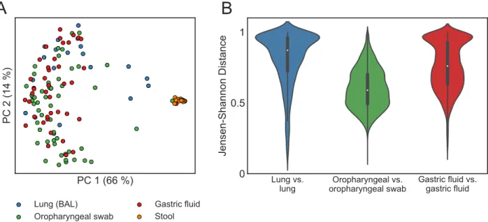
despite genus-level similarities, OTU-level aerodigestive communities are distinct and highly variable across people. The overall community composition was significantly different between sites (PERMANOVA on JSDs between BAL, gastric fluid, and oropharyngeal samples in the
two sequencing batches separately, p < 0.001,Fig 2A). Furthermore, lung communities were
very different across people (median lung-lung JSD = 0.87) while oropharyngeal communities
tended to be more similar (median oropharyngeal-oropharyngeal JSD = 0.59,Fig 2B).
Aerodigestive microbiome within people
We compared aerodigestive communities within patients who had multiple sites sequenced (Table 2,Fig 3). Oropharyngeal and gastric fluid communities are similar within patients
(median JSD = 0.56), reflecting that the mouth seeds the gastric microbiome [12,13]. The majority of patients had very different lung and oropharyngeal communities (median
JSD = 0.90), and these differences were significantly higher than either the lung-gastric fluid or
gastric fluid-oropharyngeal beta diversities (p < 1× 10⁻⁸,Fig 3A). Surprisingly, lung and
stomach communities were as similar to each other as stomach and oropharyngeal communi-ties (median JSD = 0.56 for both comparisons, p = 0.6).
We next identified specific microbes which exchange between aerodigestive sites within people. To do this, we reasoned that an actively exchanging microbe’s abundances in two
sites should be correlated across patients (S2 FigandMethods). In other words, if an OTU
is exchanged between two sites, if we observe that its abundance is low in both sites of one patient and high abundance in one site of another patient, then we would expect that its abundance in the second site of that second patient will also be high. We identified 13 OTUs exchanged between lung and oropharyngeal, 76 between gastric fluid and lung, and 117 between oropharyngeal and gastric fluid communities. These results were statistically signifi-cant: we found a maximum of 2 exchanged OTUs between sites in our null analysis. The low number of directly exchanged OTUs between the oropharynx and lungs supports the finding that these sites are more distinct than others in the aerodigestive tract. The lungs and stomach exchange fewer OTUs than the oropharynx and stomach even though they have comparable
Fig 1. Aerodigestive communities have similar predominant genera. Bar plots showing relative abundances of aerodigestive microbiomes collapsed to the genus level for the 66 patients with all sequencing data from all three aerodigestive sites. Each column corresponds to one patient who had all three aerodigestive sites sequenced (N = 19 non-aspirators, 23 aspirators, 24 untested). Phyla in legend are those with mean abundance > 0.01 across all patients. Any other phyla are colored gray.
intra-patient similarities, suggesting that factors other than specific bacterial exchange contrib-utes to the similarity between lungs and stomachs within patients.
Random Forest classifiers trained to distinguish between sites (ensuring that samples from the same patient were in the same train/test set) were able to identify a generalizable oropha-ryngeal microbial signature that distinguishes the oropharynx from other sites across people
(AUC = 0.95 for both gastric fluid and lung comparisons,Fig 3B). Interestingly, when we
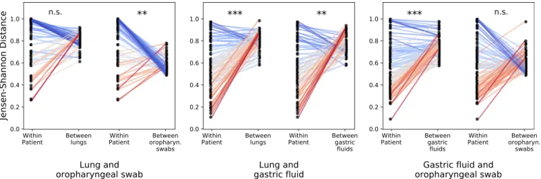
com-pared within-patient similarities across sites to across-patient similarities for the same sites, we found that lung and stomach communities within patients were more similar than lungs across
patients and than stomachs across patients (Fig 4, p < 1× 10⁻⁷,S3 Table). Thus, while there
exists a “core” oropharyngeal microbiome across people, lung and gastric communities are more variable and driven primarily by the person rather than body site. These results challenge the prevailing hypothesis that human-associated microbial communities are primarily driven by body habitat and instead suggest that patient-specific relationships may be equally, if not
more, important in determining community structure in the aerodigestive microbiome [31,
32,33].
Aspiration modulates the relationship between lung and oropharyngeal
microbiomes but not the lung and stomach
Next, we investigated the impact of oropharyngeal dysphagia and aspiration on the aerodiges-tive microbiome. To assess whether there were large-scale differences in the microbiomes of aspirators and non-aspirators, we compared the alpha diversity for each aerodigestive site between these patient groups. Aspirators did not have significantly different alpha diversity in
any of the aerodigestive sites for any of the metrics we compared (S3 Fig). Next, we attempted
to identify individual OTUs which were differentially abundant between aspirators and non-aspirators. No OTUs or genera were significant in any aerodigestive site after correcting for
multiple tests (S4 Table).
Fig 2. Lung and gastric communities are more variable across people than oropharyngeal communities. (A) PCoA plot of aerodigestive and stool microbial communities for all patients in the one sequencing batch (N = 21 BAL, 52 oropharyngeal swab, 43 gastric fluid, and 14 stool samples). The PCoA plot of the samples in the other sequencing batch are included inS1 Fig. (B) Violin plots of the Jensen-Shannon distance (JSD) between samples from the same site across different patients. A JSD close to 1 indicates that communities are very different (less similar).S4andS5
Figs show these results with the Bray Curtis distance metric instead of JSD.
We next leveraged our within-patient sampling to investigate the effect of aspiration on the relationships between sites in the aerodigestive tract. Aspirators had significantly more similar
lung and oropharyngeal communities than non-aspirators (Fig 5A, p = 0.04) and were much
more likely to have the pre-defined oropharyngeal-lung microbes in both their oropharynx
and lungs than non-aspirators (p = 4× 10⁻⁵) (Fig 5B). Lung-oropharynx exchanged OTUs
co-occurred in a median of 40% of aspirators’ lung and oropharyngeal communities but only 17% of non-aspirators’. Aspirators were not more likely to have stomach-lung microbes present in
both the lungs and gastric fluid than non-aspirators (Fig 5B, p = 0.5), and lung and gastric
communities of aspirating patients were not necessarily more similar to each other than those
of non-aspirating patients (Fig 5A, p = 0.6).
To identify potential microbial biomarkers of aspiration, we looked at the exchanged OTUs which were most frequently present in the lung and oropharyngeal communities of aspirators relative to non-aspirators. In the oropharyngeal-lung exchanged OTUs, these were an
unknown OTU in theFlavobacteriaceae family, OTUs in the Fusobacterium, Rothia,
Fig 3. Within patients, aerodigestive communities are similar but lung and oropharynx remain most distinct. (A) Jensen-Shannon distances between samples from different sites from the same patient. Comparisons between stool and oropharynx are included to contextualize these results, as these are expected to be very different. All comparisons are significant (Wilcoxon rank sums test calculated with Python’s scipy.stats.ranksums function) except the lung and gastric fluid vs. gastric fluid and oropharyngeal swab beta diversities (p = 0.6). Lung and oropharyngeal vs. oropharyngeal and stool, p = 0.005. All other comparisons:
p < 1 × 10−8.S6 Figshows these results with the Bray Curtis distance metric. (B) ROC curve of classifiers distinguishing different aerodigestive sites. Mean areas under the ROC curve (AUCs) are reported in parentheses in the legend.
Veillonella genera, and an unknown OTU in the Prevotellaceae family, among others (Table 3,
gastric-lung OTUs inS5 Table).
We used Random Forest classifiers trained on the presence of exchanged OTUs in different sites and on the entire aerodigestive communities in order to test their potential as diagnostics for aspiration. We evaluated these classifiers by calculating the Fisher’s exact p-values and the area under the ROC curve (AUC) on leave-one-out predictions, where an AUC of 1.0 indicates
a perfect classifier and an AUC of 0.5 is a classifier which assigns labels randomly [34]. The
concordant presence or absence of exchanged OTUs in the two sites slightly improved
Fig 4. Lung and gastric microbial communities are driven primarily by person rather than body site. We compared the within-patient JSD for all pairs of aerodigestive sites with the average across-patient JSD between each of the sites in the within-patient comparison. Each panel shows different aerodigestive pairs; the two slope graphs correspond to different across-site comparisons; and each point corresponds to one patient. The left points in each slope graph show the within-patient JSD for the respective pair of sites for each within-patient (and are the same within each panel). The right points show the average JSD between the corresponding patient’s site X and all other patients’ site X. For example, the middle panel shows that the JSD between lung and gastric fluid communities within patients is lower than the average JSD between different lungs (left slope graph) and the average JSD between different gastric fluid samples (right slope graph). P values were calculated with a Wilcoxon signed-rank p-values using Python’s scipy.stats.wilcoxon function.���:p < 10−10;��:10−10<p < 10−7, table of comparisons
and p-values can be found inS3 Table.S7 Figshows these results with the Bray Curtis distance metric.
https://doi.org/10.1371/journal.pone.0216453.g004
Fig 5. Dysphagia increases aspiration of microbes from the oropharynx but not the stomach. (A) Intra-patient Jensen Shannon distance for different aerodigestive site comparisons in non-aspirators (brown) and aspirators (pink). Each point represents one patient. P-values (Wilcoxon rank sums test, calculated with Python’s scipy.stats.ranksums function): lung and oropharyngeal swabp = 0.04, lung and gastric fluid p = 0.5, gastric fluid and oropharyngeal swab p = 0.8.S8 Figshows these results with the Bray Curtis distance metric. (B) Percentage of patients with the previously defined exchanged microbes present in both of the respective sites (x-axis) in non-aspirators (brown) and aspirators (pink). Each pair of points represents one exchanged OTU. P-values (paired t-test onlog10
prevalence values, calculated with Python’s scipy.stats.ttest_rel function: lung and oropharyngeal swabp = 4 × 10⁻⁵, lung and gastric fluidp = 0.5, gastric
fluid and oropharyngeal swabp = 0.1.
classifiers based on the oropharyngeal-lung OTUs but not the ones based on the lung-gastric OTUs, relative to classifiers based on the presence of the exchanged OTUs in either site alone (Table 4; classifiers trained on the abundance of exchanged OTUs presented inS6 Table). However, these marginal results suggest that additional work will be necessary to develop these exchanged OTUs into reliable diagnostic biomarkers.
Using Random Forest classifiers trained on the entire microbiomes, we found that combin-ing the oropharynx and lung communities resulted in a better classifier than either community
alone (Table 5). Surprisingly, the classifiers trained on oropharyngeal and gastric communities
performed well, despite our expectation that aspiration-induced changes in the microbiome would manifest in the lungs rather than the oropharynx or stomach. We confirmed that the
Table 3. Prevalence of lung-oropharynx exchanged OTUs.
Family Genus Non-aspirator Aspirator Difference
Flavobacteriaceae 8.7 48.0 39.3 Fusobacteriaceae Fusobacterium 30.4 68.0 37.6 Micrococcaceae Rothia 8.7 44.0 35.3 Veillonellaceae Veillonella 26.1 60.0 33.9 Prevotellaceae 43.5 76.0 32.5 Porphyromonadaceae Porphyromonas 39.1 68.0 28.9 Streptococcaceae Streptococcus 13.0 40.0 27.0 Veillonellaceae Centipeda 8.7 32.0 23.3 Prevotellaceae Prevotella 17.4 36.0 18.6 Leptotrichiaceae Streptobacillus 21.7 40.0 18.3 Fusobacteriaceae Fusobacterium 17.4 32.0 14.6 Aerococcaceae Abiotrophia 21.7 28.0 6.3 Neisseriaceae Neisseria 17.4 20.0 2.6
Prevalence is calculated as the percentage of patients who have the OTU present in both their lungs and oropharynx, calculated separately among aspirators (N = 25) and non-aspirators (N = 23). OTUs are ordered by their differential prevalence in aspirators relative to non-aspirators, and are labeled with their family- and genus-level taxonomies. Blank genus names indicate OTUs which were not annotated at the genus level. A similar table for the lung-gastric exchange OTUs can be found inS1 Table.
https://doi.org/10.1371/journal.pone.0216453.t003
Table 4. Classifiers based on the presence of exchanged OTUs.
Lung-oropharynx OTUs (13) AUC p N (non-asp/asp)
Lung 0.66 0.08 33/33 Oropharyngeal 0.57 0.35 43/36 Concordance 0.63 0.05 23/25 Lung-gastric OTUs (76) Lung 0.60 0.14 33/33 Gastric fluid 0.65 0.03 48/41 Concordance 0.53 1.0 28/29
(Top) Classifiers built from the presence of lung-oropharynx exchanged OTUs. (Bottom) Classifiers built from the presence of lung-gastric exchanged OTUs. Rows indicate which microbial community was used to train each classifier. In the “concordance” classifiers, OTUs which were either present or absent in both sites were coded as 1 and OTUs which were present in one site but absent in the other were coded as 0. AUCs are calculated as the area under the ROC curve from leave-one-out predictions. Fisher’s exact p values are calculated on the leave-one-out predictions. Similar classifiers built from the abundance of exchanged OTUs are shown inS6 Table.
patients’ aspiration status was not confounded with proton pump inhibitor usage (Fisher exact
p-value = 0.8,S1 Table), but there may be other co-morbidities or unmeasured confounders
that could be driving the differences detected in these communities. However, taken together, these results suggest that identifying a biomarker for aspiration based on bacteria in both the lungs and oropharynx may be possible, and that these two sites together contain more infor-mation about a patient’s aspiration status than either site alone.
Reflux may impact the relationship between lung and stomach
microbiomes
Reflux profiles for the 28 patients are shown inTable 6. The percent of full column, distal, and
proximal reflux were slightly negatively correlated with gastric-lung JSD, indicating that patients with more frequent reflux may have more similar gastric and lung microbial
commu-nities (Fig 6). However, the large range of gastric-lung JSDs across all patients and relatively
weak correlation suggests that other non-reflux factors likely contribute more to the similari-ties between gastric and lung communisimilari-ties that are observed across all people. Similarly, we
were not able to identify relationships between gastric-lung JSD and PPI usage (S9andS10
Figs).
Table 5. Classifiers based on perturbed relationship between lung and oropharyngeal microbiota can distinguish aspirators from non-aspirators.
Sites AUC Fisher p-value N (non-asp/asp)
Lung 0.63 0.32 33/33
Oropharyngeal swab 0.69 0.02 43/36
Gastric fluid 0.66 0.08 48/41
Lung and oropharyngeal swab 0.79 0.01 23/25
Lung and gastric fluid 0.66 0.02 28/29
Oropharyngeal swab and gastric fluid 0.73 0.003 35/32
All three sites 0.78 0.01 19/23
Areas under the ROC curve (AUC) and Fisher p-values calculated from classifiers trained on the entire microbial communities. Each row is a different classifier based on different combinations of aerodigestive communities, indicated in the “Sites” column. In the multi-site classifiers, the abundances of OTUs in different sites were used as separate features. AUCs and Fisher’s p values were calculated from the leave-one-out predictions for each sample.
https://doi.org/10.1371/journal.pone.0216453.t005
Table 6. Reflux characteristics for 28 patients measured by pH-MII.
Mean (std)
Number of acid episodes 23.1 (25.5)
Number of nonacid episodes 15.4 (16.4)
Number of pH only episodes 16.3 (12.5)
Number of total reflux episodes 38.0 (32.4)
Percent time proximal reflux 0.48 (0.46)
Percent time distal reflux 1.3 (1.1)
Percent time pH < 4 5.4 (5.2)
Number abnormal by pH-metry 9/28
Number abnormal by MII 3/28
Conclusion
In this study, we characterized the relationships between the oropharyngeal, lung, and gastric microbiomes in a large pediatric cohort with and without swallowing dysfunction. Leveraging our simultaneous sampling of multiple sites per patient, we find that there exists a “core” oro-pharyngeal microbiome across patients, but lung and gastric communities vary and are dis-tinct to individuals. Within patients, lung and oropharyngeal communities remain most distinct. We show for the first time that in patients with impaired swallowing, the lung micro-biome shifts toward oropharyngeal flora rather than gastric flora. Our results also suggest that identifying biomarkers for aspiration based on the presence of certain bacteria in both the lungs and oropharynx may ultimately be possible.
There are several limitations to our study. First, because it is unethical to perform bronchos-copies on healthy children, our patients in this study had respiratory symptoms. Furthermore,
these patients were on variety of medications (Table 1), which may affect microbial community
compositions and relationships. However, we believe that our patient population represents patients typically seen in aerodigestive centers and that understanding the degree of microbial exchange is most clinically relevant in patients with symptoms. The microbial populations we found in this study are similar to those of previously published studies of both healthy and
symptomatic adults which reinforces the validity of our results [12,13,17,18]. We also
con-firmed that medication use and symptoms were not confounded with aspiration status (S1
Table). Second, the number of patients undergoing pH-MII testing was relatively small which limits our conclusions about the impact of gastroesophageal reflux on the lung. However, our study raises enough concerns about the significance of oropharyngeal-lung exchange in
Fig 6. Reflux severity may correlate with the similarity between lung and gastric communities. Each plot shows the correlation between different reflux measures and the within-patient Jensen-Shannon distance between BAL and gastric fluid samples. Points are colored according to aspiration status. All reflux measures include both acid- and non-acid reflux. Spearman correlation and p-values: total number of reflux episodesρs=−0.25, p = 0.2, percentage of full column reflux events ρs=
−0.40, p = 0.04, percent of time reflux was proximal ρs=−0.55, p = 0.002, percent of time reflux was distal ρs=−0.45, p = 0.02.
children with impaired swallowing that gastroesophageal reflux should not be considered as the primary source of microbial exchange causing pulmonary symptoms. Third, the diagnosis of oropharyngeal dysphagia in this study was based on VFSS. While this only categorizes patients based on a “one-point-in-time” study, it is the gold standard test to diagnose oropha-ryngeal dysphagia in children and therefore we feel it is appropriate for use in this study. Finally, the low biomass of BAL and gastric fluid samples could lead to sequencing artifacts or contamination. We did not explicitly remove potential background environmental or sampling sequences from our data, though our sampling methods was carefully developed in order to
minimize potential contaminants [12,16]. The low biomass of BAL and gastric fluid samples
also resulted in fewer total sequencing reads than the oropharyngeal swabs (S11 Fig), perhaps
contributing partially to the high variability we observed between these communities. However, many of our conclusions depend upon within-patient analyses, which reduce spurious results.
Despite these limitations, our findings have broad clinical implications for the understand-ing and treatment of oropharyngeal dysphagia with resultant aspiration. Our clinical findunderstand-ing that the lung microbiome in children with aspiration shifts toward the oropharynx rather than the stomach highlights the importance of understanding the primary driver of microbial exchange so that therapies can be tailored accordingly. For example, if the mechanism of lung symptoms and disease in aspirating children results from a microbial shift towards oropharyn-geal flora, anti-reflux surgery will be of no benefit to preventing oropharynoropharyn-geal-lung exchange. Instead, therapies may need to be tailored to focused on changing oropharyngeal flora or sali-vary properties.
While there are no existing pediatric microbiome studies of the aerodigestive microbiome in patients with dysphagia, there is evidence that children with oropharyngeal dysphagia are predisposed to pneumonia and that this could be due to increased aspiration of microbes from the oral microbiome. In a study of 382 children undergoing VFSS, evidence of aspiration pre-dicted pneumonia risk, though the causative organisms for these pneumonias were not known
[35]. In cohort of elderly aspirating patients, oral colonization by respiratory pathogens was
associated with increased risk of pneumonia, highlighting the potential importance of oral
flora in influencing the lung outcomes [36]. Finally, a previous study of healthy adults found
that individuals with oropharyngeal bacteria in their lungs had increased evidence of inflam-matory metabolomic signals, suggesting that even a change of lung flora to commensal
oro-pharyngeal bacteria can trigger inflammation even in healthy patients [22]. Our results add to
these findings and suggest that changes in the lung microbiome towards oropharyngeal flora merit additional study to determine if these shifts result in increased morbidity or worse clini-cal outcomes, including the development of pneumonia.
From a microbial perspective, we identified bacterial families and genera that are more commonly exchanged between the oropharynx and lungs of children that aspirate than of children with intact swallowing mechanism. While there are no other 16S sequencing studies determining aspiration pneumonia risk in children, there is evidence from the adult literature that similar bacteria are involved in aspiration pneumonia risk. For example, oropharyngeal Streptococci were found to be more abundant in the lungs of adults with pneumonia and
aspi-ration risk factors than without aspiaspi-ration risk [37]. In a study of 173 adults in long term care
facilities, patients with oropharyngealPrevotella and Veillonella had increased risk of death
from pneumonia compared to patients who had oropharyngealNeisseria and Fusobacterium
[38]. Our study is a critical first step toward identifying bacteria present in the oropharynx
and lungs of aspirating children that may result in higher risk for pneumonias, with additional studies needed to determine their impact on pediatric outcomes.
In summary, our findings suggest that interventions to reduce aspiration-related respira-tory complications due to increased microbial exchange should target aspiration from the
oropharynx rather than the stomach. This microbial data supports the clinical observation that antireflux surgery fails to prevents pulmonary complications such as pneumonias or
hospitali-zations [3,7,8,9,10,11]. By simultaneously sampling multiple sites per patient, we show that
the lung and stomach microbiomes are highly variable across patients and determined primar-ily by patient rather than body site. If aerodigestive microbial communities are indeed specific to each individual, interventions targeting the aerodigestive microbiome may benefit from personalized medicine approaches. Finally, understanding the relationships between aerodi-gestive communities in aspirating and non-aspirating patients provides insight into the poten-tial pathophysiology behind aspiration-related respiratory outcomes and suggests potenpoten-tial diagnostics and therapeutics for future investigation.
Supporting information
S1 Table. Patient demographics separated by aspiration status. (PDF)
S2 Table. Number of patients with each combination of body sites sequenced, separated by aspiration status.
(PDF)
S3 Table. Lung and gastric microbial communities are driven primarily by person rather than body site. P-values associated with analysis presented inFig 4.
(PDF)
S4 Table. Differential abundance analysis results. (PDF)
S5 Table. Prevalence of lung-gastric exchanged OTUs in aspirators and non-aspirators. (PDF)
S6 Table. Classifiers based on the abundance of exchanged OTUs. (PDF)
S1 Fig. PCoA plot of aerodigestive and stool microbial communities for patients in the sequencing batch not shown in the main text.
(PDF)
S2 Fig. Schematic illustrating definition of exchanged OTUs. (PDF)
S3 Fig. Alpha diversity of each aerodigestive site compared between aspirators and non-aspirators.
(PDF)
S4 Fig. Bray-Curtis PCoA plots of aerodigestive and stool microbial communities for patients in the samples shown inFig 2A.
(PDF)
S5 Fig. Violin plots of the Bray-Curtis distance between samples from the same site across different patients.
(PDF)
S6 Fig. Bray-Curtis distances between samples from different sites from the same patient. (PDF)
S7 Fig. Comparison between within-patient and between-patient Bray Curtis distances, as inFig 4.
(PDF)
S8 Fig. Intra-patient Bray Curtis distance for different aerodigestive site comparisons in non-aspirators and aspirators.
(PDF)
S9 Fig. Lung-gastric JSD vs. PPI status. (PDF)
S10 Fig. Lung-gastric JSD vs. reflux, colored by PPI status. (PDF)
S11 Fig. Sequencing reads per sample. (PDF)
Acknowledgments
We thank Scott Olesen and Manu Kumar for helpful discussions on statistical analyses, and Nathaniel Chu and members of the Alm lab for helpful discussions on presenting and visualiz-ing the results.
This work was supported by NIH R01 DK097112 (R.R.), the Boston Children’s Hospital Translational Research Program (R.R.), and the North American Society for Pediatric Gastro-enterology, Hepatology and Nutrition Grant for Diseases of the Upper Gastrointestinal Tract (R.R.). Computational resources for this work were supported by the Center for Micro-biome Informatics and Therapeutics (E.A.). C.D. acknowledges support through the National Defense Science & Engineering Graduate Fellowship (NDSEG).
Author Contributions
Conceptualization: Rachel Rosen. Data curation: Claire Duvallet.Formal analysis: Claire Duvallet, Eric Alm. Funding acquisition: Rachel Rosen.
Investigation: Kara Larson, Scott Snapper, Katherine Freer, Kara May. Methodology: Eric Alm.
Resources: Sonia Iosim, Ann Lee. Supervision: Eric Alm, Rachel Rosen. Visualization: Claire Duvallet.
Writing – original draft: Claire Duvallet, Rachel Rosen.
Writing – review & editing: Claire Duvallet, Eric Alm, Rachel Rosen.
References
1. Holas MA, DePippo KL, Reding MJ. Aspiration and Relative Risk of Medical Complications Following Stroke. Archives of Neurology. 1994; 51(10):1051–1053.https://doi.org/10.1001/archneur.1994. 00540220099020PMID:7945003
2. Thomson J, Hall M, Ambroggio L, Stone B, Srivastava R, Shah SS, et al. Aspiration and Non-Aspiration Pneumonia in Hospitalized Children With Neurologic Impairment. Pediatrics. 2016; 137(2):e20151612– e20151612.https://doi.org/10.1542/peds.2015-1612PMID:26787045
3. Jancelewicz T, Lopez ME, Downard CD, Islam S, Baird R, Rangel SJ, et al. Surgical management of gastroesophageal reflux disease (GERD) in children: A systematic review. Journal of Pediatric Surgery. 2017; 52(8):1228–1238.https://doi.org/10.1016/j.jpedsurg.2016.09.072PMID:27823773
4. Lasser MS, Liao JG, Burd RS. National Trends in the Use of Antireflux Procedures for Children. PEDI-ATRICS. 2006; 118(5):1828–1835.https://doi.org/10.1542/peds.2006-1185PMID:17079551
5. Hatch LD, Scott TA, Walsh WF, Goldin AB, Blakely ML, Patrick SW. National and regional trends in gas-trostomy in very low birth weight infants in the USA: 2000–2012. Journal of Perinatology. 2018; 38 (9):1270–1276.https://doi.org/10.1038/s41372-018-0145-4PMID:29925865
6. Goldin AB, Garrison M, Christakis D. Variations Between Hospitals in Antireflux Procedures in Children. Archives of Pediatrics & Adolescent Medicine. 2009; 163(7):658.https://doi.org/10.1001/archpediatrics. 2009.103
7. Barnhart DC, Hall M, Mahant S, Goldin AB, Berry JG, Faix RG, et al. Effectiveness of Fundoplication at the Time of Gastrostomy in Infants With Neurological Impairment. JAMA Pediatrics. 2013; 167(10):911. https://doi.org/10.1001/jamapediatrics.2013.334PMID:23921627
8. Lee SL, Shabatian H, Hsu JW, Applebaum H, Haigh PI. Hospital admissions for respiratory symptoms and failure to thrive before and after Nissen fundoplication. Journal of Pediatric Surgery. 2008; 43 (1):59–65.https://doi.org/10.1016/j.jpedsurg.2007.09.020PMID:18206456
9. Goldin AB, Sawin R, Seidel KD, Flum DR. Do Antireflux Operations Decrease the Rate of Reflux-Related Hospitalizations in Children? Pediatrics. 2006; 118(6):2326–2333.https://doi.org/10.1542/ peds.2006-2212PMID:17142515
10. Yeh J, McGrath-Morrow SA, Collaco JM. Oxygen weaning after hospital discharge in children with bronchopulmonary dysplasia. Pediatric Pulmonology. 2016; 51(11):1206–1211.https://doi.org/10. 1002/ppul.23442PMID:27093064
11. Srivastava R, Downey EC, O’Gorman M, Feola P, Samore M, Holubkov R, et al. Impact of Fundoplica-tion Versus Gastrojejunal Feeding Tubes on Mortality and in Preventing AspiraFundoplica-tion Pneumonia in Young Children With Neurologic Impairment Who Have Gastroesophageal Reflux Disease. PEDIAT-RICS. 2009; 123(1):338–345.https://doi.org/10.1542/peds.2007-1740PMID:19117901
12. Bassis CM, Erb-Downward JR, Dickson RP, Freeman CM, Schmidt TM, Young VB, et al. Analysis of the Upper Respiratory Tract Microbiotas as the Source of the Lung and Gastric Microbiotas in Healthy Individuals. mBio. 2015; 6(2):e00037–15.https://doi.org/10.1128/mBio.00037-15PMID:25736890
13. Charlson ES, Bittinger K, Haas AR, Fitzgerald AS, Frank I, Yadav A, et al. Topographical Continuity of Bacterial Populations in the Healthy Human Respiratory Tract. American Journal of Respiratory and Critical Care Medicine. 2011; 184(8):957–963.https://doi.org/10.1164/rccm.201104-0655OCPMID: 21680950
14. Rosen R, Hu L, Amirault J, Khatwa U, Ward DV, Onderdonk A. 16S Community Profiling Identifies Pro-ton Pump Inhibitor Related Differences in Gastric, Lung, and Oropharyngeal Microflora. The Journal of Pediatrics. 2015; 166(4):917–923.https://doi.org/10.1016/j.jpeds.2014.12.067PMID:25661411
15. Venkataraman A, Bassis CM, Beck JM, Young VB, Curtis JL, Huffnagle GB, et al. Application of a Neu-tral Community Model To Assess Structuring of the Human Lung Microbiome. mBio. 2015; 6(1): e02284–14.https://doi.org/10.1128/mBio.02284-14PMID:25604788
16. Segal LN, Alekseyenko AV, Clemente JC, Kulkarni R, Wu B, Chen H, et al. Enrichment of lung micro-biome with supraglottic taxa is associated with increased pulmonary inflammation. Micromicro-biome. 2013; 1 (1):19.https://doi.org/10.1186/2049-2618-1-19PMID:24450871
17. Erb-Downward JR, Thompson DL, Han MK, Freeman CM, McCloskey L, Schmidt LA, et al. Analysis of the Lung Microbiome in the “Healthy” Smoker and in COPD. PLoS ONE. 2011; 6(2):e16384.https://doi. org/10.1371/journal.pone.0016384PMID:21364979
18. Morris A, Beck JM, Schloss PD, Campbell TB, Crothers K, Curtis JL, et al. Comparison of the Respira-tory Microbiome in Healthy Nonsmokers and Smokers. American Journal of RespiraRespira-tory and Critical Care Medicine. 2013; 187(10):1067–1075.https://doi.org/10.1164/rccm.201210-1913OCPMID: 23491408
19. Tantipong H, Morkchareonpong C, Jaiyindee S, Thamlikitkul V. Randomized Controlled Trial and Meta-analysis of Oral Decontamination with 2% Chlorhexidine Solution for the Prevention of Ventilator-Asso-ciated Pneumonia. Infection Control & Hospital Epidemiology. 2008; 29(02):131–136.https://doi.org/ 10.1086/526438
20. Koeman M, van der Ven AJAM, Hak E, Joore HCA, Kaasjager K, de Smet AGA, et al. Oral Decontami-nation with Chlorhexidine Reduces the Incidence of Ventilator-associated Pneumonia. American
Journal of Respiratory and Critical Care Medicine. 2006; 173(12):1348–1355.https://doi.org/10.1164/ rccm.200505-820OCPMID:16603609
21. bo Hu H, ju Huang H, ying Peng Q, Lu J, yun Lei X. Prospective study of colonization and infection because of Pseudomonas aeruginosa in mechanically ventilated patients at a neonatal intensive care unit in China. American Journal of Infection Control. 2010; 38(9):746–750.https://doi.org/10.1016/j.ajic. 2010.02.012
22. Segal LN, Clemente JC, Tsay JCJ, Koralov SB, Keller BC, Wu BG, et al. Enrichment of the lung micro-biome with oral taxa is associated with lung inflammation of a Th17 phenotype. Nature Microbiology. 2016; 1(5).https://doi.org/10.1038/nmicrobiol.2016.31PMID:27572644
23. Rosen R, Nurko S. The Importance of Multichannel Intraluminal Impedance in the Evaluation of Chil-dren with Persistent Respiratory Symptoms. The American Journal of Gastroenterology. 2004; 99 (12):2452–2458 PMID:15571595
24. Wang Q, Garrity GM, Tiedje JM, Cole JR. Naive Bayesian classifier for rapid assignment of rRNA sequences into the new bacterial taxonomy. Applied and environmental microbiology. 2007; 73 (16):5261–5267.https://doi.org/10.1128/AEM.00062-07PMID:17586664
25. Endres DM, Schindelin JE. A new metric for probability distributions. IEEE Transactions on Information Theory. 2003; 49(7):1858–1860.https://doi.org/10.1109/TIT.2003.813506
26. Preheim SP, Perrotta AR, Friedman J, Smilie C, Brito I, Smith MB, et al. Computational Methods for High-Throughput Comparative Analyses of Natural Microbial Communities. In: Methods in Enzymology. Elsevier; 2013. p. 353–370. Available from:https://doi.org/10.1016/b978-0-12-407863-5.00018-6.
27. Gajer P, Brotman RM, Bai G, Sakamoto J, Schutte UME, Zhong X, et al. Temporal Dynamics of the Human Vaginal Microbiota. Science Translational Medicine. 2012; 4(132):132ra52–132ra52.https:// doi.org/10.1126/scitranslmed.3003605PMID:22553250
28. Heintz-Buschart A, May P, Laczny CC, Lebrun LA, Bellora C, Krishna A, et al. Integrated multi-omics of the human gut microbiome in a case study of familial type 1 diabetes. Nature Microbiology. 2016; 2(1). https://doi.org/10.1038/nmicrobiol.2016.180
29. Jones E, Oliphant T, Peterson P, et al. SciPy: Open source scientific tools for Python; 2001–. Available from:http://www.scipy.org/.
30. Pedregosa F, Varoquaux G, Gramfort A, Michel V, Thirion B, Grisel O, et al. Scikit-learn: Machine Learning in Python. Journal of Machine Learning Research. 2011; 12:2825–2830.
31. Costello EK, Lauber CL, Hamady M, Fierer N, Gordon JI, Knight R. Bacterial Community Variation in Human Body Habitats Across Space and Time. Science. 2009; 326(5960):1694–1697.https://doi.org/ 10.1126/science.1177486PMID:19892944
32. Huttenhower C, Gevers D, Knight R, Abubucker S, Badger JH, Chinwalla AT, et al. Structure, function and diversity of the healthy human microbiome. Nature. 2012; 486(7402):207–214.https://doi.org/10. 1038/nature11234
33. Lozupone CA, Stombaugh J, Gonzalez A, Ackermann G, Wendel D, Vazquez-Baeza Y, et al. Meta-analyses of studies of the human microbiota. Genome Research. 2013; 23(10):1704–1714.https://doi. org/10.1101/gr.151803.112PMID:23861384
34. Fawcett T. An introduction to ROC analysis. Pattern Recognition Letters. 2006; 27(8):861–874.https:// doi.org/10.1016/j.patrec.2005.10.010
35. Weir K, McMahon S, Barry L, Ware R, Masters IB, Chang AB. Oropharyngeal aspiration and pneumo-nia in children. Pediatric Pulmonology. 2007; 42(11):1024–1031.https://doi.org/10.1002/ppul.20687 PMID:17893917
36. Ortega O, Sakwinska O, Combremont S, Berger B, Sauser J, Parra C, et al. High prevalence of coloni-zation of oral cavity by respiratory pathogens in frail older patients with oropharyngeal dysphagia. Neu-rogastroenterology & Motility. 2015; 27(12):1804–1816.https://doi.org/10.1111/nmo.12690
37. Akata K, Yatera K, Yamasaki K, Kawanami T, Naito K, Noguchi S, et al. The significance of oral strepto-cocci in patients with pneumonia with risk factors for aspiration: the bacterial floral analysis of 16S ribo-somal RNA gene using bronchoalveolar lavage fluid. BMC Pulmonary Medicine. 2016; 16(1).https:// doi.org/10.1186/s12890-016-0235-zPMID:27169775
38. Kageyama S, Takeshita T, Furuta M, Tomioka M, Asakawa M, Suma S, et al. Relationships of Varia-tions in the Tongue Microbiota and Pneumonia Mortality in Nursing Home Residents. The Journals of Gerontology: Series A. 2017.