HAL Id: hal-02192633
https://hal.archives-ouvertes.fr/hal-02192633
Submitted on 24 Jul 2019
HAL is a multi-disciplinary open access
archive for the deposit and dissemination of
sci-entific research documents, whether they are
pub-lished or not. The documents may come from
teaching and research institutions in France or
abroad, or from public or private research centers.
L’archive ouverte pluridisciplinaire HAL, est
destinée au dépôt et à la diffusion de documents
scientifiques de niveau recherche, publiés ou non,
émanant des établissements d’enseignement et de
recherche français ou étrangers, des laboratoires
publics ou privés.
Daniele Armaleo, Olaf Müller, François Lutzoni, Guillaume Blanc, Helge
Bode, Frank Collart, Francesco Dal Grande, Fred Dietrich, Igor Grigoriev,
Suzanne Joneson, et al.
To cite this version:
Daniele Armaleo, Olaf Müller, François Lutzoni, Guillaume Blanc, Helge Bode, et al.. The lichen
sym-biosis re-viewed through the genomes of Cladonia grayi and its algal partner Asterochloris glomerata.
BMC Genomics, BioMed Central, 2019, 20 (1), pp.605. �10.1186/s12864-019-5629-x�. �hal-02192633�
R E S E A R C H A R T I C L E
Open Access
The lichen symbiosis re-viewed through the
genomes of Cladonia grayi and its algal
partner Asterochloris glomerata
Daniele Armaleo
1*, Olaf Müller
1,2, François Lutzoni
1, Ólafur S. Andrésson
3, Guillaume Blanc
4, Helge B. Bode
5,
Frank R. Collart
6, Francesco Dal Grande
7, Fred Dietrich
2, Igor V. Grigoriev
8,9, Suzanne Joneson
1,10, Alan Kuo
8,
Peter E. Larsen
6, John M. Logsdon Jr
11, David Lopez
12, Francis Martin
13, Susan P. May
1,14, Tami R. McDonald
1,15,
Sabeeha S. Merchant
9,16, Vivian Miao
17, Emmanuelle Morin
13, Ryoko Oono
18, Matteo Pellegrini
19,
Nimrod Rubinstein
20,21, Maria Virginia Sanchez-Puerta
22, Elizabeth Savelkoul
11, Imke Schmitt
7,23, Jason C. Slot
24,
Darren Soanes
25, Péter Szövényi
26, Nicholas J. Talbot
27, Claire Veneault-Fourrey
13,28and Basil B. Xavier
3,29Abstract
Background: Lichens, encompassing 20,000 known species, are symbioses between specialized fungi (mycobionts), mostly ascomycetes, and unicellular green algae or cyanobacteria (photobionts). Here we describe the first parallel genomic analysis of the mycobiont Cladonia grayi and of its green algal photobiont Asterochloris glomerata. We focus on genes/predicted proteins of potential symbiotic significance, sought by surveying proteins differentially activated during early stages of mycobiont and photobiont interaction in coculture, expanded or contracted protein families, and proteins with differential rates of evolution.
Results: A) In coculture, the fungus upregulated small secreted proteins, membrane transport proteins, signal transduction components, extracellular hydrolases and, notably, a ribitol transporter and an ammonium transporter, and the alga activated DNA metabolism, signal transduction, and expression of flagellar components. B) Expanded fungal protein families include heterokaryon incompatibility proteins, polyketide synthases, and a unique set of G-proteinα subunit paralogs. Expanded algal protein families include carbohydrate active enzymes and a specific subclass of cytoplasmic carbonic anhydrases. The alga also appears to have acquired by horizontal gene transfer from prokaryotes novel archaeal ATPases and Desiccation-Related Proteins. Expanded in both symbionts are signal transduction components, ankyrin domain proteins and transcription factors involved in chromatin remodeling and stress responses. The fungal transportome is contracted, as are algal nitrate assimilation genes. C) In the mycobiont, slow-evolving proteins were enriched for components involved in protein translation, translocation and sorting.
(Continued on next page)
© The Author(s). 2019 Open Access This article is distributed under the terms of the Creative Commons Attribution 4.0 International License (http://creativecommons.org/licenses/by/4.0/), which permits unrestricted use, distribution, and reproduction in any medium, provided you give appropriate credit to the original author(s) and the source, provide a link to the Creative Commons license, and indicate if changes were made. The Creative Commons Public Domain Dedication waiver (http://creativecommons.org/publicdomain/zero/1.0/) applies to the data made available in this article, unless otherwise stated.
* Correspondence:darmaleo@duke.edu
Dedicated to Chicita Culberson, a mentor and friend who laid the groundwork on Cladonia grayi
1Department of Biology, Duke University, Durham, USA
(Continued from previous page)
Conclusions: The surveyed genes affect stress resistance, signaling, genome reprogramming, nutritional and structural interactions. The alga carries many genes likely transferred horizontally through viruses, yet we found no evidence of inter-symbiont gene transfer. The presence in the photobiont of meiosis-specific genes supports the notion that sexual reproduction occurs in Asterochloris while they are free-living, a phenomenon with implications for the adaptability of lichens and the persistent autonomy of the symbionts. The diversity of the genes affecting the symbiosis suggests that lichens evolved by accretion of many scattered regulatory and structural changes rather than through introduction of a few key innovations. This predicts that paths to lichenization were variable in different phyla, which is consistent with the emerging consensus that ascolichens could have had a few
independent origins.
Keywords: Algal virus, Coculture, Fungi, Gene expression, Gene family evolution, Horizontal gene transfer, Plant-fungal interactions, Symbiont autonomy, Symbiosis genes
Background
Simon Schwendener in 1869 [1] correctly recognized lichens as intimate symbioses between specialized fungi and phototrophic unicellular green algae as the main symbionts. Cyanobacteria were later recognized also as primary phototrophs in many lichens. In addition to the main symbiotic partners, lichens har-bor diverse communities of prokaryotes and fungi as cohabitants [2–6]. Recently, highly coevolved basidio-mycete yeasts were discovered in the cortex of many lichens [7], sometimes causing disease [8]. The de-tailed interactions of the various cohabitants with the main symbionts are being investigated [9]. Typically, lichens thrive in above-ground niches with limited water in diverse environments, often withstanding ex-treme heat, desiccation, or cold [3, 10]. Widespread across terrestrial ecosystems, often dominant carbon and nitrogen fixers in alpine, subalpine, and high lati-tude habitats, the estimated 18,000 to 20,000 lichen species [11], mostly ascomycetes, represent about 20% of all known fungi [12]. There are only about 120 li-chen phototroph species (photobionts) [10, 13], far fewer than the 20,000 known lichen fungal species (mycobionts). Lichens are named based on their mycobiont since the fungus is the most conspicuous partner and since the same photobiont species (alga or cyanobacterium) can be found in several different lichens.
Lichens can reproduce somatically through propagules comprising both symbionts, or sexually through meiotic fungal spores that must combine with the appropriate photobiont to re-form a lichen. Sexual reproduction is not commonly seen in trebouxoid lichen algae, although evi-dence supporting it has been found (Heterothallism prob-ably evolved from homothallism in Cladonia; genetic evidence for sex in Asterochlorissection). Lichens are well known for their unique and abundantly produced second-ary metabolites [14, 15]. The genetic, physiological, and structural integration of mycobionts and photobionts has
produced a vast array of beautifully differentiated partner-ships [16], with only occasional instability [17–19]. The fun-gal and lichen fossil record [20, 21] has placed fossils resembling extant lichen taxa in the Devonian-early Car-boniferous, 415–350 million years ago, and perhaps simpler mycobiont-photobiont associations even earlier [22, 23]. Despite their intimate coexistence for hundreds of millions of years and the construction of complex interfaces be-tween them [10, 24], lichen symbionts have not lost their genetic and cellular independence. Cell membranes are not breached in their interactions and genomes are not merged, although some have extended to lichens the concept of genome acquisition [25]. There is no evidence of horizontal gene transfer (HGT) between the symbionts, yet both have acquired genes from other sources (A low-GC region in Asterochloris is a remnant of a large virus insertion, an HGT-mediator, Inferences from differential transcription about nutritional fluxes at the symbiotic interface, and
Photobiont expanded families sections). The partners of
many lichens have been isolated and grown separately in axenic culture, but free-living stages of lichen fungi and their algae remain mostly cryptic in nature [18, 26–29]. However, these stages are not insignificant for the lichen life cycle [29]. The laboratory reconstitution of cultured lichen symbionts into fully developed lichens has a checkered his-tory [30], where the reproducibility needed for molecular investigations is still elusive. Molecular studies addressing functional aspects of this mutualistic symbiosis are few [9, 31–35], but the recent publication of several lichen -omics papers and datasets [9, 36–46] heralds expansion of this field.
Here we present the first parallel genomic analysis of both primary symbionts in a lichen, the fungus (myco-biont) Cladonia grayi and the alga (photo(myco-biont) Astero-chloris glomerata, and use several approaches to identify genes/proteins of potential symbiotic relevance. Our analysis is based exclusively on the symbionts’ nucleic acid sequences, and the proteins involved are predicted. Cladonia grayi (Fig. 1) belongs to a genus with
worldwide distribution, part of the class of Lecanoro-mycetes that includes 70% of the known lichens [47] (phylum Ascomycota, subphylum Pezizomycotina). The unicellular photobiont, A. glomerata, belongs to the most common order of lichen algae, the Treboux-iales [13, 48]. There are few sequenced genomes from unicellular chlorophyte algae [49–55]: some are natur-ally free-living and some, like Coccomyxa subellipsoi-dea [54] and Chlorella variabilis, are facultative symbionts [50, 54, 56]. Genomic analysis of lichens will not only increase the molecular and ecological understanding of a large and understudied portion of the fungal and algal phyla but also complement the emerging genomics of other symbioses involving mycorrhizal [57–60], endophytic [61, 62], or plant pathogenic fungi [63].
Results and discussion
General characteristics of the C. grayi and A. glomerata genomes
Genome sizes and gene organization
The mycobiont is a single-spore isolate from C. grayi and the photobiont is Asterochloris glomerata isolated from C. grayi soredia [64]. Table 1 includes basic fea-tures of the two nuclear and the three organelle ge-nomes. Organelle genomes are briefly discussed in Additional file1, and details are in Xavier et al. [65] and Xavier’s thesis [66]. Whole genome assemblies and an-notations are at [67] for the mycobiont and at [68] for the photobiont. Relationships of C. grayi and A.glomer-ata within broad phylogenetic contexts, genome sizes, and proportions of repeated and unique sequences are shown in Fig. 2. The nuclear genomes of lichen symbi-onts are not reduced in size nor gene content compared to free-living relatives, in contrast to the reductions ob-served in many host-dependent bacteria [69]. With its 35 Mb genome and 11,400 gene models, the C. grayi mycobiont falls in the average size range for most Asco-mycota [70]. Other lichen fungi fall in the same range, between 26 and 59 Mb [46]. Large increases in the num-ber of transposable elements significantly affect genome size in many biotrophic fungi [71], including the ecto-mycorrhizal ascomycetes T. melanosporum [58], E. gran-ulatus [72], and C. geophilum [73] but this was not observed in C. grayi (Fig. 2). Like other Chlorophyta, A. glomerata has more and larger introns than fungi. Its genome (56 Mb and 10,000 gene models) is significantly
Fig. 1 The lichen Cladonia grayi. The most conspicuous parts of the Cladonia thallus are the goblet-shaped podetia that support the sexual and vegetative reproductive structures: the goblets’ upper margins are covered with brown fungal apothecia, sites of meiotic spore production and ejection into the air; the podetial surfaces are covered with green vegetative propagules called soredia, which are tiny alga-fungus packets detached by rain and wind and able to grow and differentiate into full thalli. Soredia are continuously produced and extruded onto the podetial surface from the underlying fungal tissue, which has algae embedded in it. The ground is covered with the less conspicuous, leaf-shaped parts of Cladonia called squamules (yellow arrowhead), which are tiny but fully differentiated lichen thalli with typical medullar, algal, and cortical layers. The grass-like bodies are bryophyte initials. The focus-stacked photograph was taken in D.A.’s lab by Thomas Barlow, who holds the copyright and consents to its use in this study
Table 1 Genome Basics
Nuclear Genome Cladonia grayi Asterochloris glomerata Coverage 15x 24.8 x Number of scaffolds 414 151 Genome size (Mb) 35 56 Number of predicted genes 11,388 10,025 Number expressed in thallus 9800 (86%) 7700 (77%) Genes per million bases 288 173 Average # of introns per
gene
3 9
Average gene (mRNA) length
1800 (1650) 4240 (1400) Intergenic DNA ~ 45% ~ 26% Repetitive DNA ~ 10% ~ 5% Organelle Genomes Basepairs Proteins Unknown
ORFs
tRNAs Fungal mitochondrion 50,836 15 1 26 Algal mitochondrion 11,0932 32 18 25 Chloroplast 217,546 73 1 30
Fig. 2 Phylogenies, genome sizes and sequence distribution. Left side: Fungal (top) and algal (bottom) PhyML trees (LG + G + F + I) for C. grayi and A. glomerata involving, respectively, a random sample of 6000 and 4000 ungapped sites extracted from a concatenated alignment of 2137 and 683 orthologous protein families containing 794,828 and 159,356 ungapped sites. Bootstrap support values label internodes. Scales indicate nucleotide substitutions per site. Right side: Bars are proportional to genome size, and different shadings indicate the proportions of recent and older sequence replicas or of unique sequences. Duplicated sequences in genomes were revealed by BLAST alignment of the genomic sequence against itself at the nucleotide (BLASTN) or amino acid (TBLASTX) levels. The duplicated regions include regular genes as well as repeated elements (not yet fully characterized), but microsatellites and low complexity sequences were filtered out. Sequences that matched in both BLASTN and TBLASTX searches were only counted in the BLASTN category. Only alignments with e-values <1e− 15in both the BLASTN and TBLASTX analyses were considered
smaller than that of C. reinhardtii (120 Mb) [49] but is larger than that of other Trebouxiophyceae like C. subel-lipsoideae C-169 (49 Mb) [54] and C. variabilis NC64A (46.2 Mb) [50] (Fig. 2). C. subellipsoideae C-169 is free-living, but the genus includes lichenized species [56, 74]. Chlorella NC64A is a facultative symbiont of cili-ates and is host to large dsDNA viruses. Our analyses suggest that A. glomerata has also been host to large DNA viruses (A low-GC region in Asterochloris is a remnant of a large virus insertion, an HGT-mediator section), although a live virus has not yet been iso-lated from it. Over evolutionary time, chromosomal rearrangements left little synteny among the genomes of A. glomerata, Coccomyxa C-169 and Chlorella NC64A (Additional file 2).
Additional file 3 shows a KEGG-based categorization of the mycobiont and photobiont gene models. In this broad overview only the environmental information pro-cessing category (signal transduction) appears overrepre-sented in both symbiotic partners. Among the free-living Aspergillus species, from the closely related class Euro-tiomycetes (Fig.2), the signal transduction genes consti-tute between 1.4 and 1.84% of the annotated genes [75, 76], while in C. grayi the proportion is 6.2%. In A. glo-merata the proportion of signal transduction genes is 7.8%, while among other Chlorophyta they represent 5– 6% of the total [77]. These broad comparisons are only suggestive of an expansion of signal transduction com-ponents in the C. grayi partners because methodologies and annotations differ. A specific analysis of signal trans-duction functions (Specific survey of mycobiont and
photobiont signal transduction componentssection) also
reveals diversification in some of the C. grayi and A. glomer-atacomponents. This bilateral restructuring may underpin the multifaceted interactions between partners [10].
A low-GC region in Asterochloris is a remnant of a large virus insertion, an HGT-mediator
In 98% of the Asterochloris nuclear genome the GC con-tent is between 56 and 62%. However, it is significantly lower (49%) in two large genomic regions, each located at one end of two scaffolds (~ 441 Kb on scaffold 80 and ~ 102 Kb on scaffold 120). Each low-GC region reaches the scaffold’s extremity with an array of duplicated 141-bp sequence units (totaling 1300 bp on scaffold 80 and 2053 bp on scaffold 120). These repeated sequences are found nowhere else in the Asterochloris genome. Figure 3a has the two scaffolds joined at the repeats, forming a single low-GC contiguous chromosomal re-gion. Genomic contiguity has been confirmed by PCR and sequencing across the junction (Armaleo, not shown). The joined low-GC regions contain 462 predicted protein coding genes (Additional file4), 236 of which exhibit sig-nificant matches in GenBank (BLASTP e-value <1e− 5). Of
these, 45% have their best match in double-stranded DNA viruses [78]. While most genes in the algal genome have many introns, only 36 of the 462 protein coding genes in this region are predicted to have them, and only 24 of 462 (5.2%) match chlorophyte genes. This differs markedly from the rest of the genome (Fig.3b), where most genes have best matches in chlorophytes (69%). The sharp switch in nucleotide composition and phylogenetic affinity strongly suggest that the low-GC region is a remnant of a large integrated viral genome, about 540 kb long. Nucleo-cytoplasmic large DNA viruses (NCLDV) form a monophyletic class of viruses that infect a variety of eukaryotes [78, 79], including other algae and protists [80–82]. A phylogenetic analysis places the Asterochloris virus within the Phycodnaviridae family, sister to viruses that infect other green algae (Additional file4). The gen-ome of the A. glgen-omerata virus may be the largest among alga-infecting NCLDVs sequenced to date (ranging from 154 Kb for a Feldmannia sp. virus [83] to 473 Kb for a Chrysochromulina ericinavirus [84]). Viral DNA thus is a major vehicle of HGT in A. glomerata and other algae [85]. The significance of this group of virally transferred genes for the symbiosis is unclear at this time. However, some of the genes in the viral region are actively tran-scribed, which suggests that they may eventually become functional in the photobiont. Other genes with potential symbiotic significance have been introduced into treboux-ioid algae probably by HGT from bacteria [44] and archaea (Photobiont expanded familiessection). Trebouxioid algae ancestors may have even acquired genes from fungi before the origins of lichens [86].
Heterothallism probably evolved from homothallism in Cladonia; genetic evidence for sex in Asterochloris
Typically in ascomycetes, two kinds of mating genes, MAT1–1 and MAT1–2, cooperate in mating [87]. They are referred to as idiomorphs because, while they share the same locus, MAT1, their encoded proteins are differ-ent transcription factors: MAT1–1 is characterized by an α1-domain [88] and MAT1–2 by a MAT A_ HMG do-main [88]. These may be linked also to other idiomorph-specific genes. When both MAT1–1 and MAT1–2 are in the same genome, the fungus is self-fertile (homothallic), but when a genome contains only MAT1–1 or only MAT1–2, the fungus is self-sterile (heterothallic) and mating occurs only between different mating types; both heterothallic and homothallic species of lichen fungi have been found [89–93]. C. grayi pro-duces typical ascomycetous fruiting bodies with apothe-cia (Fig. 1); the MAT locus in the single-spore isolate Cgr/DA2myc/ss, like in many Pezizomycota [87, 94], is located between Apn2 and Sla2, genes for a nuclease and a cytoskeleton assembly protein, respectively (Fig. 4a). The locus in Cgr/DA2myc/ss contains the core
gene MAT1–1 and an associated gene [87], MAT1–1-7, but no MAT1–2 (Additional file5and Fig.4a), as do the sequenced mating-type loci of single-spore isolates from two other Cladonia species (Additional file 5). This ex-pands earlier RAPD-PCR and RFLP data on three other Cladoniaspecies, also heterothallic [91]. PCR amplifica-tion and sequencing of the region between Sla2 and Apn2from DNA of a natural C. grayi thallus revealed a single MAT1–2 gene (Additional file 5 and Fig. 4a; sequence accession MH795990), further supporting heterothallism. The sequences of the two loci revealed the presence of vestigial sequences suggesting that, in Cladonia, heterothallism might have evolved from ho-mothallism (Additional file 5 and Fig. 4b). This appears to diverge from the trends in other ascomycete genera
where homothallism is thought to have evolved from heterothallism [94–96], although not in all cases [97].
Sexual reproduction is generally assumed not to occur in trebouxoid algae like Asterochloris [98]. They reproduce vegetatively, both in the lichen thallus or in culture, through non-motile autospores and aplanospores or through flagel-lated zoospores [99]. Law and Lewis proposed that sexual reproduction in the photobiont, the inhabitant in the mu-tualism, should be selected against [100]. However, the oc-currence of sex in these algae is indicated by isolated microscopic observation of presumptive gametes in the 1920s [99], 1960s (figure 28 [101]; page 135 and figure 28 [102]) and in 2015 [99], and molecular evidence of genetic recombination was also uncovered through phylogenetic analysis of a Trebouxia population in Letharia lichens
A
B
Fig. 3 A viral insertion in the Asterochloris genome. a GC content and gene distribution. The diagram represents a 1 Mb genomic region produced by joining scaffolds 120 and 80 at their inverted repeat-containing edges (purple triangles). The % GC content is proportional to the height and color intensity of the orange-yellow band. Genes are indicated by rectangles whose color represents the category of their best match in Genbank (BLASTP e-value <1e− 5). The blue or red segments perpendicular to the Kb line are repeated sequences or gaps, respectively. The low GC region in yellow represents the remnant of a viral insertion (Additional file4). b Origins of best matches. Most genes in the low GC region of A. glomerata are viral or prokaryotic in origin, in contrast to those in the genome as a whole
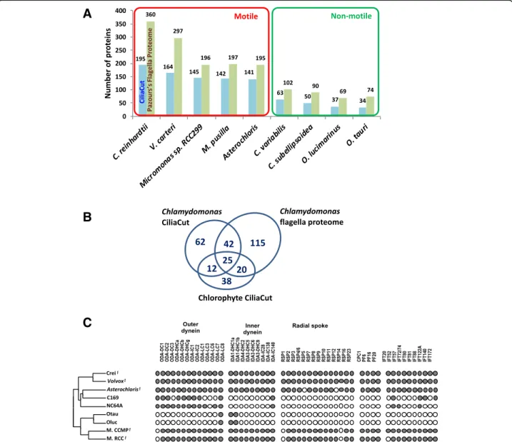
[103]. The identification of many meiotic genes in the A. glomerata genome and their expression in coculture (Additional file5) add further evidence for the occurrence of meiosis in trebouxoid algae, probably in their free-living stages [29]. The gametes observed in Astero-chloris [99] are flagellated, as are its vegetative zoo-spores (9 + 2 type [104]). Not surprisingly, the motility proteins present in Asterochloris match those of other flagellated chlorophytes but are mostly absent from non-motile chlorophytes (Fig. 5 and Additional file 5). The critical implications of fungal and algal sexual reproduction for the lichen symbiosis and for symbiont autonomy are discussed in Conclusions.
Search for symbiosis-specific genes I: differential expression in coculture vs. monoculture
Table 2 lists the genes discussed in the next three sec-tions. The system to survey the gene sets differentially transcribed when fungus and alga first contact each other is based on three parallel cultures on filters placed on low nutrient agar medium [33]: the aposymbiotic fungus, the aposymbiotic alga, and a coculture of the two (Fig. 6 and Additional file 6). RNA was extracted from the individual cultures after 21 days of growth on the filters. Differential expression data (Methods) were analyzed in two ways. In the first, we identified GO an-notations and metabolic pathways significantly enriched
A
B
Fig. 4 Cladonia MAT loci and their evolution. a Configurations of the MAT loci in three Cladonia species. The top diagrammed alignment is based on the alignment between the annotated C. grayi MAT1–1 region (scaffold_00075:76000–90,000 at [67]) and a provisional sequence of the C. grayi MAT1–2 region (accession MH795990). The C. grayi MAT1–1 and MAT1–2 regions are drawn above the basepair indicator line. Under the line are the MAT1–1 regions derived from the genomes of two other Cladonia species (Additional file5). In C. grayi, the conserved flanking regions are gray, while the unrelated central regions are stippled differently for each mating type. Dark or gray arrows represent genes and gene-segments. CLAGR_008123-RA is considered a putative MAT1–1-7 ortholog because of its location and its BLAST hits to MAT1–1-7 orthologs from
Trichophyton and other fungi. b Evolutionary model. Horizontal colored arrows represent MAT idiomorphs. The central line represents the MAT locus configuration of a possible homothallic Cladonia ancestor, and the vertical arrows represent the putative transitions towards the present heterothallic MAT1–1 (orange) or MAT1–2 (blue) configurations (Additional file5). The graded shading in the deletion triangle leading to MAT1–2 symbolizes the deletion’s undefined left boundary beyond MAT1–1-7
among genes induced or repressed more than 1.5x (0.6 > log2>− 0.6) in coculture relative to monoculture. In the second, a more extensive approach, we focused only on induced genes and added information from PFAM domains and literature searches to define each gene’s putative function in more detail than was possible through GO terms alone.
GO term-centered analysis of transcription in coculture
The limited results are listed in Additional file 6. In co-culture, the alga shows only differential activation of
ubiquitdependent catabolic processes. The fungus in-duces redox-active enzymes and proteases, a finding also reflected in the data from the more extensive approach. Surprisingly, however, in presence of the alga the fungus also down-regulates several genes involved in respiratory ATP generation; the biological significance of this remains to be determined, pending experimental confirmation.
Extended analysis of transcription induced in coculture
In this analysis we used day-21 ratios of coculture (Co) vs. monoculture (Mo) RPKM directly rather than the
A
B
C
Fig. 5 Flagellar proteins. a Number of candidate flagella proteins in chlorophytes. Reference C. reinhardtii proteins of the CiliaCut protein set (blue bars) and flagella proteome (green bars) were searched for putative orthologs in sequenced motile and non-motile chlorophytes using the reciprocal best BLASTP hit criterion. b The 314 candidate A. glomerata flagella proteins identified from multiple sources of evidence (see Methods). c Distribution of flagella proteins across Chlorophytes. The left cladogram shows the likely evolutionary relationships of sequenced Chlorophytes. Theƒ mark indicates organisms known to build motile flagella. Crei: Chlamydomonas reinhardtii; Volvox: Volvox carteri; C169: Coccomyxa subellipsoidea C-169; NC64A: Chlorella variabilis NC64A; Otau: Ostreococcus tauri; Oluc: Ostreococcus lucimarinus; M. CCMP: Micromonas pusilla CCMP1545; M. RCC: Micromonas sp. RC299. Presence (dot) or absence (circle) of putative orthologs identified by reciprocal best BLASTP hit of C. reinhardtii outer dynein proteins, inner dynein proteins, radial spoke proteins, central pair proteins and intraflagellar transport proteins
log2 values. Ratios are abbreviated here as Co/Mo. In each symbiont, a few hundred genes changed expression in coculture relative to monoculture, while most genes remained unaffected (Co/Mo≅ 1) (Fig.7). The extended transcription analysis was limited to the genes induced in coculture. Due to differences in Co/Mo ranges for the symbionts (Additional file 6), we defined two different Co/Mo induction thresholds:≥ 2 for the fungus, ≥1.3 for the alga (Fig.7). This yielded 795 up-regulated genes out of 11,388 in C. grayi (7%) and 471 out of 10,024 in A. glomerata (4.7%) (Additional file 6). Induced gene
products were inferred by BLAST [105] and, based on GO terms, PFAM domains and literature searches, were grouped into three categories summarized in Fig.8: un-known and unique to each symbiont, insufficiently de-fined, and better defined (Additional file6).
Relative to their overall genomic frequency of 7.2% (821/11388), mycobiont secreted proteins are dispropor-tionately enriched to 18.5% (147/795) in coculture (p = 2.8E-28). The genomic average protein length is 477 AA, while the induced and secreted proteins are smaller, averaging 341 AA (Fig. 9 and Additional file 6), with
Table 2 Compilation of genes/proteins of potential symbiotic significance discussed in Search for symbiosis-specific genes I, II, and III sections
Fungus: Cladonia grayi Gene groups induced in coculture
Small secreted proteins / Transcription / Cell wall turnover / Protein turnover / Metabolism / Membrane transport / Defense / Extracellular hydrolases
Selected examples Polyol transporter / Ammonium transporter / Calcium channel inhibitor / lectins / DNA methyltransferase / Gα, RGS protein, dual specificity phosphatase
Expanded protein families HET domain / Ank domain / Met permeases / Unknown transmembrane proteins / Fructosamine kinases / Polyketide synthases / Signal transduction components / Stress-related TFs
Contracted protein families Transportome: Carbohydrate transporters / Major Facilitator Superfamily (MFS) / ATP Binding Cassette (ABC) superfamily / Aminoacid-Polyamine organo Cation (APC) family / Oligopeptide Transporter (OPT) family / Proton-dependent Oligopeptide Transporter (POT) family
Slow evolvers Proteostasis maintenance / Aldehyde dehydrogenases / Major Facilitator Superfamily (MFS) Selected examples Mechanosensitive calcium channel / Sugar transporters
Fast evolvers Signal transduction / Membrane trafficking / Stress protection Selected examples Superoxide dismutase / Trehalose synthase
Alga: Asterochloris glomerata Gene groups induced in co-culture
Secreted proteins / Transcription / Cell wall turnover / Protein turnover - ubiquitin / DNA processes / Signal transduction / Protein trafficking / Flagellum synthesis
Selected examples Thioredoxin / Kinesin / Fasciclin domain proteins / Mechanosensitive calcium channel Expanded/new protein
families
Fam_16: DNA binding-recombination proteins / Kinases / Carbohydrate active enzymes (CAZ) / Ank domain proteins / Archaeal ATPases / Desiccation-Related-Proteins / Magnesium transporters / Signal transduction components / Stress-related TFs
Contracted protein families Nitrate assimilation
Slow evolvers Two kinases and one clathrin vesicle adaptor Fast evolvers Seven diverse proteins
Fig. 7 Differential fungal (C. grayi) and algal (A. glomerata) gene expression in coculture vs. monoculture. RPKM expression ratios are sorted from high to low. Genes considered unaffected in coculture are labeled gray (Co/Mo ~ 1). Those labeled black above or below the gray range are considered induced or repressed, respectively. Induction and repression thresholds correspond respectively to 2 and 0.5 for the fungus and 1.3 and 0.77 for the alga. Notice the smaller range of differential expression induction displayed by the alga under our experimental conditions (Additional file6)
Fig. 8 Classes of genes differentially induced during early fungus-alga interactions in coculture. The pie charts divide the induced genes for each symbiont into three broad classes (numbers of genes in parentheses). The“better defined” genes are subdivided in groups roughly comparable between the symbionts (gray and white boxes). The area of each box is proportional to the percent of genes it contains relative to all better defined genes (265 for the fungus and 243 for the alga). The number behind each group’s name indicates its enrichment factor relative to the whole genome (see Methods). The hatched areas represent groups with less than 10 genes each. The p values for the enrichment of the indicated groups within the induced genes are all < 0.05, and most are << 10− 3
most unknown and unique to C. grayi. The smallest pro-teins tend to be also among the most strongly induced. Cladonia shares biotrophic overexpression of small se-creted proteins with the mycorrhizal basidiomycete Lac-caria bicolor [57], the mycorrhizal ascomycete Tuber melanosporum [58], and with pathogenic fungi [106]. The Cladonia secretome (821 proteins) is comparable in size to the secretomes of ectomycorrhizal and many other fungi [107]. Also in the alga, relative to their over-all genomic frequency of 3.6% (365/10025), secreted pro-teins appear significantly enriched (p = 0.00003) among the 471 induced proteins (32/471 = 6.8%), but they are not significantly smaller than average (Fig. 9 and Additional file6). It remains to be seen whether the algal secretome in early symbiosis shares any features with the secretion responses of higher plants to fungal infec-tion [108].
The better-defined gene products induced in coculture, 265 in C. grayi and 243 in A. glomerata, were subdivided for enrichment analysis in broad functional subgroups of at least 10 genes each (Fig. 8, Additional file 6). Ex-cept for the limited enrichment (1.4-fold) of signal transduction and defense genes in the fungus, most subgroups in both symbionts were enriched between 2- and 9-fold and with high significance in the set of induced genes relative to the whole genome (Fig. 8). At these early stages, both partners respond to the other’s presence by activating transcription, cell wall metabolism and protein turnover. The mycobiont’s
specific responses center on upregulating membrane transporters, secreted hydrolases, and small proteins, broadly resembling the symbiotic responses of the EM fun-gus L. bicolor [112]. The photobiont’s specific responses center on growth and motility through the predominant activation of DNA and signal transduction processes, as well as protein trafficking and zoospore formation (flagella). This correlates with the alga’s rapid initial growth visible in coculture compared to monoculture (Additional file 6), possibly reflecting the growth ob-served in nature in free-living trebouxoid algae near suit-able fungal hyphae [29] and increasing the chances of successful formation of new lichen initials. This issue is taken up again in Conclusions, where the importance for li-chen evolution of lili-chenization of free-living algae re-leased from natural thalli is discussed. It needs underlining that this coculture system and similar ones [113] do not proceed beyond formation of poorly differentiated liche-noids (Additional file6), and thus they do not capture the complete interaction network extended in space and time needed for proper lichenization in nature. Individual genes of potential symbiotic significance are discussed in Infer-ences from differential transcription about nutritional fluxes at the symbiotic interfacesection and in Additional file6.
Inferences from differential transcription about nutritional fluxes at the symbiotic interface
Whereas hexose sugars are the carbohydrates transferred from plant to fungus in mycorrhyzae [114], polyols are the
Fig. 9 Secreted proteins among the proteins induced in coculture. The small black symbols coalescing into a curve represent the Co/Mo ratios of the genes induced in coculture; the circles represent the corresponding protein sizes, gray for non-secreted, black for secreted proteins. The average sizes (# of amino acids) of a) all genome proteins, b) all induced proteins, c) all induced and secreted proteins are a) 477, b) 381, c) 341 for the fungus and a) 447, b) 420, c) 436 for the alga. Proteins were considered secreted only if they scored as such in all three programs SignalP [109], TargetP [110], and TMHMM [111]. In TMHMM, a transmembrane domain prediction program, a protein was considered compatible with secretion only if it had either no predicted TM domains or only one at the N terminus. The data used in this figure are in Additional file6
means by which trebouxoid algae transfer carbon to the fungus in lichens [115], and the first putative polyol trans-porter gene in a lichen fungus was identified recently [116]. We identified a family of five putative ribitol transporters in C. grayi(Additional file 6 and Fig. 10); in coculture, only one of the five was induced (Table3and Fig.10), suggesting its involvement in importing ribitol to the fungus. The alga has dozens of putative sugar/MFS transporters whose ex-pression remains mostly unchanged in coculture, except for seven that are weakly induced (Co/Mo 1.13–1.27). Their in-volvement, if any, in ribitol export to the fungus remains to be determined.
It has been known since the 1970s that nitrogen is transferred from lichen mycobionts to eukaryotic photo-bionts [121, 122], and more recently it was suggested that the fungus acts as a nitrogen gateway for the alga [123]. This hypothesis is consistent with the results of a study of the lichen Cladonia portentosa, which found that the response of the algal proteome to nitrogen ex-cess was more muted than that of the fungal proteome [43]. As is thought to be the case with mycorrhizal fungi [114], a primary form of nitrogen released by the lichen mycobiont to the alga is likely to be ammonium, al-though in lichens amino acids might be involved as well
[124]. Ammonium transporters generally import NH4+ into the cell, but they can also export it [125–128]. Of particular interest are two ancient likely horizontal gene transfers of ammonium transporters from prokaryotes to fungi [129]. Transporters from the earliest transfer are found in all fungi, whereas transporters from the later transfer were derived from hyperthermoacidophilic Ar-chaea and were retained primarily by lichenizing fungi [46, 129], suggesting a specific role in the symbiosis. Their selective retention might have been driven by the li-chens’ frequent exposure to high temperatures and by the very low pH of their apoplast due to the high content of acidic secondary metabolites. Of the four MEthylammo-nium Permease-type (MEP) ammoMEthylammo-nium transporters in the C. grayi mycobiont, two belong to the general fungal class (CLAGR_000407-RA and CLAGR_009781-RA) and two to the class primarily retained by lichens (CLAGR_005848-RA and CLAGR_003366-RA). The Cla-donia mycobiont also has four transporters belonging to the Gpr1/FUN34/YaaH family, whose functions are de-bated [130] but include the postulated ability to export NH4+in yeast [128], where they are named ATO (Ammo-nia Transport Outward). The differential transcription of these eight transporter genes is interesting: relative to
A
C
B
Fig. 10 A predicted ribitol transporter in C. grayi. a Differential transcription in coculture vs. monoculture of five putative sugar transporters in C. grayi. They were the top five BLAST hits obtained by querying the genome with the sequences of two functionally validated fungal D-sorbitol/D-mannitol/ribitol transporters [117]. Only CLAGR_004844-RA is induced in coculture (Co/Mo on Y axis; 1 means no induction.). b CLAGR_004844-RA amino acid sequence. The 12 transmembrane domains [118] are indicated in bold purple. Consensus amino acids for sugar transporters [119,120] are highlighted in cyan. c Protein phylogeny (PhyML, 100 bootstraps) of the five C. grayi transporters. The green branches correspond to nodes with bootstrap support≥70%. The C. grayi proteins are labeled brown. The other transporters are identified by GI number and by fungal species. Taxa are also labeled as ascomycetes (Asco) or basidiomycetes (Basidio), the latter used as outgroup. The CLAGR_004844-RA clade is highlighted yellow. Bar indicates amino acid substitutions/site
fungal monoculture, in coculture with the alga five are re-pressed, two are induced (one strongly), and one is un-changed (Table 3). The repression of a majority of these fungal transporters in coculture echoes the selective re-pression by the ectomycorrhizal fungus A. muscaria [131] of a fungal NH4+ importer at the symbiotic interface, thought to prevent re-absorption by the fungus of exported NH4+. Awaiting experimental verification, we speculate that the ATO transporter CLAGR_005116-RA (Co/Mo 5.6) and perhaps the Mep1b-like ammonium transporter CLAGR_003366-RA (Co/Mo 1.5) are candi-dates for mediating ammonium export to the alga, while the others, probably importers, are repressed to reduce re-absorption by the fungus. In the cocultured alga relative to monoculture, expression of four of its five ammonium transporters does not change and that of the fifth is undetectably low (Table 3). Overall, however, the transcriptional shifts in coculture of the fungal ammonium transporters are consistent with NH4+ being a major mediator of nitrogen transfer from mycobiont to photobiont. They are also consist-ent with the inability of freshly isolated A. glomerata to grow on nitrate (Specific survey of photobiont proteins involved in nitrate and CO2 assimilationsection).
Phosphate is an important nutrient known to be transferred from mycorrhizal fungi to their plants [114], but there is no suggestive transport polarity or coculture induction pattern among the many
putative phosphate transporters in A. glomerata and C. grayi that could shed light on this system. (For other individual genes induced in coculture, see Table 3, Additional file 6).
Search for symbiosis-specific genes II: evolution of gene family size
Lineage-specific expansion or contraction of multigene families is often associated with lineage-specific func-tional shifts in eukaryotes [57, 58, 132–134]. We under-took one broad and one circumscribed analysis of multigene families. For the broad survey, multigene fam-ilies in C. grayi and A. glomerata were identified using MCL [135] and analyzed for changes in family size using CAFE [136], with the taxa listed in Methods. In C. grayi, 390 families were expanded, 3369 showed no change, and 769 families had undergone contraction by compari-son to a putative common ancestor. In Asterochloris, 648 families were expanded, 2729 showed no change, and no families were contracted. The circumscribed ana-lysis was limited to the C. grayi transportome and pre-dicted 458 C. grayi membrane transporters using the TransportTP online tool [137] with the Transporter Classification Database [138]. We discuss only families for which we can suggest symbiotic roles and highlight whenever a significant fraction of a gene family is also induced in coculture. We assume that the overlap of in-duction with expansion/contraction (Additional file 1)
Table 3 Selected fungal (CLAGR) and algal (Aster) genes of potential symbiotic significance differentially expressed in coculture Gene name Hypothetical function Co/Mo ratio Gene name Hypothetical function Co/Mo ratio A. Genes potentially involved in C and N exchange
CLAGR_004844-RA Ribitol transporter 2.6 Aster-02360 Ammonium transporter 1.0 CLAGR_005116-RA ATO (Amm. exporter) 5.6 Aster-02123 Ammonium transporter 0.9 CLAGR_006601-RA ATO (Amm. exporter) 0.9 Aster-04935 Ammonium transporter 1.0 CLAGR_003233-RA ATO (Amm. exporter) 0.3 Aster-08051 Ammonium transporter 1.0 CLAGR_006538-RA ATO (Amm. exporter) 0.4 Aster-08052 Ammonium transporter n.d. CLAGR_000407-RA Mep2-type Amm. transp. 0.9
CLAGR_009781-RA Mep3-type Amm. transp. 0.6 CLAGR_005848-RA Mep1a-type Amm. transp. 0.6 CLAGR_003366-RA Mep1b-type Amm. transp. 1.5 B. Other genes discussed in Additional file 6
CLAGR_010764-RA KP4 killer toxin-like 150 Aster-04252 Thioredoxin 73 CLAGR_008646-RA Lectin 10 Aster-01625 Kinesin motor 2.3 CLAGR_010932-RA Lectin 4 Aster-01936 Fasciclin domain protein 1.7 CLAGR_011186-RA Gα signaling subunit 2 Aster-06761 Fasciclin domain protein 1.5 CLAGR_002910-RA Regulator of Gα signaling 4 Aster-03695 Calcium channel 1.3 CLAGR_002710-RA Dual specificity phosphatase 2
CLAGR_007359-RA DNA methyltransferase 3 CLAGR_000113-RA Calcium transporter 2
symbiotic roles. We did not find significantly contracted families in A. glomerata, although a separate search fo-cused on nitrogen assimilation revealed a reduced set of nitrate assimilation genes in the alga (Specific survey of photobiont proteins involved in nitrate and CO2 assimilation section).
Mycobiont expanded families
In the C. grayi fungus, the most notable expansions involve 204 HET incompatibility proteins, 13 of which are also induced in coculture; 156 Ankyrin domain proteins; an 80-member family (Fam_5) of multi-transmembrane-domain proteins of unknown function with 10 coculture-induced members; Fam_668, a fructosa-mine kinase family with 7 members of which 3 are cocul-ture induced; the family of polyketide synthases (PKSs) and Non-Ribosomal Peptide Synthases with at least 29 members. HET proteins might prevent incompatible C. grayi hyphae from fusing with a C. grayi mycobiont already lichenized with its photobiont. Ankyrin domains could be involved in inter-protein contacts at the symbi-otic interface. Fructosamine kinases are known to reverse aging-associated protein damage produced by glycation [139, 140]. A few of the PKSs [34,141] have been shown to be involved in the synthesis of the primary and most abundant secondary metabolites well known in lichens, but the large number of PKSs in C. grayi (Fig.11and Add-itional file 7) points to a vast and still uncharacterized metabolic potential. Further description of these expanded families and elaboration of the hypotheses on their pos-sible symbiotic significance are found in Additional file7. A separate analysis identified expanded families of signal transduction components (Specific survey of mycobiont
and photobiont signal transduction components
sec-tion and Addisec-tional file 8), some also differentially expressed in coculture (Additional files6and8).
Mycobiont contracted families
Carbohydrate transporter Fam_1, MFS (Major Facilitator Superfamily) Fam_2 and Fam_7, and amino acid perme-ase Fam_22 are dramatically reduced, and some mem-bers of these families are also induced in coculture (Additional file7) or score as slow-evolvers as discussed
in Slow-evolving proteins and anti-stress strategies in
the mycobiont section. The contraction of these families
is part of the overall contraction observed in the specific analysis of the mycobiont’s transportome (Additional file7). A reduced transportome has been observed also in the ectomycorrhizal ascomycete T. melanosporum [58]. This is consistent with the loss of generalist nutrient uptake and the reliance of the fungus on a few specialized carbon sources from the photobiont, which are polyols in
from differential transcription about nutritional fluxes at the symbiotic interface section).
Photobiont expanded families
Additional file7: The most notable expansions in A. glo-merata involve the 100-member Fam_16 of unknown proteins unique to this alga, with 20% of the members induced in coculture; Kinase families totaling 52 mem-bers; Carbohydrate Active Enzymes (CAZ) with a total of 40 members; a 29-member Ankyrin domain protein family; a probably HGT-derived family of 26 archaeal ATPase-like proteins (Additional file 7 and Fig. 12). A survey using Phyre2, a protein structure prediction pro-gram [142, 143], suggests DNA-related functions for some Fam_16 members. The expansion of kinases matches the expanded signal transduction capability mentioned in Genome sizes and gene organization sec-tion. The expanded CAZ families might enhance the structural and biochemical versatility of the photobiont’s cell surface. The parallel expansion of Ank-domain pro-teins in C. grayi and A. glomerata might mediate recipro-cal boundary interactions. Finally, the putative ATPases of archaeal origin and the Desiccation-Related-Proteins of bacterial origin might be involved in the lichen alga’s re-sistance to desiccation and elevated temperatures. Further description of these expanded families and elaboration of the hypotheses on their possible symbiotic significance can be found in Additional file7.
Specific survey of mycobiont and photobiont signal transduction components
This analysis confirms the suggestion from the data in Additional file3that signal transduction protein families are expanded in C. grayi and A. glomerata (Genome sizes and gene organization section). For C. grayi, these include PTH11-type receptors [147] and components of MAPK pathways described in Additional file8, together with coculture expression data collected to identify sig-nal transduction candidates potentially involved in early fungus-alga interactions (Additional files 6 and 8). The analysis also revealed an expanded set of five divergent Gα subunit paralogs in C. grayi (Fig. 13). The Astero-chlorisexpansions (Additional file 8) involve a more di-verse set of families than Cladonia’s.
Specific survey of mycobiont and photobiont transcription factor (TF) families
Overall results are displayed in Fig.14, and specific fam-ilies are listed in Additional file9. With regard to overall TF family gain or loss, neither Cladonia nor Asterochloris stand out within their respective phylogenies. With regard to family expansion and contraction, Asterochloris also conforms to the general algal phylogeny trend of
expansions outnumbering contractions. In contrast, Cla-doniaappears to have experienced more expansions of TF families than not lichenized fungi. One Cladonia expan-sion involves a group of proteins characterized by PCI do-mains [148] (Additional file 9). PCI-domain proteins are involved in many diverse processes, and we could not find specific roles for these TFs. With regard to families with known functions, both Cladonia and Asterochloris have expanded TFs involved in chromatin remodeling (SWI/ SNF [149] in Cladonia, SWI/SNF_Baf60 [150] and Sir2 [151] in Asterochloris) and stress responses (SGT [152] and CSD [153] in Cladonia, WRKY [154] in Astero-chloris). Asterochloris has also contracted the SET family and the CCHC zinc finger family. The contracted SET
family comprises protein lysine methyltransferases, in-volved in methylation of histones and other proteins [155]. We hypothesize that selection on both symbionts’ ability to shuttle reversibly between free-living and symbi-otic states [31] and interact in the thallus in different ways across time and space [10] produced the needed genomic plasticity through the parallel expansion of chromatin-remodeling functions. In contrast, the signifi-cant SET family contraction seems odd, given the multifa-ceted effects of protein lysine methylation [156]. We suggest that the stresses placed on lichens by re-peated, rapid and large oscillations in their exposure to light, temperature and hydration might have led to the expansion of stress-related TFs in both symbionts.
A
B
C
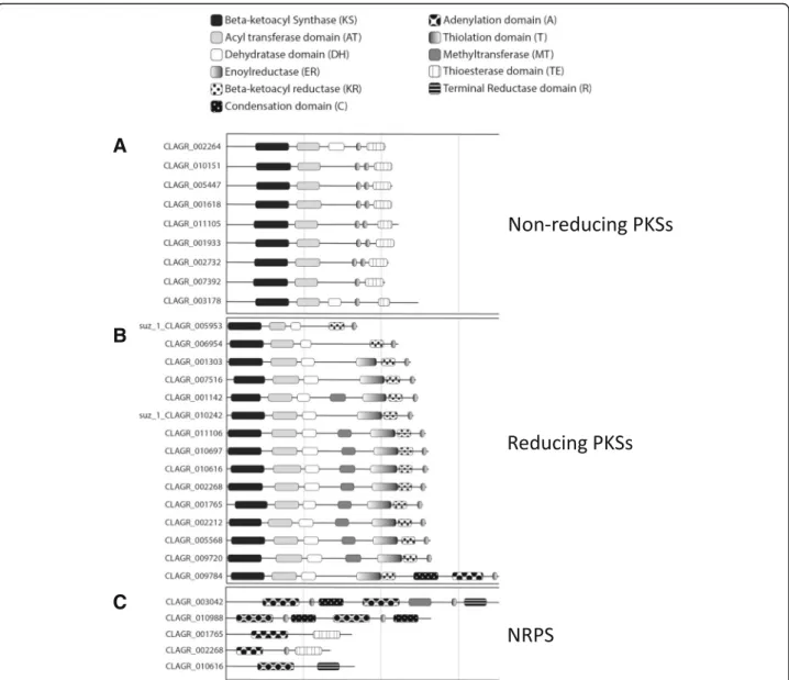
Fig. 11 Polyketide Synthase (PKS) and Non-Ribosomal Peptide Synthetase (NRPS) genes in C. grayi. The three protein categories in a, b, c are named on the right. CLAGR_009784 is a PKS-NRPS hybrid. The only PKS whose likely downstream product (grayanic acid) is known is CLAGR_002732 [34]. The length of the horizontal lines is proportional to gene length (vertical gray lines delimit 3-Kb segments). Graphic symbols for protein domains are indicated at top. Genes with a suz_1 prefix were reannotated manually. See Additional file7for further details
These environmental stresses appear to affect the evo-lution of many lichen adaptations [44, 157–160] as discussed in Slow-evolving proteins and anti-stress
strategies in the mycobiont section for the fungus
(where several converge on maintaining the resilience of the proteome) and in Additional file 7 for the alga.
Specific survey of photobiont proteins involved in nitrate and CO2assimilation
We compared A. glomerata to related algae for possible adaptations involving nitrogen and carbon assimilation
proteins that did not surface in the protein family screens. The top of Fig. 15 displays the types and organization of nitrate assimilation genes linked to a phylogenetic tree for Chlorophytes. A. glomerata has a reduced set of nitrate assimilation genes compared to other green algal genomes in this figure. The nitrate as-similation cluster (HANT-AC) [161] present in most microalgae [162] is reduced to nitrite reductase (NIR) and the NRT3 transporter in A. glomerata, and the total number of NNP transporters is reduced in Coccomyxa subellipsoideaC-169 and A. glomerata. A. glomerata has
Fig. 12 Protein family tree of archaeal ATPases. The phylogram was constucted using FastTree [144] on a MAFFT [145] amino acid alignment of 91 putative archaeal ATPases from prokaryotes and eukaryotes. Branches with bootstrap values≥0.77 are thickened. Bar indicates amino acid
substitutions/site. To the ATPases present in the published Galdieria phylogeny [146], we added all the proteins above Methanocaldococcus_j__MJ0632 in this figure. The eukaryotic taxa shown represent most of those currently known to harbor putative archaea-derived ATPases. ATPases from Galdieria (Gs) are marked red, from green algae and plants green, from fungi brown. All branches in black are prokaryotic, except for the amoeba Dictyostelium at the base. Branch labels include the taxon name or symbol and a protein identifier. The Asterochloris (Aster) proteins are indicated by their gene names in the JGI database [68]. The phylogeny suggests several independent HGT events, but it cannot exclude a very ancient HGT from Archaea to a common eukaryotic ancestor followed by losses in most eukaryotes. Asterochloris, Galdieria, and Selaginella have the largest families of archaeal ATPases (with 26, 12, and 7 members, respectively). See also Additional file7
lost the clustered nitrate reductase (NAR), but it retains a nitrate reductase paralog (NAR-P) also found in C. subellipsoidea. Nitrate cannot be taken up directly by the lichenized alga enclosed in fungal tissue but has to be first metabolized into other compounds by the mycobiont [123]. These compounds may include amino acids, excreted by the fungus and presumably taken up by the alga [124], and ammonium (seeInferences from differential transcription about nutritional fluxes at
the symbiotic interface section). Congruent with these
findings, A. glomerata isolated from C. grayi can grow on nitrate only after a period of adaptation to axenic culture (Armaleo, unpublished). Day 21 gene expression in mono-and coculture is graphed on the bottom of Fig.15. Inter-estingly, nitrate assimilation genes in A. glomerata are
turned down about 50% in coculture relative to monocul-ture, which may contribute to the regulation of growth ex-perienced by the alga when it lichenizes with the fungus [163]. We hypothesize that, while A. glomerata is capable of autonomous nitrate assimilation utilizing nitrate trans-porters and NAR-P when free-living, suppression of these mechanisms during symbiosis has relaxed selection on their strict maintenance, resulting in the loss of a full HANT-AC and a reduction in the number of nitrate transporters. The extreme reduction of the nitrate assimilation toolkit in A. glomerata relative to other non-symbiotic chlorophytes could be viewed as a parallel to the transportome contrac-tion in C. grayi (Mycobiont contracted familiessection and Additional file7), each contraction resulting from the almost exclusive nutritional reliance of each partner on the other.
Fig. 13 A unique set of Gα subunits is present in Cladonia. The protein phylogeny (PhyML, 100 bootstraps) of the eight C. grayi Gα subunits clusters into three major MAG A, MAG B, and MAG C clades (highlighted). The unique MAG C paralogs are shown on the bottom. The green branches correspond to nodes with bootstrap support≥67%. The C. grayi proteins are labeled brown. Bar indicates amino acid substitutions/site. See also Additional file8
As algal photosynthesis is central to the lichen symbi-osis, we surveyed the carbonic anhydrases (CAs) of Asterochloris, enzymes that catalyze the interconversion between CO2and HCO3−. This adds to previous exten-sive work published on CAs in lichen algae [165, 166]. Delivery of CO2 to the enclosed lichen photobionts is extremely variable, depending on the specific anatomy and physiology of its mycobiont-photobiont combination [167] and on rapid changes in temperature and in the sup-ply of nutrients, water, and light [168]. When lichen thalli become fully saturated with water, the diffusion of CO2to the algae is further limited by the swollen hyphal tissue surrounding them. This reduces photosynthetic rates [169,170] even if the hydrophobicity of the lichen interior maintains air-filled spaces for rapid CO2 diffusion [10]. Our data, detailed in Additional file10, suggest that Aster-ochlorisparallels Chlamydomonas and Chlorella in having oneα CA functioning in the pyrenoid but differs in having
expanded a specific subclass of cytoplasmicβ CAs, whose potential relationship to the symbiosis needs exploring.
Search for symbiosis-specific genes III: proteins with anomalous rates of evolution
We hypothesize that, after a burst of adaptive selection at the origin of Lecanoromycetes 300–350 million years ago [171–174], proteins that acquired fundamental symbiotic roles would have since stabilized under purifying selection and evolved at slower rates, as suggested for a polyketide synthase specific for a secondary metabolite unique to C. grayi[34]. Conversely, faster evolving proteins are likely to represent more recent changes. We compared amino acid substitution rates of C. grayi and A. glomerata proteins to those of eleven free-living ascomycetes and six unicellular chlorophytes respectively using three methods (Methods). We focus on the proteins whose rates were“slow” or “fast” relative to nonlichen species by at least two methods to
Fig. 14 Evolution of transcription factor/regulator families in fungi (left) and algae (right). All the species used and numerical data are listed in Additional file9. The C. grayi and A. glomerata abbreviations are bolded. Area of symbols is proportional to the change observed. Green circles: number of families gained, red circles: number of families lost. Green triangles: number of expanded families, red triangles: number of contracted families
reduce false positives. This yielded 38 slow- and 11 fast-evolving candidates in the fungus and 3 slow- and 7 fast-evolving candidates in the alga (Slow-evolving proteins and anti-stress strategies in the mycobiontandFast-evolving proteinssections).
Slow-evolving proteins and anti-stress strategies in the mycobiont
Additional file 11 (Excel sheet “Fungus slow-evolvers”) lists a total of 72 proteins that score in multiple ways (highlighted in orange) for potential symbiotic signifi-cance. The first 38 were identified as slow-evolving by at least two of the rate methods. Of the 38, 22 are universal eukaryotic components of the protein translation, trans-location and sorting machinery (Additional file11). This machinery maintains proteostasis, the dynamic equilib-rium of the proteome, through a complex network
guiding protein folding and functionality from synthesis to modification, to sorting, and to degradation [175– 177]. Studies with several eukaryotic systems, including yeast, show that downturns in protein translation and translocation are essential to protect cells from dehydra-tion, heat, and hyperosmotic stresses [178–184]. In yeast for instance, many temperature-sensitive mutations impairing a variety of ribosome assembly steps also dra-matically increase desiccation tolerance [185], highlight-ing how slowdowns in ribosomal assembly may reduce protein misfolding and aggregation during desiccation. However, lichens must withstand daily cycles of dehy-dration, rehydehy-dration, thermal and UV radiation stress so extreme that they would kill most other organisms [157, 186]. Therefore, we hypothesize that such ex-ceptional circumstances in early lichen evolution se-lected for “upgraded” fungal components of the
Fig. 15 Nitrate assimilation gene clustering in Chlorophytes. In the upper part of the figure, the algal phylogeny (left) and the corresponding taxa (right) bracket the gene clusters and unclustered paralogs in each taxon. Gene and cluster lengths are to scale; color codes and acronyms are listed below the 5 kb bar. Phylogeny and clusters were obtained as described in Methods. The lower part of the figure displays as vertical bars the expression levels of the nitrate assimilation genes in the alga grown alone or with the fungus. The full names of the taxa listed from top to bottom are: Micromonas pusilla CCMP1545; Micromonas RCC299; Ostreococcus tauri; Ostreococcus lucimarinus; Ostreococcus sp. RCC809; Chlamydomonas reinhardtii; Volvox carteri; Chlorella variabilis NC64A; Coccomyxa subellipsoidea C-169; Asterochloris glomerata. All the corresponding genome data are at [164]
ficking networks, which were already part of the normal Environmental Stress Response (ESR) [184]. The upgrades were then stabilized under purifying selection and inte-grated with other defenses [157] involving, for instance, an-tioxidants [159, 187] and synthesis of photoprotective anthraquinones [188] that lichens share with non-extremo-philes. There is some correlative support in the literature for the centrality of ribosomal function in the lichen re-sponse to stress. In the lichen Lobaria pulmonaria [37] as much as 35% of the expressed fungal proteome is involved in ribosomal and protein turnover functions. In the lichen Cladonia rangiferina [39] global transcriptional responses to desiccation and rehydration also suggest involvement of the protein translation machinery. In the cultured lichen fungus Endocarpon pusillum, ribosomal protein genes are highly induced during PEG-induced dehydration stress [159]. We speculate that the unusually high number of nu-clear rDNA introns (Additional file 12) present in lichen fungi [189, 190] could be another possible adaptation to control the rate of ribosomal assembly for stress (e.g. desic-cation) protection. A major consequence may be the well-known slow growth phenotype of lichens and lichen mycobionts. Other double-scoring slow evolving proteins of interest are listed and discussed in Additional file 11. These include a putative osmosensing calcium channel and two homogeneous groups of proteins: 18 membrane trans-porters and 7 aldehyde dehydrogenases (ALDHs).
Fast-evolving proteins
Additional file 11 includes a description of 11 putative fast-evolving mycobiont proteins involved in signal transduction, membrane trafficking and stress protec-tion, as well as slow- and fast- evolving proteins in the photobiont.
Conclusions
Symbiotically relevant genes affect stress resistance, nutritional, signaling, and structural interactions
Our analysis identified several proteins expected to influ-ence the symbionts’ environmental stress resistance. They include slow evolving mycobiont proteins (Slow-evolving proteins and anti-stress strategies in the mycobiontsection, Additional file 11) enriched for universal eukaryotic com-ponents of the protein translation, translocation and sorting machinery which manages the proteome under normal as well as stress conditions like dehydration and heat [178– 182]. Also, some of the faster evolving C. grayi proteins, probably under adaptive selection, appear to be involved in protection from stress (Additional file11). In A. glomerata, the likely horizontally transferred archaeal ATPases and Desiccation-Related Proteins may contribute to its heat and desiccation resistance (Photobiont expanded
families section, Additional file 7). Transcription
chromatin remodeling and stress responses in both symbionts (Specific survey of mycobiont and photo-biont transcription factor (TF) families section).
We also identified proteins governing several symbiot-ically relevant nutrient interactions. The fungus in cocul-ture induced two transporters potentially central in the carbon and nitrogen exchange at the symbiotic interface: one an importer for ribitol, the carbon source provided by trebouxoid algae to their fungal partners [115] ( Infer-ences from differential transcription about nutritional fluxes at the symbiotic interfacesection and Additional file6), the other a possible ammonium exporter (Inferences from dif-ferential transcription about nutritional fluxes at the
symbi-otic interface section), pointing at NH4+ as a major
nitrogen source provided by the mycobiont to the photo-biont. Reliance of each partner on the other as a restricted nutrient source is also reflected by the contraction of the sugar transportome in C. grayi (Mycobiont contracted fam-iliessection) and by the reduced nitrate assimilation poten-tial in A. glomerata (Specific survey of photobiont proteins
involved in nitrate and CO2 assimilation section).
Un-known is the significance for symbiotic carbon fixation of the expansion of a specific A. glomerata subclass of cyto-plasmic carbonic anhydrases, enzymes that catalyze the interconversion between CO2and HCO3− (Specific survey of photobiont proteins involved in nitrate and CO2 assimi-lationsection and Additional file10).
Gene families whose characteristics suggest involve-ment in other symbiotically relevant interactions com-prise: signal transduction proteins, expanded in both symbionts, which include a unique new set of five MAG C paralogs in the mycobiont (Specific survey of myco-biont and photomyco-biont signal transduction components section and Additional file 8); ankyrin domain protein families (Mycobiont expanded families and Photobiont
expanded familiessections), also expanded in both
part-ners, perhaps involved in increased proteprotein in-teractions at the boundaries between them; algal glycosyl transferase families, whose expansion could be necessary to adapt extracellular surfaces to the varied contacts in which the photobiont engages (Photobiont expanded
families sections, and Additional file 7); the expanded
set of mycobiont polyketide synthases producing com-pounds for a mostly undiscovered array of biochemical functions (Mycobiont expanded families section); HET incompatibility protein families in C. grayi, some pos-sibly involved in the competition among fungal geno-types to secure the appropriate alga (Mycobiont
expanded families section). Also the induction in
cocul-ture of secreted proteins in the fungus and less promin-ently in the alga (Extended analysis of transcription
induced in coculturesection) suggests their involvement
The evolution of lichenization involved changes in many conserved genes scattered throughout the symbionts’ genomes
Most of the symbiotically relevant genes suggested here have homologs in non-lichen fungi and algae, and we as-sume that they are variants modified by symbiosis. This indicates that lichens evolved mainly through the accu-mulation of scattered regulatory and structural changes in available genes rather than through sudden key innova-tions. This in turn suggests that, to establish a basic and reversible nutritional dependency at very early evolution-ary stages, the free-living ancestors of myco- and photo-bionts might have required at first only a few or even no changes. A model of such basic interactions was devel-oped experimentally using the fungus S. cerevisiae and the alga C. reinhardtii [191]. Selection towards increasing sta-bility and environmental adaptasta-bility would have then transformed such precarious mutualistic/antagonistic and reversible states into lichens over evolutionary time. This scenario suggests that there were multiple pathways for fungi, algae and cyanobacteria to evolve into lichens, which is consistent with the emerging consensus that ascolichens could have had a few independent evolution-ary origins [171, 192]. It is also compatible with the fact that lichens display a wide array of structures with differ-ent levels of complexity, from leprose and crustose to fru-ticose and foliose, and with the overall staggering variety of interactions throughout the biosphere between fungi and photosynthetic organisms [193,194].
Sexual reproduction, symbiont autonomy and equivalence
While the advantages accrued by lichens through sex-ual reproduction of their mycobionts are fairly clear [91, 92, 97, 195–197] and consistent with the behav-ior of a mutualistic exhabitant [100], the advantages of sex for the algal inhabitant were previously expected to be limited [100], mostly based on the assumption that the lichen alga is asexual and that it dies when its lichen thal-lus dies [198]. As indicated in Heterothallism probably evolved from homothallism in Cladonia; genetic evidence for sex in Asterochlorissection, however, there is strong evidence that sex in trebouxoid algae does occur and that free photobiont populations exist on the substrates near li-chen thalli [27, 29, 199–201], so that sexually produced variation and algal adaptations to lichenization are likely to be incorporated and selected for in lichenized popula-tions. W.B. Sanders proposed in 2005 [29] that the transi-ent free-living state is necessary for completion of the algal sexual cycle, which is consistent with the fact that the rare direct observations of algal sexual stages have al-ways involved aposymbiotic cells (Heterothallism probably evolved from homothallism in Cladonia; genetic evidence
for sex in Asterochloris section). Encounters of
germinating mycobiont spores and free-living photobionts [199] could produce lichens with new combinations of both genomes, expanding and fine-tuning a lichen’s adap-tation to different ecological niches. Photobiont contribu-tions to lichen adaptability are in fact highlighted by several studies. For a given lichen, the correlation of algal genotypic variation with habitat appears stronger than that of the fungal genotype [103, 202–205]. Other ana-lyses indicate that both fungal and algal genotypes substantially influence a lichen’s ecological adaptability [206, 207], and that multiple algal genotypes can co-exist within single thalli [208–210] and move horizon-tally among fungal genotypes [207, 208, 211–214] even when the predominant means of photobiont transmission is vertical through vegetative propagules [201, 208]. We propose therefore that, over hundreds of millions of years of tight coexistence, genomic and functional autonomy in each partner were maintained by the benefits of periodically detaching each partner from the symbiosis for sex and for partner switching, which increased the overall adaptability of the liche-nized symbionts. Beyond sex, the list of phycobiont investments in the symbiosis is long. It includes the adap-tations suggested inPhotobiont expanded families,Specific survey of mycobiont and photobiont signal transduction
components,Specific survey of mycobiont and photobiont
transcription factor (TF) families,Specific survey of photo-biont proteins involved in nitrate and CO2 assimilation sec-tions, the intrinsic resilience of the free-living lichen alga to desiccation [44, 160], the increased resistance to PSII photoinhibition in the symbiotic vs. free-living alga [215], and correlated structural and physiological adaptations: algal morphology changes significantly between lichenized and free-living states ([216] and references therein) and, when the lichen is hydrated, about 50% of the fixed carbon can be converted to lichen biomass [217] with an energy conversion efficiency comparable to that with which chloroplast photosynthesis translates into plant biomass [218]. Ribosomal DNA introns occur in lichenized treboux-oid algae [219–221], including the Group IB intron in the LSU gene of A. glomerata [221]. We speculate, as for the mycobiont rDNA introns (Slow-evolving proteins and anti-stress strategies in the mycobiont section), that these photobiont rDNA introns may be involved in mediating desiccation tolerance. The evolutionary introduction of A. glomerataor Trebouxiales to lichenization is not yet known but, based on the only published broad Chlorophyta chro-nogram [222] (which does not include Trebouxiales), the timing could be compatible with that estimated for lichen fungi [173, 174]. It is therefore possible that the Astero-chloris/Trebouxialineage has been adapted to symbiosis for hundreds of millions of years. Metaphorically, the lichen alga is not the“second sex” [223]: it deserves a full seat at the symbiotic table.