HAL Id: hal-03021336
https://hal.archives-ouvertes.fr/hal-03021336
Submitted on 24 Nov 2020
HAL is a multi-disciplinary open access
archive for the deposit and dissemination of
sci-entific research documents, whether they are
pub-lished or not. The documents may come from
teaching and research institutions in France or
abroad, or from public or private research centers.
L’archive ouverte pluridisciplinaire HAL, est
destinée au dépôt et à la diffusion de documents
scientifiques de niveau recherche, publiés ou non,
émanant des établissements d’enseignement et de
recherche français ou étrangers, des laboratoires
publics ou privés.
Loss of Asb2 Impairs Cardiomyocyte Differentiation and
Leads to Congenital Double Outlet Right Ventricle
Abir Yamak, Dongjian Hu, Nikhil Mittal, Jan Buikema, Sheraz Ditta, Pierre
Lutz, Christel Moog-Lutz, Patrick Ellinor, Ibrahim Domian
To cite this version:
Abir Yamak, Dongjian Hu, Nikhil Mittal, Jan Buikema, Sheraz Ditta, et al.. Loss of Asb2 Impairs
Cardiomyocyte Differentiation and Leads to Congenital Double Outlet Right Ventricle. iScience,
Elsevier, 2020, 23 (3), pp.100959. �10.1016/j.isci.2020.100959�. �hal-03021336�
E9.0-9.5 Looped Heart FHF AHF E7.5 Cardiac Crescent polyubiquitinated Substrate Cullin5 E2
Asb2
Flna Smad2 E3X
Heart Failure Embryonic Lethality DORV Negative regulation Positive regulation LV RV OFTLutz, Patrick T.
Ellinor, Ibrahim J.
Domian
fyamak@mgh.harvard.edu (A.Y.) idomian@mgh.harvard.edu (I.J.D.) HIGHLIGHTSFlna removal partially rescues embryonic lethality of Asb2-heart-specific knockout
AHF-Asb2 knockouts harboring one Flna allele have double outlet right ventricle
Asb2-Flna regulate TGFb-Smad2 signaling in the heart
Conserved role of Asb2 in heart morphogenesis between mice and humans
DATA AND CODE AVAILABILITY GSE145495
Yamak et al., iScience23, 100959
March 27, 2020ª 2020 The Author(s).
https://doi.org/10.1016/ j.isci.2020.100959
Article
Loss of Asb2 Impairs Cardiomyocyte
Differentiation and Leads to Congenital
Double Outlet Right Ventricle
Abir Yamak,
1,2,3,9,*
Dongjian Hu,
2,4Nikhil Mittal,
1,2Jan W. Buikema,
2,5Sheraz Ditta,
2,6Pierre G. Lutz,
7Christel Moog-Lutz,
7Patrick T. Ellinor,
1,2,3and Ibrahim J. Domian
1,2,8,*
SUMMARY
Defining the pathways that control cardiac development facilitates understanding the pathogenesis of congenital heart disease. Herein, we identify enrichment of a Cullin5 Ub ligase key subunit, Asb2, in myocardial progenitors and differentiated cardiomyocytes. Using two conditional murine knockouts, Nkx+/Cre.Asb2fl/fland AHF-Cre.Asb2fl/fl, and tissue clarifying technique, we reveal Asb2
requirement for embryonic survival and complete heart looping. Deletion of Asb2 results in upregu-lation of its target Filamin A (Flna), and concurrent Flna deletion partially rescues embryonic lethality. Conditional AHF-Cre.Asb2 knockouts harboring one Flna allele have double outlet right ventricle (DORV), which is rescued by biallelic Flna excision. Transcriptomic and immunofluorescence analyses identify Tgfb/Smad as downstream targets of Asb2/Flna. Finally, using CRISPR/Cas9 genome editing, we demonstrate Asb2 requirement for human cardiomyocyte differentiation suggesting a conserved mechanism between mice and humans. Collectively, our study provides deeper mechanistic under-standing of the role of the ubiquitin proteasome system in cardiac development and suggests a pre-viously unidentified murine model for DORV.
INTRODUCTION
Congenital heart diseases (CHDs) are prenatal defects that affect the heart’s structure and/or function and are the leading cause of infant mortality under 1 year of age. Approximately 1%–2% of human babies are born with cardiac malformations that pose as major risk factors for adult cardiovascular problems (Bruneau, 2008; Nemer, 2008). The heart, the first functional organ in the developing embryo, starts to form early on during development, before the end of gastrulation. The first and second heart fields (FHF and SHF, respectively) as well as the proepicardial organ and the cardiac neural crest are the major contributors to the forming heart (Martinsen and Lohr, 2015). The FHF gives rise primarily to the left ventricle and most of the atria; the SHF contributes to the right ventricle, outflow tract, and parts of the atria (Srivastava, 2006; Yamak and Nemer, 2015). Induction of the cardiac fate and the proper morphogenesis of the verte-brate heart are controlled by a well-characterized and highly conserved combinatorial network of transcrip-tion factors and signaling molecules that act together to orchestrate the embryonic development of the four-chambered mammalian heart and the subsequent post-natal maturation. Of important note, the adult heart has minimal intrinsic regenerative capacity (Mercola et al., 2011). As a result, significant stressors on the heart can result in loss of viable or functional myocardial tissue and ultimately heart failure. This renders cardiovascular disease a leading cause of death worldwide and highlights an unmet clinical need for novel approaches for heart regeneration. One major approach is the use of stem cells that can be induced to give rise to the different cell types that constitute the heart. Understanding the cellular processes and signaling pathways that governin vivo heart formation and maturation is necessary for the generation of functional mature cardiac tissue for clinical and preclinical applications (Hu et al., 2018).
Targeted protein degradation by the ubiquitin proteasome system (UPS) is important for the regulation of cellular physiology and is required for normal organ formation (Glickman and Ciechanover, 2002). The UPS consists of three enzymes: Ubiquitin (Ub) activating enzyme, E1, which transfers activated Ub to the Ub conjugating enzyme, E2. This then interacts with the E3 Ub ligase that covalently links the Ub or Ub chain to a lysine residue in the substrate thus targeting it for degradation by the proteasome. The E3 Ub ligase is responsible for substrate specificity (Jung et al., 2009). Recent evidence points to a role of the UPS in heart disease, particularly in myocardial remodeling, familial cardiomyopathies, chronic heart failure, and
1Harvard Medical School,
Boston, MA 02115, USA 2Cardiovascular Research Center, Massachusetts General Hospital, 185 Cambridge Street, CPZN3200, Boston, MA 02114, USA 3Cardiovascular Disease
Initiative, Broad Institute of MIT and Harvard, Cambridge, MA 02142, USA
4Department of Biomedical
Engineering, Boston University, Boston, MA 02215, USA
5University Medical Center
Utrecht, 3584 CX Utrecht, Netherlands 6Department of Pharmaceutical Sciences, Utrecht University, 3512 JE Utrecht, Netherlands 7Institut de Pharmacologie et
de Biologie Structurale, IPBS, Universite´ de Toulouse, CNRS, UPS, Toulouse, France
8Harvard Stem Cell Institute,
Cambridge, MA 02138, USA 9Lead Contact *Correspondence: fyamak@mgh.harvard.edu (A.Y.), idomian@mgh.harvard.edu (I.J.D.) https://doi.org/10.1016/j.isci. 2020.100959
E
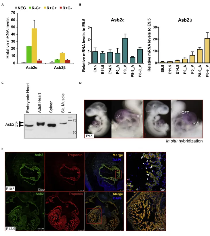
Figure 1. Asb2 Is Expressed in the Developing and Adult Heart and Undergoes Isoform Switching during Differentiation
(A) qPCR analysis of embryonic cardiomyocytes reveals predominant Asb2a expression in the R-G+ and the R+G+ populations. R-G+: Mef2c-.Nkx2-5+; R+G+: Mef2c+.Nkx2-5+; R+G-: Mef2c+.Nkx2-5-; NEG: Mef2c-.Nkx2-5-.
(B) qPCR analysis of Asb2a and Asb2b on RNA from murine hearts of different embryonic stages as well as neonates and postnatal day 8–9. Note that the a isoform is equally expressed at all stages, whereas theb isoform expression increases with development.
(C) Western blot analysis on whole tissue extracts from embryonic and adult heart, spleen, and skeletal muscle using Asb2-specific antibody. Note that Asb2 corresponding band in the embryonic heart co-migrates with that in the spleen where only thea isoform is expressed, whereas that in the adult heart co-migrates with that in the skeletal muscle that is known to express on theb isoform. These data are consistent with the qPCR data in (B).
ischemia-reperfusion injury (Pagan et al., 2013). Pharmacological inhibition of the proteasome is a new and promising means for cardioprotection (Pagan et al., 2013). Paradoxically, enhancing UPS activity has in some cases also provided protection against heart disease (Bulteau et al., 2001; Li et al., 2011; Powell et al., 2007) highlighting the importance of defining the role of the UPS as a therapeutic target in cardiac disease. In addition to its role as a protein quality control, the UPS has also been shown to regulate the turnover of sarcomere proteins, including the myofibrillar proteins myosin, actin, and troponin. Examples of this include the E3 Ub ligases MuRF1 (muscle-specific RING finger 1, targeting troponin I) and F box pro-tein Fbx122 (targetinga-actinin and filamin C) (Kedar et al., 2004; Spaich et al., 2012). E3 ligases have also been shown to regulate important signaling pathways in the heart, such as the JNK (c-Jun N terminal kinase) (Laine and Ronai, 2005), calcineurin (Fan et al., 2008), and the VEGF (vascular endothelial growth factor) signaling pathways (Murdaca et al., 2004). This important role of the UPS in the heart and its poten-tial for a therapeutic target in cardiac disease brings about a need to understand its specific function in heart development and disease. Using our previously described transgenic reporter system (Domian et al., 2009), we identified Asb2 (ankyrin repeat-containing protein with a suppressor of cytokine signaling box [SOCS box] 2) as being enriched in FHF and SHF cardiac progenitors. Asb2, which encodes specificity subunit of Cullin 5 RING E3 Ub ligase, exists in two isoforms: Asb2a and Asb2b in mouse, corresponding to variants 2 and 1 in humans, respectively (Bello et al., 2009). It has previously been shown to regulate differ-entiation of myeloid leukemia cells and skeletal myogenesis through proteasomal degradation of filamin proteins (Bello et al., 2009; Guibal et al., 2002; Heuze´ et al., 2008). Filamins (Flna, Flnb, and Flnc in mice) are actin-binding proteins important for the stabilization of the actin-cytoskeleton (van der Flier and Son-nenberg, 2001). Flnc is the only isoform expressed in the heart muscle where it is required for normal contractility (Fujita et al., 2012). Flna expression in the heart is restricted to endocardial and mesenchymal cells of the cardiac cushions during development and to valve leaflets in the adult heart (Norris et al., 2010). FLNA and FLNC mutations have been linked to cardiac defects in humans (de Wit et al., 2011, 2009; Kyndt et al., 2007; Valde´s-Mas et al., 2014). In zebrafish, an additional Asb2 target TCF3 was very recently identi-fied where TCF3 was negatively regulated by Asb2 during cardiogenesis (Fukuda et al., 2017). Asb2 down-regulation was also shown to be a mediator of follistatin-induced muscle hypertrophy and SMAD2/3 regu-lation of skeletal muscle mass in young adults in mice. The repression of Asb2 was, however, ameliorated in aging mice, some of which also displayed increasing Asb2 baseline levels (Davey et al., 2016). Asb2 over-expression was also shown to drive skeletal muscle atrophy in mice (Davey et al., 2016). A recent study also showed that Asb2 knockout is embryonic lethal and that Asb2a targets Flna for proteasomal degradation during early cardiomyocyte differentiation (Me´tais et al., 2018). The embryonic lethality of Asb2 mutants was shown to be primarily due to heart defects (Me´tais et al., 2018).
Herein, we show that Asb2 knockout in the FHF and SHF are both embryonic lethal by E10.5 and E12.5, respectively. Using tissue clearing combined with immunofluorescence technique, we show that Asb2 mutant hearts have incomplete looping. Moreover, Asb2 regulates cardiac morphogenesis partly through Flna turnover, and we hereby propose a model where Asb2-Flna controls TGFb-SMAD signaling to drive early cardiac formation. Additionally, Asb2 lethality in the anterior heart field (AHF) is partially rescued by Flna removal from these hearts. We also show that Asb2 ablation in the AHF leads to double outlet right ventricle (DORV), which is corrected upon further deletion of Flna from these hearts. Finally, we reveal that Asb2 role in cardiomyocyte differentiation is conserved in human cardiomyocytes as well. Collectively, our results shed light on the UPS regulation of heart development and its role as a cardio-therapeutic target and provide evidence for the first time for the role of the UPS in the rare congenital heart defect, DORV.
RESULTS
Asb2 Is Highly Enriched in the Embryonic Heart
We have previously characterized a transgenic reporter system for the isolation of three distinct mouse cardiac progenitor cells from developing embryos: FHF population, marked by Nkx2.5+.Mef2c- expression, and two SHF population subsets: Nkx2.5-.Mef2c+ and Nkx2.5+.Mef2c+ (Domian et al., 2009). Genome-wide Figure 1. Continued
(D)In situ hybridization on E9.5 mouse embryo showing robust Asb2 expression in the LV, RV, and OFT and to a lesser extent the IFT. LV, left ventricle; RV, right ventricle; OFT, outflow tract; IFT, inflow tract.
(E) Immunohistochemistry on E10.5 and E12.5 mouse embryos using Asb2-specific antibody (green) and Troponin T (red). DAPI (blue) marks nuclei. Note Asb2 expression colocalizes with Troponin T in the myocardium (arrows) and no expression is seen in the endocardial cells (arrow heads). Scale bar is equivalent to 250mm in the first three heart images left to right at E10.5 (top) and E12.5 (bottom), 25 mm in the E10.5 heart image top far right, and 50 mm in the E12.5 heart image bottom far right as indicated in the figure.
C
D
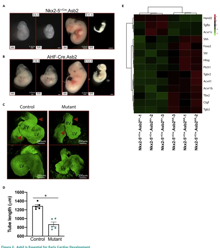
Figure 2. Asb2 Is Essential for Early Cardiac Development
(A) Nkx2-5+/Cre.Asb2 E9.5 and E11.5 knockout (KO) embryos (fl/fl) versus wild-type littermates (Wt). Note the resorbing KO embryo at E11.5. Scale bar is
equivalent to 0.4 mm for E9.5 and 0.5 mm for E11.5 as indicated.
(B) AHF-Cre.Asb2 E10.5 and E12.5 knockout (KO) embryos (fl/fl) versus wild-type littermates (Wt). Note the resorbing KO embryo at E12.5. Scale bar is equivalent to 0.5 mm for E10.5 and E12.5 as indicated in the figure.
(C) 3D reconstruction of CUBIC-cleared, Troponin-T-stained E9.5 whole control and Asb2 mutant embryos showing both ventral and dorsal views. Note the bulging in the right ventricle of the control heart that is lacking in the mutant (indicated by the red arrow heads). Scale bar is equivalent to 200mm as indicated.
transcriptional profiling and real-time PCR (qPCR) reveal Asb2 transcripts enrichment in the three popula-tions (Figure 1A) (Domian et al., 2009). To investigate the temporal expression of Asb2 in the developing heart, we performed qPCR analysis on RNA from mouse hearts at different stages of embryonic develop-ment. Our data show that Asb2a is expressed similarly throughout heart development, whereas Asb2b expression increases with development (Figure 1B). This is further confirmed by western blot analysis, which shows that the Asb2 band in the embryonic heart co-migrates with that in the spleen (which expresses Asb2a [Spinner et al., 2015]), whereas the Asb2 band in the adult heart co-migrates with that in the skeletal muscle (known to express Asb2b [Bello et al., 2009]) (Figure 1C). To further investigatein vivo spatial cardiac expres-sion of Abs2, we performedin situ hybridization on E9.5 embryos. Our data reveal robust expression of Asb2 transcripts predominantly in the left (LV) and right ventricles (RV) and to a lower extent in inflow (IFT) and outflow tracts (OFT) (Figure 1D). Furthermore, immunostaining of E10.5 and E11.5 (Figure 1E, upper and lower panels, respectively) embryonic sections using Asb2-specific antibody shows that, in the heart, Asb2 expression (green) is restricted to the myocardium overlapping with cardiac Troponin T (red). White arrows in the zoomed merged image at E10.5 (right panel) indicate overlap of Asb2 and Troponin T in the myocardial layer, but no expression is seen in the endocardial layer indicated by arrow heads. Asb2 Is Required for Early Cardiac Formation
To investigate the role of Asb2 during cardiac development, we generated two conditional knockout lines (KO): Nkx2-5+/Cre(a mouse line with the Cre recombinase knocked into the Nkx2-5 locus) and AHF-Cre (a mouse line with a transgene placing Cre under the transcriptional control of the AHF enhancer of the Mef2c gene). These mouse lines allow for the targeted removal of (Lombardi et al., 2009) Asb2 from the whole heart and the SHF, respectively (Lombardi et al., 2009). The floxed alleles are in common region and inactivate both Asb2 isoforms. Both conditional KOs have pericardial edema and are embryonic lethal: Nkx2-5+/Cre.Asb2fl/flmice die at E10.5–11 and AHF-Cre.Asb2fl/fldie at E11.5–12 (Figures 2A and 2B, respec-tively). AHF-Cre.Asb2fl/flmice analyzed at E10.5 also have shorter OFT compared with their control
litter-mates (Figure S1D). For Nkx2-5+/Cre.Asb2fl/fl, mice were analyzed at E8.5 (3 litters), E9.5 (23 litters), E10.5 (3 litters), and E11.5 (2 litters); for AHF-Cre.Asb2fl/fl, mice were analyzed at E9.5 (3 litters), E10.5 (4 litters),
and E12.5 (2 litters). Each litter consists of 8–11 embryos in total. All embryos were genotyped.Figure S1A shows the reduced level of Asb2 in the heterozygotes (Nkx2-5+/Cre.Asb2fl/+) and the complete loss of Asb2
in the knockouts (Nkx2-5+/Cre.Asb2fl/fl).
In order to perform a phenotypic analysis of the Nkx2-5+/Cre-Asb2fl/flmutant embryos, we used
state-of-the-art tissue clearing technique CUBIC combined with immunostaining. CUBIC can effectively clear mice embryos and embryonic hearts while preserving immunolabels (Kolesova´ et al., 2016; Tainaka et al., 2014). Nkx2-5+/Cre-Asb2fl/fland control littermates e9.5 mice embryos were cleared with CUBIC and stained for Troponin T to mark cardiomyocytes as well as DAPI for nuclei. Confocal microscopy with optical sectioning followed by 3D-reconstruction allowed the precise visualization of the developing hearts without disruption of underlying anatomy. During cardiac morphogenesis, the straight heart tube undergoes sequential looping steps to get to the fully looped heart. The fully looped heart acquires a he-lical shape in mice that is also referred to as the mature S-loop in chicks (Le Garrec et al., 2017; Ma¨nner, 2009). In Le Garrec et al. paper, they used computer modeling to simulate the biological process of mouse cardiac looping, incorporating in their model the left-right asymmetry and mechanical constraints seen in the looping heart. Their findings suggest that the lack of any of these parameters would lead to a C-shaped heart loop rather than the helical structure. In the chick, the heart is first transformed into a C-shaped heart, a process known as dextral looping. The C-loop is then converted into an immature S-loop that then trans-forms into a mature S-looped heart where the ventricular segments are curved outward to generate the left and right chambers (Ma¨nner, 2009). As shown inFigure 2C, the mutant embryos do not form the full helical structure seen in the control littermates. Instead, they have partially looped hearts that resemble the C-shaped hearts in the chick (Ma¨nner, 2009). Moreover, measurement of the heart tube length in mutant versus control hearts reveals statistically significant shorter tubes in the mutant hearts (Figure 2D).Video S1is a z stack of stained control and mutant e9.5 embryos showing the incomplete looping in the mutant embryo.Figure S1C represents four images from the z stack at different depth in the embryo. Four to five Figure 2. Continued
(D) Measurement of the heart tube of control and Asb2 mutant hearts. Note the statistically significant shorter heart tubes of the mutants. N = 5 per group. Data are represented as meanG SEM. * = p < 0.005. Unpaired t test was used using GraphPad Prism; p < 0.05 is considered statistically significant. (E) Heatmap analysis of a subset of cardiac looping differentially expressed genes in RNA-seq data from control (Nkx2-5+/Cre.Asb2fl/+) versus Nkx2-5+/Cre.Asb2
embryos were analyzed for each condition. The efficiency of the CUBIC/immunostaining technique on e9.5 mouse embryo is evidenced by the clearly visible striations of the cardiac muscle fibers (Figure S1B). In order to identify Asb2 downstream targets in the heart, RNA sequencing (RNA-seq) analysis was per-formed on Nkx2-5+/Cre.Asb2fl/fland control littermates (Figure 3B) (Figure S2C shows reduced levels of Asb2 transcripts in the Nkx2-5+/Cre.Asb2fl/flknockout compared with the Nkx2-5+/Cre.Asb2fl/+heterozygote
control). The gene expression profile was greatly altered in the Asb2 mutant hearts compared with their control littermates (Group 2 versus Group 1) (Figure 3B). Of note, a number of genes that are mis-expressed in the Asb2 cardiac mutant hearts have been previously linked to abnormal cardiac looping in mice (Figure 2E) (Azhar et al., 2003; Bardot et al., 2017; Chen et al., 1997; Le Garrec et al., 2017; Mine et al., 2008; Ribeiro et al., 2007; Vincentz et al., 2011). Ingenuity Pathway Analysis also shows that ‘‘cardiovascular system devel-opment and function’’ as well as ‘‘cardiovascular disease’’ are among the top pathways altered in the Asb2 mutant hearts (Table S1, yellow highlights).Table S2is an upstream analysis with the ones with a positive activationZ score > 1.5 highlighted in yellow. This list shows the pathways whose downstream targets are altered (upregulated or downregulated) in our knockouts versus controls. Targets with a positiveZ score suggest upregulation pathways in the Asb2 mutant hearts.
Asb2 Controls Cardiac Morphogenesis Partly through Regulating Filamin A
Since Asb2 targets filamin proteins for degradation (Me´tais et al., 2018) and Flna perturbations lead to car-diac defects and embryonic lethality (Feng et al., 2006), we investigated cardiac Flna expression in the Nkx2-5+/Cre.Asb2fl/fl. Flna expression in the control heart (Figure 3A, top panel) is restricted to endocardial
and pericardial layers (red staining, white arrow heads). In the knockout embryos (Figure 3A, third panel), Flna’s expression domain is abnormally expanded to include the myocardial layer (white arrows), co-local-izing with Troponin T expression (green for Troponin and yellow for the co-localization). Moreover, in Nkx2-5+/Cre.Asb2fl/+ heterozygous hearts (Figure 3A, second panel), Flna is abnormally expressed in some cardiomyocytes of the OFT myocardium (yellow arrows) suggesting that Asb2 regulation of Flna turn-over is dose dependent. We then hypothesized that, if Asb2 cardiac mutant phenotype is due to overexpression of Flna, then concurrently deleting Flna along with Asb2 should suppress the Asb2 phenotype. (Please note that Flna is an x-linked gene so a knockout is denoted by fl/fl for female or fl/y for male, whereas a heterozygous is denoted by fl/x or fl/+.) To examine this hypothesis, we developed Nkx2-5+/Cre.Asb2fl/fl.Flnafl/ydouble mutants. Removal of Flna from the hearts of Nkx2-5+/Cre.Asb2fl/fldid
not rescue lethality (Figure S2A). Approximately 16 litters were analyzed at E9.5 and 3 litters at E10.5. As expected, Nkx2-5+/Cre.Asb2fl/fl.Flnafl/fldouble knockouts no longer harbor ectopic Flna expression in the
myocardium (Figure S2B) as was previously seen with the Nkx2-5+/Cre.Asb2fl/flsingle knockouts (Figure 3A).
Instead, the double knockouts have normal endocardial expression of Flna similar to their control litter-mates (Figure S2B). RNA-seq analysis on e9.5 hearts from these mice show that their gene expression pro-file is closely related to the Nkx2-5+/Cre.Asb2fl/fl group (Group 2 versus Group 4) (Figure S2C shows
reduced levels of Asb2 transcripts in the Nkx2-5+/Cre.Asb2fl/fl.Flnafl/y double knockout compared with
the Nkx2-5+/Cre.Asb2fl/+heterozygote control). However, some genes whose expression was altered in the Nkx2-5+/Cre.Asb2fl/flgroup are restored to normal in the Nkx2-5+/Cre.Asb2fl/fl.Flnafl/yhearts (indicated
by arrows and shown inTable S3). These results suggest that Flna concurrent deletion can restore the normal expression level of a subset of genes in the Asb2 mutant hearts. Among these genes are the
Figure 3. Asb2 Targets Flna for Proteasomal Degradation in the Developing Heart and Asb2-Mutant Hearts Have an Altered Gene Expression Profile
(A) Immunohistochemistry on E9.5 Abs2 heterozygote (Nkx2-5+/Cre.Asb2fl/+, middle pane) and mutant hearts (Nkx2-5+/Cre.Asb2fl/fl, lower panel) as well as Wt
controls (top panel) using Flna (red) and Troponin-T (green)-specific antibodies. Note that FlnA expression is restricted to the endocardial layer (white arrow heads) in the Wt heart, whereas it is abnormally expressed in the myocardial layer in the Asb2-mutant hearts co-localizing with Troponin-T expression there (white arrows). Moreover, some cardiomyocytes in the outflow tract of the Asb2-heterozygous hearts also express Flna (yellow arrows) suggesting a dose-dependent regulation. Scale bar is equivalent to 250mm in the first column (left), 100 mm in the second, third, and fourth columns, and 25 mm in the fifth (far right) column as indicated in the figure.
(B) Heatmap analysis of RNA-seq data from control (Group1: Nkx2-5+/Cre.Asb2fl/+), Asb2 mutant (Group2: Nkx2-5+/Cre.Asb2fl/fl), Flna mutant (Group3:
Nkx2-5+/Cre.Flnafl/y), and Asb2-Flna double mutant (Group4: Nkx2-5+/Cre.Asb2fl/fl.Flnafl/y) E9.5 murine hearts. Note the high level of differentially expressed
genes in the Asb2-mutant and Asb2-Flna double mutant versus the control groups. A small subset of genes (indicated by arrows) that are perturbed in the Asb2-mutant hearts are restored to normal in the Asb2-Flna double mutants. N = 3 in each group (each sample is in itself a combination of three to four hearts to account for heterogeneity among different litters).
(C) Heatmap analysis of a subset of genes from the RNA-seq data in (B) that are part of the Tgfb/Smad signaling pathway. Note that the Foxa genes expression levels (indicated with a yellow line) that are downstream of the Tgfb/Smad are restored to normal in the Asb2-Flna double mutants versus the Asb2-mutant hearts.
B
Foxa genes, which are downstream of the Tgfb/Smad signaling (Figure 3C, yellow line) (Tang et al., 2011). Other genes in the Tgfb/Smad pathway are also altered in both Asb2-mutant and Asb2.Flna double mutant hearts (Figure 3C).Figure S2D is a qPCR analysis confirming some of these altered genes. Tgfbr1 and InhA
(which encodes a member of the Tgfb superfamily) are also among the positively regulated targets in the upstream analysis of the RNA-seq data of Asb2-mutant hearts versus control (Table S2). Both genes are no longer positively regulated in the upstream analysis of the list of genes corrected in the Asb2-Flna double mutant hearts (Table S3, yellow highlights). These data prompted further analysis of the Asb2/Flna regula-tion of TGFb/Smad signaling in the heart of these mice.
Asb2 Regulates TGFb/Smad Signaling through Regulating Filamin A Protein
TGFb signaling is initiated upon ligand-stimulated activation of serine/threonine receptor kinases that in turn lead to phosphorylation and activation of Smad proteins. Activated Smads interact with common signaling transducer Smad4, translocate to the nucleus, and activate downstream targets (Shi and Massague´, 2003). Flna directly associates with Smad2 and Smad2 phosphorylation, and TGFb/Smad2
signaling is impaired in Fln-null human melanoma cells (Sasaki et al., 2001; Zhou et al., 2011). Moreover, FLNA mutations were linked to x-linked myxomatous valvular dystrophy, a multivalve degeneration disor-der, and disrupted TGFb/Smad2/3 signaling was implicated in the disease pathogenesis (Geirsson et al., 2012; Norris et al., 2010). Using the ‘‘Build Network’’ module in MetaCore Clarivate Analytics software, we investigated the Asb2-Flna-Smad2 interaction. As shown in Figure 4A, Asb2 negatively regulates Flna through ubiquitination and Flna positively regulates Smad2 through direct binding. Asb2, Flna, and Smad2 are shown in red for visualization. In order to investigate further Asb2/Flna regulation of TGFb/ Smad2 signaling in cardiac development, we immunostained E9.5 Asb2-mutant hearts with antisera directed against pSmad2 (Figure 4B) and then quantified the pSmad2-positive nuclei.Figure 4C shows significant increase in the percentage of pSmad2-positive nuclei in the Nkx2-5+/Cre.Asb2fl/flmyocytes
(pre-viously shown to have overexpression of Flna [Figure 3A]) compared with their littermate controls. This increase was not seen in the endocardial cells where Flna expression is normal (Figure 3A). Interestingly, pSmad2 levels were restored to normal in the Nkx2-5+/Cre.Asb2fl/fl.Flnafl/fl(double mutant) myocytes further
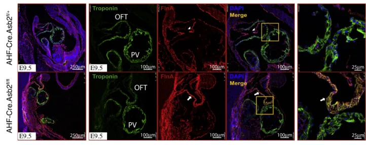
confirming that Asb2 regulates pSmad2 in the heart through the regulated turnover of Flna. Flna Removal from AHF-Cre.Asb2 Mutant Hearts Partially Rescues Embryonic Lethality To examine Flna expression in the AHF-Cre.Asb2fl/flhearts (where Asb2 is knocked out in the RV and OFT
only), Flna immunostaining was performed. As shown inFigure 5A, Flna expression (red) is restricted to the endocardial and epicardial layers in the control hearts (top panel, white arrow heads), whereas it is aber-rantly expressed in the myocardial layer of the OFT and RV only (red staining lower panel, white arrows), co-localizing with TroponinT expression (yellow staining lower panel) there. Flna expression was normal in the myocardial layer of the primitive left ventricle (PV) that harbors normal Asb2 expression and acts as an internal control in these mice. We then sought to examine the effect of further knocking out Flna from the AHF-Cre.Asb2-mutant hearts. To do this, we crossed Asb2fl/fl.Flnafl/flwith AHF-Cre.Asb2fl/+
mice. Our results show that AHF-Cre.Asb2fl/fl.Flnafl/y are born with the expected Mendelian ratios (Figure 5B); however, newborn pups die between P0.5 and P1.5. These results show that Flna deletion partially rescues Asb2 lethality. The AHF-Cre.Asb2fl/fl.Flnafl/+also survive to birth albeit at a lower percent-age from what is expected by Mendelian ratios; these mice also die right after birth at P0.5.
Figure 4. Tgfb/Smad Signaling Activity Is Downstream Asb2-Flna in the Developing Heart
(A) Schematic representation of Asb2-Flna-Smad2 interaction network using MetaCore Clarivate Analytics software. Note that Asb2 ubiquitinates and negatively regulates Flna, whereas Flna binds directly to and positively regulates Smad2.
(B) Immunohistochemistry on Nkx2-5+/Cre.Asb2 mutant (middle panel) and Nkx2-5+/Cre.Asb2-Flna double mutant (last panel) murine hearts as well as
wild-type controls (top panel) using pSmad2-specific antibody (green) and Troponin T (red). Note the nuclear localization of pSmad2 as a sign of Tgfb/Smad2 cycle activation. Examples of positive (purple arrowhead) and negative (yellow arrowheads) nuclei are indicated in the Wt sample (red box). DAPI marks all nuclei (AV, atrioventricular canal; V, primitive ventricle; OFT, outflow tract; Myo, myocytes; Endo, endocardial cells). Myocardial cells are marked by white arrows; endocardial cells are marked by white arrowheads. Scale bar is equivalent to 75mm in the first two columns from the left and 25 mm in the third, fourth, and fifth columns as indicated in the figure.
(C) Quantification of the immunostaining in (B) of percentage of pSmad2-positive nuclei in cardiomyocytes ((AV+V) Myo and OFT Myo) as well as endocardial cells (AV endo). Note the increased level of pSmad2-positive nuclei in Asb2-mutant myocytes that are restored to normal in the Asb2-FlnA double mutants. This regulation is not seen in the endocardial cells that do not express Asb2 (Figure 1E). n = 7 for Wt; n = 4 for Nkx2-5+/Cre.Asb2fl/fl; n = 3 for Nkx2-5+/Cre.Asb2fl/fl.Flnafl/fl. Data are
represented as meanG SEM. * = p < 0.05 Control versus Nkx2-5+/Cre.Asb2fl/fl; # = p < 0.05 Nkx2-5+/Cre.Asb2fl/flversus Nkx2-5+/Cre.Asb2fl/fl.Flnafl/fl; NS = not
Asb2 Removal from the Anterior Heart Field Leads to Double Outlet Right Ventricle) in Mice To determine the cardiac defects of the AHF-Cre.Asb2fl/fl.Flnafl/+, we examined these mice at e16.5–e17.5 after
the completion of cardiac morphogenesis but prior to the perinatal mortality associated with this genotype. Five litters were analyzed.Figure 6A shows the survival rate of these mice at E16.5. Gross examination of these hearts revealed that both the aorta and the pulmonary artery originate in the RV (Figure 6B, middle panel, yellow circle). In contrast, both the control hearts (Figure 6B, left panel) and those with AHF-Cre.Asb2-Flna double mutant ( Fig-ure 6B, right panel) were grossly normal with the pulmonary artery originating in the RV and the aorta originating in
Expected
Observed at P0.5
Asb2
fl/fl.FlnA
fl/+12.5%
1/31 (3.2%)
Asb2
fl/fl.FlnA
fl/y12.5%
4/31 (12.9%)
Asb2
fl/+.FlnA
fl/+12.5% 6/31
(19.3%)
Asb2
fl/+.FlnA
fl/y12.5% 6/31
(19.3%)
50% 14/31
(45.2%)
AHF
Cre
Control
AHF-Cre.Asb2
fl/+X Asb2
fl/fl.FlnA
fl/flExpected
Observed at P0.5
AHF
CreAsb2
fl/fl25%
0/29 (0%)
AHF
CreAsb2
fl/+25% 13/29
(44.8%)
50% 16/29
(55.1%)
Control
AHF-Cre.Asb2
fl/+X Asb2
fl/flDead at P0.5-1.5
B
Figure 5. Flna Removal from AHFCre.Asb2 Mutant Hearts Partially Rescues Their Lethality
(A) Immunohistochemistry on E9.5 AHF-Cre.Abs2 mutant hearts (AHF-Cre.Asb2fl/fl, lower panel) and littermate controls (AHF-Cre.Asb2fl/+, top panel) using
Flna (red)- and Troponin-T (green)-specific antibodies. Note overexpression of Flna in the OFT (white arrows) of the AHF-Cre.Asb2 mutant hearts but not the primitive left ventricle (PV) that harbors normal Asb2 levels thus serving as an internal control. Flna expression in the control hearts is restricted to the endocardial layer (white arrow heads). Scale bar is equivalent to 250mm in the first column (left); 100 mm in the second, third, and fourth columns; 25 mm in the fifth column (far right) as indicated.
(B) Table showing the survival of AHF-Cre.Asb2 mutant (top) and AHF-Cre.Asb2-Flna double mutant (bottom) mice. Note that no AHF-Cre.Abs2 mutant mice are observed at P0.5. However, the AHF-Cre.Asb2-Flna double mutant (Asb2fl/fl.Flnafl/y) mice are born at the expected Mendelian ratios.
AHF-Cre.Asb2-mutant mice harboring one copy of Flna (Asb2fl/fl.Flnafl/+) are also born yet at lower percentage than what is expected by Mendelian genetics. These mice
the LV. Serial sections of mutant and control hearts (Figure 6C) further confirm that the AHF-Cre.Asb2fl/fl.Flnafl/x mice have DORV (Figure 6C middle panel, yellow oval). This is also accompanied by a ventricular septal defect (Figure 6C middle panel right, indicated by asterisk), a feature commonly associated with DORV in patients with congenital heart disease (Obler et al., 2008). As shown inFigure 6D, the DORV phenotype appeared to be fully penetrant in the AHF-Cre.Asb2fl/fl.Flnafl/xhearts. Notably, the DORV phenotype is corrected in the
AHF-Cre.Asb2-Flna double mutant hearts (AHF-Cre.Asb2fl/fl.Flnafl/y,Figure 6C lower panel).
Asb2 Is Required for Human Embryonic Stem Cell-Derived Cardiomyocyte Differentiation To further investigate if the requirement for Asb2 for cardiac development is conserved during human cardiomyo-cyte differentiation, we turned to human embryonic stem cell (hESC)-derived cardiomyocardiomyo-cyte in vitro differentiation. Both ASB2 variants 1 (Asb2b in mice) and 2 (Asb2a in mice) are expressed at different stages of cardiomyocyte differentiation (Figure 7A, top and bottom graphs, respectively). Using CRISPR/Cas9 genome ed-iting technology, we then generated ASB2-null hESCs. The guides were designed in exon 2 (targeting variant 1 specifically) or exon 4 (targeting variants 1 and 2) (Figure S3A). Four wild-type (Wt) (received the CRISPR/Cas9 con-structs but failed to generate an in/del) and four knockout (KO) lines were generated. The genotype of all lines was confirmed by sequencing (refer toTransparent Methods), and the knockouts were confirmed by qPCR (Figure 7B, right panel). Wt clones were able to differentiate into beating cardiomyocytes, whereas all four KO lines failed to do so (Video S2, top panels for Wt clones and bottom panels for KO clones). Calcium cycling was also impaired in the Asb2-null derived hESCs (Video S3, left panel for Wt and right panel for KO, andFigure S3B). Two Wt and two KO lines were used for further investigation. qPCR analysis on RNA from cardiomyocytes derived from these cells
A C
B
D
*
Figure 6. AHF-Asb2 Mutant Hearts Have Double Outlet Right Ventricle and Ventricular Septal Defect
(A) Table showing the survival of AHF-Cre.Asb2-Flna double mutant mice at E16.5.
(B) E16.5 whole hearts of AHF-Cre.Asb2-mutant embryos with one copy of Flna (AHF-Cre.Asb2fl/fl.Flnafl/x), AHF-Cre.Asb2.Flna double mutants (AHF-Cre.Asb2fl/fl.Flnafl/fl), and wild-type control. Note that both the pulmonary artery (PA)
and the aorta (Ao) are open in the right ventricle (RV) of the AHF-Cre.Asb2fl/fl.Flnafl/xhearts (yellow circle). Scale bar is
equivalent to 0.02 mm as indicated.
(C) Masson trichrome staining of E16.5 heart sections of control (Wt) (top), AHF-Cre.Asb2fl/fl.Flnafl/x(middle), and
AHF-Cre.Asb2fl/fl.Flnafl/fl(bottom) embryos. Note that both the pulmonary artery and the aorta open in the right ventricle of the
AHF-Cre.Asb2fl/fl.Flnafl/xhearts (yellow circle, middle panel) but not the Wt or the AHF-Cre.Asb2fl/fl.Flnafl/flhearts. The
AHF-Cre.Asb2fl/fl.Flnafl/xalso have a VSD indicated by asterisk (middle panel, right). Ao, aorta; PA, pulmonary artery; RV,
right ventricle; LV, left ventricle; IVS, interventricular septum. Scale bar is equivalent to 250mm.
(D) Number of E16.5 hearts with DORV in Wt, AHF-Cre.Asb2fl/fl.Flnafl/x, and AHF-Cre.Asb2fl/fl.Flnafl/flembryos. Note that
shows that cardiac Troponin T transcript levels (TNNT2, marker of cardiomyocyte differentiation) are greatly reduced in the KO lines at d15 of differentiation (Figure 7B, right). On the other hand, both NKX2-5 and MESP1 (markers of cardiac progenitors) are normally expressed at d7 of differentiation (Figure 7B, left). This was further confirmed at the protein level by immunostaining that shows great reduction in cardiac Troponin T (red) expression at d15 (Figure 7D) and normal NKX2-5 levels (green) at days 15 and 8 (Figures 7C and 7D, respec-tively). These data suggest that Asb2-null hES cells can commit to the cardiac lineage but arrest in differentiation prior to the generation of functional cardiomyocytes.
We then examined if ASB2 regulation of the TGFb/SMAD signaling seen in mice hearts is conserved in the human cells. As discussed above, upon TGFb/Smad activation, the signaling transducer Smad4 is
C D
Figure 7. ASB2 Is Essential for Human Embryonic Stem Cell (hESC)-derived Cardiomyocytes Differentiation
(A) qPCR analysis of RNA from hESC-derived cardiomyocytes at different stages of differentiation. Note that both ASB2 variants are expressed at the different stages. N = 4 for D0, n = 5 for all other stages. Data are represented as meanG SEM.
(B) qPCR analysis of RNA extracted from two different wild-type (Wt) clones and two ASB2 mutant (KO) clones d7 (left) and d15 (right) differentiated hESCs. Note reduced Troponin T (differentiation marker) transcripts in the mutants at d15 and no difference in MESP1 and NKX2-5 (cardiac progenitor markers) levels at d7. N = 3 per sample for d7 and N = 4 per sample for d14. Data are represented as meanG SEM. * = p < 0.05. Two-way ANOVA was used for analysis using GraphPad Prism. p < 0.05 is considered statistically significant.
(C and D) Immunostaining of d8 (C) and d15 (D) differentiated Wt and ASB2 mutant cells (KO). Note reduced Troponin T (red)-positive mutant cells at d15 but no difference in NKX2-5 (green) levels between Wt and mutant cells at both stages. DAPI (blue) marks all nuclei. Scale bar is equivalent to 8mm in (C) and 10mm in (D) as indicated.
translocated to the nucleus to activate downstream targets.Figure 8A shows an increase in nuclear SMAD4 (green) in the ASB2-null hES-derived cardiomyocytes. The nuclear versus total SMAD4 was quantified ( Fig-ure 8A, right graph) showing that nuclear SMAD4 signal is doubled in the ASB2-null cells, whereas total Smad4 levels remain the same. Western blot analysis on total protein extracts from these cells also confirms significant increase in both SMAD4 and pSMAD2 protein levels (Figures 8B and 8C). This further confirms that the TGFb/SMAD signaling pathway is activated in Asb2-null cardiomyocytes.
DISCUSSION
In this study, we provide strong evidence for the role of Asb2 in controlling heart morphogenesis partly through its regulation of the actin-binding protein, Filamin A (Flna), and Tgfb/Smad signaling. We further show that this regulation is part of the DORV disease pathogenesis.
Using CUBIC clearing technique combined with immunofluorescence and confocal microscopy, we show that the Asb2-mutant hearts have shorter heart tubes and do not form the fully looped helical structure. RNA-seq analysis also reveals that a number of genes that have been linked to cardiac looping defects
A
B
C
Figure 8. ASB2 Is an Upstream Regulator of TGFb/SMAD Pathway in hESC-derived Cardiomyocytes
(A) Immunostaining of d15 Wt and ASB2 mutant (KO) hESC-derived cardiomyocytes using SMAD4-specific antibody (green). Note reduced level of nuclear but not total mean fluorescence intensity of SMAD4-positive cells in the KOs (quantification graphs on the right). DAPI (blue) marks all nuclei. Scale bar is equivalent to 75mm in the first column and 25mm in the second and third columns as indicated. *: p < 0.005 significant versus Wt. Unpaired t test was used for analysis using GraphPad Prism.
(B and C) (B) Western blot analysis of Wt and ASB2 mutant (KO) hESC-derived cardiomyocytes using SMAD4, SMAD2, and pSMAD2 antibodies. Note increase of SMAD4 and pSMAD2 in the mutant clones. Data are representative of three separate experiments (C) Quantification of the western blot analysis in (B). Data are average of quantification from three separate experiments. *: significant versus Wt1 and Wt2. p < 0.05 is considered statistically significant. One-way ANOVA was used for analysis using GraphPad Prism.
that Asb2 regulates SMAD signaling through the Flna pathway. RNA-seq analysis also reveals that regula-tion of the Tgfb-Smad pathway in the Asb2-mutant hearts and the Foxa genes, which are downstream effectors of the Tgfb/Smad signaling (Tang et al., 2011), is in fact restored to normal in the Asb2-Flna dou-ble mutants. Flna has been previously shown to associate with Smad2 signaling (Sasaki et al., 2001). Moreover, Tgfb/Smad2/3 signaling is impaired in the multivalve degeneration disorder, X-linked myxoma-tous valvular dystrophy, in which FLNA mutations were reported (Geirsson et al., 2012; Norris et al., 2010). Our data provide further evidence for regulation of the Tgfb/Smad cycle by Flna and show that Asb2 is upstream of this regulatory pathway in the developing heart.
Using human embryonic stem cell (hESC)-derived cardiomyocytes, we further show that the Asb2 role in embryonic heart differentiation is conserved in humans. Although ASB2-null hESCs are able to form cardiac progenitor cells (marked by expression of MESP1 and NKX2-5), they have an impaired ability to differen-tiate into beating TNNT+ cardiomyocytes. It is important to note here that the difference between Troponin T levels in the Asb2-null hESCs and the Asb2 mutantsin vivo could be due to the total knockout in the cells that is more severe than the conditionalin vivo knockout. Additionally, the cell system lacks signaling coming from the endocardium, which could also explain this difference. These results demon-strate that, in human PSCs differentiatingin vitro, ASB2-mediated targeted degradation is required for the differentiation from NKX2-5+ progenitors to beating TNNT+ cardiomyocytes and that deletion of ASB2 results in a differentiation arrest at the progenitor stage. Moreover, the finding that these cells have increased levels of SMAD4 and pSMAD2, markers of TGFb/SMAD pathway activation, provides further evidence that ASB2 is an upstream regulator of the TGFb/SMAD pathway during the differentiation of human cardiomyocytes. These considerations become increasingly important given the potential of pluripotent stem cell-derived CMs to serve as a renewable cell source for cardiac regeneration in the injured heart.
Given that Flna is a direct target of Asb2 that is aberrantly upregulated in Asb2-mutant cardiomyocytes, we then investigated whether Asb2 cardiac mutant embryonic lethality can be rescued by the concurrent deletion of Flna. Accordingly, we generated AHF-Cre.Asb2fl/fl.Flnafl/ydouble mutants. Our data show that, as opposed to the AHF-Cre.Asb2fl/flsingle mutants that die by E11.5, the AHF-Cre.Asb2fl/fl.Flnafl/ydouble mutants are born with
the expected Mendelian ratios (Figure 5B) but die shortly after birth. This suggests partial rescue of lethality seen in the AHF-Cre.Asb2fl/flmutant hearts. Of note, we also generated Nkx2-5+/Cre.Asb2fl/fl.Flnafl/Ydouble
mutants that did not rescue the Asb2 lethality (Figure S2A), suggesting a greater Asb2 dependency or a more complex phenotype in these mice. These findings are not surprising owing to earlier and broader expression domain of the Nxk2-5Crecompared with the AHF-Cre line. Moreover, AHF-Asb2-mutant mice with one Flna allele
(AHF-Cre.Asb2fl/fl.Flnafl/x) sometimes also survive to P0, albeit at significantly lower than expected ratios ( Fig-ure 5B). Not only do these results suggest a dose dependency of Asb2 and its target Flna but they also allow us to identify DORV associated with ventricular septal defect (VSD) as a penetrant cardiac phenotype. More inter-estingly, this phenotype was rescued when Flna was abrogated by the concurrent deletion of Flna showing that both Asb2 and Flna play a functional role in the pathogenesis of DORV.
During cardiac development, the heart first forms as a primitive heart tube that then elongates and starts to loop by addition of cells from the anterior, posterior, and second heart field at both the venous and arterial poles. At the onset of looping, left-right asymmetry in the heart becomes morphologically evident and any defects in this process can lead to complex congenital heart problems, including DORV and VSDs ( Rams-dell, 2005). The heart is the first organ to break the left-right symmetry in the developing embryo, and it has been shown that the actin-cytoskeleton is fundamental for laterality and modulation associated with heart looping. It was shown to provide the built-in mechanism required for cells to acquire left-right asymmetry (Linask and Vanauker, 2007; Tee et al., 2015). Abnormalities in the control of construction of the cytoskel-eton has been previously shown to result in looping defects and ultimately lead to congenital heart prob-lems (Langdon et al., 2012; Linask and Vanauker, 2007). Our data and the data from Metais et al. provide solid evidence for the Asb2-Flna regulation of the actin cytoskeleton during heart morphogenesis (Me´tais et al., 2018). Our data extend this regulation to show that it is important for normal heart tube and OFT development and, if perturbed, leads to DORV and VSD in the developing mammalian heart. Additionally, we suggest a mechanism where Asb2 downregulation leads to abnormal overexpression of Flna that ulti-mately leads to increased activity of the Tgfb/Smad2 signaling in the myocardium thus causing growth/ elongation defects and DORV in the mammalian heart. Indeed, prior reports have implicated the Tgfb superfamily and Smad2/3 in left-right asymmetry, and Tgfb2 mutant mice have been shown to develop DORV and die right after birth (Azhar et al., 2003; Sanford et al., 1997; Whitman and Mercola, 2001). In humans, a missense mutation in Flna (c.5290G>A (p.A1764T) has been reported in a patient with DORV (de Wit et al., 2011). Since missense mutations can result in both loss and gain of function, future studies will be required to determine the effect of this mutation on Flna expression and function. Thus, our data demonstrate a link between targeted protein turnover and the development of DORV and highlights the potential of the ASB2/FLNA axis as a diagnostic, prognostic, and/or therapeutic target for patients with DORV.
Limitations of the Study
Although our data show that the role of Asb2 in heart morphogenesis is conserved between mice and human, a limitation is that thein vivo murine system is a conditional knockout compared with the total knockout in the human cells system. Additionally, as we know, a cross talk between the endocardium and the myocardium occurs during heart morphogenesis, and this again is lacking in our human cell system.
METHODS
All methods can be found in the accompanyingTransparent Methods supplemental file.
DATA AND CODE AVAILABILITY
RNA-seq data have been deposited in NCBI’s Gene Expression Omnibus (GEO). The accession number for the RNA-seq data reported in this paper is GEO: GSE145495.
SUPPLEMENTAL INFORMATION
Supplemental Information can be found online athttps://doi.org/10.1016/j.isci.2020.100959.
ACKNOWLEDGMENTS
We thank all members of the Domian Lab for valuable insight and suggestions. We also thank the his-tology core at Dana Farber Cancer Institute/Harvard Medical School and the NextGen Sequencing core at Massachusetts General Hospital for technical support. This work was supported by the American Heart Association (17GRNT33630170), the Centre National de la Recherche Scientifique, and the Univer-sity of Toulouse. A.Y. is a recipient of the Fund for Medical Discovery (FMD) Award from the Massachu-setts General Hospital/Harvard Medical School.
AUTHOR CONTRIBUTIONS
Conceptualization, A.Y. and I.J.D.; Methodology, A.Y. and I.J.D.; Investigation A.Y., D.H., N.M., J.W.B., and S.D.; Writing – Original Draft, A.Y.; Writing – Review & Editing, A.Y., P.G.L., C.M.-L, P.T.L., and I.J.D.; Visu-alization, A.Y.; Supervision, A.Y. and I.J.D.; Funding Acquisition, A.Y. and I.J.D.
cardiac progenitor population with ventricular differentiation potential. Nat. Commun.8, 14428.
Bello, N.F., Lamsoul, I., Heuze´, M.L., Me´tais, A., Moreaux, G., Calderwood, D.A., Duprez, D., Moog-Lutz, C., and Lutz, P.G. (2009). The E3 ubiquitin ligase specificity subunit ASB2beta is a novel regulator of muscle differentiation that targets filamin B to proteasomal degradation. Cell Death Differ.16, 921–932.
Bruneau, B.G. (2008). The developmental genetics of congenital heart disease. Nature451, 943–948.
Bulteau, A.L., Lundberg, K.C., Humphries, K.M., Sadek, H.A., Szweda, P.A., Friguet, B., and Szweda, L.I. (2001). Oxidative modification and inactivation of the proteasome during coronary occlusion/reperfusion. J. Biol. Chem.276, 30057–30063.
Chen, J.N., van Eeden, F.J., Warren, K.S., Chin, A., Nu¨sslein-Volhard, C., Haffter, P., and Fishman, M.C. (1997). Left-right pattern of cardiac BMP4 may drive asymmetry of the heart in zebrafish. Development124, 4373–4382.
Davey, J.R., Watt, K.I., Parker, B.L., Chaudhuri, R., Ryall, J.G., Cunningham, L., Qian, H., Sartorelli, V., Sandri, M., Chamberlain, J., et al. (2016). Integrated expression analysis of muscle hypertrophy identifies Asb2 as a negative regulator of muscle mass. JCI Insight1,https:// doi.org/10.1172/jci.insight.85477.
de Wit, M.C.Y., de Coo, I.F.M., Lequin, M.H., Halley, D.J.J., Roos-Hesselink, J.W., and Mancini, G.M.S. (2011). Combined cardiological and neurological abnormalities due to filamin A gene mutation. Clin. Res. Cardiol.100, 45–50.
de Wit, M.C.Y., Kros, J.M., Halley, D.J.J., de Coo, I.F.M., Verdijk, R., Jacobs, B.C., and Mancini, G.M.S. (2009). Filamin A mutation, a common cause for periventricular heterotopia, aneurysms and cardiac defects. J. Neurol. Neurosurg. Psychiatry80, 426–428.
Domian, I.J., Chiravuri, M., van der Meer, P., Feinberg, A.W., Shi, X., Shao, Y., Wu, S.M., Parker, K.K., and Chien, K.R. (2009). Generation of functional ventricular heart muscle from mouse ventricular progenitor cells. Science326, 426–429.
Natl. Acad. Sci. U S A103, 19836–19841.
Fujita, M., Mitsuhashi, H., Isogai, S., Nakata, T., Kawakami, A., Nonaka, I., Noguchi, S., Hayashi, Y.K., Nishino, I., and Kudo, A. (2012). Filamin C plays an essential role in the maintenance of the structural integrity of cardiac and skeletal muscles, revealed by the medaka mutant zacro. Dev. Biol.361, 79–89.
Fukuda, R., Gunawan, F., Beisaw, A., Jimenez-Amilburu, V., Maischein, H.-M., Kostin, S., Kawakami, K., and Stainier, D.Y.R. (2017). Proteolysis regulates cardiomyocyte maturation and tissue integration. Nat. Commun.8, 14495.
Geirsson, A., Singh, M., Ali, R., Abbas, H., Li, W., Sanchez, J.A., Hashim, S., and Tellides, G. (2012). Modulation of transforming growth factor-b signaling and extracellular matrix production in myxomatous mitral valves by angiotensin II receptor blockers. Circulation126, S189–S197.
Glickman, M.H., and Ciechanover, A. (2002). The ubiquitin-proteasome proteolytic pathway: destruction for the sake of construction. Physiol. Rev.82, 373–428.
Guibal, F.C., Moog-Lutz, C., Smolewski, P., Di Gioia, Y., Darzynkiewicz, Z., Lutz, P.G., and Cayre, Y.E. (2002). ASB-2 inhibits growth and promotes commitment in myeloid leukemia cells. J. Biol. Chem.277, 218–224.
Heuze´, M.L., Lamsoul, I., Baldassarre, M., Lad, Y., Le´veˆque, S., Razinia, Z., Moog-Lutz, C., Calderwood, D.A., and Lutz, P.G. (2008). ASB2 targets filamins A and B to proteasomal degradation. Blood112, 5130–5140.
Hu, D., Linders, A., Yamak, A., Correia, C., Kijlstra, J.D., Garakani, A., Xiao, L., Milan, D.J., van der Meer, P., Serra, M., et al. (2018). Metabolic maturation of human pluripotent stem cell-derived cardiomyocytes by inhibition of HIF1a and LDHA. Circ. Res.123, 1066–1079.
Jung, T., Catalgol, B., and Grune, T. (2009). The proteasomal system. Mol. Aspects Med.30, 191–296.
Kedar, V., McDonough, H., Arya, R., Li, H.-H., Rockman, H.A., and Patterson, C. (2004). Muscle-specific RING finger 1 is a bona fide ubiquitin
Kyndt, F., Gueffet, J.-P., Probst, V., Jaafar, P., Legendre, A., Le Bouffant, F., Toquet, C., Roy, E., McGregor, L., Lynch, S.A., et al. (2007). Mutations in the gene encoding filamin A as a cause for familial cardiac valvular dystrophy. Circulation 115, 40–49.
Laine, A., and Ronai, Z. (2005). Ubiquitin chains in the ladder of MAPK signaling. Sci. STKE2005, re5.
Langdon, Y., Tandon, P., Paden, E., Duddy, J., Taylor, J.M., and Conlon, F.L. (2012). SHP-2 acts via ROCK to regulate the cardiac actin cytoskeleton. Development139, 948–957. Le Garrec, J.-F., Domı´nguez, J.N., Desgrange, A., Ivanovitch, K.D., Raphae¨l, E., Bangham, J.A., Torres, M., Coen, E., Mohun, T.J., and Meilhac, S.M. (2017). A predictive model of asymmetric morphogenesis from 3D reconstructions of mouse heart looping dynamics. Elife6,https:// doi.org/10.7554/eLife.28951.
Li, J., Horak, K.M., Su, H., Sanbe, A., Robbins, J., and Wang, X. (2011). Enhancement of proteasomal function protects against cardiac proteinopathy and ischemia/reperfusion injury in mice. J. Clin. Invest.121, 3689–3700.
Linask, K.K., and Vanauker, M. (2007). A role for the cytoskeleton in heart looping.
ScientificWorldJournal7, 280–298.
Lombardi, R., Dong, J., Rodriguez, G., Bell, A., Leung, T.K., Schwartz, R.J., Willerson, J.T., Brugada, R., and Marian, A.J. (2009). Genetic fate mapping identifies second heart field progenitor cells as a source of adipocytes in arrhythmogenic right ventricular cardiomyopathy. Circ. Res.104, 1076–1084.
Ma¨nner, J. (2009). The anatomy of cardiac looping: a step towards the understanding of the morphogenesis of several forms of congenital cardiac malformations. Clin. Anat.22, 21–35.
Martinsen, B.J., and Lohr, J.L. (2015). Cardiac development. In Handbook of Cardiac Anatomy, Physiology, and Devices, P.A. Laizzo, ed. (Springer), pp. 23–33.
Mercola, M., Ruiz-Lozano, P., and Schneider, M.D. (2011). Cardiac muscle regeneration: lessons from development. Genes Dev.25, 299–309.
Me´tais, A., Lamsoul, I., Melet, A., Uttenweiler-Joseph, S., Poincloux, R., Stefanovic, S., Valie`re, A., Gonzalez de Peredo, A., Stella, A., Burlet-Schiltz, O., et al. (2018). Asb2a-filamin A axis is essential for actin cytoskeleton remodeling during heart development. Circ. Res.122, e34–e48.
Mine, N., Anderson, R.M., and Klingensmith, J. (2008). BMP antagonism is required in both the node and lateral plate mesoderm for mammalian left-right axis establishment. Development135, 2425–2434.
Murdaca, J., Treins, C., Monthoue¨l-Kartmann, M.-N., Pontier-Bres, R., Kumar, S., Van Obberghen, E., and Giorgetti-Peraldi, S. (2004). Grb10 prevents Nedd4-mediated vascular endothelial growth factor receptor-2 degradation. J. Biol. Chem.279, 26754–26761.
Nemer, M. (2008). Genetic insights into normal and abnormal heart development. Cardiovasc. Pathol.17, 48–54.
Norris, R.A., Moreno-Rodriguez, R., Wessels, A., Merot, J., Bruneval, P., Chester, A.H., Yacoub, M.H., Hage`ge, A., Slaugenhaupt, S.A., Aikawa, E., et al. (2010). Expression of the familial cardiac valvular dystrophy gene, filamin-A, during heart morphogenesis. Dev. Dyn.239, 2118–2127.
Obler, D., Juraszek, A.L., Smoot, L.B., and Natowicz, M.R. (2008). Double outlet right ventricle: aetiologies and associations. J. Med. Genet.45, 481–497.
Pagan, J., Seto, T., Pagano, M., and Cittadini, A. (2013). Role of the ubiquitin proteasome system in the heart. Circ. Res.112, 1046–1058.
Powell, S.R., Davies, K.J.A., and Divald, A. (2007). Optimal determination of heart tissue 26S-proteasome activity requires maximal stimulating ATP concentrations. J. Mol. Cell. Cardiol.42, 265–269.
Ramsdell, A.F. (2005). Left-right asymmetry and congenital cardiac defects: getting to the heart of
the matter in vertebrate left-right axis determination. Dev. Biol.288, 1–20.
Ribeiro, I., Kawakami, Y., Bu¨scher, D., Raya, A., Rodrı´guez-Leo´n, J., Morita, M., Rodrı´guez Esteban, C., and Izpisu´a Belmonte, J.C. (2007). Tbx2 and Tbx3 regulate the dynamics of cell proliferation during heart remodeling. PLoS One 2, e398.
Sanford, L.P., Ormsby, I., Gittenberger-de Groot, A.C., Sariola, H., Friedman, R., Boivin, G.P., Cardell, E.L., and Doetschman, T. (1997). TGFbeta2 knockout mice have multiple developmental defects that are non-overlapping with other TGFbeta knockout phenotypes. Development124, 2659–2670.
Sasaki, A., Masuda, Y., Ohta, Y., Ikeda, K., and Watanabe, K. (2001). Filamin associates with Smads and regulates transforming growth factor-beta signaling. J. Biol. Chem.276, 17871–17877.
Shi, Y., and Massague´, J. (2003). Mechanisms of TGF-beta signaling from cell membrane to the nucleus. Cell113, 685–700.
Spaich, S., Will, R.D., Just, S., Spaich, S., Kuhn, C., Frank, D., Berger, I.M., Wiemann, S., Korn, B., Koegl, M., et al. (2012). F-box and leucine-rich repeat protein 22 is a cardiac-enriched F-box protein that regulates sarcomeric protein turnover and is essential for maintenance of contractile function in vivo. Circ. Res.111, 1504–1516.
Spinner, C.A., Uttenweiler-Joseph, S., Metais, A., Stella, A., Burlet-Schiltz, O., Moog-Lutz, C., Lamsoul, I., and Lutz, P.G. (2015). Substrates of the ASB2a E3 ubiquitin ligase in dendritic cells. Sci. Rep.5, 16269.
Srivastava, D. (2006). Making or breaking the heart: from lineage determination to morphogenesis. Cell126, 1037–1048.
Tainaka, K., Kubota, S.I., Suyama, T.Q., Susaki, E.A., Perrin, D., Ukai-Tadenuma, M., Ukai, H., and Ueda, H.R. (2014). Whole-body imaging with
single-cell resolution by tissue decolorization. Cell159, 911–924.
Tang, Y., Shu, G., Yuan, X., Jing, N., and Song, J. (2011). FOXA2 functions as a suppressor of tumor metastasis by inhibition of epithelial-to-mesenchymal transition in human lung cancers. Cell Res.21, 316–326.
Tee, Y.H., Shemesh, T., Thiagarajan, V., Hariadi, R.F., Anderson, K.L., Page, C., Volkmann, N., Hanein, D., Sivaramakrishnan, S., Kozlov, M.M., and Bershadsky, A.D. (2015). Cellular chirality arising from the self-organization of the actin cytoskeleton. Nat. Cell Biol.17, 445–457.
Valde´s-Mas, R., Gutie´rrez-Ferna´ndez, A., Go´mez, J., Coto, E., Astudillo, A., Puente, D.A., Reguero, J.R., A´lvarez, V., Morı´s, C., Leo´n, D., et al. (2014). Mutations in filamin C cause a new form of familial hypertrophic cardiomyopathy. Nat. Commun.5, 5326.
van der Flier, A., and Sonnenberg, A. (2001). Structural and functional aspects of filamins. Biochim. Biophys. Acta1538, 99–117.
Vincentz, J.W., Barnes, R.M., and Firulli, A.B. (2011). Hand factors as regulators of cardiac morphogenesis and implications for congenital heart defects. Birth Defects Res. A Clin. Mol. Teratol.91, 485–494.
Whitman, M., and Mercola, M. (2001). TGF-beta superfamily signaling and left-right asymmetry. Sci. STKE2001, re1.
Yamak, A., and Nemer, M. (2015). Role of embryonic and differentiated cells in cardiac development. In Biomaterials for Cardiac Regeneration, E.J. Suuronen and M. Ruel, eds. (Springer), pp. 37–70.
Zhou, A.-X., Toylu, A., Nallapalli, R.K., Nilsson, G., Atabey, N., Heldin, C.-H., Bore´n, J., Bergo, M.O., and Akyu¨rek, L.M. (2011). Filamin a mediates HGF/c-MET signaling in tumor cell migration. Int. J. Cancer128, 839–846.