HAL Id: hal-02125415
https://hal.archives-ouvertes.fr/hal-02125415
Submitted on 10 May 2019
HAL is a multi-disciplinary open access
archive for the deposit and dissemination of
sci-entific research documents, whether they are
pub-lished or not. The documents may come from
teaching and research institutions in France or
abroad, or from public or private research centers.
L’archive ouverte pluridisciplinaire HAL, est
destinée au dépôt et à la diffusion de documents
scientifiques de niveau recherche, publiés ou non,
émanant des établissements d’enseignement et de
recherche français ou étrangers, des laboratoires
publics ou privés.
Ultrasound modulated optical tomography in scattering
media: flux filtering based on persistent spectral hole
burning in the optical diagnosis window
Caroline Venet, Maïmouna Bocoum, Jean-Baptiste Laudereau, Thierry
Chanelière, François Ramaz, Anne Louchet-Chauvet
To cite this version:
Caroline Venet, Maïmouna Bocoum, Jean-Baptiste Laudereau, Thierry Chanelière, François Ramaz,
et al.. Ultrasound modulated optical tomography in scattering media: flux filtering based on persistent
spectral hole burning in the optical diagnosis window. Optics Letters, Optical Society of America
-OSA Publishing, 2018, 43 (16), pp.3993. �10.1364/OL.43.003993�. �hal-02125415�
Ultrasound modulated optical tomography in
scattering media: flux filtering based on persistent
spectral hole burning in the optical diagnosis window
C
AROLINE
V
ENET
1,2,*,
M
AÏMOUNA
B
OCOUM
1,
J
EAN
-B
APTISTE
L
AUDEREAU
1,
T
HIERRY
C
HANELIERE
2,
F
RANÇOIS
R
AMAZ
1,
A
NNE
L
OUCHET
-C
HAUVET
21
Institut Langevin, Ondes et Images, ESPCI ParisTech, PSL Research University, CNRS UMR 7587, INSERM U979, Université Paris VI Pierre et Marie Curie, 1 rue Jussieu, 75005 Paris, France
2
Laboratoire Aimé Cotton, CNRS, Univ. Paris-Sud, ENS Cachan, Université Paris-Saclay, Bât.505, Campus d’Orsay, 91400 Orsay France *Corresponding author: caroline.venet@espci.fr
Received XX Month XXXX; revised XX Month, XXXX; accepted XX Month XXXX; posted XX Month XXXX (Doc. ID XXXXX); published XX Month XXXX
Ultrasound modulated optical tomography (UOT) is a powerful imaging technique to discriminate healthy from unhealthy biological tissues based on their optical signature. Among the numerous detection techniques developed for acousto-optic imaging, only those based on spectral filtering are intrinsically immune to speckle decorrelation. This paper reports on UOT imaging based on spectral hole burning in Tm:YAG crystal under a moderate magnetic field (200G) with a well-defined orientation. The deep and long-lasting holes translate into a more efficient UOT imaging with a higher contrast and faster imaging frame rate. We demonstrate the potential of this method by imaging calibrated phantom scattering gels.
OCIS codes: (170.1065) Acousto-optics; (120.2440) Filters;
(160.5690) Rare-earth-doped materials; (110.7050) Turbid media; (110.6150) Speckle imaging; (020.7490) Zeeman effect;
(110.5120) Photoacoustic imaging; (110.7170) Ultrasound.
http://dx.doi.org/xxxxxxxxx
Non-invasive optical imaging is an active field of research because local absorption and scattering of tissues are critical to medical diagnosis. For example, histology of malignant breast tumors shows they have an optical signature which differs from benign cysts [1]. Optical imaging of biological tissues at depths greater than a few mm is however challenging because of light multiple scattering, making imaging techniques relying on ballistic light inapplicable. Over the last twenty years, ultrasound modulated optical tomography (UOT) has emerged as one of the bimodal-imaging technique allowing to bypass this limitation. UOT exploits the acousto-optic effect [2] which occurs between ballistic ultrasounds (US) focused along a chosen direction and diffused light. As it propagates, an US burst of carrier frequency fUS
on the MHz range modulates both the refractive index and the scattering particle positions [2]. This results in the creation of ultrasonically tagged photons shifted from the carrier frequency fL
by ±fUS [3][see Figure 1(a)]. By filtering these tagged photons, we
retrieve a signal proportional to the local optical irradiance along the US path, with the spatial resolution of the US [4]. The ratio of tagged photons to the total number of photons lies between 10-3
and 10-4 with short bursts [5].
Two parameters are critical for imaging applications. First, because of scatterers random motion, the technique used to detect the tagged photons must handle speckle decorrelation. Second, the signal to noise ratio (SNR) should be maximal to ensure a large imaging frame rate (>Hz). A good SNR is reached when the tagged and transmitted photons are effectively discriminated for large detection etendue.
The most mature techniques for the detection of tagged photons are based on self-adaptive wavefront holography. Holographic response times close to 1ms or below have been reported, which is promising but still insufficient for in vivo imaging [6-7]. Digital holography is far less sensitive to speckle decorrelation [8]. However, the CCD camera needs to process multiple speckles in parallel thus imposes a frame rate too slow for real-time tracking of the US propagation. As opposed to holographic techniques, the confocal Fabry-Perot detection consists in spectrally filtering the tagged photons. It is intrinsically immune to speckle decorrelation but operates with limited etendue [9].
Spectral Hole Burning (SHB) allows, among other applications, the generation of narrow-band filters, which can discriminate ultrasonically tagged photons over a large etendue. In rare-earth doped crystals at cryogenic temperature, the inhomogeneous absorption linewidth is at least one order of magnitude larger that the homogeneous linewidth. This is why narrow spectral classes of atoms within the inhomogeneous broadening can be selected with SHB. To burn a spectral hole, a spectrally narrow laser saturates the homogeneous line absorption thereby creating a transparency
Fig. 1. (a) Spectral filtering of UOT tagged photons. Dashed line: initial absorption spectrum. Solid line: absorption spectrum after spectral holeburning at two frequencies. Colored area: spectrum of the light coming out of the scattering medium (blue photons at the carrier frequency and green tagged photons). is the ultrasound frequency (typically 5 MHz). (b) Tm:YAG level scheme in the absence of magnetic field. Optically excited atoms are stored in the metastable 3F4 level for 8ms. (c) Level scheme under magnetic field: optically excited atoms are stored in one of the two ground state sublevels with a 30s lifetime. Δg and Δe are the Zeeman splitting dependence of the ground and excited levels, respectively [19].
window in the absorption profile, called spectral hole. Biological imaging requires the SHB transition to be in the optical diagnosis window [10]. Among many of the materials exhibiting the SHB property, Tm3+ and Nd3+ doped crystals operate at appropriate
wavelengths (793nm and 883nm respectively).
UOT-SHB in Tm-doped YAG has been reported in [11-13]. Under laser excitation the resonant atoms at 793nm in Tm:YAG are promoted to the excited state, as depicted in Figure 1(b) without magnetic field. In about 200µs [14], 30% of the atoms relax straight to the ground state while 70% remain in the metastable level , whose lifetime is 8ms [14]. This time defines the spectral hole lifetime, which is a key parameter for UOT because it will set the maximum number of signals acquired for a given US burst repetition rate. Because refreshing the hole is a time consuming step, a longer lifetime would in overall speed up the imaging process.
If a ground state level substructure exists (hyperfine or nuclear Zeeman structure), the hole lifetime can be extended much above the excited state lifetime, typically above a few seconds. This is called the Persistent spectral hole burning (PSHB) regime [15], and allows a more efficient optical pumping process, together with deeper holes. This was demonstrated for example in praseodymium-doped YSO, where a hole lifetime of several thousand seconds was observed at 606nm [16]. Additionally, the optical dispersion due to the steep absorption variation induces a slowing of the tagged photons going through the filter compared to untagged photons. This so-called slow light approach has been used to enhance the filter sensitivity. So far, most significant PSHB-UOT experiments were conducted using slow light at the wavelength (606nm) which is not well adapted for deep tissue imaging [17-18].
In the case of thulium, PSHB can be achieved by applying a static magnetic field, revealing the Zeeman structure [19], as shown in Figure 1(c). As a consequence, atoms relax to their ground state sublevel which acts as a new long lived shelving state. PSHB at
convenient operation wavelength is therefore possible using thulium.
In this article, we report the first demonstration of UOT-imaging based on PSHB in thulium, without slow light. To improve the detection dynamics, two spectral holes are tuned at the two tagged photons’ frequencies as illustrated in Figure 1(a). We first compare the SHB and PSHB filters on a weakly scattering sample, and then demonstrate mm imaging resolution on a 1cm scattering sample with two embedded absorbing inclusions.
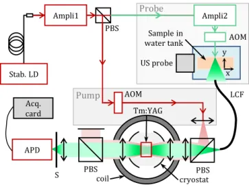
Fig. 2. Optical setup, with a probe beam (green) and a pump beam (red). Stab. LD stabilized laser diode, Ampli 1, 2: laser amplifiers, PBS: polarizing beam splitter, AOM: acousto-optic modulator, US probe: ultrasound probe, x: longitudinal coordinates along US propagation, y: transverse coordinates perpendicular to US propagation, LCF liquid core fiber with high numerical aperture, S: mechanical shutter, APD: avalanche photodiode, Acq. card: acquisition card.
The experiments are conducted using a 10x10x2 mm3 2% at.
Tm3+:YAG single crystal (Scientific Materials Corp.) with an optical
thickness close to = 6 along the 2mm dimension, where and L are respectively the absorption coefficient and the crystal length. The temperature of the crystal is set at about 2K with a variable liquid helium cryostat (SMC-TBT) so that the homogeneous linewidth is a few kHz, and the measured inhomogeneous bandwidth 30GHz. The 234G magnetic field is created by a pair of water-cooled Helmholtz coils. It is oriented along the [001] crystallographic axis [19]. Under these conditions, the lifetime of Zeeman sublevels is about 30s [20]. Indeed, both heteronuclear cross-relaxation with neighboring Al ions observed at 30 and 60G [21] and spin-lattice relaxation due to phonons observed above 500G [22] are prevented. By controlling the power, duration and spectral width of the pump pulse, the spectral holes width (FWHM) is adjusted to 1.7MHz. This corresponds to a tradeoff between the US pulse width (2.4MHz) and an efficient blocking of untagged photons.
The master laser is an extended cavity diode laser stabilized on a Fabry-Perot cavity [23], leading to a measured frequency drift of about 0.1MHz over 30s. The laser is amplified and split into two optical paths (see Figure 2). In the first path, a 200mW probe beam
Pump APD Acq. card S PBScoil cryostatPBS Tm:YAG Stab. LD Ampli1 PBS AOM Probe Ampli2 US probe AOM Sample in water tank x y LCF B≠0 // [001] 793 nm Δg = 285 MHz/T Δe = 60 MHz/T B=0 8ms 793 nm 3F4 3H 4 6H 4 Ab so rp ti on Frequency fUS Ca rr ie r an d tagged phot on s in ten sity (b) (c) (a)
illu 11 tem El ele re an di sc 10 tis in 0.5 su wi ete illu an m Ha 12 un du 70 pu be th liq be de de de th VS an wa tra re int at ac ab de re Tm (5 pu co th eff ca ab de or uminates the s 10mW/cm² pum mporally shape ectronic MT80) ement transduc esolution calcula nd the lateral res We prepare tw fferent intralipi cattering coeff 0 (sample ssues [24]. A 2 side sample 1 5x0.5x0.5mm3 urface.
The probe scatt ith a 1m-long
tendue. The fibe
uminate its who nd focused on module C12703-andyscope HS5) 2sr.mm² by the ndergoes signific ue to the lense 0%) and to unco We choose a co ump beams to m eam is expanded he light cone of quid core fiber. eamsplitter (PB epolarized light eviates most of epolarized pump herefore it must S14). The AOMs nd the acquisitio aveform genera Our 2D UOT anslating the US epresents the co to x coordinates t 1500 m.s-1, an cquisition of one bout 1 epicted on Figu emains closed w m:YAG crystal at 5ms per hole). T
ulses are emitt ontinuously illum he end of the cy
fficiency is , th ase of SHB, the 8 bove which the egrades the sign rders of magnitu scattering samp mp beam burns d with acousto-o ). US pulses are er (Olympus 5M ated for a two-c solution set by th wo agar gel ph id solution con ficients μ 2), the latter b 2x2x2mm3 black 1, while samp separated by 2 tered light is col liquid core fibe er output is slig ole volume. Ligh an avalanche -01) connected ). The etendue of e cryostat windo
cant losses betw es and cryostat oated cryostat w o-propagating g maximize their o d with an 8mm f f the divergent
The two beams BS), despite th t. At the outpu f the pump pow
p light is still tra t be blocked wit s, the ultrasoun on card are all ator (Tektronix A T-images are S probe in the onversion of tem
s, using the bal nd it is avera e line, the US pr 185ms to its p ure 1(b) and d while the pump p
t ±5MHz from th The shutter then
ted every minated by the p ycle. The key pa he number of U 8ms lifetime allo e exponential d
nal. In the case ude longer, allow
ple, and in the the spectral hol optic modulator e emitted with a MHz) of 5cm foca cycle US pulse a he US waist is 0.6 hantoms of 7x7 ncentrations, lea 1 (sample being equal to k Indian ink in ple 2 has tw 2mm, close to t llected at the out er (LOT-Oriel) w ghtly focused in ht exiting the cry photodiode (H to an acquisit f the overall colle ows. In addition ween the fiber ou
optical apertur windows (61%).
geometry betwe overlap on the c focal lens to app scattered light s are combined he 50% losses ut of the cryos
wer, but the re ansmitted and s th a mechanical nd probe, the m synchronized w AWG5004). acquired in transverse dire mporal traces o listic propagatio ged time robe is mechanic position. One ac defined as foll pulse burns 2 sp he carrier freque n opens in 4ms, a = 0.1ms whil probe beam. Th arameter to qua S pulses emitted ows only = 5 decay of the h e of PSHB, the wing for a comm
e second path e. Both beams a rs (AOM, AA Opt a spherical singl al length. The ax at 5MHz is 0.6m 6mm. 7x1cm3 gels wi ading to differe 1) and μ that of biologic nclusion is buri wo inclusions
the sample inp tput of the samp with a 45sr.mm nto the crystal
yostat is collect Hamamatsu AP tion card (Tiep ection is limited n, the probe lig utput and the AP res (estimated een the probe an
crystal. The pum proximately mat coming from th with a polarizin s on the prob stat, another PB emaining partia saturates the AP l shutter (Unibl mechanical shutt with an arbitra = 51 lines b ection . Each lin of the probe pul
on of the US pul es. To begin th
cally translated cquisition cycle ows: the shutt pectral holes in th
ency during 10m and a series of U le the sample
e shutter closes antify the imagi
d per cycle. In th 5 pulses per cyc hole transmissio hole lifetime is mensurate increa a are to- le-xial mm ith ent cal ed of put ple m² to ed PD pie to ght PD, to nd mp tch the ng be BS ally PD, itz ter ary by ne lse lse the in is ter the ms US is at ng the cle, on 3 ase of , e the dec blocked the me 10s, w measur photon untagge repetiti cycle w cycle, d repeate line. Fig. 3. ( spectral ultrasou photons pulses a shutter normali Fig. 4. U (no ma field). E along th data, blu longitud transmi We f on the respect = are req reduced present especia relevan to S. P (a) e.g. = f
cay of the record d at = 0. After etastable level, th which is consist
red using indep ns are attenuated
ed light. One cyc ion rate, allowin with a period eq
data are transfe
ed = /
(a) Pulse cycle f l holes at the t und pulse (blue s at repetit are exploitable d opening, SC: ized tagged photo
UOT image of sam agnetic field) and Each image is ave he white arrows
ue line: fit, red d dinal coordinates
itted light detecte first image samp e imaging per tively shown on 100 times. The quired to gener d to = 3 t case digitized ally for high nt time dedicate = Filter lifet .O. US pulse Pump Filter lifet
for the following ded tagged phot
r a rapid 25% de he filter transm tent with the pendent time-res
d by a factor of -cle lasts = ng a faster fram qual to the hole ferred to the co / times to rea
for UOT. The pu tagged photons e) and probe lig tion period. Whe due to the short shutter closing tons. mple 1 ( = 1 d (b) with a per eraged 100 time in images (a) an dashed line: Gaus
s (mm), y: transv ed by the APD (m ple 1 to study th rformance. SHB n Figure 4(a) and
first striking re rate an image u 0s in the case o after each cycl , for a fair co ed to imaging on an time B = 0 S.C. time Probe light es time B ≠ 0 // g images. Figure tons when the p
ecay due to part mission loses ano 35s spectral h solved spectrosc -20dB, which is 6 100 as set b me rate than in lifetime. At the omputer. One c ach avera
ump pulse (red) frequency. The ght (green) creat en = 0, only t filter lifetime (g g. (b) Transmis ) (a) with a rsistent hole (23 es. (c) and (d) ar nd (b) respective sian fit (see text verse coordinates mV). he effect of the m B and PSHB d (b), with each l esult is that using SHB while of PSHB. Since da
le, which is tim omparison we nly. It fundamen nd gives a Ta gg ed ph ot on s tr an sm issi on ( no rm .) Ti (b) e 3(b) shows pump beam is tial storage in other 20% in hole lifetime copy. Tagged 6dB less than by the shutter the case of a e end of each cycle will be aging for each
creates the 2 simultaneous te the tagged the 5 first US gray area). SO: ssion of the
a spectral hole 34G magnetic re the profiles ly. Black dots: for details), x: s (mm) and s: magnetic field images are line averaged = 8.5min e this time is ata are in the e consuming calculate the ntally reduces duration of ime (s)
th wi us m ev th (C PS tra th in Fig av fit co re im co to de th to fit en in (1 in ex un sim m Tm str te pr by [2 Fu cry Fin = 111s ( he number o ithout refreshin remains no sing a transdu mechanical scann very = 0.5m hen be shorten by The second st CNR) of the inclu SHB. This is due ansmission. The he double benefi an increase of th g. 5. Imaging res veraged 200 tim = 4.4 . Blac t (see text for deta oordinates (mm) In a second esolution of our mage of samp orresponding = 47s a espite a modest he strong scatter a lower signal tted with a wid nvelope minus a clusions (same 1.8±0.2mm)², si clusions size a xplained by the f ncontrolled diffu milarity of the i modest CNR, this m:YAG PSHB f rongly scatterin The performan chnical issues. I robe array to red y increasing the 25] and anti-re urthermore, a la rystal would lea nally, although t
= 20) and of successive U ng the hole is cru onetheless a lim ucer array to ning in the trans
ms and = y ∙ = triking result is usion increases fr e to a tenfold im erefore, with th it of PSHB filter he tagged photo
solution with per mes. (b) Profile a ck dots: data, blu ails), x: longitudin and s: transmitte experiment, we PSHB-UOT set ple 2, averag acquisition time CNR (7.5 0.4 ring coefficient o collection comp de 2D-Gaussian a pair of narrow method used fo ignificantly larg at the time of finite US resolut usion of the ink inclusions and t s experiment v filter to image g samples. nce of this proof
ndeed, in additi duce the scannin
light probe pow eflection-coated
rger pump pow ad to a deeper s
the etendue of ou
= 14s US pulses which ucial for fast acqu miting time and
perform electr sverse direction, = 0. The acquis
185ms ∙ 51 = that the contr from 3.3±0.6 for mprovement of t his experiment, in terms of hole on signal) and im rsistent SHB (a) I along = 57 ue line: fit, red das
nal coordinates ( ed light detected b e evaluate the tup. In Figure 5, ged over 200 . The inclusions 4). This CNR is of sample 2 (µs’ pared to sampl function repres wer 2D-Gaussian or Fig. 4). We ob ger than the f fabrication. Th
tion (0.6mm) an k inside the gel the surrounding validates the po e millimeter-siz
f of principle is ion to switching ng time, one can wer within the m cryostat wind wer and a more a spectral hole, an ur setup is close
( = 1). Hen
h can be emitt uisition. The del could be reduc ronic rather th
, e.g. one US pul sition time wou
9s.
rast-to-noise rat SHB to 32±0.7 f the spectral hol we demonstra e depth (resultin maging speed.
Image of sample . (c) Profile alo shed line: Gaussi (mm), y: transver by the APD (mV). e spatial imagin
, we show a UO 0 single imag
are clearly visib mainly limited b ’=10cm-1), leadin
le 1. The image senting the bea n functions for th btain a FWHM si (0,5mm)2 init his is essentia nd by the possib l due to chemic g gel. In spite of ossibility of usin zed inclusions mostly limited b g to an ultrasoun n improve the CN medical safety lim ows and cryst absorbing Tm:YA nd a larger CN e to the state of th nce ted lay ed hat lse uld tio for les ate ng e 2, ng ian rse . ng OT ges ble by ng is am the ize tial ally ble cal f a ng in by nd NR mit tal. AG NR. the art [17] optical In s demon Tm:YAG 2K. Our the pos tissues deeper photon extende refresh imaging UOT se living ti Fundin CNRS (ANR-1
Refere
1. R. C Pat S. R Ro 2. L. V 3. J. G (20 4. A. W Bio 5. G. D Sci 6. J.-B M. 7. B. Ja 8. M. G Du 9. G. R 10. R. 55 11. Y. Ph 12. X. 66 13. J. W (20 14. G. 20 15. W Ap 16. K. 14 17. H. He 18. A. 19. F. Go 20. N. (20 ], it could still b access. summary, we nstration of UOT G, by applying a r experiment pe ssibility to perfo at 793nm. Ind r spectral holes ns by a factor 1 ed hole lifetime hing cycle. Our wg at video rate. S etup where Tm:Y
issues. ng. ITMO C (Défi Instrume 10-LABX-24 and
ences
Choe, S. D. Konecky thak, B. J. Czerniec R. Arridge, M. Schw osen, and A. G. Yod V. Wang, Phys. Rev. unther and S. And 017).Walther, L. Rippe, L
omed. Opt. Express
D. Mahan, W. E. En i. 95, 14015–1401 B. Laudereau, E. B. à . Tanter, J.-L. Genn ayet, J.-P. Huignard Gross, P. Goy, B. C unn, Opt. Lett. 30, 1 Rousseau, A. Blouin Richards-Kortum a 55–606 (1996).
Li, H. Zhang, C. Kim
hys. Lett. 93, 11111 Xu, S.-R. Kothapal 6018-5 (2010). W. Tay, P. M. Ledin 010). Armagan, A. M. B 0 (1992). W. E. Moerner, Persi pplications. Springe Holliday, M. Croci, 4741–14752 (1993) Zhang, M. Saboon emmer, Appl. Phys. Kinos, Q. Li, L. Ripp de Seze, A. Louche ouët, O. Guillot-No . Ohlsson, M. Nilsso 002). be improved by have conduct T imaging using a magnetic field erformed on sca orm in vivo cm-deed, compared increases the 0, leading to a allows up to 40 work therefore o Such a probe wi YAG PSHB detec Cancer AVIESAN entation aux Li d ANR-10-IDEX-y, A. Corlu, K. Lee, T cki, J. Tchou, D. L. F weiger, J. P. Culver dh, J. Biomed. Opt., . Lett. 87 (2001). dersson-Engels, Fro L. V. Wang, S. Ande s 8, 4523 (2017). ngler, J. J. Tiemann, 19 (1998). à la Guillaume, V. S nisson, F. Ramaz, J. d, and F. Ramaz, O C. Forget, M. Atlan, 1357–1359 (2005) n, and J.-P. Monch and E. Sevick-Mura m, K. H. Wagner, P. 1 (2008).
li, H. Liu, and L. V. W ngham, and J. J. Lo Buoncristiani, and B
istent Spectral Hol
er Science & Busine , E. Vauthey, and U ).
ni, L. Rippe, C. Kim,
. Lett. 100, 131102 pe, and S. Kröll, Ap et, V. Crozatier, I. L ël, and P. Goldner, on, and S. Kröll, R.K using a cryosta ted the first e
g a filter based on the crystal m attering phantom -depth imaging to anterior dem number of det larger CNR. In 0 times more acq
opens the possi ill be integrated ction will be tes
N MALT project imites 2016); L 0001-02 PSL*). T. Durduran, D. R. Fraker, A. DeMiche r, M. D. Schnall, M. , 14, 24020 (2009). ont. Optoelectron. 1 ersson-Engels, and , and E. Uzgiris, Pro Servois, P. Mariani,
Biophotonics 8, 42 Opt. Express 22, 206
F. Ramaz, A. C. Bo .
alin, Opt. Lett. 34, aca, Annu. Rev. Phy . Hemmer, and L. V Wang, J. Biomed. O ongdell, Appl. Opt. 4 B. Di Bartolo, Opt. M le-Burning: Science ess Media (2012). U. P. Wild, Phys. Re , S. Kröll, L. V. Wang 2 (2012). ppl. Opt. 55, 10442 Lorgeré, F. Bretena , Phys. Rev. B 73 (2 K.Mohan Optics Le at with better experimental d on PSHB in maintained at m gels shows of scattering monstrations tected tagged addition, the quisitions per ibility of UOT in our future sted on actual (C16027HS); LABEX WIFI Busch, S. ele, B. Chance, E. Putt, M. A. . 10, 211–238 S. Kröll,
oc. Natl. Acad.
, A. A. Grabar, 29–436 (2015). 622 (2014). occara, A. K. 3445 (2009). ys. Chem. 47, V. Wang, Appl. Opt. 15 66018-49, 4331–4334 Mater. 1, 11– e and ev. B 47, g, and P. R. 2 (2016). ker, J.-L. Le 2006). etters 28
21. L. Veissier, C. W. Thiel, T. Lutz, P. E. Barclay, W. Tittel, and R. L. Cone,
Phys. Rev. B 94 (2016).
22. R. L. Ahlefeldt, M. F. Pascual-Winter, A. Louchet-Chauvet, T. Chanelière, and J.-L. Le Gouët, Phys. Rev. B 92 (2015).
23. V. Crozatier, F. de Seze, L. Haals, F. Bretenaker, I. Lorgeré, and J.-L. Le Gouët, Opt. Commun. 241, 203–213 (2004).
24. D. Grosenick, H. Wabnitz, K. T. Moesta, J. Mucke, P. M. Schlag, and H. Rinneberg, Phys. Med. Biol. 50, 2451–2468 (2005).
25. Sécurité des appareils à laser – Partie 1 : classification des matériels –
Prescription et guide de l’utilisateur, AFNOR NF EN 608251-1 (1994).
References
[1] R. Choe, S. D. Konecky, A. Corlu, K. Lee, T. Durduran, D. R. Busch, S. Pathak, B. J. Czerniecki, J. Tchou, D. L. Fraker, A. DeMichele, B. Chance, S. R. Arridge, M. Schweiger, J. P. Culver, M. D. Schnall, M. E. Putt, M. A. Rosen, and A. G. Yodh, “Differentiation of Benign and Malignant Breast Tumors by In-Vivo Three-Dimensional Parallel-Plate Diffuse Optical Tomography,” J. Biomed. Opt., vol. 14, no. 2, p. 024020, 2009. [2] L. V. Wang, “Mechanisms of Ultrasonic Modulation of Multiply
Scattered Coherent Light: An Analytic Model,” Phys. Rev. Lett., vol. 87, no. 4, Jul. 2001.
[3] J. Gunther and S. Andersson-Engels, “Review of current methods of acousto-optical tomography for biomedical applications,” Front. Optoelectron. vol. 10, no3, pp. 211–238, 2017.
[4] A. Walther, L. Rippe, L. V. Wang, S. Andersson-Engels, and S. Kröll, “Analysis of the potential for non-invasive imaging of oxygenation at heart depth, using ultrasound optical tomography (UOT) or photo-acoustic tomography (PAT),”
Biomed. Opt. Express, vol. 8, no. 10, p. 4523, Oct. 2017.
[5] G. D. Mahan, W. E. Engler, J. J. Tiemann, and E. Uzgiris, “Ultrasonic tagging of light: Theory,” Proc. Natl. Acad. Sci., vol. 95, no. 24, pp. 14015–14019, Nov. 1998.
[6] J.-B. Laudereau, E. B. à la Guillaume, V. Servois, P. Mariani, A. A. Grabar, M. Tanter, J.-L. Gennisson, F. Ramaz, “Multi-modal acousto-optic/ultrasound imaging of ex vivo liver tumors at 790 nm using a Sn2P2S6 wavefront adaptive holographic setup,” J.
Biophotonics, vol. 8, no. 5, pp. 429–436, May 2015.
[7] B. Jayet, J.-P. Huignard, and F. Ramaz, “Fast wavefront adaptive holography in Nd:YVO_4 for ultrasound optical tomography imaging,” Opt. Express, vol. 22, no. 17, p. 20622, Aug. 2014. [8] M. Gross, P. Goy, B. C. Forget, M. Atlan, F. Ramaz, A. C.
Boccara, A. K. Dunn, “Heterodyne detection of multiply scattered monochromatic light with a multipixel detector,”
Opt. Lett., vol. 30, no. 11, pp. 1357–1359, Jun. 2005.
[9] G. Rousseau, A. Blouin, and J.-P. Monchalin, “Ultrasound-modulated optical imaging using a high-power pulsed laser and a double-pass confocal Fabry–Perot interferometer,” Opt. Lett., vol. 34, no. 21, p. 3445, Nov. 2009.
[10] R. Richards-Kortum and E. Sevick-Muraca, “Quantitative optical spectroscopy for tissue diagnosis,” Annu. Rev. Phys. Chem., vol. 47, no. 1, pp. 555–606, 1996.
[11] Y. Li, H. Zhang, C. Kim, K. H. Wagner, P. Hemmer, and L. V. Wang, “Pulsed ultrasound-modulated optical tomography using spectral-hole burning as a narrowband spectral filter,”
Appl. Phys. Lett., vol. 93, no. 1, p. 011111, 2008.
[12] X. Xu, S.-R. Kothapalli, H. Liu, and L. V. Wang, “Spectral hole burning for ultrasound-modulated optical tomography of thick tissue,” J. Biomed. Opt., vol. 15, no. 6, pp. 066018-066018-5, 2010.
[13] J. W. Tay, P. M. Ledingham, and J. J. Longdell, “Coherent optical ultrasound detection with rare-earth ion dopants,” Appl. Opt., vol. 49, no. 23, pp. 4331–4334, Aug. 2010.
[14] G. Armagan, A. M. Buoncristiani, and B. Di Bartolo, “Excited state dynamics of thulium ions in Yttrium Aluminum Garnets,”
Opt. Mater., vol. 1, no. 1, pp. 11–20, 1992.
[15] W. E. Moerner, Persistent Spectral Hole-Burning: Science and
Applications. Springer Science & Business Media, 2012.
[16] K. Holliday, M. Croci, E. Vauthey, and U. P. Wild, “Spectral hole burning and holography in an Y2SiO5:Pr3+ crystal,” Phys. Rev.
B, vol. 47, no. 22, pp. 14741–14752, Jun. 1993.
[17] H. Zhang, M. Sabooni, L. Rippe, C. Kim, S. Kröll, L. V. Wang, and P. R. Hemmer “Slow light for deep tissue imaging with ultrasound modulation,” Appl. Phys. Lett., vol. 100, no. 13, p. 131102, Mar. 2012.
[18] A. Kinos, Q. Li, L. Rippe, and S. Kröll, “Development and characterization of high suppression and high étendue narrowband spectral filters,” Appl. Opt., vol. 55, no. 36, p. 10442, Dec. 2016.
[19] F. de Seze, A. Louchet, V. Crozatier, I. Lorgeré, F. Bretenaker, J.-L. Le Gouët, O. Guillot-Noël, and P. Goldner , “Experimental tailoring of a three-level Λ system in Tm 3 + : YAG,” Phys. Rev.
B, vol. 73, no. 8, Feb. 2006.
[20] N. Ohlsson, M. Nilsson, S. Kröll, and R. K. Mohan, “Long-time-storage mechanism for Tm: YAG in a magnetic field,” Opt. Lett., vol. 28, no. 6, pp. 450–452, 2003.
[21] L. Veissier, C. W. Thiel, T. Lutz, P. E. Barclay, W. Tittel, and R. L. Cone, “Quadratic Zeeman effect and spin-lattice relaxation of Tm3+:YAG at high magnetic fields,” Phys. Rev. B, vol. 94, no. 20, Nov. 2016.
[22] R. L. Ahlefeldt, M. F. Pascual-Winter, A. Louchet-Chauvet, T. Chanelière, and J.-L. Le Gouët, “Optical measurement of heteronuclear cross-relaxation interactions in Tm:YAG,” Phys.
Rev. B, vol. 92, no. 9, Sep. 2015.
[23] V. Crozatier, F. de Seze, L. Haals, F. Bretenaker, I. Lorgeré, and J.-L. L. Gouët, “Laser diode stabilisation for coherent driving of rare earth ions,” Opt. Commun., vol. 241, no. 1–3, pp. 203–213, Nov. 2004.
[24] D. Grosenick, H. Wabnitz, K. T. Moesta, J. Mucke, P. M. Schlag, and H. Rinneberg, “Time-domain scanning optical
mammography: II. Optical properties and tissue parameters of 87 carcinomas,” Phys. Med. Biol., vol. 50, no. 11, pp. 2451– 2468, Jun. 2005.
[25] Sécurité des appareils à laser – Partie 1 : classification des matériels – Prescription et guide de l’utilisateur, AFNOR NF EN 608251-1, 112 p,