Publisher’s version / Version de l'éditeur:
Journal of Leukocyte Biology, 86, 5, pp. 1217-1226, 2009-07-22
READ THESE TERMS AND CONDITIONS CAREFULLY BEFORE USING THIS WEBSITE. https://nrc-publications.canada.ca/eng/copyright
Vous avez des questions? Nous pouvons vous aider. Pour communiquer directement avec un auteur, consultez la première page de la revue dans laquelle son article a été publié afin de trouver ses coordonnées. Si vous n’arrivez pas à les repérer, communiquez avec nous à PublicationsArchive-ArchivesPublications@nrc-cnrc.gc.ca.
Questions? Contact the NRC Publications Archive team at
PublicationsArchive-ArchivesPublications@nrc-cnrc.gc.ca. If you wish to email the authors directly, please see the first page of the publication for their contact information.
NRC Publications Archive
Archives des publications du CNRC
This publication could be one of several versions: author’s original, accepted manuscript or the publisher’s version. / La version de cette publication peut être l’une des suivantes : la version prépublication de l’auteur, la version acceptée du manuscrit ou la version de l’éditeur.
For the publisher’s version, please access the DOI link below./ Pour consulter la version de l’éditeur, utilisez le lien DOI ci-dessous.
https://doi.org/10.1189/jlb.0908517
Access and use of this website and the material on it are subject to the Terms and Conditions set forth at
Human mast cells synthesize and release angiogenin, a member of the
ribonuclease A (RNase A) superfamily
Kulka, Marianna; Fukuishi, Nobuyuki; Metcalfe, Dean D.
https://publications-cnrc.canada.ca/fra/droits
L’accès à ce site Web et l’utilisation de son contenu sont assujettis aux conditions présentées dans le site LISEZ CES CONDITIONS ATTENTIVEMENT AVANT D’UTILISER CE SITE WEB.
NRC Publications Record / Notice d'Archives des publications de CNRC:
https://nrc-publications.canada.ca/eng/view/object/?id=c17fea76-dc64-4904-97a3-9e98acbf30c0
https://publications-cnrc.canada.ca/fra/voir/objet/?id=c17fea76-dc64-4904-97a3-9e98acbf30c0
Human mast cells synthesize and release
angiogenin, a member of the ribonuclease A
(RNase A) superfamily
Marianna Kulka,*
,1Nobuyuki Fukuishi,
†and Dean D. Metcalfe
†*Institute for Nutrisciences and Health, National Research Council, Charlottetown, Prince Edward Island, Canada; and †Laboratory of Allergic Diseases, National Institute of Allergy and Infectious Diseases, National Institutes of Health, Bethesda,
Maryland, USA
RECEIVED SEPTEMBER 2, 2008; REVISED JUNE 10, 2009; ACCEPTED JUNE 13, 2009. DOI: 10.1189/jlb.0908517
ABSTRACT
ANG is a plasma protein with angiogenic and ribonu-cleolytic activity implicated in tumor growth, heart fail-ure, wound healing, asthma, and the composition of the adult gut microflora. Human mast cells (HuMC) are similarly associated with modulation of vascular perme-ability, angiogenic processes, wound healing, and asthma. We hypothesized that HuMC express and se-crete ANG in response to divergent stimuli. ANG ex-pression was evaluated in the LAD2 HMC, the HMC-1, and CD34⫹-derived HuMC, following exposure to live Escherichia coli, TLR ligands, or neuropeptides and fol-lowing FcRI aggregation. Expression and production of ANG were determined by microarray analysis, qRT-PCR, confocal microscopy, and ELISA. Microarray anal-ysis showed that ANG is up-regulated by LAD2 cells ex-posed to live E. coli. qRT-PCR analysis revealed that LAD2, HMC-1, and HuMC constitutively expressed ANG mRNA and that it was up-regulated by exposure to E. coli. Activation of HuMC by FcRI aggregation resulted in release of small amounts of ANG (⬍100 pg/mL), whereas compound 48/80, NGF, LPS, PGN, and flagel-lin activated HuMC to secrete ⬎160 pg/mL ANG. These observations demonstrate that HuMC store and se-crete ANG to a variety of stimuli and suggest that MC-derived ANG is available in the subsequent inflamma-tory response. J. Leukoc. Biol. 86: 1217–1226; 2009.
Introduction
ANG (RNase 5) was isolated originally based on its ability to stimulate vasculogenesis in the chick chorioallantoic mem-brane [1] and is one of the most potent inducers of neovascu-larization in experimental models in vivo [2]. The 14.1-kDa
ANG protein has 35% aa sequence identity with human pan-creatic RNase and displays ribonucleolytic activity [3]. ANG expression and its physiological role in human disease are not fully understood. Mice have four ANG genes clustered to-gether on chromosome 14; humans, nonhuman primates, and rats have only a single ANG gene. ANG was isolated as a tumor angiogenic factor based solely on its angiogenic activity, and subsequent studies have often focused on its angiogenic capac-ity. However, several recent reports have implicated ANG in rRNA transcription in cancer cells [4], chronic heart failure [5], wound healing [6], and asthma [7–10]. ANG may also have antimicrobial activity, although data are not consistent in this regard [11, 12].
MCs have been similarly related to innate immune re-sponses, wound healing, and asthmatic inflammation through production of proinflammatory cytokines, leukotrienes, and chemokines. MCs also release vascular endothelial growth fac-tor, which can modulate vascular permeability [13] and con-tribute to tumor growth [14]. Because of these associations, we questioned whether MCs express and secrete ANG [2]. As will be shown, we found that HuMC express, store, and secrete ANG, which was released in response to FcRI aggregation, TLR ligands, and G-protein-coupled receptor activation.
MATERIALS AND METHODS
HuMC culture
LAD2 MCs [15] were cultured in serum-free media (StemPro-34 SFM, Life Technologies, Gaithersburg, MD, USA), supplemented with 2 mM L-glutamine, 100 U/ml penicillin, 50 g/ml streptomycin, and 100 ng/ml SCF. The cell suspensions were seeded at a density of 105cells/ml and maintained at 37°C and 5% CO2. Cells were fed by hemi-depletion of me-dia once/week. HMC-1 MCs [16] were cultured in Iscove’s medium con-taining 10% FBS, 100 U/mL penicillin, and 100 g/mL streptomycin (Bio-source International, Rockville, MD, USA) in a humidified atmosphere of 5% CO2in air at 37°C.
Human peripheral blood-derived CD34⫹cells were cultured in StemPro-34 SFM, supplemented with 2 mM L-glutamine, 50 g/ml streptomycin, 100
1. Correspondence: National Research Council, 550 University Ave., Char-lottetown, PE, Canada. E-mail: marianna.kulka@nrc.ca
Abbreviations: ANG⫽angiogenin, CGRP⫽calcitonin gene-related peptide, Ct⫽threshold cycle, h⫽human, HMC-1⫽human mast cell line-1,
HuMC⫽human cultured mast cell(s), LAD⫽laboratory of allergic disease mast cell line, MC⫽mast cell, NGF⫽nerve growth factor, NIAID⫽National Institute of Allergy and Infectious Diseases, PGN⫽peptidoglycan, qRT-PCR⫽quantitative RT-PCR, SCF⫽stem cell factor, SFM⫽serum- and feeder-free medium
Article
IU/ml penicillin, 100 ng/ml SCF, and 100 ng/ml rhIL-6 (PeproTech, Inc., Rocky Hill, NJ, USA). rhIL-3 (30 ng/ml) was added for the first week. Half of the culture medium was replaced every 7 days. Cultures at 8–10 weeks con-sisted of ⬎99% HuMC [17]. In cases where ANG expression was measured, MCs were grown in StemPro-34 media lacking the supplement component.
Escherichia coli
The E. coli K12 strain purchased from American Type Culture Collection (Manassas, VA, USA) was cultured in 8 g/l Trypton (Fisher Scientific, Fair Lawn, NJ, USA) and 0.5 g/l NaCl (Sigma-Aldrich, St. Louis, MO, USA). This media (20 mL), in a sterilized, 50-ml tube, were inoculated with E. coli, incubated at 37°C for 16 h with constant shaking at 250 rpm and spread on a 1.5% agar plate containing Luria-Bertani broth (Molecular Biologicals, Inc., Columbia, MD, USA) using a platinum loop. Single colo-nies of bacteria were picked and placed in 50 ml sterilized tubes containing 20 ml bacteria culture media. These cultures were incubated at 37°C for 16 h and then centrifuged at 3000 rpm for 15 min. The supernatant was discarded, and the E. coli pellet was resuspended in 20 ml PBS and centri-fuged at 3000 rpm for 15 min. The supernatant was discarded, and the concentration of E. coli was adjusted to 5 ⫻ 109bacteria/ml with PBS. To measure E. coli concentration in liquid culture, the turbidity of the culture was measured as light absorbance readings on a spectrophotometer at 600 nm. A standard curve of E. coli CFU was established and used to determine the number of E. coli CFU/mL culture. CFU measurements were per-formed by making tenfold serial dilutions of the liquid culture, which was seeded onto agar plates, and the numbers of colonies/plate were deter-mined subsequently. Additionally, the Alexa Fluor 488-labeled E. coli K12 strain (Molecular Probes, Inc., Eugene, OR, USA) was used for flow cytom-etry and in some confocal experiments.
E. coli
internalization by MCs
LAD or CD34-derived HuMC cultures were centrifuged at 1000 rpm for 5 min, the supernatant discarded, and the cells resuspended in cell culture media adjusted to 5 ⫻ 105cells/ml. The cell suspension (5 ml) was then added to a 25-cm2culture flask, incubated for 30 min at 37°C in a 5%-CO
2 incubator, and 5 l 5 ⫻ 109bacteria/ml added (bacteria:cell ratio of 10:1). The cell-bacteria suspension was then maintained at 37°C in a 5%-CO2 in-cubator for times specified. After incubation, the cell-bacteria suspension was centrifuged at 1000 rpm for 5 min, and the supernatant was discarded. The pellet was resuspended in 20 ml cell culture media containing antibi-otics and centrifuged at 1000 rpm for 5 min. The procedure was repeated and the pellet resuspended in 2.5 ml culture media containing antibiotics and incubated for times specified.
MTT assay
The MTT assay was performed to determine cell viability. Briefly, MTT (Sigma-Aldrich) was dissolved at 5 mg/ml in PBS and stored in the dark at 4°C. MCs were cocultured with live E. coli for 24 h at 37°C in complete StemPro media in a 96-well plate. A 10-l aliquot of MTT (5 mg/ml in PBS) was added to each well, and the plate was incubated at 37°C for 5 h. The plate was spun at 400 g for 10 min, and the media were removed. Sol-ubilizing solution [100 l; 20% (w/v) SDS and 50% (v/v) N,N-dimethyl formamide)] in deionized water] was added to each well, and the plate was incubated overnight at 37°C. OD of the formazan crystals was measured at 570 nm.
Flow cytometry
E. coli internalization by MCs was evaluated by incubating cells with or with-out cytochalasin D or mannose for 30 min in a CO2incubator. Cells were next incubated with or without Alexa Fluor 488-labeled E. coli for 30 min in a CO2incubator. The suspension was centrifuged at 1000 rpm for 5 min, and the supernatant was discarded. The pellet was resuspended in 1 ml PBS and centrifuged at 1000 rpm for 5 min, and the supernatant was dis-carded. This procedure was repeated, and the pellet was then suspended in
0.25 ml PBS. The cells were analyzed by FACScan (BD Biosciences, Moun-tain View, CA, USA) after addition of 0.75 ml 0.4% trypan blue solution (Sigma-Aldrich) to a 0.25-ml cell suspension to quench fluorescence associ-ated with bacteria on the cell surface.
Microarray analysis
The array chips are custom-made by NIAID (human sequence chip series “sa”) and consist of 13,971 oligonucleotides, each of which represents a unit gene cluster. All of these elements are 70-mer oligonucleotides synthe-sized by Qiagen Operon Inc. (Valencia, CA, USA), which can hybridize hu-man cDNA synthesized from a huhu-man mRNA library.
Total RNA was extracted from LAD3 HuMC incubated with or without E. coli for the times specified. RNA was then purified using an RNeasy mini kit (Qiagen Operon Inc.). For probe generation, RNA was converted to dscDNA by RT and Cy3- or Cy5-labeled. Oligo dT 20-mer was first annealed to the RNA, and then reverse transcription was performed using Super-script II RT (Invitrogen Corp., Carlsbad, CA, USA). Cy3-labeled dUTP (Amersham Biosciences AB, Uppsala, Sweden) was added along with unla-beled dNTPs to make a Cy3-launla-beled probe for E. coli-nonexposed samples (control), and Cy5-labeled dUTP (Amersham Biosciences AB) was added along with unlabeled dNTPs to make a Cy5-labeled probe for E. coli-ex-posed samples. The probes were then purified using a Vivaspin 30K centrif-ugal filter device (Vivascience AG, Hannova, Germany) in Tris-EDTA buffer. Probes were quantitated at 550 nm for Cy3 or at 650 nm for Cy5. The microarray chip was incubated with a blocking mixture that contained 5⫻ SSC, 1% BSA, and 0.1% SDS at 42°C for 1 h, and the hybridization was performed by adding 50 pmol-labeled probes to a reaction mix that in-cluded 10 g human Cot-1 DNA, 1 g Poly dA40-60, 4 g yeast transfer-RNA (Invitrogen Corp.), 5⫻ SSC, and 0.1% SDS in 25% formamide solu-tion. The mixture was heated at 98°C for 2 min, applied to a microarray slip after cooling, and incubated overnight at 42°C. The arrays were then washed sequentially in 1⫻ SSC and 0.05% SDS and 0.1⫻ SSC. Next, the arrays were centrifuged for 5 min at 500 rpm for drying and scanned with a GenePix 4000A microarray scanner (Axon Instruments, Inc., Union City, CA, USA). Data were analyzed using National Institutes of Health-designed soft-ware called “mAdb” located on the website at http://nciarray.nci.nih.gov/, and the data with a P value ⱕ0.02, compared with control, with a signal-to-noise ratio 2.0 or greater, were selected for further analysis.
qPCR
Total RNA was purified from MCs as described in “Microarray analysis”. Contaminating genomic DNA was digested and thus removed by incubat-ing 10 g total RNA with 2 U DNase (amplification grade; Life Technolo-gies) in DNase buffer (200 mM Tris-HCl, 20 mM MgCl2, 500 mM KCl, pH 8.4; Life Technologies) and RNase-free H2O for 10 min at room temp. RNA was then precipitated with 3 M C2H2O2Na (pH 5.2; Sigma-Aldrich).
Treated RNA (1 g) was incubated with 0.5 g oligo(dT) (Life Technolo-gies) at 70°C for 10 min and then added to a mixture containing First-Strand buffer (50 mM Tris-HCl, 75 mM KCl, 3 mM MgCl2, pH 8.3; Life Technolo-gies), 10 mM DTT, 10 mM each dNTP, sterile water (Sigma-Aldrich), and 200 U Moloney murine leukemia virus RT enzyme (Life Technologies). This mix-ture was incubated at 37°C for 1 h and then at 70°C for 10 min.
Duplex qPCR amplification of the ANG and -actin genes in each sam-ple was performed using qPCR master mix containing AmpliTaq Gold威 DNA Polymerase Ultra Pure, Uracil-DNA glycosylase, dTNPs with dUTP, and optimized buffer components (Applied Biosystems, Foster City, CA, USA). cDNA (100 ng) was used in each qPCR assay, and primers were de-signed using Primer Express software (Applied Biosystems). All reactions were performed in triplicate for 40 cycles. Samples were normalized using the Ctof the internal control gene (-actin) and target gene using the ⌬⌬Ctmethod, according to the formula ⌬⌬Ct⫽ ⌬Ctsample– ⌬Ctcontrol, where the controls are untreated cells. Each “n” represents an experiment executed independently, representing a different RNA sample.
Confocal microscopy
After 4 or 8 weeks of culture, CD34⫹-derived HuMC were washed with PBS, and cytospin slides were prepared. Slides were incubated in 2% paraformaldehyde in PBS (pH 7.4) for 15 min, rinsed with PBS, and incubated with 0.1% saponin in PBS for 15 min. Slides were washed once with PBS and incubated for 1 h with TBS containing 5 g/ml mouse mAb for human ANG (Sigma-Aldrich) or rabbit mAb for human tryptase (Calbiochem, San Diego, CA, USA). Primary antibody-binding was detected using 20 g/ml Texas Red-conjugated goat anti-mouse
an-tibody (Abcam, Cambridge, UK) or Alexa Fluor 488-conjugated anti-rabbit IgG (Molecular Probes Inc.). Rabbit and mouse IgG (5 g/ml) was used as an isotype control (R&D Systems, Minneapolis, MN, USA). Slides were examined using a ⫻100 objective under a TCS-NT/SP laser-scanning confocal microscope (Leica, Heidelberg, Germany), as de-scribed.
Statistical analysis
Each experiment was performed at least three times, and values represent mean of n ⫽ 3 ⫾ sem. P values were determined by Student’s t-test (be-tween groups) or one-way ANOVA (comparing more than two groups).
RESULTS
HuMC express angiogenin following exposure to E. coli
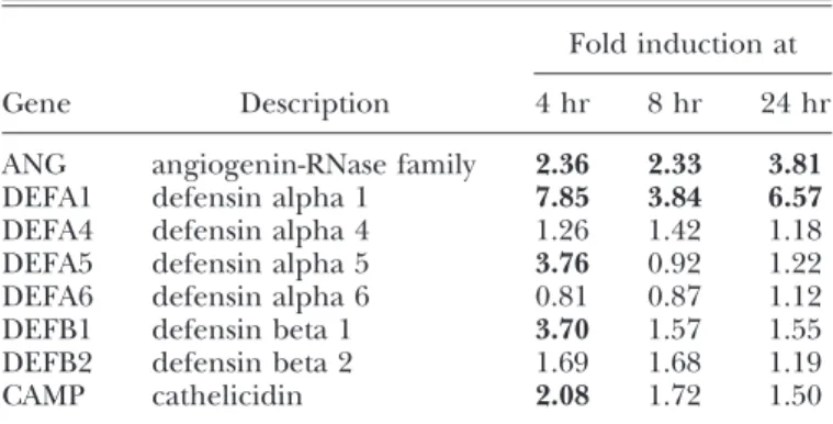
An important consequence of HuMC exposure to E. coli would be the production of molecules that would promote bacterial killing and promote vascularization to clear these pathogenic or-ganisms. The expression of some antimicrobial genes, such as defensins, following E. coli exposure for 4, 8, and 24 h, was con-firmed [18] by microarray analysis We found that E. coli exposure up-regulated members of the defensin family (␣1, ␣5, and 1), cathelicidin, as well as ANG (Table 1). As ANG expression had not been examined heretofore in MCs, we selected this gene for
0 2 4 6
incubation with E. coli (hr)
fold induction
A
B
**
**
*
0 5 10 15 20 25 30 35 E. coli exposure (hr) fo ld o ver u n tr eat ed co n tr o lC
tryptase ANGD
tryptase ANG tryptase + ANG
**
**
**
*
tryptase ANG tryptase + ANG
Figure 1. Microarray and qPCR analysis of ANG expression by HuMC (LAD2) coincubated with E.
coli.(A) Microarray data showing
the expression of ANG by LAD2 cells exposed to live E. coli (n⫽3; P⬍0.01). (B) qPCR analysis of ANG and -actin expression by HuMC following coincubation with E. coli. RNA isolated from untreated HuMC was used as a control (n⫽5). *, Significance of P ⬍ 0.05; **, significance of P ⬍ 0.01. (C) Confocal analysis of ANG and tryptase expression in mature (8-week-old) HuMC showing partial colocalization with tryptase. Cells were prepared as described in Ma-terials and Methods. (D) Confocal analysis of ANG and tryptase ex-pression in immature (4-week-old) HuMC showing small amounts of tryptase expression and no detect-able ANG expression.
TABLE 1. Antimicrobial Gene Expression by HuMC following Exposure to E. coli
Gene Description
Fold induction at 4 hr 8 hr 24 hr ANG angiogenin-RNase family 2.36 2.33 3.81 DEFA1 defensin alpha 1 7.85 3.84 6.57 DEFA4 defensin alpha 4 1.26 1.42 1.18 DEFA5 defensin alpha 5 3.76 0.92 1.22 DEFA6 defensin alpha 6 0.81 0.87 1.12 DEFB1 defensin beta 1 3.70 1.57 1.55 DEFB2 defensin beta 2 1.69 1.68 1.19 CAMP cathelicidin 2.08 1.72 1.50
Kulka et al. Human mast cells express angiogenin
further study. Microarray analysis showed that ANG expression was up-regulated more than twofold at all time-points analyzed but with the greatest up-regulation at 24 h (Fig. 1A). qPCR analy-sis confirmed that E. coli up-regulated ANG mRNA expression in CD34⫹-derived HuMC after 24 h of exposure (Fig. 1B). To
deter-mine if exposure to E. coli induced cell death, HuMC were ex-posed to live E. coli, and cell viability was analyzed by the MTT assay. HuMC showed 72 ⫾ 12% viability after exposure to E. coli for 24 h compared with untreated cells (94⫾4%). HuMC showed 96 ⫾ 2% viability after exposure to E. coli for 16 h compared with untreated cells (94⫾4%).
To determine whether HuMC stored ANG and whether it was localized to their granules, we analyzed ANG expression by confo-cal microscopy using antibodies to ANG and tryptase, and tryptase expression was used as a granule marker. As shown in Figure 1C, this imaging was consistent with the conclusion that HuMC store ANG intracellularly, localized to tryptase-positive granules. In our analysis, the granules surrounded the nucleus, attributed to the cytospin process in these mature cells. Immature (4-week-old) HuMC did not express ANG, although they ex-pressed low levels of tryptase in the cytoplasm (Fig. 1D). The
ap-pearance of aggregation of granules around the nucleus was not observed, likely as the granules are not yet as complex and dense as in the mature 8-week-old HuMC.
Binding and internalization of E. coli by HuMC
Murine and cord blood-derived HuMC have been found to at-tach to and internalize bacteria [19, 20]. Therefore, the ability of LAD2 HuMC to recognize and internalize bacteria was confirmed by incubating LAD2 with fluorescinated E. coli. As can be seen in Figure 2, fluorescent intensity increased significantly (histogram shifted to the right in Fig. 2B compared with Fig. 2A; dot plot analysis Fig. 2D vs. Fig. 2E) following exposure of MCs to fluo-rescinated E. coli. Furthermore, there was a dose-response rela-tionship between the number of bacteria added to the cell cul-tures and the fluorescence intensity that plateaued when the ra-tio reached approximately 50 bacteria to one MC (Fig. 2C). Pretreatment of the MCs with paraformaldehyde (Fig. 2F) or with cytochalasin D (a specific inhibitor of actin and contractile micro-filaments; Fig. 2, G and H) resulted in a decrease in the internal-ization of fluorescinated bacteria into these cells in a
dose-re-A
B
C
D
E
F
G
H
Fluorescence intensity Fluorescence intensity
Forward Scatter Forward Scatter
Forward Scatter
Forward Scatter Ratio of bacteria to LAD cells
Fluorescence intensity
Fluorescence intensity Fluorescence intensity
Counts Counts
Fluorescence intensity
Mean of fluorescence intensity
Fluorescence intensities
Cell alone 0 0.2 1 4
E. coli + Cytochalasin D (µg/mL)
Figure 2. Flow cytometric analysis of phagocytosis of fluorescinated E.
coliby LAD2 HuMC.(A) No
bacte-ria added to MCs. (B) MCs incu-bated for 30 min with 50-fold-fluoro-labeled E. coli particles. (C) Rela-tionship between fluorescence intensity of the MCs and the num-ber of E. coli particles added (per one MC). (D) A dot plot presenta-tion of MCs without fluorolabeled E. coli. (E) A dot plot of MCs incu-bated for 30 min with fluorolabeled E. coli particles (10 particles/cell). (F) Same conditions as in E except that cells were pretreated with para-formaldehyde. (G) Same conditions as in E except that cells were pre-treated with 4 g/ml cytochalasin D. (H) Dose-dependent effects of cytochalasin D pretreatment on in-ternalization of fluorolabeled E. coli by MCs.
sponse manner. Thus, LAD2 HuMC are able to internalize E. coli through a process requiring cytoskeletal rearrangement.
Primary cultured HuMC express and store angiogenin
in granules
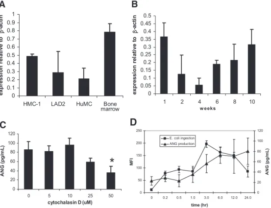
The expression of ANG in LAD2 was compared with other types of MCs such as the HMC-1 and CD34⫹-derived HuMC. qPCR
analysis showed further that the HMC-1 and primary-cultured HuMC derived from peripheral blood also expressed ANG (Fig. 3A). To determine the kinetics of ANG expression during MC development, we cultured CD34⫹peripheral blood progenitors
in SCF and IL-6 for 8 weeks and analyzed ANG expression at 4, 6, 8, and 10 weeks of culture. Immature 4-week-old MCs do not ex-press ANG, but at 6 weeks, they begin to exex-press mRNA for ANG, and by 10 weeks of culture, MCs express the highest levels of
ANG mRNA (Fig. 3B). To determine whether the process of E. coli ingestion is related to ANG production, HuMC were
incu-bated with E. coli in the presence of cytochalasin D, and ANG production was measured. Our data show that cytochalasin D in-hibited ANG production by ⬃50% (50 uM cytochalasin D, Fig. 3C). This suggests that ANG production may be related to the process of E. coli internalization.
To explore further the connection between E. coli ingestion and ANG production, a kinetic study was performed in which HuMC ingestion of fluorescinated E. coli and ANG production was measured simultaneously (Fig. 3D). E. coli interaction/inges-tion was measured by flow cytometry as in Figure 2, and ANG production from the same samples was measured by ELISA. The data in Figure 3D show that these two events correlate.
HuMC secrete angiogenin upon stimulation
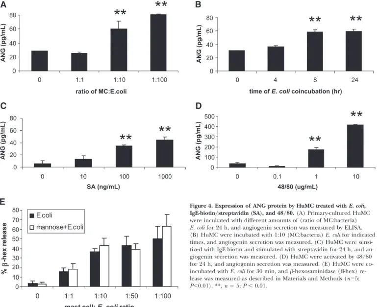
As HuMC were found to store ANG, we determined whether activation of HuMC would induce ANG secretion. First, we activated HuMC with different ratios of E. coli for 24 h (Fig. 4A). HuMC secreted significant amounts of ANG protein when exposed to E. coli bacteria, where the number of bac-teria was 10 times or 100 times the number of MCs (Fig. 4A). Maximum release of ANG in response to E. coli (1:10) stimulation occurred at 8 h of exposure (Fig. 4B). Activa-tion of HuMC via FcRI (Fig. 4C) induced similar levels of ANG secretion with those induced by incubation with a 1:10 ratio of E. coli (60.2⫾11.4 pg/mL with E. coli and 44.5⫾4.7 with streptavidin; Fig. 4A). Stimulation of HuMC with 48/80 induced the most significant amount of ANG (Fig. 4D), sug-gesting that direct activation of degranulation via G-protein activation also induces ANG secretion. It has been shown that E. coli interaction with MCs can induce degranulation [21]. To determine whether HuMC similarly degranulate in response to E. coli interaction, -hexosaminidase release in HuMC and E. coli cocultures was measured (Fig. 4E). E. coli induced HuMC degranulation, but this process was not blocked by mannose.
HuMC release TNF when activated by E. coli [22]. Therefore, the production of cytokines by HuMC incubated with E. coli was characterized. Compared with ANG levels shown in Figure 4, HuMC incubated with E. coli produced similar amounts of TNF (Fig. 5A), IL-10 (Fig. 5B), and IL-1 (Fig. 5C). However, HuMC produced significantly more IL-6 and IL-8 than ANG when bated with E. coli. HuMC did not produce IL-12p70 when
incu-0 0.1 0.2 0.3 0.4 0.5 0.6 0.7 0.8 0.9 1
HMC-1 LAD2 HuMC Bone
exp ressi o n r el at ive to β -act in
A
0 20 40 60 80 100 120 0 5 10 25 50 cytochalasin D (uM) AN G ( p g /m L )*
C
0 0.05 0.1 0.15 0.2 0.25 0.3 0.35 0.4 0.45 0.5 1 2 4 6 8 10 w eeks ex p re ssi o n r e la ti ve t o β -a cti nB
0 50 100 150 200 250 0 0.2 0.5 1.0 3.0 6.0 12.0 24.0 time (hr) MF I 0 20 40 60 80 100 120 ANG ( p g /m L ) E. coli ingestion ANG productionD
marrowFigure 3. Analysis of ANG expression.
(A) HMC, HMC-1, LAD2, and CD34⫹-derived HuMC expression of ANG and -actin by qRT-PCR. RNA isolated from untreated HuMC was used as a control. (B) CD34⫹ HuMC with ANG expres-sion measured during 10 weeks of devel-opment. PCR-amplified ANG bands from an ethidium bromide-stained gel were excised and sequenced and found to have a sequence corresponding with human ANG. (C) Confocal analysis of ANG expression in HuMC showing par-tial colocalization with tryptase. Cells were prepared as described in Materials and Methods. HuMC were incubated with E. coli (1:100 HuMC:bacteria ratio) in the presence of cytochalasin D (0, 5, 25, and 50 M) for 24 h, and ANG pro-duction was measured by ELISA. *, n ⫽ 5; P ⬍ 0.05. (D) HuMC were incubated with fluorescinated E. coli as in Figure 2 for indicated times. E. coli particle inges-tion was measured by flow cytometry, and supernatants from the same samples were analyzed for ANG production by ELISA (n⫽5; P⬍0.01). MFI⫽Mean fluorescence intensity.
Kulka et al. Human mast cells express angiogenin
bated with E. coli. Thus, the amount of ANG produced by HuMC is comparable with TNF, IL-10, and IL-1, all of which are impor-tant MC-derived immunomodulatory cytokines.
TLR ligands also induce angiogenin expression
and secretion
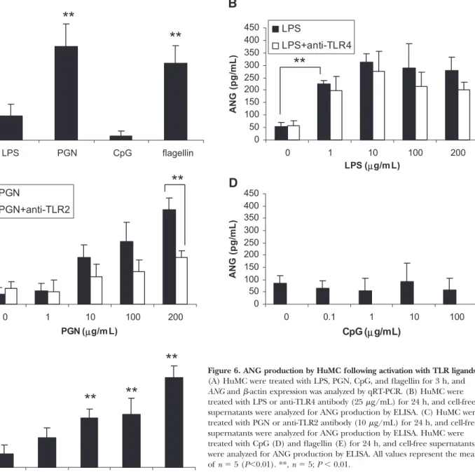
As E. coli could up-regulate ANG expression and induce secre-tion, we hypothesized that TLR ligands associated with bacte-rial products, such as LPS, PGN, CpG oligonucleotides, and flagellin, may also up-regulate ANG expression. qRT-PCR anal-ysis of HuMC revealed that LPS, PGN, and flagellin treatment up-regulated ANG expression (Fig. 6A). CpG oligonucleotides, however, had no effect on ANG expression.
To determine if TLR ligands could activate ANG secretion, primary HuMC were activated with LPS, PGN, CpG oligonucle-otides, and flagellin for 24 h, and ANG secretion into the su-pernatant was measured. LPS, PGN, and flagellin induced ANG secretion (Fig. 6, B, C, and E). In confirmation of qPCR
results obtained in Figure 6A, CpG oligonucleotides did not induce ANG secretion by HuMC (Fig. 6D). Anti-TLR4 antibod-ies do not significantly inhibit LPS-mediated activation of ANG production (Fig. 6B). However, anti-TLR2 significantly inhibits PGN-mediated activation of ANG production, suggesting that TLR2 is involved in this process (Fig. 6C).
NGF activates HuMC to produce angiogenin
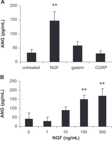
MCs respond to stimuli that are independent of FcRI or TLR, such as neuropeptides, hormones, and opiates [23, 24]. There-fore, the ability of HuMC to produce ANG in response to sub-stance P, NGF, gastrin, CGRP, and codeine was determined (Fig. 7A). NGF induced the production of significant amounts of ANG, whereas none of the other compounds induced ANG production by HuMC. Further analysis showed that NGF acti-vates NGF production dose-dependently (Fig. 7B).
0 20 40 60 80 0 1:1 1:10 1:100 ratio of MC:E.coli A N G (pg/mL) 0 20 40 60 80 0 4 8 24
time of E. coli coincubation (hr)
ANG (pg/mL) 0 20 40 60 80 0 10 100 1000 SA (ng/mL) A N G (pg/mL) 0 100 200 300 400 500 0 0.1 1 10 48/80 (ug/mL) ANG (pg/mL)
**
**
**
**
**
**
**
**
A
B
D
C
E
0 10 20 30 40 50 60 70 80 0 1:1 1:10 1:50 1:100 mast cell: E. coli ratio% β -h ex r e le ase E.coli mannose+E.coli
Figure 4. Expression of ANG protein by HuMC treated with E. coli, IgE-biotin/streptavidin (SA), and 48/80.(A) Primary-cultured HuMC were incubated with different amounts of (ratio of MC:bacteria) E. coli for 24 h, and angiogenin secretion was measured by ELISA. (B) HuMC were incubated with 1:10 (MC:bacteria) E. coli for indicated times, and angiogenin secretion was measured. (C) HuMC were sensi-tized with IgE-biotin and stimulated with streptavidin for 24 h, and an-giogenin secretion was measured. (D) HuMC were activated by 48/80 for 24 h, and angiogenin secretion was measured. (E) HuMC were co-incubated with E. coli for 30 min, and -hexosaminidase (-hex) re-lease was measured as described in Materials and Methods (n⫽5; P⬍0.01). **, n ⫽ 5; P ⬍ 0.01.
DISCUSSION
This study shows that HuMC (two HMC as well as CD34⫹-de-rived MCs) express ANG. Furthermore, HuMC store ANG in their granules and release ANG to a variety of stimuli, includ-ing FcRI-mediated signals, TLR ligands, and NGF, implyinclud-ing that MC-derived ANG is released in a variety of physiological conditions.
ANG was isolated as a tumor angiogenic factor based solely on its angiogenic activity, and subsequent studies have focused mainly on its angiogenic capacity. However, several reports have suggested that ANG is important in regulating rRNA transcription in cancer cells [4], involved in chronic heart fail-ure [5], important in wound healing [6], and elevated in pa-tients with asthma [7–10]. Although the reports are conflict-ing, ANG may also have antimicrobial activity and may be im-portant in clearing infections [11, 12].
ANG is one of several unusual members of the pancreatic RNase superfamily. It was first isolated as a 14-kDa-soluble pro-tein from culture medium conditioned by human colon carci-noma (HT-29) cells and was identified as an angiogenic sub-stance based on its capacity to induce blood-vessel formation on the chorioallantoic membrane of the chicken embryo. Al-though ANG is secreted by many tumor cells and has been shown to be essential for tumor growth, it is not a tumor-spe-cific protein. It is present at a concentration of 250 –360 ng/mL in normal human plasma [3, 5]. Higher or lower con-centrations have been seen in a variety of conditions, including endometrial cancer, pregnancy, and renal dialysis, but thus far, its plasma level has not been shown to have diagnostic relevance. Recently, a protein that inhibits the degranulation of polymor-phonuclear leukocytes was isolated from plasma ultrafiltrates of patients with uremia and shown to be identical to ANG [25].
0 10 20 30 40 50 60 70 untreated 1:1 1:10 1:100
mast cell:E. coli ratio
IL-1 0 (pg/ mL) 0 10 20 30 40 50 60 untreated 1:1 1:10 1:100
mast cell:E. coli ratio
TNF (pg /m L) 0 10 20 30 40 50 60 70 80 untreated 1:1 1:10 1:100
mast cell:E. coli ratio
IL-1 β (pg/ mL) 0 1000 2000 3000 4000 5000 6000 7000 8000 untreated 1:1 1:10 1:100
mast cell:E. coli ratio
IL-8 (pg/ mL) 0 10 20 30 40 50 untreated 1:1 1:10 1:100
mast cell:E. coli ratio
IL-1 2 p7 0 (pg/ m L ) 0 50 100 150 200 250 300 350 400 untreated 1:1 1:10 1:100
mast cell:E. coli ratio
IL-6 (pg/ mL)
F
E
**
**
**
**
**
**
**
**
**
**
**
**
**
*
*
A
B
C
D
Figure 5. Cytokine production by HuMC following exposure to E. coli.HuMC were exposed to E. coli for 24 h, and cell-free supernatants were ana-lyzed for TNF (A), IL-10 (B), IL-1 (C), IL-6 (D), IL-8 (E), and IL12p70 (F) production by ELISA. All values represent the mean of n ⫽ 5 (P⬍0.01). *, n ⫽ 5; P ⬍ 0.01; **, n ⫽ 5; P ⬍ 0.05.
Kulka et al. Human mast cells express angiogenin
One finding of this study is that HuMC produce ANG in response to activation of TLR4, TLR2, and TLR5 via LPS, PGN, and flagellin, respectively. These findings suggest that ANG may be important in MC responses to bacterial patho-gens and the associated inflammatory response leading to bac-terial clearance. MCs have been implicated in innate immune responses, in that they produce cytokines, leukotrienes, and chemokines in response to bacteria and viruses, mediated in part through the cell-surface pattern recognition receptors, such as TLR1, -2, -4, -6, and -9 [26, 27], and the FimH recep-tor CD48 for bacterial fimbriae [20]. MCs also recognize and attach to a variety of opsonized bacteria and release antimicro-bial peptides such as cathelicidin or cytokines [28], which
pro-mote the inflammatory response to bacterial insults. Other and recent evidence shows that bacterial products such as LPS and PGN modulate HuMC differentiation, cytokine produc-tion, and expression of chymase and tryptase [29]. The location and immune functions associated with MCs and observations of antimicrobial peptides in MCs of lower vertebrates led us to hy-pothesize that HuMC may also express and release ANG in re-sponse to bacterial or TLR ligand activation.
However, the precise role of ANG in bacterial infections is controversial. Hooper et al. [11] have shown that mouse and human ANG is able to reduce growth of Streptococcus
pneu-moniae and Candida albicans by at least 100-fold, yet Avdeeva et
al. [12] reported that the antimicrobial activities of
commer-0 50 100 150 200 250 300 350 400 450 0 0.1 1 10 100
flagellin (µg/mL)
ANG (p g /m L ) 0 50 100 150 200 250 300 350 400 450 0 1 10 100 200 PGN (µg/m L) ANG ( p g /m L ) PGN PGN+anti-TLR2A
C
E
0 5 10 15 20 25 LPS PGN CpG flagellin fo ld in d u c tio n (r elat ive t o u n tr eat ed )**
**
**
**
**
**
B
0 50 100 150 200 250 300 350 400 450 0 1 10 100 200 LPS (µg/m L) ANG ( p g /m L ) LPS LPS+anti-TLR4**
0 50 100 150 200 250 300 350 400 450 0 0.1 1 10 100 CpG (µg/mL) AN G ( p g /m L )D
Figure 6. ANG production by HuMC following activation with TLR ligands.
(A) HuMC were treated with LPS, PGN, CpG, and flagellin for 3 h, and ANG and -actin expression was analyzed by qRT-PCR. (B) HuMC were treated with LPS or anti-TLR4 antibody (25 g/mL) for 24 h, and cell-free supernatants were analyzed for ANG production by ELISA. (C) HuMC were treated with PGN or anti-TLR2 antibody (10 g/mL) for 24 h, and cell-free supernatants were analyzed for ANG production by ELISA. HuMC were treated with CpG (D) and flagellin (E) for 24 h, and cell-free supernatants were analyzed for ANG production by ELISA. All values represent the mean of n ⫽ 5 (P⬍0.01). **, n ⫽ 5; P ⬍ 0.01.
cially prepared human ANG were comparable with that of BSA. A study by Cognasse et al. [30] showed that E. coli-de-rived LPS inhibited constitutive ANG expression (⬃5%) by human blood-derived platelets. Yet, angiogenesis is extremely important during tissue regeneration throughout a bacterial or viral infection, and expression of angiogenic factors such as ANG increases during hepatitis C infection of the liver [31]. Therefore, it is possible that the role of ANG in infection is not antimicrobial but serves as a modulator of tissue regenera-tion and remodeling necessary for pathogen clearance and re-establishment of tissue homeostasis.
Our study has also shown that NGF stimulates HuMC to re-lease greater amounts of ANG (Fig. 7) compared with MCs stimulated by FcRI cross-linking (Fig. 4C). This observation has some interesting implications in light of some recent stud-ies in which ANG gene mutations have been associated with amyotrophic lateral sclerosis [32, 33]. Human ANG has been shown to be neuroprotective and promotes the survival and neurite extension formation of motor neurons [34]. MCs are intimately associated with nerves and are therefore ideally suited to communicate with neurons. As such, it is possible that NGF stimulation of MCs in neuroinflammatory conditions
may ultimately lead to ANG production and promotion of neuron survival.
In this study, we have shown that HuMC secrete ANG in re-sponse to a variety of stimuli. Some of the stimuli tested, namely the TLR ligands LPS, PGN, and flagellin and the neu-ropeptide NGF, induced significant levels of ANG (160 pg/mL or more), and FcRI cross-linking induced smaller amounts of ANG (⬍100 pg/mL). These findings suggest that ANG may be important in certain physiological settings, particularly those mediated by TLR ligands and NGF. MC-derived ANG may be an important immune modulator in nonallergic conditions.
ACKNOWLEDGMENTS
This work was funded by the Division of Intramural Research, NIAID, and an interest section grant from the American Acad-emy of Allergy, Asthma and Immunology. We thank Victor Karpov and Adriana Catalli for their technical assistance.
REFERENCES
1. Fett, J. W., Strydom, D. J., Lobb, R. R., Alderman, E. M., Bethune, J. L., Riordan, J. F., Vallee, B. L. (1985) Isolation and characterization of an-giogenin, an angiogenic protein from human carcinoma cells. Biochemistry
24,5480 –5486.
2. Badet, J. (1999) Angiogenin, a potent mediator of angiogenesis. Biologi-cal, biochemical and structural properties. Pathol. Biol. (Paris) 47, 345– 351.
3. Shapiro, R., Strydom, D. J., Olson, K. A., Vallee, B. L. (1987) Isolation of angiogenin from normal human plasma. Biochemistry 26, 5141–5146. 4. Tsuji, T., Sun, Y., Kishimoto, K., Olson, K. A., Liu, S., Hirukawa, S., Hu,
G. F. (2005) Angiogenin is translocated to the nucleus of HeLa cells and is involved in ribosomal RNA transcription and cell proliferation. Cancer
Res. 65, 1352–1360.
5. Patel, J. V., Sosin, M., Gunarathne, A., Hussain, I., Davis, R. C., Hughes, E. A., Lip, G. Y. (2008) Elevated angiogenin levels in chronic heart fail-ure. Ann. Med. Apr., 1– 6.
6. Steed, D. L., Trumpower, C., Duffy, D., Smith, C., Marshall, V., Rupp, R., Robson, M. (2008) Amnion-derived cellular cytokine solution: a physio-logical combination of cytokines for wound healing. Eplasty 8, e18. 7. Simcock, D. E., Kanabar, V., Clarke, G. W., Mahn, K., Karner, C.,
O’Connor, B. J., Lee, T. H., Hirst, S. J. (2008) Induction of angiogenesis by airway smooth muscle from patients with asthma. Am. J. Respir. Crit.
Care Med. 178, 460 – 468.
8. Simcock, D. E., Kanabar, V., Clarke, G. W., O’Connor, B. J., Lee, T. H., Hirst, S. J. (2007) Proangiogenic activity in bronchoalveolar lavage fluid from patients with asthma. Am. J. Respir. Crit. Care Med. 176, 146 –153. 9. Abdel-Rahman, A. M., el-Sahrigy, S. A., Bakr, S. I. (2006) A comparative
study of two angiogenic factors: vascular endothelial growth factor and angiogenin in induced sputum from asthmatic children in acute attack.
Chest 129, 266 –271.
10. Hoshino, M., Takahashi, M., Aoike, N. (2001) Expression of vascular en-dothelial growth factor, basic fibroblast growth factor, and angiogenin immunoreactivity in asthmatic airways and its relationship to angiogene-sis. J. Allergy Clin. Immunol. 107, 295–301.
11. Hooper, L. V., Stappenbeck, T. S., Hong, C. V., Gordon, J. I. (2003) An-giogenins: a new class of microbicidal proteins involved in innate immu-nity. Nat. Immunol. 4, 269 –273.
12. Avdeeva, S. V., Chernukha, M. U., Shaginyan, I. A., Tarantul, V. Z., Nar-oditsky, B. S. (2006) Human angiogenin lacks specific antimicrobial activ-ity. Curr. Microbiol. 53, 477– 478.
13. Lee, K. S., Kim, S. R., Park, S. J., Min, K. H., Lee, K. Y., Choe, Y. H., Park, S. Y., Chai, O. H., Zhang, X., Song, C. H., Lee, Y. C. (2008) Mast cells can mediate vascular permeability through regulation of PI3K-HIF-1{␣}-VEGF axis. Am. J. Respir. Crit. Care Med. 178, 787–797.
14. Conti, P., Castellani, M. L., Kempuraj, D., Salini, V., Vecchiet, J., Tete, S., Mastrangelo, F., Perrella, A., De Lutiis, M. A., Tagen, M., Theoharides, T. C. (2007) Role of mast cells in tumor growth. Ann. Clin. Lab. Sci. 37, 315–322.
15. Kirshenbaum, A. S., Akin, C., Wu, Y., Rottem, M., Goff, J. P., Beaven, M. A., Rao, V. K., Metcalfe, D. D. (2003) Characterization of novel stem cell factor responsive human mast cell lines LAD 1 and 2 established from a patient with mast cell sarcoma/leukemia; activation following ag-gregation of FcRI or Fc␥RI. Leuk. Res. 27, 677– 682.
0 50 100 150 200 untreated NGF gastrin CGRP
ANG
(
p
g
/m
L
)
A
B
0 50 100 150 200 250NGF (ng/mL)
ANG
(
p
g
/m
L
)
**
**
**
Figure 7. ANG production by HuMC following activation with neu-ropeptides, gastrin, and codeine.(A) HuMC were treated with NGF (1 g/mL), gastrin (10 g/mL), and CGRP (1 g/mL) for 24 h, and cell-free supernatants were analyzed for angiogenin production by ELISA. (B) HuMC were treated with NGF for 24 h, and ANG produc-tion was measured by ELISA. All values represent the mean of n ⫽ 5 (P⬍0.01). **, P ⬍ 0.01.
Kulka et al. Human mast cells express angiogenin
16. Nilsson, G., Blom, T., Kusche-Gullberg, M., Kjellen, L., Butterfield, J. H., Sundstrom, C., Nilsson, K., Hellman, L. (1994) Phenotypic characteriza-tion of the human mast-cell line HMC-1. Scand. J. Immunol. 39, 489 – 498. 17. Kulka, M., Metcalfe, D. D. (2005) High-resolution tracking of cell division
demonstrates differential effects of TH1 and TH2 cytokines on SCF-de-pendent human mast cell production in vitro: correlation with apoptosis and Kit expression. Blood 105, 592–599.
18. Di Nardo, A., Vitiello, A., Gallo, R. L. (2003) Cutting edge: mast cell anti-microbial activity is mediated by expression of cathelicidin antianti-microbial peptide. J. Immunol. 170, 2274 –2278.
19. Munoz, S., Hernandez-Pando, R., Abraham, S. N., Enciso, J. A. (2003) Mast cell activation by Mycobacterium tuberculosis: mediator release and role of CD48. J. Immunol. 170, 5590 –5596.
20. Malaviya, R., Ikeda, T., Abraham, S. N., Malaviya, R. (2004) Contribution of mast cells to bacterial clearance and their proliferation during experi-mental cystitis induced by type 1 fimbriated E. coli. Immunol. Lett. 91, 103–111.
21. Malaviya, R., Ross, E., Jakschik, B. A., Abraham, S. N. (1994) Mast cell degranulation induced by type 1 fimbriated Escherichia coli in mice.
J. Clin. Invest. 93, 1645–1653.
22. Malaviya, R., Gao, Z., Thankavel, K., van der Merwe, P. A., Abraham, S. N. (1999) The mast cell tumor necrosis factor␣response to FimH-expressing Escherichia coli is mediated by the glycosylphosphatidylinositol-anchored molecule CD48. Proc. Natl. Acad. Sci. USA 96, 8110 – 8115. 23. Kulka, M., Sheen, C. H., Tancowny, B. P., Grammer, L. C., Schleimer,
R. P. (2008) Neuropeptides activate human mast cell degranulation and chemokine production. Immunology 123, 398 – 410.
24. Sheen, C. H., Schleimer, R. P., Kulka, M. (2007) Codeine induces human mast cell chemokine and cytokine production: involvement of G-protein activation. Allergy 62, 532–538.
25. Haag-Weber, M., Horl, W. H. (1996) Dysfunction of polymorphonuclear leukocytes in uremia. Semin. Nephrol. 16, 192–201.
26. Rocha-de-Souza, C. M., Berent-Maoz, B., Mankuta, D., Moses, A. E., Levi-Schaffer, F. (2008) Human mast cell activation by Staphylococcus aureus: IL-8 and TNF-{␣} release and the role of Toll-like receptor 2 and CD48 molecules. Infect. Immun. 76, 4489 – 4497.
27. Kulka, M., Alexopoulou, L., Flavell, R. A., Metcalfe, D. D. (2004) Activa-tion of mast cells by double-stranded RNA: evidence for activaActiva-tion through Toll-like receptor 3. J. Allergy Clin. Immunol. 114, 174 –182. 28. Ketavarapu, J. M., Rodriguez, A. R., Yu, J. J., Cong, Y., Murthy, A. K.,
Forsthuber, T. G., Guentzel, M. N., Klose, K. E., Berton, M. T., Arulanan-dam, B. P. (2008) Mast cells inhibit intramacrophage Francisella tularensis replication via contact and secreted products including IL-4. Proc. Natl.
Acad. Sci. USA 105, 9313–9318.
29. Kirshenbaum, A. S., Swindle, E., Kulka, M., Wu, Y., Metcalfe, D. D. (2008) Effect of lipopolysaccharide (LPS) and peptidoglycan (PGN) on human mast cell numbers, cytokine production, and protease composi-tion. BMC Immunol. 9, 45.
30. Cognasse, F., Hamzeh-Cognasse, H., Lafarge, S., Delezay, O., Pozzetto, B., McNicol, A., Garraud, O. (2008) Toll-like receptor 4 ligand can differen-tially modulate the release of cytokines by human platelets. Br. J.
Haema-tol. 141, 84 –91.
31. Mas, V. R., Maluf, D. G., Archer, K. J., Yanek, K. C., Fisher, R. A. (2007) Angiogenesis soluble factors as hepatocellular carcinoma noninvasive markers for monitoring hepatitis C virus cirrhotic patients awaiting liver transplantation. Transplantation 84, 1262–1271.
32. Gellera, C., Colombrita, C., Ticozzi, N., Castellotti, B., Bragato, C., Ratti, A., Taroni, F., Silani, V. (2008) Identification of new ANG gene muta-tions in a large cohort of Italian patients with amyotrophic lateral sclero-sis. Neurogenetics 9, 33– 40.
33. Schymick, J.C., Talbot, K., Traynor, B.J. (2007) Genetics of sporadic amyotrophic lateral sclerosis. Hum. Mol. Genet. 16 (Spec. No. 2), R233– R242.
34. Subramanian, V., Crabtree, B., Acharya, K. R. (2008) Human angiogenin is a neuroprotective factor and amyotrophic lateral sclerosis associated angiogenin variants affect neurite extension/pathfinding and survival of motor neurons. Hum. Mol. Genet. 17, 130 –149.
KEY WORDS: