HAL Id: tel-01299031
https://tel.archives-ouvertes.fr/tel-01299031
Submitted on 7 Apr 2016HAL is a multi-disciplinary open access archive for the deposit and dissemination of sci-entific research documents, whether they are pub-lished or not. The documents may come from teaching and research institutions in France or abroad, or from public or private research centers.
L’archive ouverte pluridisciplinaire HAL, est destinée au dépôt et à la diffusion de documents scientifiques de niveau recherche, publiés ou non, émanant des établissements d’enseignement et de recherche français ou étrangers, des laboratoires publics ou privés.
Expression of wild type and variants of human
apolipoprotein A-I in Pichia pastoris
Vignesh Narasimhan Janakiraman
To cite this version:
Vignesh Narasimhan Janakiraman. Expression of wild type and variants of human apolipoprotein A-I in Pichia pastoris. Immunology. Université de Bordeaux; Vellore Institute of Technology (Vellore, Inde), 2015. English. �NNT : 2015BORD0450�. �tel-01299031�
THÈSE EN COTUTELLE PRÉSENTÉE POUR OBTENIR LE GRADE DE
DOCTEUR DE
L’UNIVERSITÉ DE BORDEAUX, FRANCE
ET DE VIT UNIVERSITY, INDE
ÉCOLE DOCTORALE UBX
ÉCOLE DOCTORALE DU VIT UNIVERSITY SPÉCIALITÉ MICROBIOLOGIE-IMMUNOLOGIE
Par Vignesh Narasimhan JANAKIRAMAN
Expression de type sauvage et des variantes de
l’Apolipoprotéine A-I humaine chez Pichia pastoris
Expression of wild type and variants of human apolipoprotein A-I in Pichia pastoris Sous la direction de Xavier SANTARELLI
et de Krishnan VENKATARAMAN
Soutenue le 11 décembre 2015 Membres du jury :
M. DE LAMOTTE-GUÉRY, Fréderic INRA, Montpellier, France Rapporteur
M. JAYARAMAN, Guhan IIT-Madras, Chennai, India Rapporteur
M. SANTARELLI, Xavier Université de Bordeaux, France Directeur de Thèse M. VENKATARAMAN, Krishnan CBST, VIT University, Vellore, India Directeur de Thèse
i
Avant-propos / Acknowledgement
This doctoral thesis has been an exciting journey, over the past three years with more experiences than just experimental data: I won a couple of national fellowships, spent a year in France, learnt to converse fluently in French, got married, published two reasonably good papers, won a couple of presentation awards, wrote grants, and am now on my way to becoming an entrepreneur with my own biotech start-up. I am writing this small note to try and thank every person who has enriched this journey, and have added value and meaning to my thesis and my life.
To begin with, I cannot thank enough Prof. M.A. Vijayalakshmi, Director, CBST, VIT University, who has been my mentor (and mother when the times required), and has shaped me from the little boy that I was to what I am today. None of my milestones would have been possible without her constant support and criticism, which has really shaped me for the world. Viji’s energy and enthusiasm even today (while she’s 72 years young) is an important daily reminder for me not to slag in my work and other aspects of life.
I wish to convey my sincere thanks to my thesis directors, Prof. Xavier Santarelli (Professor, EA-4135, ENSTBB/IPB, Université de Bordeaux, France) and Prof. Krishnan Venkataraman (Professor, CBST, VIT University, India). Krishnan has been great support with my work, and has provided much advice from his life experiences. The excitement he shows while discussing science and data still inspires me to work more and be more productive! Xavier has been an inspiration on handling multiple things swiftly and his carrying out experiments like a young post-doc always reminds me not to sleep away from the bench. I also thank Dr. Majid Noubhani (Maître de conférences, ENSTBB/IPB, Université de Bordeaux), who has been second to Xavier in helping me with my thesis and in the experimental work, especially with handling P. pastoris.
I thank Dr. M. Balasubramanyam (Asst. Director, MDRF, Chennai) and Dr. Suvro Chatterjee (Faculty Scientist, AU-KBC Research Centre, Chennai) who have been kind in acting as my external doctoral committee members and have provided valuable inputs throughout my thesis during my presentations. I also thank Dr. Ayesha Noor, Asst. Professor (Senior), CBST, VIT University, who in addition to being my doctoral committee’s internal member also took care of coordinating with the research office and in organising the logistics for presentations, which made life at CBST very easy. I thank Dr. Rajasekar Prasanna (now with ThermoScientific, India), Dr. Stephane Chaignepain (CBMN, UMR5248, Université de Bordeaux) and Ms. Goldy Sachdev (CBST, VIT University) for their support in my experimental work for proteomic analyses and expression in protease deficient strain of P. pastoris.
I must extend my gratitude to my friends and colleagues at both research teams (EA-4135, ENSTBB/IPB, Université de Bordeaux, France and CBST, VIT University, Vellore, India) for their support and cooperation throughout this journey.
ii
I thank VIT University’s Chancellor Dr. G. Viswanathan, for his approval, encouragement and support in executing this jointly guided doctoral thesis, which happens to be VIT’s first international co-directional doctoral thesis. His forward thinking and hard work with commitment will remain something I aspire to follow all through my life.
I sincerely acknowledge the fellowships from the governments of India (University Grants Commission, for NET Research Fellowship) and France (Ministre des Affaires étrangères for the Eiffel Scholarship), which enabled me to peacefully carry out this work.
A special thanks to Ms. Laure Bataille, Ingineur de Recherche, LCPO, Bordeaux, who has been my French guru and a great friend, who taught many valuable lessons that have shaped my approaches to handling situations.
It would be cruel if I do not thank my dear friends who are pursuing their PhDs across the globe: Mr. Avinash Kumar Shanmugam (University of Michigan, USA), Mr. Ram Kumar Selvaraju (Uppsala University, Sweden), Mr. Shashank Masaldan (Deiken University, Australia), Mr. Atulya Prasad (SUNY Stony Brook, USA), Mr. Nikhil Rajagopalan (University of Cincinnati, USA). They have been great support when needed I hope this mutual help and collaboration would continue in the long run.
No milestone in life is achievable without support from one’s family. I thank my parents Mr. T.N. Janakiraman and Ms. Vidya Janakiraman, who have encouraged me to choose the
career and life path that I wish, and have been completely supportive in every decision that I have made.
Finally, none of this would have been possible without my great love and my better half: my wife Aayushi. She has stood by my side through all the ups and downs in this amazing journey. She has been a constant conscience for me to never lose my targets and stay focussed.
iii
Table de Matières /
Table of Contents
Résumé vii Abstract viii Résumé de thèse en Français 1 R.1. Introduction 2 R.1.1. L’introduction générale 2 R.1.2. Transport inverse du cholesterol 2 R.1.3. Les objectives 3 R.2. Clonage et expression de type sauvage ApoA1 humaine recombinante chez P. pastoris X-33 4 R.2.1. Méthodes 4 R.2.2. Résultats et discussion 5 R.3. Purification de type sauvage rhApoA1 exprimée chez P. pastoris X-33 par Chromatographie en mode mixte 6 R.3.1. Méthodes 6 R.3.2. Résultats et discussion 6 R.3.2.1. Purification de l’ApoA1 avec HEA HyperCel 7 R.3.2.2. Purification de l’ApoA1 avec Capto™ MMC 7 R.3.2.3. Comparaison des méthodes de purification avec méthodes publiées 8 R.3.2.4. Identification de l’ApoA1 purifiée par spectrométrie de masse 9 R.4. Clonage, l'expression et la purification de rhApoA1 chez P. pastoris souche SMD-1168 (déficiente en protéase) 9 R.4.1. Méthodes 9 R.4.2. Résultats et discussion 10 R.5. Mise à l'échelle de production et de purification de rhApoA1 chez P. pastoris X-33 11 R.5.1. Méthodes 11 R.5.2. Résultats et discussion 11 R.6. Génération de variantes de l’ApoA1 : Milano & Paris 13 R.6.1. Méthodes 13 R.6.2. Résultats et discussion 13 R.7. Conclusion 14 Chapter 1: General Introduction & Review of Literature 16 1.1. General Introduction 17 1.1.1. Cardiovascular Disorders and Atherosclerosis 17 1.1.2. Cholesterol and Its Metabolism 17 1.2. Lipoproteins 19 1.2.1. Structure and Classification of Lipoproteins 19 1.2.2. High-Density Lipoproteins (HDL) 20 1.2.3. Reverse Cholesterol Transport (RCT) 22 1.2.4. Lipoprotein Ratio & Cardiovascular Wellbeing 23 1.3. Apolipoprotein A-I 25 1.3.1. Proteins of HDL 25 1.3.2. Life Cycle of ApoA1 26 1.3.3. Structure of ApoA1 26iv 1.3.4. ApoA1 Variants 27 1.3.5. Modifications on Apoa1 28 1.3.6. Clinical Applications of ApoA1 – Diagnostic and Therapeutic 29 1.4. Expression Host 29 1.4.1. Introduction to Expression Systems 29 1.4.2. Bacterial Expression Systems 30 1.4.3. Eukaryotic Expression Systems 30 1.4.4. Pichia Pastoris Expression System 31 1.4.5. Heterologous Expression of Proteins in P. Pastoris: The AOX1 System 31 1.5. Goal and Broad Objectives 33 Chapter 2: Generation of Wild-Type rhApoA1 in P. Pastoris 34 2.1. Introduction to Cloning in P. Pastoris 35 2.2. Experimental 35 2.2.1. Materials 35 2.2.2. Cloning of rhApoA1 Gene into pPICzαA 36 2.2.3. Transformation into P. Pastoris 36 2.2.4. Colony PCR Analysis 36 2.2.5. Expression Studies On Flask Cultures 37 2.2.6. Expression in 2l Benchtop Bioreactor 37 2.2.7. SDS-PAGE and Western Blotting 38 2.3. Results 38 2.3.1. Cloning of rhApoA1 Gene in pPICzαA and Transformation into P. Pastoris 38 2.3.2. Expression of rhApoA1 in P. Pastoris 41 2.3.3. Expression of rhApoA1 in P. Pastoris (2l Bioreactor) 42 2.4. Conclusion 43 Chapter 3: Purification of Wild-Type rhApoA1 Expressed in P. Pastoris by Mixed-Mode Chromatography 44 3.1. Introduction to Mixed-Mode Chromatography 45 3.2. Experimental 48 3.2.1. Materials 48 3.2.2. Experimental Conditions For Derived Purification Methods 48 3.2.2.1. Purification of rhApoA1 by Cloud-Point Extraction 48 3.2.2.2. Purification of rhApoA1 by Cold-Acetone Precipitation 49 3.2.2.3. Purification of rhApoA1 by Ion-Exchange Chromatography 49 3.2.3. Chromatographic Conditions For Novel Purification Methods Using Mixed-Mode Chromatography 50 3.2.3.1. Purification of rhApoA1 by HEA HyperCel 50 3.2.3.2. Purification of rhApoA1 by PPA HyperCel 50 3.2.3.3. Purification of rhApoA1 by Capto MMC 51 3.2.4. Validation of Purified rhApoA1: Mass Spectrometry 51 3.3. Results 52 3.3.1. Purification of rhApoA1 by Methods Based On Published Literature 52 3.3.1.1. Purification of rhApoA1 by Cloud Point Extraction 52 3.3.1.2. Purification of rhApoA1 by Cold-Acetone Precipitation 54 3.3.1.3. Purification of rhApoA1 by Ion-Exchange Chromatography 55 3.3.2. Purification of rhApoA1 by Mixed-Mode Chromatography 56 3.3.2.1. Purification of rhApoA1 by HEA HyperCel 57 3.3.2.2. Purification of rhApoA1 by PPA HyperCel 58 3.3.2.3. Purification of rhApoA1 by Capto MMC 60
v 3.3.3. Comparison of Novel Purification Methods with Previously Published Methods 61 3.3.4. Mass Spectrometric Analysis of Purified rhApoA1 62 3.4. Conclusion 64 Chapter 4: Cloning, Expression & Purification of rhApoA1 in P. Pastoris Protease Deficient Strain SMD-1168 66 4.1. Introduction to Strains of P. Pastoris 67 4.1.1. Protease Deficient Strains of P. Pastoris 68 4.2. Experimental 68 4.2.1. Materials 68 4.2.2. Transformation of Apoa1 Gene into Competent P. Pastoris SMD-1168 68 4.2.3. Expression of rhApoA1 in P. Pastoris SMD-1168 69 4.2.4. Purification of rhApoA1 by Mixed-Mode Chromatography 70 4.2.5. Comparison of rhApoA1 Expressed in Various P. Pastoris Strains 70 4.3. Results 70 4.3.1. Transformation of pPICzα-ApoA1 and Selection of Resistant Clones 70 4.3.2. Expression of rhApoA1 in P. Pastoris SMD-1168 71 4.3.3. Purification of rhApoA1 by Mixed-Mode Chromatography 72 4.3.4. Comparison of rhApoA1 Expressed in X-33 and SMD-1168 73 4.4. Conclusion 75 Chapter 5: Scale-Up of Production and Purification of Wild Type rhApoA1 Using P. Pastoris 76 5.1. Introduction to Scale-Up of Industrial Processes 77 5.1.1. Scale-Up of Expression of Recombinant Proteins 77 5.1.2. Scale-Up of Purification Systems 78 5.1.3. Expanded-Bed Adsorption For The Purification of Proteins 79 5.2. Experimental 80 5.2.1. Materials 80 5.2.2. Scale-Up of Production of rhApoA1 to 5l Bench-top Bioreactor 80 5.2.3. Purification of rhApoA1 by Direct CST-I in Expanded-Bed Mode 81 5.3. Results 82 5.3.1. Scaled-Up Expression of rhApoA1 82 5.3.2. Purification of rhApoA1 by Direct CST-I 85 5.3.3. Development of an Integrated Process For The Production & Purification of rhApoA1 87 5.4. Conclusion 88 Chapter 6: Generation of Apoa1 Variants: Milano & Paris 89 6.1. Introduction to Variants of ApoA1 90 6.1.1. Importance of ApoA1 mutants: Milano and Paris 90 6.1.2. Structural Changes Due to Point Mutation and Potential Impact On Function 91 6.2. Experimental 91 6.2.1. Materials 91 6.2.2. Site-Directed Mutagenesis of Apoa1 Gene to Generate Milano & Paris Constructs 91 6.2.3. Transformation and Screening of rhApoA1-Milano & rhApoA1-Paris 92 6.2.4. Flask-Culture Expression of rhApoA1-Milano & Rh-Apoa1-Paris 92 6.2.5. Scale-Up of Expression in 2l Benchtop Bioreactor 93 6.2.6. Preliminary Purification of rhApoA1-Milano & rhApoA1-Paris 93 6.3. Results 94 6.3.1. Generation of rhApoA1-Milano & rhApoA1-Paris 94 6.3.2. Flask-Culture Expression of rhApoA1-Milano & rhApoA1-Paris 95
vi 6.3.3. Scale-Up of Expression in 2l Benchtop Bioreactor 96 6.3.4. Binding Patterns of rhApoA1-Milano 97 6.3.5. Binding Patterns of rhApoA1-Paris 100 6.4. Conclusion 102 Chapter 7: General Conclusions and Perspectives 103 Références Bibliographiques 106 Liste des Publications 115
vii
Résumé
Expression de type sauvage et des variantes de
l’Apolipoprotéine A-I humaine chez Pichia pastoris
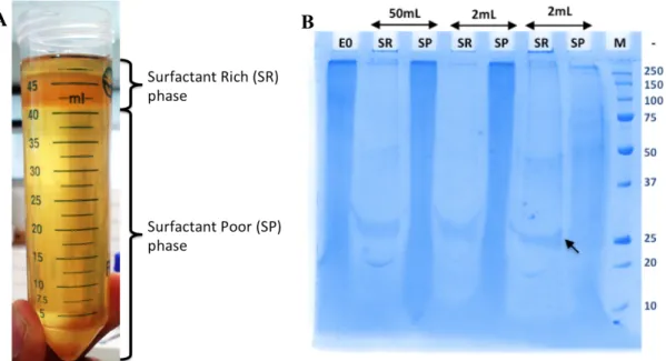
Les lipoprotéines de haute densité (High Density Lipoprotein, HDL) permet de réduction de risque de maladies cardio-vasculaires principalement en raison de leur capacité à éliminer le cholestérol accumulé des artères (via transport inverse du cholestérol). Les effets protecteurs des HDL sont médiés par l'apolipoprotéine AI (ApoA1), qui est le La protéine la plus importante quantitativement du HDL. L’ApoA1 favorise l'efflux de cholestérol vers le foie pour l'excrétion. Une augmentation des niveaux plasmatiques de l’ApoA1 est généralement acceptée d'être cardioprotecteur, ce qui en fait un potentiel thérapeutique. Deux variantes naturelle (mutants) de l’ApoA1, Milano et Paris, sont caractérisées par une mutation ponctuelle unique a permis l'introduction d'un résidu cystéine. Populations avec ApoA1-Milano ont été rapportés d'avoir un système cardiovasculaire, même avec de faibles niveaux de plasma de ApoA1 et HDL. Il est donc d'intérêt pour générer recombinante de type sauvage et des variantes de ApoA1 humaine pour des applications thérapeutiques potentielles. Dans cette étude, de type sauvage rhApoA1 a été produit chez P. pastoris et purifié par chromatographie en mode mixte en une seule étape. Par la suite, un processus intégré a été le développement de la production et la récupération rapide de type sauvage rhApoA1 chez P. pastoris par chromatographie par lit expansée. En outre, les variantes de l'ApoA1, Milano & Paris, ont été générées par mutagenèse dirigée et ont été exprimés chez P. pastoris. Les motifs d’adsorption de rhApoA1-Milano et rhApoA1-Paris ont été comparés à celle de type sauvage ApoA1 et les différences ont été discutées.Mots clés : apolipoprotéine a-I (ApoA1), Pichia pastoris, Milano et ApoA1-Paris, chromatographie par mode-mixte, HEA HyperCel, PPA HyperCel, Capto MMC
Unités de recherche
EA-4135, Biotechnologie des protéines recombinantes à visée santé, Université de Bordeaux, France Centre for Bio-Separation Technology (CBST), VIT University, Vellore, India
viii
Abstract
Expression of Wild Type and Variants of Human
Apolipoprotein A-I in Pichia pastoris
The high-density lipoprotein (HDL) complex helps reduce the risk of cardiovascular disorders mainly due to its ability to remove accumulated cholesterol from arteries via reverse cholesterol transport. These protective effects of HDL are known to be mediated by Apolipoprotein A-I (ApoA1), which is the major protein component of HDL. ApoA1 is a lipid binding protein and promotes cholesterol efflux from peripheral tissues to the liver for excretion. An increase in the plasma levels of ApoA1 is generally accepted to be cardioprotective, making it a potential therapeutic. Two naturally occuring variants of ApoA1, namely the Milano & Paris mutants, are characterised by a single point mutation resulting in the introduction of a Cysteine residue. Populations with ApoA1-Milano have been reported to have a healthier cardiovascular system even with low plasma levels of ApoA1/HDL. It is hence of interest to generate recombinant wild type and variants of human ApoA1 for potential therapeutic applications. In this study, wild type rhApoA1 was produced in P. pastoris and purified by mixed-mode chromatgraphy in a single step. Subsequently, an integrated process has been development for the production and rapid recovery of wild type rhApoA1 in Pichia pastoris. This has paved way to the establishment of a scalable integrated process that could be further developed to industrial levels. In addition, the cysteine variants of ApoA1, Milano & Paris, have been generated by site directed mutagenesis and have been successfully expressed in P. pastoris. The binding patterns of rhApoA1-Milano and rhApoA1-Paris have been compared with that of wild-type ApoA1 and the differences have been discussed. Keywords: apolipoprotein a-I (ApoA1), Pichia pastoris, Milano and ApoA1-Paris, mixed-mode chromatorgraphy, HEA HyperCel, PPA HyperCel, Capto MMCResearch Units
EA-4135, Biotechnologie des protéines recombinantes à visée santé, Université de Bordeaux, France Centre for Bio-Separation Technology (CBST), VIT University, Vellore, India
1
Résumé de thèse en français
L’expression de type sauvage et des
variantes de l’Apolipoprotéine A-I
humaine chez Pichia pastoris
2
R.1. INTRODUCTION
R.1.1. L’INTRODUCTION GÉNÉRALE
Les troubles métaboliques, notamment le diabète, l'athérosclérose et les maladies cardiovasculaires sont les principales causes de morbidité et de mortalité dans les pays développés et en développement. L'obésité et la dyslipidémie sont les principaux facteurs de risque particulier dans les maladies cardiovasculaires (Miller, 1978).
Il est généralement accepté que les lipoprotéines de haute densité (High Density Lipoprotein, HDL) permet de réduire le risque de maladies cardio-vasculaires, car il se déplace le cholestérol au foie par le transport inverse du cholestérol (Reverse Cholesterol Transport, RCT) (Tall, Costet, & Wang, 2002). En plus, le HDL contribue également au bien-être général du système cardio-vasculaire grâce à ses propriétés anti-inflammatoires, anti-oxydantes et anti-thrombotiques (Assmann & Gotto, 2004). On pense que ces effets protecteurs des HDL être médiée principalement par l'apolipoprotéine AI (ApoA1) qui est le composant protéique majeur des HDL (Heinecke, 2010).
R.1.2. TRANSPORT INVERSE DU CHOLESTEROL
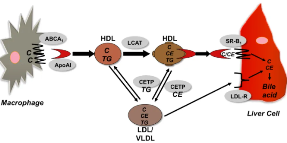
L’ApoA1 favorise l'efflux de cholestérol des tissus vers le foie (Oram, 2003) (Fig. R.1). ApoA1 prend cholestérol des cellules à travers l'ABCA1 (ATP-Binding Cassette A1) et forme des particules de HDL naissantes. Les particules HDL plus matures lors de l'activation de la Lécithine-cholestérol acyltransférase (LCAT) qui convertir le cholestérol à des esters de cholestérol (Vanloo et al., 1992). Ces esters de cholestérol de HDL sont échangés avec des triglycérides provenant d'autres lipoprotéines (LDL/VLDL) par l'action de la protéine de transfert des esters de cholestérol (Cholesteryl Ester Transfer Protein, CETP). Le transport inverse du cholestérol est complétée par le dépôt d'esters de cholestérol dans le foie, soit directement par Scavenging récepteur B1 (SR-B1) ou par LDL et le LDL-récepteur (Lewis & Rader, 2005).
3
Figure R.1. Le mécanisme de transport inverse de cholestérol
On sait que des niveaux élevés dans le plasma de l’ApoA1 contribue à la réduction des risques cardio-vasculaires chez les humains (Gordon et al., 1989), ce qui rend l’ApoA1 du potentiel thérapeutique. En outre, deux variants naturels de ApoA1 : ApoA1-Milano et ApoA1-Paris ont été signalés dans les populations qui ont réduit risque d'athérosclérose (Alexander et al., 2009). Les variantes Milano et de Paris sont caractérisées par des mutations ponctuelles au R173C et R151C respectivement. L’ApoA1 de type sauvage ne contient pas de résidus de cystéine, et donc l'introduction de ces mutants cystéine dans leur permet de former des homo-dimères à liaison disulfure sur le HDL (Klon, Jones, Segrest, & Harvey, 2000).
R.1.3. LES OBJECTIVES
Dans ce travail, nous avons mis en place un processus simplifié pour générer l’ApoA1 recombinante chez Pichia pastoris et de la purifier par chromatographie en mode mixte.
Le système d'expression choisi pour cette étude est la levure méthylotrophe
Pichia pastoris, en raison d'un certain nombre d'avantages qu'elle pose : la
capacité d'atteindre des niveaux élevés d'expression de protéines hétérologues (Cereghino & Cregg, 2000; Sreekrishna et al., 1997), la capacité à sécréter des protéines hétérologues dans le milieu (Brake et al., 1984), et étant «généralement considéré comme sûr" (Generally Regarded As Safe, GRAS) micro-organismes (Klein, 1998). La surexpression de protéines hétérologues
4
chez P. pastoris est atteinte par le promoteur AOX1 qui est activé dans des conditions de carbone faim (Daly & Hearn, 2005).
Après l'expression, l’ApoA1 recombinante a été ensuite purifié par Chromatographie en mode mixte. Chromatographie par mode mixte ou « multimodal » sont généralement émis l'hypothèse d'agir par une combinaison d'interactions électrostatiques et hydrophobes (Chung, Freed, Holstein, McCallum, & Cramer, 2010). Donc, les objectifs suivants sont envisagés dans ce travail de thèse: • Clonage et expresion de type sauvage l’ApoA1 recombinante (rhApoA1) dans P. pastoris X-33 • Purification de rhApoA1 de la culture de P. pastoris, et scale-up de la purification à grande échelle avec l’adsorption a lit expansée (Expanded Bed Adsorption, EBA) • Le clonage, l'expression et la purification de rhApoA1 chez P. pastoris SMD-1168 (souche de P. pastoris déficiente en protéase) • Génération de variantes de l’ApoA1: Milano (R173C) et Paris (R151C)
R.2. CLONAGE ET EXPRESSION DE TYPE SAUVAGE DE L’ApoA1
HUMAINE RECOMBINANTE DANS P. pastoris X-33
R.2.1. MÉTHODESLa séquence correspondant à l’ApoA1 a été amplifié en utilisant des amorces spécifiques sa séquence, et a été clone dans le vecteur pPICZαA et transformé en Pichia pastoris X-33 compétente, en suite les transformants ont été sélectionnés la résistance à la Zéocine™ (jusqu'à 2 mg / ml). Un étude préliminaire d'expression a été réalisée sur plusieurs clones de haute résistance sur les cultures en flasques, avec induction effectuée en utilisant 0,5% de méthanol chaque 24h pour 120hrs. Par la suite, le clone exprimant le plus élevé a été en outre pour l'expression dans un bioréacteur de 2L dans conditions régulées: Temp 30°C, pH 6,0, 15% saturation d’oxygène, induction avec 0,5% de
5
méthanol chaque 12hrs. L'expression a été analysée par SDS-PAGE, dot-blot et Western blot.
R.2.2. RÉSULTATS ET DISCUSSION
Après le clonage, le construit (pPICZα-ApoA1) a été soumis à un séquençage d'ADN et a été vérifiée. La construit ete transforme par électroporation dans compétentes cellules de Pichia pastoris X-33, un certain nombre de transformants présentait une haute résistance à la zéocine et cinq d'entre eux ont été repris pour les études d'expression sur les cultures en flacons.
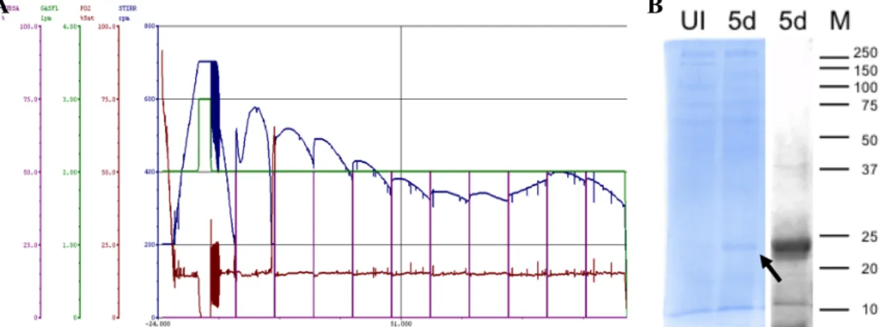
Suite les études sur les cultures en flacons, un clone a été prise pour l'expression dans un bioréacteur. Une colonie unique a été inoculée et cultivée dans des flacons à déflecteurs de 100mL de milieu de glycérol-complexe (Buffered Complex Glycerol Medium, BMGY) jusqu'à ce que la DO (600 nm) atteint de 4 au 8, ce qui a été inoculé dans 2 litres d'BMGY dans un BIOSTAT® Bplus 2l bioréacteur. Les paramètres ont été maintenus constamment, et lors de la consommation complète du glycérol dans le milieu, une phase méthanol fed-batch a été initiée par l'addition de methanol toutes les 12 heures jusqu'à une concentration finale de 0,5% pour 120hrs. L'expression des protéines a été vérifiée par dot-blot, SDS-PAGE et analyse western blot.
Figure R.2. Profil (A) et l’analyse SDS-PAGE et western blot (B) de l’expression de rhApoA1 expression in P. pastoris X-33.
Après vérification de l'expression, la milieu de P. pastoris contenant rhApoA1 a été utilisé pour les expériences de purification.
6
R.3. PURIFICATION DE TYPE SAUVAGE rhApoA1 EXPRIMÉE CHEZ
P. pastoris X-33 PAR CHROMATOGRAPHIE EN MODE MIXTE
R.3.1. MÉTHODES Un certain nombre de méthodes de purification publiés ont été testés pour leur aptitude à récupérer rhApoA1 exprimée dans P. pastoris (Feng, Cai, Song, Dong, & Zhou, 2006; Marco Aurélio Zezzi Arruda, Lopes, Marcelo Anselmo Oseas da Silva, & Gozzo, 2011). Ces méthodes ont été conservés en tant que méthodes de référence pour évaluer l'efficacité des procédés de purification développé dans cette étude.
Deux méthodes de chromatographie en mode mixte ont été testés pour leur capacité à capturer rhApoA1 directement à partir du milieu d'expression de P.
pastoris : HEA HyperCel (Pall Life Sciences) et Capto MMC (GE Healthcare), dont
les structures sont à la Fig. R.3. Les conditions de chromatographiques ont été établies sur la base des directives de leur fabricant respectif, et les différentes fractions recueillies ont été analysées en utilisant SDS-PAGE et techniques Western blot.
Figure R.3. Structures de ligands (A) HEA HyperCel et (B) Capto™ MMC
Après avoir optimisé avec succès conditions chromatographiques pour récupérer rhApoA1, les protéines purifiées ont été digérés par la trypsine et analysées sur un-ESI-Q-TOF LC spectromètre de masse (modèle Agilent G6540A) pour évaluer la couverture de la séquence, afin de vérifier la protéine purifiée .
R.3.2. RÉSULTATS ET DISCUSSION
Des méthodes publiées pour la purification de l’ApoA1 ont été adaptées pour travailler avec de milieu de P. pastoris. Après les trois procédés décrits ont été testés, leur efficacité pour « scale-up » a été évaluée. Étant donné que toutes les méthodes précédemment rapportées ne sont pas réalisables pour scale-up, il était impératif d'explorer d'autres procédés chromatographiques de colonnes
7
efficaces qui pourraient être utilisés pour purifier l'rhApoA1 exprimée chez P.
pastoris..
R.3.2.1. PURIFICATION DE L’ApoA1 AVEC HEA HYPERCEL
La résine en mode mixte HEA HyperCel généralement activé l’adsorption et l'élution de protéines par attraction hydrophobe et répulsion électrostatique, respectivement. En cas de rhApoA1, l’adsorption a été réalisé à pH neutre avec moins conductivité promouvoir l'attraction hydrophobe. Par la suite, le sel a été éliminé et le pH a été réduit, ce qui pourrait avoir exposé charges positives sur la surface de la protéine qui faciliter élution par répulsion électrostatique. Le profil de purification est rapporté dans Fig. R.4.
Figure R.4. Purification de l’rhApoA1 a l’aide de l’HEA HyperCel : (A) Chromatogram et (B) analyse de SDS-PAGE 12%.
Un désavantage de cette méthode était l'emploi de pH bas (4,0) pour éluée de l’ApoA1, comme il est connu que ApoA1 forme amyloïdes aux basses pH. (Ramella et al. 2012).
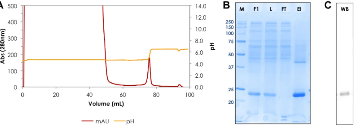
R.3.2.2. PURIFICATION DE L’ApoA1 AVEC CAPTO™ MMC
Le ligand Capto™ MMC favorise l’adsorption des protéines par des interactions hydrophobes, ioniques et thiophiles. Comme il marche comme un échangeur de cations faible, il a été prévu pour fonctionner d'une manière inverse à la HEA HyperCel (qui contient un groupe à charge positive). Comme l’adsorption a été effectuée à pH 5,0 (le pH du milieu d'expression de P. pastoris), aucun prétraitement de l'échantillon était nécessaire. En outre, la protéine liée a été
8
élue à pH neutre (Fig. R.5), de faciliter le maintien de la fonctionnalité maximale. Ce processus était plus compatible pour scale-up et pour la développement d’un procède industrielle.
Figure R.5. Purification de l’rhApoA1 a l’aide de Capto MMC : (A) Chromatogram, (B) analyse de SDS-PAGE 12% et (C) Blot western
R.3.2.3. COMPARAISON DES MÉTHODES DE PURIFICATION AVEC MÉTHODES PUBLIÉES
Les méthodes chromatographiques développés dans ce travail ont été comparées avec les méthodes déjà publiées pour la purification de rhApoA1 (Tableau R.1). Tableau R.1. Comparaison des méthodes de purification Méthode de purification Nombres d’étapes Rendement Purité d’rhApoA1 Reference 1. Extraction par « Cloud-point » avec Triton X-114 2 55.97% 57.1% (Marco Aurélio Zezzi Arruda et al., 2011) 2. Précipitation avec acétone froid 14 60.00% 71.9% (Feng et al., 2006) 3. Chromatographie par mode mixte (HEA HyperCel) 1 56.25% 70.2% Présente travail 4. Chromatographie par mode mixte (PPA HyperCel) 1 52.50% 76.3% Présente travail 5. Chromatographie par mode mixte (CaptoMMC) 1 68.89% 84.0% Présente travail A B A C
9
Il était évident que les deux méthodes de purification développées dans cette étude ont été mieux par rapport aux méthodes déjà publiées.
R.3.2.4. IDENTIFICATION DE L’ApoA1 PURIFIÉE PAR SPECTROMÉTRIE DE MASSE
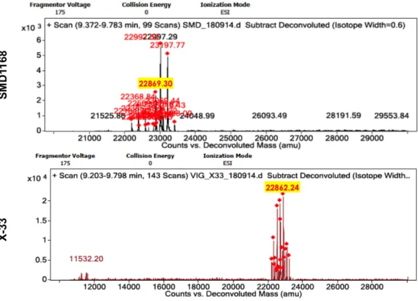
La rhApoA1 purifié par les deux procédés de purification ont été digérés avec trypsine et analysée par spectrométrie de mass sur ESI-Q-TOF MS/MS (Agilent). La rhApoA1 purifié par des deux procédés montré couverture de séquence substantielle (~ 65%) avec la séquence d'ApoA1 humaine disponible sur NCBI. Ce fut une validation globale des méthodes de purification utilisées. Bien que la protéine purifiée a été vérifié pour être ApoA1, le poids moléculaire sur des gels SDS-PAGE a été constamment inférieur (~ 25-26kDa) que le poids moléculaire attendue (28 kDa). Cela a conduit à la spéculation sur une éventuelle troncature de la protéine due à l'activité de la protéase de P. pastoris. Pour vérifier cette hypothese, le gène de l’ApoA1 a ensuite été transformé en une souche déficiente de la protéase de P.
pastoris (SMD1168) et a induit avec du méthanol pour exprimer rhApoA1. Les
détails expérimentaux et les résultats sont discutés en détail dans la section 4.
R.4. CLONAGE, L'EXPRESSION ET LA PURIFICATION DE rhApoA1
CHEZ P. pastoris SOUCHE SMD-1168 (DEFICIENTE EN PROTEASE)
Le poids moléculaire de rhApoA1 obtenu lors de l'expression de type sauvage souche P. pastoris X-33 a été constamment inférieur au poids moléculaire attendu, au cours des analyses SDS-PAGE. Nous regardé si il y avait une activité de protéase conduisant à cette troncature hypothétique, et essayé à exprimer la rhApoA1 dans une souche déficiente en protéase de P. pastoris : SMD1168. Les protéines exprimées étaient comparées entre les deux souches, de conclure globalement si la protéine a en effet été tronquée.
R.4.1. MÉTHODES
Compétente P. pastoris SMD1168 cellules ont été préparés et la construit pPICZα-ApoA1 a été transformé. Les transformants présentant une forte
10 résistance à la Zéocine ont été choisis pour analyse de l'expression. L'expression a été effectuée d'une manière similaire à celle de la souche de type sauvage (X-33). Les niveaux d'expression ont été analysés par SDS-PAGE. Suite de la production, l'rhApoA1 exprimé de P. pastoris SMD1168 a été purifié par chromatographie en mode mixte avec de ligand Capto™ MMC. Le procédé de purification était similaire que la procédé developpe pour la rhApoA1 exprimé sur le P. pastoris type sauvage, et les protéines purifiées à partir de APOA1 deux souches ont été comparées pour leur taille et leur séquence.
R.4.2. RÉSULTATS ET DISCUSSION
L'expression de rhApoA1 chez P. pastoris SMD1168 a été vérifiée par SDS-PAGE. Aucune différence significative du poids moléculaire été observée entre la rhApoA1 exprimée par X-33 et SMD-1168.
Figure R.6. Analyse spectrométrie en mass (ESI-Q-TOF) de fractions contenant la rhApoA1 purifiées à partir P. pastoris X-33 et SMD1168
11
La profil de purification de la rhApoA1 chez P. pastoris SMD1168 à l'aide Capto™ MMC a été similaires de purification profil obtenu en purifiant rhApoA1 de P.
pastoris X-33.
En analyse par spectrométrie en mass, les rhApoA1 purifiée à partir de deux souches des P. pastoris, il a été observé qu'il n'y avait pas de différence significative entre les poids moléculaires des deux souches de rhApoA1 (Fig. R.6). Cela a conduit à croire que la protéine était en fait complète et intact.
R.5. MISE A L'ECHELLE DE PRODUCTION ET DE PURIFICATION DE
rhApoA1 CHEZ P. pastoris X-33
R.5.1. MÉTHODES
Suite a l’expression et la purification de rhApoA1 en échelle laboratoire, les études pour la mise à l'échelle de la production et la purification de rhApoA1 a été envisagé. La première étape vers l'intensification de l'expression a été effectuée dans un bioréacteur de 5l capacité. Les paramètres de l’expression ont été réglés comme ils l'étaient dans les lots de bioréacteurs 2l (voir section 2.1), et l'expression des protéines ont été analysées par SDS-PAGE et dot-blot. À la fin du lot de production, toute la culture de P. pastoris a été passé à travers une colonne équilibrée directe CST-I en mode "adsorption par lit expansé". Les conditions de tampon ont été maintenues comme il été optimisée dans des colonnes a l’echelle laboratoire, et le profil a été surveillée par l’absorbance à 280 nm. Les différentes fractions ont été recueillies et analysées par SDS-PAGE.
R.5.2. RÉSULTATS ET DISCUSSION
La profil de production de la rhApoA1 a ete suivre par SDS-PAGE et les niveaux de production de la rhApoA1 été comparables ont obtenus dans le bioréacteur de 2l (section R.2.2). Un taux d’addition de méthanol plus fréquente (chaque 8 heures) assurée l’induction suffisant de cellules en croissance. L'examen microscopique périodiques d'échantillons a vérifié l'absence de contamination dans la culture. La production en bioréacteur était très reproductible.
12
Tableau R.2. Comparaison des échelles de l’expression de la rhApoA1
Paramètre\Échelle Flasque Bioréacteur 2L Bioréacteur 5L Volume de culture 150 mL 1800 mL 4000 mL
DO600nm finale 27.56 41.34 44.71
pH d’induction 6,0 5,0 5,0
Système de
tampon Phosphate + acide/base phosphate + acide/base phosphate Température de l’induction 30°C 28°C 28°C Rendement de la rhApoA1 22,4 mg/l 37,5 mg/l 43,8 mg/l Le profil chromatographique de la purification (Fig. R.7) en utilisant Direct CST-I (la même chimie de ligand comme Capto™ MMC), était similaire à celle obtenue dans des conditions à colonne paquée (Section R.3.2). Analyse par SDS-PAGE a confirmé la capture de tous rhApoA1 du milieu à une très haute concentration. Figure R.7. Purification de rhApoA1 par Direct CST-I en mode « adsorption lit expansee ». (A) Chromatogramme et (B) 12% SDS-PAGE de la charge (L), les fractions non retenues (FT) et éluée à pH 7,0 (fractions 1 - 4) et 8,5 (fractions 5 - 8). (C) 12% de l'analyse SDS-PAGE de fractions provenant Resource Q échangeuse d'ions de polissage étape: charge (L),
13
non retenu (FT), et les fractions éluées à 5%, 15%, 30% et 100% de tampon d'élution (20 mM de tampon phosphate, pH 7,0, 1M NaCl).
Quelques impuretés ont été éliminées par une deuxième étape de chromatographie avec échange d'anions. Ce processus de purification évolutive montre très prometteur pour la production industrielle à grande échelle de rhApoA1.
R.6. GENERATION DE VARIANTES DE L’ApoA1 : MILANO & PARIS
Deux variantes naturelles de l’ApoA1, Milano et Paris, sont caractérisés par une seule substitution de la pointe de l'arginine à cystéine à positions différentes. Ces deux variantes ont été rapportées chez des populations avec un risque réduit de troubles cardio-vasculaires. Une étude comparative de ces variantes pourrait donner une meilleure idée de leur mécanisme d'action, et aider à générer potentiellement thérapeutiques.
R.6.1. MÉTHODES
La mutagenèse dirigée a été utilisée pour introduire des mutations dans le gène de la rhApoA1. Les Milano (R173C) et Paris (R151C) variantes ont été générés en utilisant des amorces spécifiques de la séquence d'intégrer les mutations souhaitées. Les constructions ont été ensuite transformé par électroporation dans des cellules compétentes de P. pastoris X-33, et les transformants ont été criblés pour la résistance à la Zéocine. Quelques clones ont ensuite été essayé pour l'expression par induction avec du méthanol en cultures flacons et le bioréacteur 2L, et l'expression a été suivre par dot-blot.
Suite de l'expression, des expériences préliminaires de purification ont été réalisées pour évaluer les différences dans l’adsorption des variantes de l’ApoA1 au ligand Capto™ MMC en mode mixte.
R.6.2. RÉSULTATS ET DISCUSSION
Les constructions de l’ApoA1-Milano et l’ApoA1-Paris ont été générées par mutagénèse dirigée, et vérifiés par séquençage d'ADN. Par la suite, les
14
constructions ont été transformés de individuellement dans compétentes X-33 cellules de P. pastoris et les transformants en résistante à 2 mg/mL Zéocine ont été sélectionnés pour les études de l’expression. Trois clones chacun de Milan et de Paris ont été testés dans des cultures en ballon agité, et tous ont montré expression réussie de variantes rhApoA1 (Fig. R.8). Un clone de chaque ont ensuite été poussée plus loin pour l'expression au niveau de bioréacteur 2L.
Figure R.8. Analyse « Dot-Blot » de l’expression de Milano et l’ApoA1-Paris surexprimé chez P. pastoris X-33
Purification préliminaire des deux variantes de l’ApoA1, Milano et Paris ont été testés à l'aide de deux supports de chromatographie HEA HyperCel et Capto MMC. Les profils d’adsorption de l’ApoA1-Milano et l’ApoA1-Paris étaient significativement différents de l’ApoA1 type sauvage. Ce permis de mieux comprendre les changements importants dans la structure de liaison induite-cystéine à partir du résidu cystéine introduit.
R.7. CONCLUSION
Cette thèse a focalisé sur la génération de type sauvage et des variantes de l'apolipoprotéine AI humaine chez la levure Pichia pastoris. La méthode de production et de purification de type sauvage de l’ApoA1 était mise en échelle. La purification de l’ApoA1 été fait par l'adsorption en lit expansée (Expanded Bed
Adsorption, EBA). La comparaison des ApoA1 exprimé en type sauvage P. pastoris
X-33 et la souche protéase déficiente P. pastoris SMD1168 ont confirmé la production de rhApoA1 complet. Les études sur la génération de variantes
15
Milano et Paris de ApoA1 ouvre de nouvelles avenues pour effectuer des études comparatives entre le type sauvage et des mutantes de l’ApoA1.
16
Chapter 1
General Introduction
& Review of Literature
17
1.1. GENERAL INTRODUCTION
1.1.1. CARDIOVASCULAR DISORDERS AND ATHEROSCLEROSIS
With great advances in healthcare, the major causes of death in the industrialised world have shifted from infectious diseases to degenerative ones such as cardiovascular disorders (CVD), this shift being termed as “the epidemiologic transition” (Yusuf et al. 2001). Over the years, studies have reflected on the migration of the global burden of Ischemic Heart Diseases from high-income countries to middle- & low-income countries, India included (Finegold, Asaria, and Francis 2013). Indians are one of the more vulnerable populations in the world, and deaths due to CVD/stroke is very high due to a number of factors, not limiting to genetic factors, lifestyle habits and inadequate healthcare policies (Reddy and Yusuf 1998). Owing to the rising number of CVD cases, India is slated to become the world’s CVD capital within the next ten years (Gupta et al. 2008). Atherosclerosis is one of the major components of CVD, and is characterised by the hardening of arteries due to invasion and accumulation of macrophages and subsequent build-up of cholesterol, lipids and lipoproteins (Epstein and Ross 1999). Advanced stages of atherosclerosis often occlude blood flow causing several mortal conditions such as myocardial infarction, cardiovascular stroke, or form emboli that could affect other tissues. One of the major causative reasons for the progression of atheroma is the dysregulation of cholesterol metabolism in the body (Assmann and Gotto 2004).
1.1.2. CHOLESTEROL AND ITS METABOLISM
Cholesterol is a sterol molecule, which is an essential structural component of higher eukaryotic cells, in addition to being a precursor molecule for the biosynthesis of steroid hormones, bile acids, etc. (Hanukoglu 1992). Cholesterol molecules are used to modulate the membrane fluidity of cells: by varying their concentration with changes in temperature. The structure of cholesterol has been elucidated in Fig. 1.1.
18
Figure 1.1. Structure of Cholesterol (Reproduced from Wikipedia EN: http://en.wikipedia.org/wiki/Cholesterol)
The cholesterol molecule contains a hydroxyl group that enables it to interact with the polar heads of membrane phospholipids and sphingolipids; whereas the bulky steroid and hydrocarbon chain sink into the membrane along with they hydrophobic fatty acid chains of lipids (Yeagle 1991). This ability of cholesterol to blend in with membrane lipids enables it to provide fluidity to the cell membrane when required. In addition to providing membrane fluidity, cholesterol is also a precursor molecule for the synthesis of steroid hormones and bile acid (Berg et al. 2002).
The average cholesterol intake is approximately 400mg per day, with primary sources being de novo biosynthesis and diet. About 50% of the cholesterol from dietary sources is actually absorbed (the balance is directly excreted), and hence there is a bulk production through biosynthesis pathways for catering to the body’s cholesterol requirements (Lehninger, Nelson, and Cox 2005).
As cholesterol is only mildly soluble in water, it is primarily transported in blood through protein carriers called lipoproteins, which are complex structures with a hydrophilic exterior (with polar lipid heads and proteins) and an apolar core (explained in greater detail in Section 1.2). Lipoproteins are classified based on their density, and vary in protein compositions and functions.
19
1.2. LIPOPROTEINS
Lipoproteins are complex biological assemblies of lipids and proteins that enable the transport of fatty acids and cholesterol through water inside and outside cells (Jonas 2002). The proteins enable the emulsification of lipid particles, facilitating the smooth movement of cholesterol and triglycerides through blood.
1.2.1. STRUCTURE AND CLASSIFICATION OF LIPOPROTEINS
Lipoprotein molecules vary from discoidal to spherical forms, with a hydrophobic core and a hydrophilic surface (Jonas 2002).
As marked in Fig. 1.2, the proteins and the hydrophilic head of lipid molecules form the polar outer shell encapsulating the hydrophobic core made of cholesterol, fatty acids and triglycerides. The most important protein component of lipoproteins is apoproteins. Apoproteins bind to lipids to form nascent lipoproteins (Jackson, Morrisett, and Gotto 1976). There are a number of apoproteins associated with various sizes of lipoproteins; for example, apolipoprotein B associates with LDL and chylomicrons and apolipoprotein A associates with HDL.
Figure 1.2. Lipoprotein structure highlighting components comprising hydrophilic surface (proteins, polar lipid head) and hydrophobic core (non-polar lipid, cholesterol, cholesterylester, triglycerides and fatty acids), reproduced from (Wasan et al. 2008). Lipoproteins can be classified based on three different parameters: (i) Based on density/size (ii) Based on electrophoretic mobility (iii) Based on nature of Apo- protein content
20
Among the above classification methods, classification of lipoproteins based on their density is most commonly adopted. The density of lipoproteins decreases with an increase in the ratio of lipids to proteins in them. Based on the density of the lipoprotein molecule, they are classified into high-density lipoproteins (HDL), low-density lipoproteins (LDL), intermediate-density lipoproteins (IDL), very low-density lipoproteins (VLDL) and chylomycrons. The relative sizes and densities of these various lipoproteins have been highlighted in Fig. 1.3. Figure 1.3. Classification of lipoproteins based on diameters (nm) and density (g/ml). The lipoproteins of various densities play key roles in cholesterol metabolism in the body. Chylomycron are created from intestinal absorption of triacylglycerol & other lipids, VLDL/LDL particles are derived from the liver for the export of cholesterol, and HDL particles are formed from cholesterol effluxed from peripheral cells such as macrophages. There is a constant exchange of cholesterol, cholesterol-esters and triglycerides between lipoproteins, which play a key role in regulating cholesterol homeostasis.
1.2.2. HIGH-DENSITY LIPOPROTEINS (HDL)
The High Density Lipoprotein, or HDL, is the densest and smallest of lipoproteins. HDL particles range from 7 to 12nm in diameter, and from 1.063 to 1.25 g/ml in density (P. Barter et al. 2003).
21 Table 1.1. Classification of High Density Lipoprotein (HDL) S. No. Parameter for classification Sub-populations (Tabet and Rye 2009) 1. Shape of the HDL particle 2. Composition of Apolipoproteins 3. Size of the HDL particle 4. Density of HDL particle HDL2: 1.063 < d < 1.125 g/ml HDL3: 1.125 < d < 1.210 g/ml 5. Electrophoretic
Mobility Pre-β HDL: lipid-poor/lipid-free ApoA1 & discoidal HDL α-HDL: Spherical HDL particles (HDL2 & HDL3)
γ-HDL: Large spherical particles with ApoE
Compared to other lipoproteins, HDLs have the highest proportion of proteins (>50%) relative to their lipid content. The hydrophobic core of HDL is mainly composed of cholesterol/cholesterol-esters and a small amount of triglyceride (Rifai, Warnick, and Dominiczak 2000). The major lipid composition of HDL includes phospholipids (50%), cholesteryl esters (30%), free cholesterol (10%) and triglycerides (10%).
One of the key characteristics of HDL is their heterogeneity: in size, density and apolipoprotein composition. HDLs are classified into five sub-populations according to their size (row 3, Table 1.1), and two major subfractions based on their density (row 4, Table 1.1). Furthermore, they are also classified based on
22
their apolipoprotein composition into those that contain only ApoA1 (A-I HDL) and ones that contain both ApoA1 & ApoA2 (A-I/A-II HDL), highlighted in row 2 (Table 1.1).
HDL is generally perceived as cardio-protective, primarily due to its role in Reverse Cholesterol Transport (RCT), in addition to oxidant, anti-inflammatory, anti-apoptotic, anti-thrombotic and anti-platelet activating effects (Tabet and Rye 2009). 1.2.3. REVERSE CHOLESTEROL TRANSPORT (RCT) The process by which cholesterol is moved from peripheral tissues to the liver for catabolism is known as Reverse Cholesterol Transport (RCT, Fig. 1.4). RCT is the major cardioprotective function of the HDL particle (Fielding and Fielding 1995).
Figure 1.4. Schematic of Reverse Cholesterol Transport (RCT)
Briefly, either lipid-free ApoA1 or lipid-poor pre-β HDL particles (from liver or intestine) accept cholesterol from macrophages through the effluxor ABCA1 (ATP-Binding Cassette A1) to form discoidal HDL particles. This discoidal HDL particle accepts further cholesterol and trigliceride molecules to form a spherical HDL molecule. The key step in RCT is the activation of HDL by Lecithin-Cholesterol Acyl Transferase (LCAT), an enzyme that catalyses the conversion of cholesterol to cholesterylester, resulting in the maturation of HDL (Vanloo et al. 1992). From the mature HDL, there are two routes for the cholesterol to reach
23
the liver: one directly through Scavenging Receptor B1 (SR-B1); second through the LDL to LDL-receptor. The exchange of cholesterol/cholsesterylester/triglycerides is achieved by the action of Cholesteryl Ester Transfer Protein (CETP), which is a key actor in maintaining the ratio of HDL to LDL in the blood (P. J. Barter et al. 2003). Once cholesterol/ cholesterylester reaches the liver, it is subsequently converted to bile acid and excreted via feces.
The RCT represents one of the key pathways by which cholesterol homeostasis is maintained in the body, and a number of interventions have been proposed to intervene/enhance various stages of this pathway, which will be discussed in greater detail in the following sub-section.
1.2.4. LIPOPROTEIN RATIO & CARDIOVASCULAR WELLBEING
Over the decades, several epidemiological studies have demonstrated that increasing levels of HDL are inversely correlated to risk of CHD/CVD and atherosclerosis (Assmann and Gotto 2004; P. Barter et al. 2003; William B. Kannel et al. 1971). The Framingham Study was the first to report a correlation between serum cholesterol levels and coronary heart disease risk (William B. Kannel et al. 1971), classifying cardiovascular risk based on the ratio of total cholesterol to HDL-cholesterol (W. B. Kannel 1983). This has been subsequently followed by numerous epidemiological and clinical studies across the globe, resulting in the inclusion of HDL-C in the list of risk-predictors for atherosclerosis and cardiovascular disorders (Assmann and Gotto 2004). It is estimated that a 1mg/dl increase in HDL-cholesterol correlates to a 2-3% reduction in the risk of cardiovascular events (Gordon et al. 1989).
On the other hand, high levels of LDL and LDL-C (LDL cholesterol) were correlated with an increased risk of CHD/CVD. However, risk factors based on absolute values of LDL/LDL-C weren’t sufficient to accurately predict disposition to CHD, especially for patients with intermediate risk (Superko and King 2008). Subsequently, ratios of total cholesterol (TC) to HDL cholesterol (HDL-C), and LDL-C to HDL-C were considered to provide a more reliable risk assessment.
24 Typical risk levels and target levels to be achieved to bring down risk have been summarised in Table 1.2. Table 1.2. Risk categories and target levels for CHD (Millán et al. 2009). Ratio Primary prevention Secondary prevention Risk level Target Risk level Target Men Women Men Women Men Women Men Women TC/HDL-C >5.0 >4.5 <4.5 <4.0 >4.0 >3.5 <3.5 <3.0 LDL-C/HDL-C >3.5 >3.0 <3.0 <2.5 >3.0 >2.5 <2.5 <2.0 ApoB/ApoA-I >1.0 >0.9 <0.9 <0.8 >0.8 >0.7 <0.7 <0.6
A key point to note in table 1.2 is the distinction between primary and secondary prevention ratios. In persons without established CHD, risk levels and target ratios are as defined under primary prevention. Once a CHD event has taken place, there is a significant increase in cardiovascular risk, and hence the target levels for various parameters are much lower than for primary prevention persons (Grundy et al. 1999).
Although absolute values and ratios of various lipoproteins and cholesterol prove to be a reasonable indicator for cardiovascular risk, there have been several exceptions to it. Some studies have reported the occurrence of CVD in certain individuals despite high levels of HDL (Manninen et al. 1992).
Furthermore, reports at the beginning of this millennium showcased the propensity of HDL and its major protein component apolipoprotein A-I to undergo oxidation (Lemin Zheng 2004; Panzenböck et al. 2000; Shao et al. 2008). Oxidised ApoA1/HDL showed reduced ability in cholesterol binding and LCAT activation. In addition, for several decades, studies have shown the presence of better quality ApoA1/HDL in certain population, through naturally occurring mutations (Alexander et al. 2009; Weisgraber et al. 1983). This brought in a paradigm shift in thinking that the mere quantity of HDL wasn’t sufficient for determining cardiovascular risk, but the quality of the HDL particle also had to be considered for determining risk. Subsequently, a number of diagnostic targets have been envisaged, which have been detailed in the next section (Smith 2010).
25
1.3. APOLIPOPROTEIN A-I
Human Apolipoprotein A-I (ApoA1) is the major protein component of the HDL, constituting about 70% of the total HDL protein (Rogers et al. 1997). In addition to providing major structural support to HDL particles, it also plays two important functional roles: the extraction of cholesterol from peripheral tissues by interacting with ABCA1; and in the activation of LCAT, which is a key factor in reverse cholesterol transport (Borhani et al. 1997). This section will cover all aspects of ApoA1, its life cycle, structure, variants and sensitivity to modifications.
1.3.1. PROTEINS OF HDL
The High Density Lipoprotein has been described to be cardio protective in nature, primarily owing to the positive effects exhibited by its various proteins (Heinecke 2010). In addition to the major protein ApoA1, other HDL proteins such as ApoA2, ApoD, ApoE, paraoxonase 1 (PON1), are also involved in various functions of HDL. Some of the key functions of the HDL enzymes are detailed in Table 1.3.
Table 1.3. HDL proteins and their major functions
S.No. Protein Function Reference
1. Apolipoprotein A-I
(ApoA1) Main structural protein, lipid binding, ABCA1 binding for cholesterol efflux, LCAT activation (Borhani et al. 1997) 2. Apolipoprotein A-II (ApoA2) Enhances hepatic lipase activity 3. Apolipoprotein D
(ApoD) Associated with LCAT, progesterone binding 4. Apolipoprotein E (ApoE) LDL-receptor binding (Barbier et al. 2006, 2) 5. Apolipoprotein M (ApoM) Transport of Sphingosine-1 Phosphate 6. Serum Paraoxonase
1 (PON1) Anti-oxidant and anti-inflammatory effects (Heinecke 2010)
As is evident from Table 1.3, ApoA1 is a key effector of major protective functions of HDL, especially in reverse cholesterol transport (Fielding and Fielding 1995).
26 1.3.2. LIFE CYCLE OF ApoA1
The overall life cycle of ApoA1 is closely linked to that of the HDL molecule itself. ApoA1 molecules are produced by the liver or intestines, or are released from lipolysed VLDL and chylomicrons (von Eckardstein, Nofer, and Assmann 2001). These free ApoA1 molecules accept cholesterol from peripheral cells through ABCA1 to form discoidal HDL molecules. They move through the RCT as explained in section 1.2.3, and are eventually cleared in the liver at the end of the reverse cholesterol transport, wherein they are recycled (Fielding and Fielding 1995).
The ApoA1 protein is expressed as a single polypeptide chain of 243 amino acids, with a 24 amino acid signal peptide sequence for its secretion. Once in circulation, it self-associates into a 4-helix bundle linked via their hydrophobic faces to transport cholesterol, triglycerides and lipids (Lewis and Rader 2005).
1.3.3. STRUCTURE OF ApoA1
ApoA1 is a 28kDa monomer that is synthesised by the liver and intestine. The first crystal structure of ApoA1 suggested a horseshoe like shape, constituted almost entirely of amphipathic α-helix that is punctuated by kinks at regular intervals introduced by proline residues (Borhani et al. 1997). This amphipathic α-helix enables it to bind with lipids and cholesterol through its hydrophobic face and with the aqueous exterior with its polar face (Murphy 2013). A schematic ribbon structure of ApoA1 based on its crystal structure (PDB Accession Number 1AV1) is shown below in Fig. 1.5.
Figure 1.5. Ribbon structure of ApoA1 (PDB accession # 1AV1).
27
This structure was later confirmed by a subsequent study, revealing addition details. The structure of ApoA1 further showed a solvent-exposed loop from residues 159 to 180, which plays a key role in activation of LCAT (Wu et al. 2007).
1.3.4. ApoA1 VARIANTS
The ApoA1 gene has been well documented to have several polymorphisms: more than forty naturally occurring variants of ApoA1 have been reported in literature (Matsunaga et al. 2010). Some of the variants have been listed in Table 1.4.
Table 1.4. Variants of ApoA1 S.No. ApoA1
Variant Mutation Physiological Consequence Reference 1. Milano R173C Reduced HDL but healthy cardiovascular system (Weisgraber et al. 1983) 2. Paris R151C Reduced HDL but healthy cardiovascular system (Bruckert et al. 1997) 3. Pisa L141R Absence of HDL cholesterol (Miccoli et al. 1996) 4. Finland K159R Hypoalphalipo-proteinemia (Miettinen et al. 1997) 5. Iowa G026R Amyloidosis (Benson et al.
1991)
6. Helsinky K107Δ Amyloidosis (Ramella et al. 2012)
7. Fukuoka E110K (Takada et al.
1990) Of the above listed variants, two variants, Milano and Paris, are characterised by a single point variation of Arginine to Cysteine at 173 and 151 respectively (Klon et al. 2000). Both these variants have been reported in populations where people had a healthy cardiovascular system despite low HDL levels (Rocco et al. 2010). An important point to note is that wild type ApoA1 contains no cysteine residues, and hence these cysteine variants are hypothesised to be more stable due to formation of homodimers through disulphide bridges (Klon et al. 2000). Hence,
28
in this thesis work, special focus has been laid on the generation of these two ApoA1 cysteine variants, to enable their future applications in therapeutic areas.
1.3.5. MODIFICATIONS ON ApoA1
In addition to naturally occurring variations, ApoA1 is also highly susceptible to modifications under oxidising conditions. Myeloperoxidase (MPO), a defensive enzyme, is expressed by neutrophil granulocytes to fight bacterial infections (Klebanoff 2005). However, elevated levels of MPO have been reported in atherosclerotic plaques, leading to a number of modifications on HDL/ApoA1, thereby rendering them dysfunctional (Smith 2010). Table 1.5 lists some of the characterised modifications on ApoA1 by MPO, along with functional consequences that have been reported so far. Table 1.5. MPO-mediated modifications of ApoA1 and its consequences S.No Type of Modification Modified amino acid generated Residues modified Mechanism Functional Consequences in vitro Reference 1. Chlorination
of tyrosine 3-chloro tyrosine Tyr-166 Tyr-192 MPO + H2O2 + Cl- ion generated (HOCl) hypochloite Loss of Cholesterol acceptor activity (Shao et al. 2005) 2. Nitration of
tyrosine 3-nitro tyrosine Tyr-166 Tyr-192 MPO + H2O2 + NO2 generated (ONOO-) peroxynitrite
Loss of LCAT
activation (Shao et al. 2005)
3. Oxidation of
Methionine Methionine sulfoxide Met-86 Met-112 Met-148 MPO + H2O2 + Cl- ion generated HOCl hypochlorite Loss of cholesterol acceptor activity Loss of LCAT activation (Shao et al. 2008) 4. Hydroxylation
of tryptophan Mono and Dihydroxy tryptophans Trp-8 Trp-50 Trp-72 Trp-108 MPO + H2O2 + Cl- ion generated HOCl (hypochlorite) Loss of Cholesterol acceptor activity (Peng et al. 2008) 5. Carbamylation
of lysine Lys-226 MPO + H2O2 + SCN- (thiocyanate) Generate Proatherogenic and pro-inflammatory particles (Brubaker et al. 2006)
29
Among the residues affected by MPO-oxidation, Tryptophan-modifications have been shown to be most consequential. Subsequently, an MPO-mediated oxidation-resistant mutant of ApoA1 was generated by substituting the four Trp residues with Phe residues (Peng et al. 2008). Despite extensive studies on the effects of oxidation on wild type ApoA1, there is yet not conclusive data to showcase the behaviour of ApoA1 variants, especially Milano & Paris, under oxidative stress. It is hence of extreme interest to generate robust processes for the production of both wild type and cysteine variants of ApoA. 1.3.6. CLINICAL APPLICATIONS OF APOA1 – DIAGNOSTIC AND THERAPEUTIC Recent studies, based on the sensitivity of ApoA1 to MPO-mediated modification, have established oxidised ApoA1 as a diagnostic target for the early detection of atherosclerosis. Antibodies specific to oxidised residues on the surface of ApoA1 are currently being explored for use in the early diagnosis of atherosclerosis (Nakano and Nagata 2003).
High levels of ApoA1 have been correlated with a decreased risk of atherosclerosis, and it has long since been hypothesised that elevating plasma ApoA1 levels could lead to regression of atherosclerosis (Fazio and Linton 2003). In addition to the above main function, ApoA1 has recently also been suggested to be used during in other therapeutic interventions like cancer therapy (Su et al. 2010). Keeping in mind the vast diagnostic and therapeutic applications of ApoA1, this thesis focuses on the development of an integrated process for the generation of wild type and variants of ApoA1 in a suitable recombinant protein-producing host.
1.4. EXPRESSION HOST
1.4.1. INTRODUCTION TO EXPRESSION SYSTEMSThe advent of recombinant DNA (rDNA) technology enabled the industrial production of heterologous proteins, making affordable therapeutics and
30
reducing the cost of several diagnostic and therapeutic interventions. Human insulin was the first heterologous protein produced in the laboratory in 1977 (Porro et al. 2011). Several expressions systems are currently being used for the expression of heterologous proteins: like bacterial prokaryotic expression systems (by far E. coli) and eukaryotic systems (yeast and mammalian expressions systems). In addition, plant systems and transgenic animals are being studied for the expression of recombinant therapeutics despite their difficulties, extreme high-cost and ethics involved in their production.
1.4.2. BACTERIAL EXPRESSION SYSTEMS
The bacterial expression system Escherichia coli is the most exploited system for the expression of recombinant proteins. E. coli is favoured as a host for the expression of heterologous proteins for a number of reasons: short doubling time, well established genome, ease of manipulation, GRAS (Generally Regarded As Safe) status, the ability to scale-up production to industrial scale at minimal costs (Makrides 1996). However, plasmid instability and the lack of complex post translational modification machinery like glycosylation limit their use (Porro et al. 2011). 1.4.3. EUKARYOTIC EXPRESSION SYSTEMS
The yeasts Saccharomyces cerevisiae and Pichia pastoris have been developed into highly successful systems for the expression of heterologous proteins (Cereghino and Cregg 2000). The issue of plasmid instability often faced with prokaryotic systems is overcome in eukaryotes due to stable integration of the gene of interest into the host genome, which is usually achieved through homologous recombination. The ability to grow yeasts in simple, defined media to high cell densities and their ability to perform post-translational modifications make them favourites for the expression of recombinant proteins (Demain and Vaishnav 2009). Nevertheless, hyper-mannosylation remains as a major drawback while working with proteins that require glycosylation. As the target protein in this work (ApoA1) has no glycosylation sites, this would not be affected by expressing in a lower eukaryotic host like yeast.
31 1.4.4. Pichia pastoris EXPRESSION SYSTEM
The Pichia pastoris expression system serves as a suitable system for the cost-effective production of value added proteins. Numerous heterologous proteins have been successfully expressed by the Pichia Pastoris expression system (Cereghino and Cregg 2000). The P. pastoris system is also generally regarded as being faster, easier, and less expensive to use than expression systems derived from higher eukaryotes such as insect and mammalian tissue culture cell systems and usually gives higher expression levels (Klein 1998; Sreekrishna et al. 1997). Overexpression of heterologous proteins in P. pastoris is achieved by the AOX1 promoter, which is activated under carbon starvation (Daly and Hearn 2005).
1.4.5. HETEROLOGOUS EXPRESSION OF PROTEINS IN P. pastoris: THE AOX1 SYSTEM
Pichia pastoris is one of approximately a dozen yeast species representing four
different genera capable of metabolizing methanol. The methanol metabolic pathway appears to be the same in all yeasts and involves a unique set of pathway enzymes. The first step in the metabolism of methanol is the oxidation of methanol to formaldehyde in the peroxisome, generating hydrogen peroxide in the process, by the enzyme alcohol oxidase (AOX). The H2O2 produced by the
AOX reaction is metabolised by catalase in the peroxisome (Fig. 1.6). AOX is strongly repressed by many alternate carbon sources such as glucose, glycerol and even ethanol, and is induced by carbon starvation when grown with methanol as the sole carbon source. Although alcohol oxidase is not specific for methanol and is capable of oxidizing other primary alcohols, the activity of the enzyme decreases as the number of carbons in the alcohol increases. The conversion of methanol to formaldehyde is the rate-limiting step in utilizing methanol, due to the poor affinity of AOX to O2, and is regulated by increasing the
amount of AOX enzyme in the cells (Daly and Hearn 2005).


![Figure 3.1. Cloud Point Extraction of rhApoA1 from P. pastoris expression broth under various [Triton X-114] & [NaCl]: (a) partition coefficient and (b) 12% SDS-PAGE.](https://thumb-eu.123doks.com/thumbv2/123doknet/14741111.755080/62.892.214.695.505.1055/figure-extraction-pastoris-expression-various-triton-partition-coefficient.webp)