Alexander E. Handschin Marcus Egermann Volker Wedler Otmar Trentz Sonja Hemmi Omana A. Trentz Received: 12 September 2005 Accepted: 14 December 2005 Published online: 24 February 2006
# Springer-Verlag 2006
A comparative analysis of phenotype
expression in human osteoblasts
from heterotopic ossification
and normal bone
Abstract Background and aims: Heterotopic ossification (HO) is a pathological bone formation process in which ectopic bone is formed in soft tissue. The formation of bone depends on the expression of the osteoblast phenotype. Earlier studies have shown conflicting results on the expression of phenotype markers of cells originating from HO and normal bone. The hypothesis of the present study is that cells from HO show an altered expression of osteoblast-spe-cific phenotype markers compared to normal osteoblasts. The aims of the study were to further characterize the expression of osteoblast phenotype-markers and to provide a comparison with other study results. Patients and methods: Using an in vitro tech-nique, reverse transcription polymer-ase chain reaction (RT-PCR), real-time PCR and immunohisto-chemistry, we compared the
pheno-type gene expression (pheno-type I collagen, alkaline phosphatase, Cbfa-1, osteo-calcin) of osteoblasts from resected HO and normal bone (iliac crest). Results: Cells from HO expressed the osteoblast phenotype (type I col-lagen, alkaline phosphatase) but were characterized by a depleted osteocal-cin expression. The expression of Cbfa-1 (osteocalcin transcription gene) showed a large variety in our study. Preoperative radiotherapy had no effect on phenotype expression in cells from HO. Conclusion: Our results provide a characterization of cells originating from HO and support the thesis of an impaired osteoblast differentiation underlying the forma-tion of HO. The transcripforma-tion axis from Cbfa-1 to osteocalcin could be involved in the pathogenesis of HO. Keywords Osteoblast . In vitro . Heterotopic ossification . Osteocalcin
Introduction
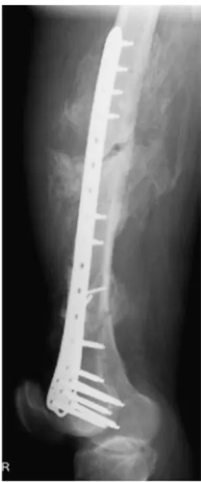
Heterotopic ossification (HO) is defined as the formation of bone in tissues which normally do not ossify [1]. HO is frequently observed in trauma patients, e.g. following long-bone fracture, hip fracture, joint dislocation, total hip arthroplasty or burns (Fig. 1) [2]. The incidence and location of bone formed may vary greatly. In severe cases, post-traumatic HO may lead to grotesque deviation of the extremities and complete ankylosis. The treatment of choice in clinical HO is surgical resection, which may include extensive soft tissue resection and reconstruction [3]. To avoid recurrence, postoperative radiotherapy is
increasingly used [4]. In addition to local trauma, HO may also occur in isolated traumatic brain injury [5]. This observation suggests a neurologic factor that may con-tribute to the development of HO, but the exact pathomechanism is poorly understood [6, 7]. Earlier studies have proposed three conditions necessary for the formation of HO: a stimulating event, the presence of mesenchymal stem cell, and an environment favourable of osteogenesis [8]. A potential mechanism underlying HO involves the differentiation from mesenchymal stem cells resulting in osteoblast formation. Thus, in vitro analysis of osteoblasts from HO may contribute to the understanding of ectopic bone formation. The hypothesis of the present A. E. Handschin (*) . M. Egermann .
S. Hemmi . O. A. Trentz Department of Surgery, Research Division,
University Hospital of Zurich, Raemistrasse 100, 8091 Zurich, Switzerland e-mail: [email protected] Tel.: +41-1-255-2213 V. Wedler Department of Surgery, Division of Plastic,
Hand and Reconstructive Surgery, University Hospital of Zurich, Zurich, Switzerland
O. Trentz
Department of Surgery, Division of Trauma Surgery, University Hospital of Zurich, Zurich, Switzerland
study is that cells from HO show an altered expression of osteoblast-specific phenotype markers compared to normal osteoblasts. The aims of the study were to further characterize the expression of osteoblast phenotypemarkers and to provide a comparison with other study results.
The recent advance in human genetics has led to the identification of many genes involved in the osteogenetic process. Parallel, improved methods to quantify mRNA have been introduced. Using the method of reverse transcription polymerase chain reaction (RT-PCR), we have analysed the expression of osteoblast-specific genes (type I collagen, alkaline phosphatase, Cbfa-1, osteocalcin) in cells originating both from normal and ectopic bone. These markers are specific for the osteoblast phenotype, and their analysis allows a comparison with the results from other studies. In addition to osteocalcin and Cbfa-1 being phenotypic markers of osteoblasts, they also play a central role in osteoblast development and the regulation of bone formation. Therefore, analysis of these genes may help better understand the genesis of HO.
Patients and methods
Patients
Cancellous bone chips were harvested from 25 trauma patients without head injuries undergoing osteosynthesis with bone grafting from the iliac crest (male/female ratio 14:11, age 37±2 years) and from 25 patients with manifest HO who underwent surgical resection (male/female ratio 17:8, age 36.5±11 years). To provide a homogeneous harvesting, known osteoporosis, age more than 50 years, corticosteroid therapy, diabetes mellitus,
immunosupres-sion, rheumatoid arthritis and underlying bone diseases were regarded as exclusion criteria. The location of the resected HO included the proximal femur (n=12), distal femur (n=8), elbow (n=4) and shoulder (n=1). The mean delay between the initial trauma and the resection of clinical manifest HO was 296±160 days (range 50– 707 days). A preoperative radiotherapy was performed in eight cases.
Primary human osteoblast culture
We used an established primary human osteoblast cell culture [9,10]; cancellous bone chips were harvested from either the iliac crest or from resected post-traumatic HO. The cleaned and minced small bone fragments were cultured in Petri dishes (Falcon) in α-modification of Eagle’s minimal essential medium (α-MEM; Invitrogen, Basel, Switzerland) supplemented with 10% foetal calf serum (FCS), 60μg/ml antibiotic–antimycotic (penicillin, streptomycin, amphotericin) (Invitrogen) and 60 μg/ml ascorbic acid (Sigma-Aldrich, Buchs, Switzerland) in a humified atmosphere of 95% air and 5% CO2at 37°C. The first passage of confluent osteoblasts was used for the study. Following trypsination, wells were seeded with 20,000 cells/ml in duplicate. The medium was changed after 3 days of incubation. After 7 days, cells were fixed and analysis of phenotype gene expression was performed. In addition, immunohistochemical analysis was performed using the method described before [9].
RNA extraction
The confluent primary osteoblasts were washed with phosphate-buffered saline (PBS) solution, and RNA was extracted using a Qiagen RNase Kit (Hilden, Germany), according to the instructor’s guidelines. After isolation, the concentration and purity were measured from the prepared RNA samples on a Gene Quant II (Amersham Pharmacia Biotech, Piscataway NJ, USA).
Semi-quantitative RT-PCR
The semi-quantitative RT-PCR was performed using the Omniscript Reverse Transciptase and HotStar Taq Master Mix PCR Kit from Qiagen according to their instructions. Oligodeoxynucleotide primers were as follows:
– Osteocalcin: forward 5′-atg aga gcc ctc aca ctc ctc-3′, reverse 5′-gcc gta gaa gcg ccg ata ggc-3′, 297 bp, T=60°C
– Alkaline phosphatase: forward 5-ccc aaa ggc ttc ttc ttg-3′, reverse 5-ctg gta gtt gtt gtg agc at-3′, 356 bp, T=52°C
Fig. 1 Heterotopic ossification (HO) in a 42-year-old male following femoral shaft fracture and plate osteosynthesis. Ectop-ic bone surrounds the femoral shaft, causing pain and immobilization
– Type I collagen: forward 5′-gcg aga gca tga ccg atg ga-3′, reverse 5′-gcg gat ctc gat ctc gtt gga-3′, 218 bp, T=55°C
– Cbfa-1: forward 5′-cag acc agc agc act cca ta-3′, reverse 5′-ttc aat atg gtc gcc aaa ca-3, 256 bp, T=52°C The RT-PCR were performed with a GeneAmp PCR System 9700 thermocycler (Applied Biosystems, Foster City, CA, USA). The master mix and the RNA solution had a total volume of 20μl; thus, the RNA was included as an aliquot of 1μg total RNA and was converted using reverse transcriptase at 37°C for 60 min. Out of this reaction mixture, 3 μl of RT product was used for the PCR. The same amounts of RNA were also used as control to amplify the housekeeping gene glyceraldehydes-3-phophate dehy-drogenase (GAPDH, forward 5′-ggg ctg ctt tta act ctg ct-3′,
reverse 5′-tgg cag gtt ttt cta gac gg-3′, 702 bp, T=60°C). The PCR reaction started with an initial heat activation step at 94°C for 15 min. The cycling parameters were as fol-lows: denaturation at 94°C for 1 min, annealing at 52–60°C (different protein) for 30 s, extension at 72°C for 1 min, 26–30 cycles (target dependent), followed by the initial extension at 72°C for 10 min. Negative control reactions for RT-PCR were performed in each assay using all the reagents as for the experimental sample without cDNA. PCR products were fractionated on a 1.5% agarose gel and visualized by ethidium bromide staining. The stained bands were photographed by a UVP Life Science Camera. All experiments were repeated at least thrice to verify the results. Positive and negative control reactions were per-formed for each primer pair.
Fig. 2 Relative mRNA expression of osteoblast phenotype genes [(reverse transcription polymerase chain reaction (RT-PCR)] follow-ing 7 days of incubation. In both groups, phenotype expression of type I collagen, alkaline phosphatase and Cbfa-1 was detected, with
no significant difference between the groups. Cells from HO (n=25, grey column) show a significant depleted expression of osteocalcin compared to cells from iliac crest (n=25, white column)
Real-time PCR (TaqMan)
Real-time PCR (TaqMan) was performed using an ABI Prism 7000 Sequence Detection System (Applied Biosys-tems). The design of primers and probes for the TaqMan assays were carried out using the primer Express software supplied by Applied Biosystems. The TaqMan primers were as follows:
– Osteocalcin: forward gaa gcc cag cgg tgc aaa, reverse tac ctc gct gcc ctc ctg, probe tcc agc aaa ggt gca gcc ttt gtg tcg
– Alkaline phosphatase: forward cct cgt tga cac ctg gaa gag, reverse ttc cgt gcg gtt cca gac, probe ttc aaa ccg aga tac aag cac tcc cac ttc
– Type I collagen: forward cca gaa gaa ctg gta cat cag caa, reverse cgc cat act cga gga atc, probe ccc caa gga caa gag gca gca tgt ctg gta
– Cbfa-1: forward gca gaa tgg atg aat ctg ttt gga, reverse gga tgt ggc ccc cag ata caa, probe cca tat tga aat tcc tca gca gtg gcc caa
The probe is dual-labeled, with a reporter dye, carboxyfluorescein (FAM), and a quencher dye, 6-carboxytetramethylrhodamin (TAMRA). PCR reactions were set up in 96-well reaction plates using a TaqMan core reagent kit (Applied Biosystems) with final volume of 25μl/well. All PCR reactions were performed in duplicate. The threshold cycle (CT) values are then calculated by determining the point at which the fluorescence exceeds this chosen threshold limit. The CTvalue for each reaction reflects the amount of PCR needed to identify the target gene; thus, high values reflect a low gene expression, while low values resemble high expression of the corresponding gene. Stained 18S rRNA-DNA (VIC) was used as internal control gene. Calculation was performed using the internal
control level and the calibration standard using the formal 2 ΔΔCT:
Statistical analysis
Statistical significance of differences between the groups (HO vs iliac crest) was determined by the Mann–Whitney U test. Significance was declared at p<0.05. All data are presented as mean (standard deviation, SD).
Results
Figure2shows the relative mRNA expression (RT-PCR) of the phenotype markers in cells from HO and iliac crest following 7 days of incubation. The figures show the mean expression in cultures cultivated from the 25 donors in each group.
Type I collagen expression was strongly positive in both groups, with no significant differences between cells from HO and normal osteoblasts (RT-PCR: HO 156.3±18.5 vs 168.2±11.2; real-time PCR: HO 0.938±0.15 vs 1.035± 0.23).
Alkaline phosphatase expression varied greatly among the cells and was higher in cells originating from HO, but this difference did not reach statistical significance (RT-PCR: HO 42.3±2.6 vs 33.1±4.6; real-time PCR: HO 1.736±0.5 vs 1.956±0.66).
Cbfa-1 showed individual differences between the two groups with a large SD. There was no statistical significant difference between the two groups (RT-PCR: HO 80.3± 40.3 vs 88±46.4; real-time PCR: HO 1.243±0.98 vs 1.381± 0.88).
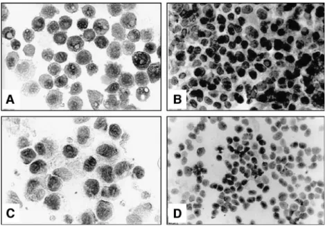
Fig. 3 Osteocalcin expression (immunohistochemistry) in-creases in human osteoblasts after 3 days (a, 1:1,000) and after 7 days (b, 1:100). In con-trast, osteocalcin expression in osteoblasts from HO is less positive (c, 1:1,000; d, 1:100)
Osteocalcin expression was significantly depleted in cells from HO compared to normal osteoblasts from iliac crest (RT-PCR: HO 37.3±12.6 vs 123.1±11.6, p=0.001; real-time PCR: HO 1.627±0.6 vs 0.593±0.3). These data were confirmed in the immunohistochemistry, which showed a depleted osteocalcin expression in cells from HO after 7 days of incubation (Fig. 3). Preoperative radiotherapy did not have an effect on any phenotype marker expression.
Discussion
The normal bone remodelling process is characterized by the tight interaction between osteoblasts (bone formation) and osteoclasts (bone resorption). In the present study, we have demonstrated that bone cells originating from HO express the osteoblast phenotypes type I collagen and alkaline phosphatase to a similar extent as osteoblasts originating from normal bone, relating these cells to the osteoblast phenotype. In accordance to this result, other studies have also found collagen expression and alkaline phosphatase expression to be similar in both cell culture types [11,12]. Mature osteoblasts exclusively produce type I collagen and only rudimentary amounts of other collagen types [11], indicating that cells from HO express the proteins characteristic of the osteoblast phenotype. In contrast, we found a significant depletion of osteocalcin expression in cells originating from HO. These data suggest that HO is associated with an imbalance of bone turnover involving osteoblasts and osteoclast, with a shift towards osteoblast activity and enhanced osteogenesis.
Osteoblasts are of mesenchymal origin and primarily arise from stromal cells found in bone marrow. Under the influence of growth factors, cytokines and other stimuli, these stromal precursor cells differentiate into mature osteoblasts and subsequently to osteocytes [13]. The
osteoblast maturation sequence is divided into three consecutive stages: proliferation, extracellular matrix mat-uration and mineralization [14]. Within these three stages, osteoblasts synthesize two types of extracellular matrix proteins: the collagens, mostly type 1 collagens which resemble 90% of the bone matrix proteins, and the non-collagenous proteins, including osteocalcin. Osteocalcin is exclusively synthesized by mature osteoblasts and is a crucial regulator of osteoblast development. [15]. The expression of osteocalcin by the osteoblast contributes to both the stimulation of undifferentiated mesenchymal cells to proliferate into osteoblast-like cells and down-regulation of further osteoblast maturation and mineralization [14]. Osteocalcin knockout mice show an alteration of the bone remodelling process with an enhanced and stronger bone formation due to a failed arrest of matrix mineralization [15]. In addition, osteocalcin may promote osteoclast activity, hence inducing bone resorption [16]. Interestingly, clinical investigators have found that changes in osteocal-cin levels in the blood may be a predictive factor for the later development of HO [17]. Following total hip arthroplasty, a rise in osteocalcin serum concentration of more than 13% had a sensitivity of 56% and a specificity of 91% for the development of HO [17]. In another clinical study, we demonstrated that patients suffering from traumatic brain injury, who carry an increased risk for the later development of HO, show a significant depletion of osteocalcin serum concentrations during the first 2 weeks after trauma [18].
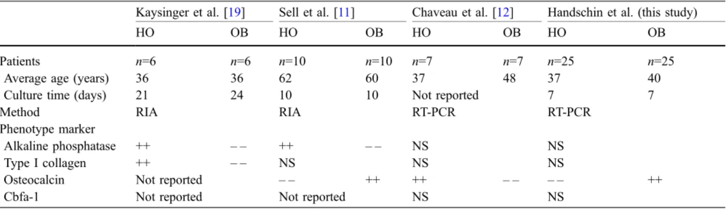
The analysis and comparison of phenotypic markers in HO cells have been investigated by few other studies that have shown conflicting results [11,12,19]. Table1shows the data of the in vitro studies on gene expression of osteoblasts from HO and normal bone. Kaysinger et al. [19] found that osteoblasts from HO showed increased synthesis of type I collagen and increased cell proliferation, while osteocalcin expression did not differ from normal
Table 1 Comparative studies on the expression of osteoblasts phenotype in cells from heterotopic ossification (HO) and normal osteoblasts (OB)
Kaysinger et al. [19] Sell et al. [11] Chaveau et al. [12] Handschin et al. (this study)
HO OB HO OB HO OB HO OB
Patients n=6 n=6 n=10 n=10 n=7 n=7 n=25 n=25
Average age (years) 36 36 62 60 37 48 37 40
Culture time (days) 21 24 10 10 Not reported 7 7
Method RIA RIA RT-PCR RT-PCR
Phenotype marker
Alkaline phosphatase ++ – – ++ – – NS NS
Type I collagen ++ – – NS NS NS
Osteocalcin Not reported – – ++ ++ – – – – ++
Cbfa-1 Not reported Not reported NS NS
Culture time: days between primary seeding and measurement of phenotype expression. (++ = strong expression vs.− − = depleted expression). RIA: Radioimmunoassay, RT-PCR: reverse transcription polymerase chain reaction, NS: not significant
osteoblasts. However, that study was performed on a smaller sample size with only six patients, which could explain why osteocalcin expression was not different in cells originating from HO. In addition, different methods (radioimmunoassay, longer culture time) were used to measure osteocalcin expression.
In another study, osteocalcin expression (RT-PCR) was significantly depleted in cells from HO [11], which is in concordance to our observation. Sell et al. [11] also found that the time for reaching confluence as measure for migration and mitosis was considerably shorter for cells from HO. The authors suggest that the balance of bone turnover in HO is shifted from a steady-state situation towards osteoneogenesis. In contrast, Chaveau et al. [12] found an elevated osteocalcin expression in cells from HO. These diverging results may be caused by differences in sample numbers, by different methods and also by the origin of HO. Our patients had suffered a local trauma with fractures to the larger joints and bones, while Chaveau et al. analysed patients with traumatic brain injury who had developed neurogenic HO without local trauma.
The expression of osteocalcin by osteoblasts is under the control of an osteocalcin transcription factor, the core-binding factor 1 (Cbfa-1), which is also referred to as Runx-2 [20, 21]. Cbfa-1 plays a critical role during osteoblasts differentiation in vertebrates. Deletion of the Cbfa-1 gene leads to a complete absence of osteoblast maturation, indicating that no other transcription factor can
fulfill Cbfa-1’s regulatory function during osteoblast differentiation [21]. In our study, we found a great variation of Cbfa-1 expression in cells from both HO and normal bone with no significant difference between the two. Similar to our observations, the only other study analyzing Cbfa-1 expression in cells from HO found no significant difference compared to normal osteoblasts [12]. A possible explanation for the difference in osteocalcin expression and the non-significant difference in Cbfa-1 expression may be that osteocalcin is expressed during a much later matura-tion stage than Cbfa-1, which encodes during the early proliferation stages [22,23]. Thus, the similar expression of Cbfa-1 in both groups may be explained by the fact that analysis was performed after 7 days at the late osteoblast differentiation stage. At this time, the osteoblasts have reached the matrix maturation stage and are starting to build bone nodules entering the mineralization stage.
In conclusion, our study provides a phenotype char-acterization of HO in vitro on a large patient population. Our in vitro experiment has confirmed the results of other studies which have shown a depleted osteocalcin expres-sion in cells from HO. Our data support the theory that HO is based on a loss of equilibrium between osteoblast and osteoclast activity with a pathological shift towards osteoblast-controlled bone formation. We conclude that the depleted expression of osteocalcin in osteoblasts from HO may represent a key mechanism in the genesis and understanding of ectopic bone formation.
References
1. Garland DE (1991) A clinical perspec-tive on common forms of acquired heterotopic ossification. Clin Orthop 263:13–29
2. Brooker AF, Bowerman JW, Robinson RA, Riley LH Jr (1973) Ectopic ossi-fication following total hip replace-ment. Incidence and a method of classification. J Bone Joint Surg Am 55:1629–1632
3. Moore TJ (1993) Functional outcome following surgical excision of hetero-topic ossification in patients with trau-matic brain injury. J Orthop Trauma 7:11–14
4. Pakos EE, Ioannadinis JP (2004) Ra-diotherapy vs. nonsteroidal anti-in-flammatory drugs for the prevention of heterotopic ossification after major hip procedures: a meta-analysis of randomized trials. Int J Radiat Oncol Biol Phys 60:888–895
5. Sarafis KA, Karatzas GD, Yotis CL (1999) Ankylosed hips caused by het-erotopic ossification after traumatic brain injury: a difficult problem. J Trauma 46:104–109
6. Pape HC, Marsh S, Morley JR, Krettek C, Giannoudis PV (2004) Current concepts in the development of het-erotopic ossification. J Bone Joint Surg Br 86:783–787
7. Gennarelli TA (1988) Heterotopic os-sification. Brain Inj 2:175–178 8. Chalmers J, Gray DH, Rush J (1975)
Observations on the induction of bone in soft tissues. J Bone Joint Surg Br 57:36–45
9. Trentz OA, Hoerstrup SP, Sun LK, Bestmann L, Platz A, Trentz O (2003) Osteoblasts response to allogenic and xenogenic solvent dehydrated cancel-lous bone in vitro. Biomaterials 24:3417–3426
10. Handschin AE, Trentz OA, Hoerstrup SP, Kock HJ, Wanner GA, Trentz O (2005) Effect of low molecular weight heparin (dalteparin) and fondaparinux (Arixtra) on human osteoblasts in vitro. Br J Surg 92:177–183
11. Sell S, Gaissmaier C, Fritz J, Herr G, Esenwein S, Kusswetter W, Volkmann R, Wittkowski KM, Rodemann HP (1998) Different behavior of human osteoblast-like cells isolated from nor-mal and heterotopic bone In vitro. Calcif Tissue Int 62:51–59 12. Chauveau C, Devedjian J-C, Blary
M-C, Delecourt C, Hardouin P, Jeanfils J, Broux O (2004) Gene expression in human osteoblastic cells from normal and heterotopic ossification. Exp Mol Pathol 76:37–43
13. Tenenbaum HC (1990) Cellular origins and theories of differentiation. In: Hall BK (ed) Bone, vol I. The osteocyte. Telford Press, New Jersey, pp 41–69 14. Lian JB, Stein GS, Stein JL, van
Wijnen AJ (1998) Osteocalcin gene promoter: unlocking the secrets for regulation of osteoblast growth and differentiation. J Cell Biochem Suppl 30/31:62–72
15. Ducy P, Desbois C, Boyce B, Pinero G, Story B, Dunstan C, Smith E, Bonadio J, Goldstein S, Gundberg C, Bradley A, Karsenty G (1996) Increased bone formation in osteocalcin-deficient mice. Nature 382:448–452
16. Chenu C, Colucci S, Grano M, Zigrino P, Barattolo R, Zambonin G, Baldini N, Vergnaud P, Delmas PD, Zallone AZ (1994) Osteocalcin induces chemotaxis, secretion of matrix proteins, and calci-um-mediated intracellular signaling in human osteoclast-like cells. J Cell Biol 127:1149–1158
17. Wilkinson JM, Stockley I, Hamer AJ, Barrington NA, Eastell R (2003) Bio-chemical markers of bone turnover and development of heterotopic ossification after total hip arthroplasty. J Orthop Res 21:529–534
18. Trentz OA, Handschin AE, Bestmann L, Hoerstrup SP, Trentz OL, Platz A (2005) Influence of brain injury on early posttraumatic bone metabolism. Crit Care Med 33:399–406
19. Kaysinger K, Ramp WK, Lang GJ, Gruber HE (1997) Comparison of human osteoblasts and osteogenic cells from heterotopic bone. Clin Orthop Relat Res 342:181–191
20. Banarjee C, McCabe LR, Choi JY, Hiebert SW, Stein JL, Stein GS, Lian JB (1997) Runt homology domain proteins in osteoblast differentiation: AML3/CBFA1 is a major component of a bone-specific complex. J Cell Biochem 66:1–8
21. Komori T, Yagi H, Nomura S, Yamaguchi A, Sasaki K, Deguchi K, Shimizu Y, Bronson RT, Gao YH, Inanda M, Sato M, Okamoto R, Kitamura Y, Yoshiki S, Kishimoto T (1997) Targeted disruption of Cbfa1 results in a complete lack of bone formation owing to maturational arrest of osteoblasts. Cell 89:755–764
22. Siggelkow H, Rebenstorff K, Kurre W, Niedhardt C, Engel I, Schulz H, Atkinson MJ, Hüfner M (1999) De-velopment of the osteoblast phenotype in primary human osteoblasts in cul-ture: comparison with rat calvaria cells in osteoblast differentiation. J Cell Biochem 75:22–35
23. Gilbert L, He X, Farmer P, Rubin J, Drissi H, van Wijnen AJ, Lian JB, Stein GS, Nanes M (2002) Expression of the osteoblast differentiation factor RUNX2 (Cbfa-1/AML3/Pebp2aA) is inhibited by tumor necrosis factor-a. J Biol Chem 277:2695–2701
![Fig. 2 Relative mRNA expression of osteoblast phenotype genes [(reverse transcription polymerase chain reaction (RT-PCR)] follow-ing 7 days of incubation](https://thumb-eu.123doks.com/thumbv2/123doknet/14841079.625095/3.892.360.803.384.982/relative-expression-osteoblast-phenotype-transcription-polymerase-reaction-incubation.webp)