HAL Id: hal-02551981
https://hal.archives-ouvertes.fr/hal-02551981
Submitted on 23 Apr 2020HAL is a multi-disciplinary open access archive for the deposit and dissemination of sci-entific research documents, whether they are pub-lished or not. The documents may come from teaching and research institutions in France or abroad, or from public or private research centers.
L’archive ouverte pluridisciplinaire HAL, est destinée au dépôt et à la diffusion de documents scientifiques de niveau recherche, publiés ou non, émanant des établissements d’enseignement et de recherche français ou étrangers, des laboratoires publics ou privés.
A Ruthenium(II) Complex Containing a Redox-Active
Semiquinonate Ligand as Potential Chemotherapeutic
Agent: From Synthesis to In Vivo Studies
Anna Notaro, Angelo Frei, Riccardo Rubbiani, Marta Jakubaszek, Uttara
Basu, Severin Koch, Cristina Mari, Mazzarine Dotou, Olivier Blacque,
Jeremie Gouyon, et al.
To cite this version:
Anna Notaro, Angelo Frei, Riccardo Rubbiani, Marta Jakubaszek, Uttara Basu, et al.. A Ruthe-nium(II) Complex Containing a Redox-Active Semiquinonate Ligand as Potential Chemotherapeutic Agent: From Synthesis to In Vivo Studies. Journal of Medicinal Chemistry, American Chemical Society, 2020, �10.1021/acs.jmedchem.0c00431�. �hal-02551981�
1
A Ruthenium(II) Complex Containing
a Redox-Active Semiquinonate
Ligand as Potential Chemotherapeutic
Agent: From Synthesis to in vivo
Studies
Anna Notaro,a,# Angelo Frei,b,# Riccardo Rubbiani,b,# Marta Jakubaszek,a, e Uttara
Basu,a Severin Koch,b Cristina Mari,b Mazzarine Dotou,a Olivier Blacque,b Jérémie
Gouyon,c Fethi Bedioui,c Nils Rotthowe,d Rainer F. Winter,d Bruno Goud,e Stefano
Ferrari,,f,g Mickaël Tharaud,h Martina Řezáčová,i Jana Humajová,j Pavel Tomšík,i
and Gilles Gassera,*
a Chimie ParisTech, PSL University, CNRS, Institute of Chemistry for Life and
Health Sciences, Laboratory for Inorganic Chemical Biology, F-75005 Paris, France.
b Department of Chemistry, University of Zurich, Winterthurerstrasse 190, 8057
Zurich, Switzerland.
c Chimie ParisTech, PSL University, CNRS, Institute of Chemistry for Life and
Health Sciences, Team Synthèse, Electrochimie, Imagerie et Systèmes Analytiques pour le Diagnostic, F-75005 Paris, France.
d Department of Chemistry, University of Konstanz, Universitätsstrasse 10, D-78457
Konstanz, Germany.
e Institut Curie, PSL University, CNRS UMR 144, F-75005, Paris, France.
f Institute of Molecular Cancer Research, University of Zurich, CH-8057, Zurich,
Switzerland.
g Institute of Molecular Genetics of the Czech Academy of Sciences, Videnska 1083,
2
h Université de Paris, Institut de physique du globe de Paris, CNRS, F-75005 Paris,
France.
i Department of Medical Biochemistry, Faculty of Medicine in Hradec Kralove,
Charles University, Šimkova 870, 500 03 Hradec Kralove, Czech Republic.
j Department of Medical Biochemistry, Faculty of Medicine in Prague, 150 06
Prague, Czech Republic.
# these authors have contributed equally to the work
* Corresponding author: E-mail: [email protected]; WWW:
www.gassergroup.com; Phone: +33 1 44 27 56 02 ORCID Number Anna Notaro: 0000-0003-0148-1160 Angelo Frei: 0000-0001-6169-2491 Marta Jakubaszek: 0000-0001-7590-2330 Uttara Basu: 0000-0002-0509-2421 Mazzarine Dotou: 0000-0001-8781-6763 Olivier Blacque: 0000-0001-9857-4042 Fethi Bedioui: 0000-0002-0063-4412 Bruno Goud: 0000-0003-1227-4159 Stefano Ferrari: 0000-0002-6607-215X Martina Řezáčová: 0000-0001-5370-2290
Jana Humajová (Mattová): 0000-0001-8099-6781 Pavel Tomšík: 0000-0002-4366-075X
Gilles Gasser: 0000-0002-4244-5097
Keywords: Bioinorganic Chemistry, Cancer, DNA, Medicinal Inorganic Chemistry, Ruthenium.
3 Abstract
Chemotherapy remains one of the dominant treatments to cure cancer. However, due to the many inherent drawbacks, there is a surge for new chemotherapeutic drugs. Many classes of compounds have been investigated over the years in order to discover new targets and synergistic mechanisms of action including multicellular targets. In this work, we designed a new chemotherapeutic drug candidate against cancer, namely [Ru(DIP)2(sq)]PF6 (Ru-sq) (DIP = 4,7-diphenyl-1,10-phenanthroline; sq =
semiquinonate ligand). The aim was to combine the great potential expressed by Ru(II) polypyridyl complexes and the singular redox and biological properties associated to the catecholate moiety. Experimental evidences (e.g. X-ray crystallography, electron paramagnetic resonance, electrochemistry) demonstrate that the semiquinonate is the preferred oxidation state of the dioxo ligand in this complex. The biological activity of Ru-sq was then scrutinised in vitro and in vivo, and the results highlight the auspicious potential of this complex as a chemotherapeutic agent against cancer.
4 Introduction
In the last decades, the search for new chemotherapeutic agents against cancer has challenged scientists worldwide. Chemotherapy, together with surgery, radiotherapy and immunotherapy, is used in a combined modality therapy to treat cancer.1 The goal
of this combination is to overcome the drawbacks of each singular treatment to afford the best chances of survival for the patients.1 Cisplatin is one of the most common
chemotherapeutic agents utilized against cancer. However, its severe side effects are limiting its clinical use.2–6 Therefore, many other platinum-based drug candidates have
been investigated over the last 40 years leading to the worldwide clinical approval of carboplatin and oxaliplatin.7,8 On the basis of these ground-breaking discoveries and
the observed occurrence of resistance with platinum treatment, a large number of metal complexes based on other metals than platinum have been examined.9–18 In this field,
ruthenium complexes play a central role due to their inherent advantages (e.g., multiple stable oxidation states, well-established chemistry, etc.).19–23 Interestingly, Ru(III) and
Ru(II) complexes usually have ligand exchange kinetics similar to those of Pt(II) complexes.19 In addition, ruthenium complexes have found applications in different
fields of medicinal chemistry against cancer since they behave differently depending on the biological settings, therefore displaying different biological targets.24–29
KP-1019, IT-139 (formerly NKP-1339) and NAMI-A are, to date, the only three Ru complexes to have reached clinical trial as anticancer agents. Their mechanism of action involves ligand exchange, resembling therefore the one of cisplatin.30–35 Of note,
TLD-1433, a substitutionally inert Ru(II) polypyridyl complex, has recently entered phase II clinical trial as a photosensitizer for photodynamic therapy (PDT).36,37 Another
very promising class of ruthenium complexes are the coordinatively saturated and substitutionally inert ruthenium polypyridyl complexes. These compounds have been
5 intensely investigated over the last years and several applications as potential chemotherapeutic agents have been unearthed.24 At first, most of the bio-activity of
these compounds was associated with interactions with DNA.38–41 However, over the
years, many other modes of action were identified, such as the trigger of mitochondrial dysfunction,42–44 Topoisomerases I and II inhibition,45,46 modification of cell
membranes47 and others.24 Due to the great opportunities offered by this class of Ru
compounds, in this work, we designed a new Ru polypyridyl complex, namely [Ru(DIP)2(sq)](PF6) (Ru-sq, Scheme 1a) where DIP is
4,7-diphenyl-1,10-phenanthroline and sq is a semiquinonate ligand, which was found to be a very interesting anticancer drug candidate. Semiquinonate is a so-called ‘non-innocent’ ligand as its electrochemical properties strongly resemble that of the metal center.48
Semiquinonate is the oxidised form of catechol, a well-known dioxo ligand, which can exist in three redox forms, namely catecholate (cat), semiquinonate (sq) and quinone (q) (Scheme 1b).49 Catecholate and its oxidation products have already been intensively
investigated as ligands.50,51 However, the focus of these studies has mostly been on the
unique electronic/redox properties of metal complexes containing such ‘non-innocent’ dioxo ligands.52–55 Catechols are also known as pan-assay interference compounds
(PAINS) due to their redox and chelating properties.56 Nevertheless, catecholate and its
derivatives have also shown potential in different fields of biological interest,57–61 such
as cancer chemoprevention,59 antifungal activity60 and the inhibition of the spontaneous
Aβ fibril formation,61 which is a key target for the treatment of Alzheimer´s disease.
Worthy of note, vanadium compounds carrying catechol-like ligands have been investigated by Crans and co-workers.62,63 During these studies, particularly potent
cytotoxic vanadium (V) catecholate complexes toward bone cancer cells were unveiled.62 The cytotoxicity on glial cells of [RuIII(NH3)4(catecholate)]+ was also
6 investigated in 2007 by Almeida and co-workers.58 In this case, the catechol was found
to be more cytotoxic than the Ru(III) complex itself with an EC50 of 0.342 mM against
rat astrocytes and 0.568 mM against human glioblastoma GL-15 cell line, while the [RuIII(NH
3)4(catecholate)]+ complex had EC50 = 1.380 mM and EC50 = 2.6 mM against
rat astrocytes and human glioblastoma, respectively.58 Further studies suggested that
depletion of glutathione and induction of apoptosis were possible explanations for the cytotoxicity observed for catechol towards mouse neuroblastoma N2a cell line.57 These
preliminary studies rationalize our choice to integrate catechol and its oxidation products into a Ru(II) polypyridyl complex. To the best of our knowledge, [Ru(DIP)2(sq)](PF6) is the first Ru(II) polypyridyl complex containing a catechol
moiety to be deeply investigated from both a physico-chemical and biological point of view. The complex was isolated as a racemic mixture of ∆ and Λ enantiomers. No effort was made in this work to isolate pure enantiomers. As described below, in vitro and in
vivo studies demonstrate a significant potential of this compound as a chemotherapeutic
7 Scheme 1.a) Synthesis of [Ru(DIP)2(sq)](PF6). I) DIP, LiCl, DMF, reflux, 24 h, 78%;
II) (i) NaOH, catechol 2-propanol, reflux, 24h; (ii) air, 2 h; (iii) NH4PF6,
2-propanol/H2O (1:8), 19%. b) Catecholate (cat) and its oxidised forms, semiquinonate
(sq) and quinone (q).
Results and Discussion
Synthesis and characterization of [Ru(DIP)2(sq)](PF6)
The synthesis of the target compound [Ru(DIP)2(sq)](PF6) was achieved in a 2-step
synthesis (Scheme 1a). Briefly, the known Ru(DMSO)2Cl264, DIP and LiCl were
refluxed in DMF to afford Ru(DIP)2Cl2 in 72% yield after precipitation with acetone.65
The compound was then refluxed in a nitrogen atmosphere overnight with catechol in the presence of NaOH in 2-propanol. The oxidation step of the catecholate to the semiquinonate was performed by exposing the solution of the Ru complex in 2-propanol to air for 2 h. [Ru(DIP)2(sq)](PF6) was obtained in 19% yield after
precipitation with a large excess of NH4PF6 and purification via silica gel
chromatography. The identity of the product was confirmed by HR-MS and NMR spectroscopy. 1H-NMR spectra showed a characteristic peak broadening in the aromatic
8 region between 7–9 ppm due to the paramagnetism of the complex. In the 13C NMR
and 2D 1H-13C HSQC spectra (Figure S1) ten inequivalent CH carbons were observed.
The purity of the product was confirmed by microanalysis.
X-ray Crystallography of [Ru(DIP)2(sq)](Cl)
The crystal structure of [Ru(DIP)2(sq)](Cl) was determined by a single crystal X-ray
diffraction study. Suitable single crystals were grown from slow diffusion of diethylether into a solution of the product prior to precipitation with NH4PF6 in MeCN.
The crystal structure revealed two independent Ru molecules (Ru-1 and Ru-2 in Figure S2), two chloride counter ions (from LiCl) and three water molecules in the asymmetric unit (monoclinic P21/c space group). Both Cl atoms are disordered over two sets of
sites with site-occupancy ratios of 0.299/0.701(3) and 0.244/0.756(5). The H atoms of the isolated water molecules could be introduced in the final refinements, but their positions were kept fixed to satisfy reliable hydrogen bonding. The molecular structure of one of the independent Ru molecules is shown in Figure 1 and a selection of the most relevant bond lengths and angles are provided in Tables S1 and S2 (additional crystallographic information can be found in the supporting information). The X-ray crystal structure determination also provided evidence for the nature of the dioxo ligand, as it can exist as catecholate, semiquinonate and quinone.49,53,66 The typical
range for the C-O bond length of such a ligand coordinated to a metal is 1.34–1.47 Å for the catecholate form, 1.27–1.31 Å for the semiquinonate form and around 1.23 Å for the quinone.49,66 The C-O bond distances of the dioxo ligand in [Ru(DIP)2(sq)](Cl)
are 1.309(4), 1.314(4), 1.315(4) and 1.319(4) Å, which suggest that it is present in its semiquinonate form.49
9 Figure 1. Molecular structure of [Ru(DIP)2(sq)](Cl). The asymmetric unit contains two
crystallographically independent Ru cations, only one of which is presented. The Cl
-counter ions, H atoms and solvent molecules are omitted for clarity. The thermal ellipsoids are shown at the 30% probability level.
Electrochemistry
The electrochemistry of [Ru(DIP)2(sq)](PF6) (abbreviated as Ru-sq) was investigated
using cyclic voltammetry (CV) and rotating disc electrode voltammetry (RDE) in DMF containing tetrabutylammonium hexafluorophosphate 0.1 M. The RDE voltammogram shown in Fig. S3 exhibits four well-defined, reversible waves, in addition to that of decamethylferrocene, which was used as internal reference with a half-wave potential of 0.030 V vs the Saturated Calomel Electrode (SCE). The four features related to the Ru-sq have the same intensity, which attests that the related redox processes involve the same number of exchanged electrons. By comparison with the data reported in the literature for closely related complexes under similar conditions,52,67,68 the underlying
redox processes were assigned as shown in Table S3. The oxidation located at + 0.647 V vs SCE can be attributed to the Ru(II)/Ru(III) redox couple while the sq/cat redox
10 couple can be associated to the first reduction process at -0.249 V vs SCE. The following two processes, at more negative potentials, can be assigned to the sequential reductions of the ancillary ligands (DIP0/-). Of note, the latter are separate couples with
quite some substantial redox splitting.52,67 These data clearly show how the presence of
the semiquinonate ligand influences the redox properties of the metal centre, causing a shift to lower potential. The couple of Ru (III)-quinone ligand is not observed in these conditions since they are possibly located outside the anodic limit of a DMF-based electrolyte. Moreover, the CV experiment (Figure S3) indicates the reversibility of the redox processes, at least on the voltammetric timescale.
Electron Paramagnetic Resonance
Ru-sq in its native state is Electron Paramagnetic Resonance (EPR) active in DCM due to the presence of an unpaired spin as already confirmed by X-ray crystallography (Figure S4a). At room temperature, a rather broad isotropic signal was observed. Its g-value of 2.0244 is in line with a ligand-centred spin density and deviates only slightly from the free electron value ge of 2.0023. This behaviour is in strong contrast to a
metal-centred spin of a Ru(III) complex, which would only become observable at low temperatures due to rapid relaxation and display a broad, axial or rhombic signal with large anisotropy.53,69 The reduced form Ru-cat (Scheme 2) was generated by the
reaction of Ru-sq with equimolar amounts of cobaltocene (Cp2Co, E1/2 = -0.880 V vs
SCE in DMF/0.1 M NBu4PF6) (Figure S4b).70 Owing to the presence of a low-spin
Ru(II) ion and a closed-shell catecholate ligand this species is EPR silent. The same holds also true for oxidized, dicationic Ru+-sq (Scheme 2), which was prepared by
treatment of Ru-sq with an excess of 1,1’-diacetylferrocenium hexafluoroantimonate (Ac2FcSbF6, E1/2 = 0.940 V vs SCE in DMF/0.1 M NBu4PF6) (Figure S4c).70,71 The
11 absence of an EPR signal indicates that the unpaired spins at the Ru(III) ion (Ru+) and
the sq ligand are antiferromagnetically coupled.
Scheme 2. Structures of Ru-cat, Ru-sq, and Ru+-sq, carrying a catecholate or a
semiquinonate ligand and Ru in oxidation state +II (Ru) or +III (Ru+), respectively.
Stability in DMSO and human plasma
The stability of a compound plays an important role in its biological activity and viability. Therefore, the integrity of Ru-sq was first assessed in DMSO-d6 using 1H
NMR spectroscopy. Ru-sq was found to be stable in DMSO over 8 days. No change in the NMR spectra of the complex was observed over 8 days (Figure S5). Next, to obtain a preliminary insight into the behaviour of Ru-sq under physiological conditions, the stability of Ru-sq in human plasma was investigated by UPLC following a procedure already established by our group.43 Ru-sq was incubated in human plasma at 37°C for
0 h, 4 h, 6 h, 12 h, 20 h and 24 h using diazepam as an internal standard. The UV traces of the UPLC analysis are shown in Figure S6a. The concentration of Ru-sq was normalized with respect to the internal standard and plotted against time. The linear trend shown in Figure S6b clearly demonstrates that between 6 and 20 h, a decomposition of 50% of the compound was observed, to reach a total degradation of the compound after 24 h.
12 Cytotoxicity Studies
After a full characterisation of Ru-sq, its potential activity as a chemotherapeutic agent was investigated starting from the biological evaluation of its behaviour against cancer cells in monolayer cell cultures. The cytotoxicity of Ru-sq towards HeLa (human cervical adenocarcinoma), A2780 (human ovarian carcinoma), A2780 cis (human cisplatin resistant ovarian carcinoma), A2780 ADR (human doxorubicin resistant ovarian carcinoma), CT-26 (mouse colon adenocarcinoma), CT-26 LUC (mouse colon adenocarcinoma stably expressing luciferase), RPE-1 (human normal retina pigmented epithelial) and MRC-5 (human normal lung fibroblast) cell lines was therefore investigated using a fluorometric cell viability assay (single graphs available in Figures S7).72 Cytotoxicity of cisplatin and doxorubicin was determined in the same cell lines
as positive controls and, as additional controls, Ru(DIP)2Cl2 and catechol were also
tested.73,74 As shown in Table 1 where IC
50 (the half maximal inhibitory concentration)
values are reported, Ru-sq displayed IC50 values between the high nanomolar and low
micromolar range on the cell lines investigated in this study, while the Ru(DIP)2Cl2
precursor and the catechol ligand itself showed much lower cytotoxicity. Very impressively, Ru-sq exerted an activity 40 times higher than cisplatin against a cisplatin resistant cell line. On the other hand, the cytotoxicity of doxorubicin and Ru-sq against a doxorubicin resistant cell line appeared to be in the same order of magnitude. Overall, complex Ru-sq displays a cytotoxicity, which is comparable to doxorubicin and much higher than the one of cisplatin. However, no selectivity was observed between cancerous and non-cancerous cell lines. This shortcoming is often faced in medicinal chemistry and could be improved by the introduction of a targeting moiety or by nanoformulation.
13 Table 1.IC50 values of Ru-sq, the Ru(DIP)2Cl2 precursor and the catechol ligand in
tested cell lines; cisplatin and doxorubicin were used as positive controls.
IC 50 (μM) HeLa A2780 A2780 ADR A2780 cis CT-26 CT-26 LUC RPE-1 MRC-5 Cisplatin 9.28 ± 0.20 4.00 ± 0.76 8.32 ± 0.71 18.33 ± 2.92 2.60 ± 0.18 2.42 ± 0.23 30.24 ± 5.11 11.20 ± 2.32 Doxorubic in 0.34 ± 0.02 0.19 ± 0.03 5.94 ± 0.58 0.54 ± 0.04 0.082 ± 0.003 0.18 ± 0.006 0.89 ± 0.17 3.37 ± 1.24 Ru-sq 0.50 ± 0.01 0.67 ± 0.04 4.13 ± 0.2 0.45 ± 0.03 1.00 ± 0.03 1.51 ± 0.14 0.90 ± 0.04 0.95 ± 0.09 Ru(DIP)2 Cl2 15.03 ± 0.4 4.69 ± 0.14 78.27 ± 4.9 6.36 ± 0.57 9.20 ± 1.22 6.65 ± 0.5 3.13 ± 0.07 5.54 ± 0.39 Catechol >100 22.80 ± 5.96 >100 54.55 ± 11.30 16 ± 4.14 11.56 ± 0.40 >100 >100
We then decided to pursue the investigation of Ru-sq behaviour in a Multi Cellular Tumour Spheroids (MCTS) model.75 It was previously shown that such a model mimics
the in vivo microenvironment and tumour metabolism.76,77 Moreover, large MCTS
develop a central necrosis core similar to that found in the inner core of tumours.78
These unique features give a better representation of a cancer model compared to a 2D model, lowering the disparity between in vitro and in vivo models.78 Table 2 shows the
IC50 values obtained via a luminescent cell viability assay for compounds that were
administered to HeLa MCTS for 48 h (single graphs are availabe in Figure S8). The Ru(DIP)2Cl2 precursor and the catechol ligand were tested as additional controls and
exhibited lower cytotoxicity than Ru-sq. Catechol showed no toxicity towards the cell line tested (IC50 > 100 μM) while the precursor displayed a cytotoxicity comparable to
cisplatin. Cisplatin was used as a positive control and the results are in line with literature data.79 The cytotoxicity of Ru-sq in HeLa MCTS was impressively high after
14 (IC50 ~ 47 µM or 39 µM, respectively). Noteworthy, the cytotoxicity of Ru-sq was
comparable to the one of doxorubicin after 72 h treatment (IC50 ~ 11 μM).80 These
studies clearly demonstrate the high potential of Ru-sq as an anticancer drug candidate. The impressive bioactivity, comparable to doxorubicin in monolayer cell culture, was also confirmed in the 3D tumour model – HeLa MCTS.
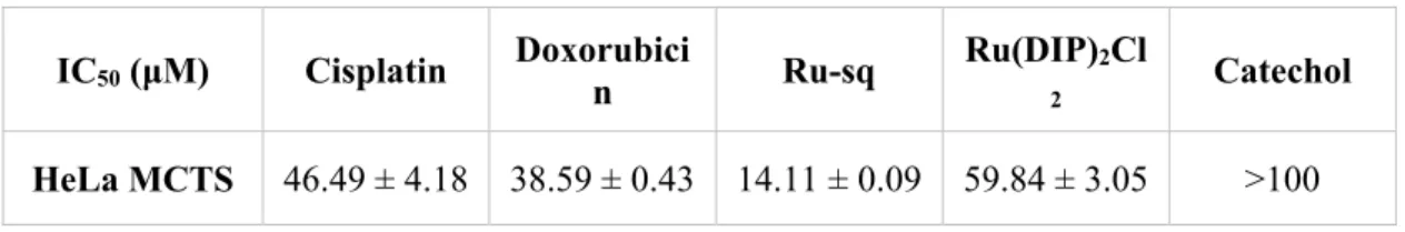
Table 2. IC50 values for Ru-sq, Ru(DIP)2Cl2 precursor and the catechol ligand in
multicellular HeLa cancer cell spheroids (approximately 400 μm in diameter); cisplatin and doxorubicin were used as positive controls.
IC50 (μM) Cisplatin Doxorubici
n Ru-sq
Ru(DIP)2Cl
2 Catechol HeLa MCTS 46.49 ± 4.18 38.59 ± 0.43 14.11 ± 0.09 59.84 ± 3.05 >100
Spheroid integrity and growth upon treatment are very useful tools to determine a potential drug activity.78 In this study MCTS were monitored over 13 days after
treatment with different concentrations of Ru-sq (Figure 2). Every 3 days, the spheroids were washed to remove dead cells and their diameters were measured (Figure 2). It is important to note that at each washing step, half of the media was removed and replaced with fresh one, diluting twice the quantity of the compound in each well. The effect of Ru-sq on growth inhibition is dose-dependent and already visible after 3 days. Low concentrations treatments (1 µM and 2.5 µM) led to regrowth of the spheroids after the first 72 h, while for 5 µM and 10 µM treatments, the regrowth is visible after 6 and 9 days, respectively. Ru-sq treatment with concentrations higher than IC50 (20 µM and
25 µM) completely inhibits the spheroids growth after 13 days of treatment. Overall, we can conclude that Ru-sq treatment at 20 µM and 25 µM concentrations, severely affect the size and the integrity of the spheroids after 13 days of treatment.
15
Figure 2.Growth kinetics of HeLa MCTS upon treatment with different concentrations
of Ru-sq (1, 2.5, 5, 10 and 20µM). a) Images collected at day 0 (before treatment) and at day 3, 6, 9 and 13. b) MCTS diameter calculated at different time points. Blue dotted line indicates day of seeding, red dashed line indicates day of treatment, green dotted lines indicate days of washing.
Cell Death Mechanism
The excellent activity displayed by Ru-sq in HeLa MCTS encouraged us to perform further experiments in order to obtain more insights into its in vitro behaviour. The first step was the evaluation of the type of cell death occurring when cancer cells were treated with Ru-sq. For this experiment, HeLa cells were analysed via flow cytometry using the Annexin V and PI (propidium iodide) staining method.81 Staurosporine, a
known inducer of apoptosis, was employed as a positive control.82 As shown in Figure
3 and Figure S9 (dot plots), Ru-sq induced significant apoptosis as early as 30 min treatment with progression from early to late apoptosis at 4 h. The level of apoptosis induction by the complex after 4 h was comparable to that caused by 24 h staurosporine treatment. These data clearly demonstrate that Ru-sq induces apoptosis as the only type of cell death in HeLa cells.
16 Figure 3. Induction of apoptosis/necrosis in HeLa cells upon treatment with Ru-sq (10 μM) and staurosporine (1 μM) at different time frames. The values are expressed as a mean ± S.D. (standard deviation) of three biological repeats.
Cellular uptake, intracellular distribution and DNA metalation studies
Next, the cellular uptake of Ru-sq and cisplatin was investigated in HeLa cells. The amount of ruthenium accumulated was detected by Inductively Coupled Plasma Mass Spectrometry (ICP-MS). Working concentrations and incubation times were chosen to avoid extended cell mass loss due to the high cytotoxicity of the complexes but considering a ruthenium final amount that could afford determination of the metal content. After 2 h treatment (5 µM), Ru-sq internalises slightly better than the drug cisplatin (Figure 4a). The low accumulation of cisplatin in these working conditions is in agreement with the literature data.83,84 To have more insights about uptake
mechanisms of Ru-sq into the HeLa cells, additional experiments were performed. Cells were pre-treated with different inhibitors of uptake pathways or kept at different
17 temperatures to assess the energy dependence of the uptake mechanism(s) (Figure S10). More specifically, low temperature (4ºC), pre-treatment with metabolic inhibitors (which decreases ATP production), pre-treatment with chloroquine or ammonium chloride (which mostly impede endocytic pathways), or pre-treatment with tetraethylammonium chloride (mostly inhibiting cation transporters) decreased Ru-sq accumulation in cells by half when compared to 37 ºC condition (see Figure S10). This outcome indicates that Ru-sq might be transported into the HeLa cells by both energy dependent (active) and energy independent (passive) pathways. Further cellular fractionation experiment showed preferential accumulation of Ru-sq inside the nucleus (Figure 4b). These findings suggest that the mode of action could be related to the damage caused to DNA and/or to prevention of replication as well as transcription.85,86
In order to identify DNA as a potential target for the complex, the genetic material was extracted from HeLa cells after 2 h treatment with Ru-sq or cisplatin and the amount of metal analysed by ICP-MS. The DNA of cells treated with Ru-sq displayed a metal content much higher when compared to cisplatin. Taken together, these findings strongly suggest direct interaction with DNA as a possible mechanism of action exerted by Ru-sq.
18 Figure 4.Cellular uptake (a), cellular fractionation (b) and DNA metalation (c) of HeLa cells after treatment with tested compounds (5 µM, 2 h). Data are presented as the mean ± SD of at least 3 biological replicates.
JC-1 Mitochondrial Membrane Potential Test and Metabolic Studies
The search for additional mechanisms of action associated to the treatment of Ru-sq led us to the investigation of the mitochondrial metabolism impairment.87 Firstly, the
mitochondrial membrane potential was studied with the use of a largely used indicator JC-1 (a membrane-permeant dye).88,89 At high potentials, the dye forms red emitting
aggregates in the mitochondria membrane, whereas at low potentials, it stays as a green emitting monomer.88,89 The membrane potential is directly connected to oxidative
phosphorylation (the main mitochondrial function).90 HeLa cells were treated for 24 h
with increasing concentrations of Ru-sq (from 0.2 µM to 0.6 µM). Figure 5a shows a slight decrease of the red fluorescence signal with increasing concentrations of Ru-sq and a significant drop in the signal around the IC50 concentration (0.5 µM, marked in
red). However, the dramatic collapse of mitochondrial membrane potential could also
be caused by ongoing apoptosis.91 Carbonyl cyanide
19
membrane potential92 was used as positive control.92 Comparison of the results obtained
with Ru-sq (0.5 µM) and FCCP treatment showed that the same loss in potential was
detected. These findings strongly suggest a contribution of the membrane potential impairment to the cell death mechanism.
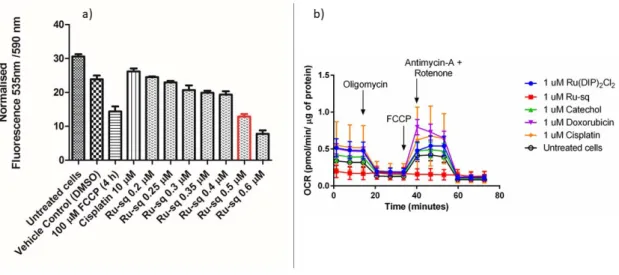
Figure 5. a) Fluorescence signal of JC-1 dye detected in HeLa cells treated for 24 h with different concentrations of Ru-sq (from 0.2 µM to 0.6 µM). Bar marked in red indicates the IC50 concentration (0.5 µM). FCCP is used as positive control, cisplatin
and DMSO (1%) are used as negative controls. b) Mito Stress Test profile in HeLa cells
after 24 h treatment; oxygen consumption rate changes after treatment with specific electron transport chain inhibitors. Oligomycin (inhibitor of ATP synthase (complex V)), FCCP (uncoupling agent), Antimycin-A (complex III inhibitor) and Rotenone (complex I inhibitor).
Inspired by these findings, further studies on the metabolic pathways that could be affected by the complex were performed. For this purpose, Seahorse XF Analyzer was used to measure, in real time, the oxygen consumption rate (OCR) and extracellular acidification rate (ECAR) of treated cells. Firstly, the effect of Ru-sq on the oxidative phosphorylation in the HeLa cell line was investigated. Mitochondrial respiration was
20 found to be severely impaired in cells treated with Ru-sq as opposed to the precursor Ru(DIP)2Cl2. This was evident from the low basal respiration and the inhibition of ATP
production compared to untreated cells. The mitochondrial membrane of the cells treated with Ru-sq, lost the capacity to restore the proton balance when treated with an uncoupling agent (FCCP). The maximal respiration (the OCR value when the mitochondrial membrane is uncoupled) and spare respiratory capacity (difference in the OCR values between maximal respiration and basal respiration) of the cells was reduced compared to untreated cells (Figure 5b and Figure S11). The combination of these effects suggests disrupted mitochondrial respiration in cervical cancer cells caused by Ru-sq. The effect on glycolysis and the possible metabolic modulation of the three primary fuel pathways (involving glucose, glutamine or fatty acids as substrates) were then examined. In contrast to that was observed for the mitochondrial respiration, the cell glycolysis, which is a cytosolic process, was not affected by Ru-sq (Figure S12). Additionally, due to the very low oxygen consumption rate in cells treated with Ru-sq, a direct effect on the 3-primary fuel pathways could not be determined (Figure S13). Overall, metabolic studies showed that the accumulation of Ru-sq in mitochondria has a significant role in the impairment of oxidative phosphorylation. This effect, together with the results obtained by the JC-1 staining, strongly suggests mitochondrial dysfunction as one of the modes of action of Ru-sq. In contrast, the chemotherapeutic drug cisplatin showed no significant effect on the mitochondrial metabolism of HeLa cells. This data suggests fundamental differences between the mode of action of Ru-sq and cisplatin. The latter covalently binds to the nuclear DNA and inhibits the replication process. It is widely known that DNA crosslinks can be repaired by different mechanisms such as the nucleotide excision repair (NER) that eventually leads to drug resistance in cancer cells. Ru-sq, with its multiple cellular
21 targets, could potentially evade these repair pathways and circumvent such drawbacks associated with cisplatin.
In vivo efficacy studies
It is very difficult to evaluate selectivity of the anticancer drugs in vitro, as the proliferation of non-malignant cells is greatly affected by non-physiological conditions of cell culture in 2D and 3D models. The promising results obtained in studies conducted in vitro justified the assessment of Ru-sq efficacy in the context of whole organism. To this end, we performed in vivo studies to evaluate the effect on both tumour growth and survival of tumour-bearing mice. The doses were selected according to the dose-finding study, which had revealed a maximum tolerated dose (MTD) of 15 mg/kg of body weight. Two distinct models for testing in vivo efficacy of antitumor drugs are possible: a syngeneic (mice) tumour growing in a naturally immunocompetent mouse, or human tumour cells growing in immunodeficient animals. As both approaches have its advantages and pitfalls, we decided to use both models in this study.
Effect of Ru-sq on the growth of Ehrlich mammary carcinoma in immunocompetent NMRI mice and survival of tumour bearing mice
Even though the use of syngeneic tumour allografts in naturally immunocompetent animals had been often considered inferior during the era of athymic mice models, this method made a comeback as the necessity of diversified, near-physiological experimental sets was recognised. In this model, we can observe the effect of the tested compound within the context of the genuine immune system that plays a key role in tumour resistance.93
22
Figure 6.Kaplan-Meier analysis of survival of immunocompetent NMRI mice bearing
Ehrlich carcinoma. Only the administration of complex Ru-sq 5 mg/kg i.p. and that of cisplatin (5 mg/kg) significantly prolonged the survival of tumour bearing mice when compared with the mixture of co-solvent and water. The compounds were administered i.p. on days 1 and 7 after tumour inoculation, n = 7 in each group.
During the study of the effect on the survival of immunocompetent NMRI mice bearing Ehrlich carcinoma (Figure 6), it was observed that the geometric mean of the overall survival of tumour bearing mice without therapy was 20.6 days. Among the three doses of Ru-sq tested, only 5 mg/kg prolonged the survival time significantly when compared with untreated tumour-bearing control mice (geom. mean = 31.9 days, P = 0.033). 10 mg and 15 mg/kg of Ru-sq seemed to exceed the optimal dose, causing a non-significant prolongation of survival (P > 0.05), with the geometric means of 30.2 and 25.5 days, respectively. The explanation may be a subclinical toxic effect that
23 negatively affects immune surveillance as well as other body functions necessary for the natural cancer defence. Although the optimal intracellular cancer-suppressing concentration may correspond to higher doses, subtle systemic toxicity does not allow to develop the desirable effect in vivo. The positive control cisplatin appeared to have similar efficacy (geom. mean = 33.7 days, P 0.014). An interesting and rare phenomenon was observed in all three groups of Ru-sq. Although the tumour was advanced in the later stage of the experiment, all mice treated with Ru-sq showed active behaviour, little cachexia and unsuppressed food consumption.
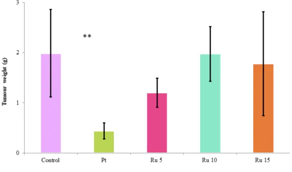
Figure 7.The weight of the solid Ehrlich tumour (in grams) on day 10 of mice injected on days 1 and 7 i.p. with pure vehicle, Ru-sq or cisplatin. Values are the means ± SEM (n = 7 in each group). Control – tumour-bearing control treated with mixture of co-solvent and water; Pt – cisplatin 5 mg/kg i.p.; Ru 5 – sq 5 mg/kg i.p.; Ru 10 – Ru-sq 10 mg/kg i.p.; Ru 15 – Ru-Ru-sq 15 mg/kg i.p. Significantly different from the controls (**P < 0.01).
24 Furthermore, the effect of Ru-sq on tumour growth was examined. Figure 7 shows the weight of tumours at day 10 in mice treated with mixture of co-solvent and water, Ru-sq at 5, 10 or 15 mg/kg, or cisplatin at 5 mg/kg, and documents differences in the effect of the used drugs. Although only cisplatin exhibited a significant inhibitory effect on tumour growth (P = 0.0011), there was a slight but insignificant suppression at 5 mg/kg Ru-sq (P = 0.108). As in the survival study, also here the optimum dose of Ru-sq seems to be in the lower part of the range tested.
Effect of Ru-sq on the growth of A2780 human ovarian cancer in immunodeficient nude mice and survival of tumour bearing mice
To compare the efficacy of the drug, therapeutic and survival experiment was repeated with athymic nude mice and human cancer line. A2780 human ovarian cancer cell line was chosen because of the use of cisplatin as comparative drug. Cisplatin is usually used for the therapy of ovarian cancer. Unfortunately, resistance often arises in treated patients. The use of human tumour xenografts in immunodeficient mice to examine therapeutic effect of potential chemotherapeutics, has several advantages. The major one is the use of actual human tumour tissue, featuring the complexity of genetic and epigenetic abnormalities that exist in the human tumour cell population.94,95 We
evaluated the growth of A2780 human ovarian cancer cells in immunodeficient nude mice and their survival. Figure 8 shows the survival of animals; the longest average day of death is surprisingly associated to the negative control (42.88 ± 16.97 days). However, there was one surviving mouse in the group treated with Ru-sq 10 mg/kg and in the group treated with cisplatin. Two surviving mice were found in the group treated with higher dose of Ru-sq (15 mg/kg). Very interestingly, one of them was completely cured with no observable tumour.
25 Figure 8. Kaplan-Meier analysis of survival of immunodeficient nude mice bearing A2780 human ovarian cancer. The treatment of Ru 15 mg/Kg led to a completely cancer free mouse. The compounds were administered i.p. on days 1 and 7 after the tumour reached 5 – 8 mm in size, n = 8 in each group.
Looking at the effect of Ru-sq on tumour growth (Figure 9), we observed that during the first days of therapy (day 4), there is a significant difference between groups treated with Ru-sq 15 mg/kg and cisplatin (P = 0.00675). Similar results between these two groups were observed at days 11 and 15 (P = 0,04246 for day 11 and P = 0,0262 for day 15). Comparison with untreated control group showed significant differences at days 11 and 15 (P = 0.024 for day 11; P = 0.00931 for day 15). Ru-sq administered in the dose of 15 mg/kg also showed decrease in tumour size over 15 days. Very interestingly, one mouse of this group was completely cured, no tumour volume was observed on the day 36 until the end of the experiment (day 60, data not shown). The longer survival of untreated mice observed on the nude model could be rationalised by
26 considering the higher sensitivity of immunocompromised animals to any kind of treatment. In addition, the intraperitoneal administration of the compound itself was found not ideal because of solubility reasons, which could have led to toxic peritonitis and eventually death.
Figure 9.Tumour growth of A2780 cancer line in nude mice in first 15 days of therapy. Tumour size is shown as volume in cm3. Control – tumour-bearing control treated with
mixture of co-solvent and water; Pt – cisplatin 5 mg/kg i.p.; Ru 5 – Ru-sq 5 mg/kg i.p.; Ru 10 – Ru-sq 10 mg/kg i.p.; Ru 15 – Ru-sq 15 mg/kg i.p. The most significant slowing down of tumour growth is observable in the group with Ru-sq 15 mg/kg.
These data demonstrate that the group treated with Ru-sq 15 mg/kg experienced a healing effect (in some points better than cisplatin), warranting further research. Ru-sq in a dose of 15 mg/kg has shown great potential to be an alternative and better drug candidate than cisplatin.
Taken together, we might conclude that in both models used, Ru-sq reduces the growth of tumour cells and prolongs tumour-bearing mice survival in immunocompetent
27 NMRI mice bearing Ehrlich carcinoma. Moreover, the optimal dose would be different depending on strain of the mice and tumour type.
Akt-1 protein levels in HeLa cells
The interesting results obtained during the in vivo studies led us to further investigate the influence of Ru-sq on cell proliferation and/or migration. Akt is a serine/threonine kinase that promotes cellular survival.96 Three isoforms of this protein exist in
mammalian cells: Akt-1, Akt-2 and Akt-3.97 Despite their high sequence similarity,
they exhibit unique functions.98 Akt-1 was found to be involved in the regulation of cell
proliferation, transformation and tumour metastasis.98 In this study, we assessed the
influence of different concentrations of Ru-sq on total Akt-1 protein levels in HeLa cells. As shown in Figures 12a and 12b, the treatment with the complex at concentrations lower than the IC50 does not change the total Akt-1 protein levels. A
similar effect is observed upon treatment with cisplatin and doxorubicin at their IC50
concentrations (IC50 =10 and 0.3 µM, respectively for cisplatin and doxorubicin). On
the other hand, Akt-1 protein levels are significantly decreasing when HeLa cells were treated with Ru-sq at IC50 concentration (0.5 µM) and higher. It is important to note
that GAPDH protein levels (loading control) are also changing at these concentrations, probably indicating ongoing cell death. Although Ru-sq complex does not change the total amount of the Akt-1 protein levels, we cannot exclude its impact on the amount of active form of this protein. It is known that Akt-1 needs to be phosphorylated for its activity99 and hyper activation of this protein is frequently found in human cancers.100
Further studies will be needed to fully understand the influence of our compound on cell proliferation and/or migration.
28 Figure 10. (a) Western blot analysis of Akt-1 protein levels in HeLa cell line after 24 h treatment with different concentrations of Ru-sq. Cisplatin, doxorubicin and untreated cells were used as controls. The positions of the nearest molecular weight markers are indicated. (b) Akt-1 protein levels normalised to GAPDH signal). Data is presented as the mean ± SD of at least 3 independent experiments. The IC50 concentration of the
29 Conclusions
Ru-sq was successfully synthesised and fully characterised. Crystal structure, electrochemical and EPR studies confirmed the oxidation state of the dioxo ligand (semiquinonate), which led to an overall positive charge of the complex. Ru-sq was found to be stable at room temperature in DMSO solution over one week and to have a half-life of 12 h upon incubation in human plasma at 37 °C. Cytotoxicity studies were performed in both, cellular monolayer (2D) and Multi Cellular Tumour Spheroids (MCTS) (3D) models. The cytotoxicity in the 2D model was tested against different cell lines showing higher activity than cisplatin with IC50 values mostly in the
nanomolar range. The cytotoxicity in HeLa MCTS confirmed the higher activity compared to cisplatin. Great tumour growth inhibition was observed after treatment with Ru-sq at 20 μM and 25 μM. Deeper investigation revealed apoptosis as the main cause of cell death. Ru-sq was found to be taken up by HeLa cells more efficiently than cisplatin and to accumulate preferentially in nucleus. DNA ruthenation studies suggest that Ru-sq might damage the DNA and/or prevent replication as well as transcription processes. Mitochondrial function upon Ru-sq treatment was also studied using an indicator of the mitochondrial membrane potential (JC-1) and mitostress test (Seahorse technology). From these studies, a severe impairement of the mitochondrial potential was observed suggesting mitochondrial disfunction contributes to the mode of action of Ru-sq. In vivo studies were performed using two different models: a syngeneic tumour growing in a naturally immunocompetent mouse, or human tumour cells growing in immunodeficient animals. Ru-sq reduces the growth of tumour cells and prolongs the survival of tumour-bearing mice. However, the optimal dose would be different depending on strain of the mouse and tumour type. In addition, during this study, especially in the case of the nude animals, the intraperitoneal administration was
30 found to be not ideal because of solubility reasons, which probably lead to some toxicity and eventually death.
Overall, Ru-sq displayed better activity than cisplatin in 2D and 3D cell cultures as well as for some conditions used in vivo. In conclusion, in this work, combining the well-known anticancer activity of Ru(II) polypyridyl complexes and the unique properties of the non-innocent ligand semiquinonate, we discovered a remarkable complex, Ru-sq, with promising potential as a chemotherapeutic agent against cancer. Further studies are currently ongoing in our group toward a different formulation of the compound prior administration. We hope that these studies might lead our compound to advance towards pre-clinical trials.
31
Experimental Section
Materials.
All chemicals were either of reagent or analytical grade and used as purchased from commercial sources without additional purification. Ruthenium trichloride hydrate was provided by I2CNS, 4,7-Diphenyl-1,10-phenanthroline, Lithium chloride (anhydrous,
99%), and catechol by Alfa Aesar, tetrabutylammonium hexafluorophosphate by Sigma-Aldrich. All solvents were purchased of analytical, or HPLC grade. When necessary, solvents were degassed by purging with dry, oxygen-free nitrogen for at least 30 min before use.
Instrumentation and methods.
Amber glass or clear glassware wrapped in tin foil were used when protection from the light was necessary. Schlenk glassware and a vacuum line were employed when reactions sensitive to moisture/oxygen had to be performed under nitrogen atmosphere. Thin layer chromatography (TLC) was performed using silica gel 60 F-254 (Merck) plates with detection of spots being achieved by exposure to UV light. Column chromatography was done using Silica gel 60-200 µm (VWR). Eluent mixtures are expressed as volume to volume (v/v) ratios. 1H and 13C NMR spectra were measured
on Bruker Avance III HD 400 MHz or Bruker Avance Neo 500 MHz spectrometers using the signal of the deuterated solvent as an internal standard.101 The chemical shifts
δ are reported in ppm (parts per million) relative to tetramethylsilane (TMS) or signals
from the residual protons of deuterated solvents. Coupling constants J are given in Hertz (Hz). The abbreviation for the peaks multiplicity is br (broad). ESI-HRMS experiments were carried out using a LTQ-Orbitrap XL from Thermo Scientific (Thermo Fisher Scientific, Courtaboeuf, France) and operated in positive ionization mode, with a spray voltage at 3.6 kV. Sheath and auxiliary gas were set at a flow rate
32 of 5 and 0 arbitrary units (a.u.), respectively. Applied voltages were 40 and 100 V for the ion transfer capillary and the tube lens, respectively. The ion transfer capillary was held at 275°C. Detection was achieved in the Orbitrap with a resolution set to 100,000 (at m/z 400) and a m/z range between 200-2000 in profile mode. Spectrum was analysed using the acquisition software XCalibur 2.1 (Thermo Fisher Scientific, Courtaboeuf, France). The automatic gain control (AGC) allowed accumulation of up to 2.105 ions for FTMS scans, Maximum injection time was set to 300 ms and 1 µscan was acquired. 5µL was injected using a Thermo Finnigan Surveyor HPLC system (Thermo Fisher Scientific, Courtaboeuf, France) with a continuous infusion of methanol at 100 µL.min-1. Purity of the compounds was determined by elemental
analysis performed at Science Centre, London Metropolitan University using Thermo Fisher (Carlo Erba) Flash 2000 Elemental Analyser, configured for %CHN confirming ≥95% purity. IR spectra were recorded with SpectrumTwo FTIR Spectrometer (Perkin– Elmer) equipped with a Specac Golden GateTM ATR (attenuated total reflection) accessory; applied as neat samples; 1/λ in cm–1. Analytical HPLC measurement was
performed using the following system: 2 x Agilent G1361 1260 Prep Pump system with Agilent G7115A 1260 DAD WR Detector equipped with an Agilent Pursuit XRs 5C18 (100Å, C18 5 μm 250 x 4.6 mm) Column and an Agilent G1364B 1260-FC fraction collector. The solvents (HPLC grade) were millipore water (0.1% TFA, solvent A) and acetonitrile (0.1% TFA, solvent B). The HPLC gradient used is the following: 0-3 minutes: isocratic 90% A (5% B); 3- 25 minutes: linear gradient from 90% A (5% B) to 0% A (100% B); 25-30 minutes: isocratic 0% A (100% B), 30-35 minutes: linear gradient from 0% A (100% B) to 95% A (5% B). The flow rate was 1 mL/min. Detection was performed at 215nm, 250nm, 350nm, 450nm, 550nm and 650nm with a slit of 4nm. Stability in human plasma was performed on HPLC (Acquity Ultra
33 Performance LC, Waters) that was connected to a mass spectrometer (Bruker Esquire 6000) operated in ESI mode. The ACQUITY UPLC BEH C18 Gravity 1.7 μm (2.1 × 50 mm) reverse phase column was used with a flow rate of 0.6 ml/min and UV-absorption was measured at 275 nm. The runs were performed with a linear gradient of A (acetonitrile (Sigma Aldrich HPLC-grade)) and B (distilled water containing 0.1% formic acid): t = 0−0.25 min, 95% A; t = 1.5 min, 100% A; t = 2.5 min, 100% A. Fractionation ICP-MS measurements were performed on an Agilent QQQ 8800 Triple quad ICP-MS spectrometer (Agilent Technologies) with a ASX200 autosampler (Agilent Technologies), equipped with standard nickel cones and a “micro-mist” quartz nebulizer fed with 0.3 ml/min analytic flow (as a 2% HNO3 aqueous solution). Celular
Uptake, mechanism of uptake and ruthenation of the DNA was performed using a High-Resolution ICP-MS Element II from ThermoScientific located within the Environmental Biogeochemistry team of the Institut de Physique du Globe de Paris. This ICP-MS enables working in different resolution modes (LR=400, MR=4000 and HR=10000) for a better discrimination between elements of interest and interferences.102
For the metabolic studies Seahorse XFe96 Analyser by Agilent Technologies was used. Synthesis and characterization.
Ru(DMSO)2Cl2. Ru(DMSO)2Cl2 was synthesised following an adapted literature
procedure.64 Spectroscopic data (1H NMR) was in agreement with literature.64
Ru(DIP)2Cl2. The complex was synthesised following an adapted literature
procedure.65 A mixture of Ru(DMSO)2Cl2 (3.0 g, 6.19 mmol),
4,7-diphenyl-1,10-phenanthroline (4.11 g, 12.38 mmol) and LiCl (2.0 g, 47.18 mmol) dissolved in DMF (100 mL) was refluxed for 24 h. After cooling to r.t., the solvent was reduced in vacuo to 8 mL and 350 mL of acetone were added. The mixture was then stored at -20 °C
34 overnight before filtration with a Buchner funnel and washed with Acetone and Et2O
to afford Ru(DIP)2Cl2 as a deep purple solid (3.76 g, 4.49 mmol, 72%). Spectroscopic
data (1H NMR) were in agreement with literature.65
[Ru(DIP)2(sq)](PF6) (Ru-sq).
Ru(DIP)2Cl2 (0.739 g, 0.88 mmol) and aq. NaOH (0.5 mL, 1 M) were dissolved in
2-propanol (40 mL). The solution was degassed for 15 min and catechol (0.155 g, 1.41 mmol) was added. The mixture was heated to reflux for 24 h under N2 atmosphere
and protected from light. After cooling to r.t., the mixture was stirred opened to air while still protected from light and the solvent was removed under vacuum. The residual solid was dissolved in 2-propanol (7 mL) and H2O (56 mL) and NH4PF6
(0.700 g, 4.3 mmol) were added. The mixture was stored in the fridge (4 °C) overnight. The precipitate was filtered with a Buchner funnel and washed with H2O (3 x 50 mL)
and Et2O (3 x 50 mL). The solid was collected with DCM and dried under vacuum to
deliver a crude product as the PF6 salt (0.70 g), which was chromatographed on silica
(DCM/MeCN 20:1 Rf: 0.3). Evaporation of the solvent under vacuum provided [Ru(DIP)2(sq)](PF6) as a deep red solid. Further wash with Et2O and Heptane were
necessary in order to obtain clean product. The solid with the washing solvent (10 mL) was sonicated for 10 min and then centrifuged. This procedure was repeated three times for each solvent. Finally the red solid was collected with DCM and dried under vacuum to afford a clean product (0.17 g, 0.167 mmol, 19%). IR (Golden Gate, cm-1): 3345w,
1710m, 1600w, 1520s, 1455s, 1335s, 1270s, 1125s, 820s, 760m. 1H NMR (400 MHz,
CD2Cl2): /ppm = 8.79–8.20 (br, 5H, arom.), 8.09–7.88 (br, 5H, arom.), 7.73–7.42 (br,
14H, arom.), 7.26–6.92 (br, 10H, arom.), 6.92 – 6.63 (br, 2H, arom.). 13C NMR (125
MHz, CD2Cl2): /ppm = 149.84, 144.68, 136.10, 133.56, 130.36, 129.89, 129.53,
35 observed in the 13C NMR spectrum where five were expected. This could be explained
by peak overlap or the signal being too weak to be detected within the acquisition time of the experiment which is common for quaternary carbons. HRMS (ESI+): m/z 874.1887 [M - PF6]+. Elemental Analysis: calcd. for C54H36F6N4O2PRu= C, 63.65; H,
3.56; N, 5.50. Found = C, 63.62; H, 3.52; N, 5.45. HPLC: TR = 31.304 min.
X-ray Crystallography.
Single-crystal X-ray diffraction data were collected at 183(1) K on a Rigaku OD XtaLAB Synergy, Dualflex, Pilatus 200K diffractometer using a single wavelength X-ray source (Mo Kα radiation: λ = 0.71073 Å)103 from a micro-focus sealed X-ray tube
and an Oxford liquid-nitrogen Cryostream cooler. The selected suitable single crystal was mounted using polybutene oil on a flexible loop fixed on a goniometer head and transferred to the diffractometer. Pre-experiment, data collection, data reduction and analytical absorption correction104 were performed with the program suite
CrysAlisPro.105 Using Olex2,106 the structure was solved with the SHELXT107 small
molecule structure solution program and refined with the SHELXL program package108
(version 2018/3) by full-matrix least-squares minimization on F2. PLATON109 was used
to check the result of the X-ray analysis.
CCDC 1950873 contains the supplementary crystallographic data for this compound, and can be obtained free of charge from the Cambridge Crystallographic Data Centre via www.ccdc.cam.ac.uk/data_request/cif.
Electrochemical Measurements.
The electrochemical experiments were carried out with a conventional three-electrodes cell (solution volume of 15 mL) and a PC-controlled potentiostat/galvanostat (Princeton Applied Research Inc. model 263A). The working electrode was a vitreous carbon electrode from Origalys (France) exposing a geometrical area of 0.071 cm2 and
36 mounted in Teflon®. The electrode was polished before each experiment with 3 and 0.3
m alumina pastes followed by extensive rinsing with ultra-pure Milli-Q water. Platinum wire was used as counter electrode and saturated calomel electrode, SCE, as reference electrode. Electrolytic solutions, DMF containing tetrabutylammonium hexafluorofosfate 0.1M (TBAPF6, Aldrich, +99 %) as supporting electrolyte, were
routinely deoxygenated by argon bubbling. All the potential values are given versus the calomel saturated electrode SCE and recalculated versus Me10Fc0/+ potential value.
EPR.
Electron paramagnetic resonance (EPR) experiments were performed on a MiniScope MS400 table-top X-band spectrometer from Magnettech. Simulation of the experimental EPR spectra was performed with the MATLAB EasySpin program.110 All
samples were dissolved in dry and N2-saturated DCM at a concentration of ca. 1 mM.
Oxidized forms were generated using 1,1’-diacetylferrocenium hexafluoroantimonate (Ac2FcSbF6, E1/2 = 0.940 V vs SCE in DMF/0.1 M NBu4PF6).70,71 Chemical reduction
was achieved by using cobaltocene (Cp2Co, E1/2 = -0.880 V vs SCE in DMF/0.1 M
NBu4PF6). 70
Stability studies.
The stability in DMSO-d6 at room temperature was assessed by 1H NMR over 8 days.
The stability of Ru-sq in human plasma at 37 °C was evaluated following a slightly modified procedure already reported by our group.43 The human plasma was provided
by the Blutspendezentrum, Zurich, Switzerland. Diazepam (internal standard) was obtained from SigmaAldrich. Stock solutions of the complexes (20 mM) and diazepam (3.2 mM) were prepared in DMSO. For a typical experiment, an aliquot of the respective stock solutions and DMSO were then added to the plasma solution (975 μL)
37 to a total volume of 1000 μL and final concentrations of 40 μM for the complexes and diazepam. The resulting plasma solution was incubated for either: 0, 4, 6, 12, 20, 24 or 48 h at 37°C with continuous and gentle shaking (ca. 600 rpm). The reaction was stopped by addition of 2 mL of methanol, and the mixture was centrifuged for 45 min at 650g at room temperature. The methanolic solution was evaporated and the residue was suspended in 500 μL of 1:1 (v/v) acetonitrile/H2O solution. The suspension was
filtered and analyzed using UPLC−MS with a total injection volume of 2 μL.
Cell culture.
HeLa and CT-26 cell lines were cultured in DMEM media (Gibco). CT-26 LUC cell line was cultured in DMEM media (Gibco) supplemented with 1.6 mg/mL of Genticin. RPE-1 cell line was cultured in DMEM/F-12 media (Gibco). MRC-5 cell line was cultured in DMEM/F-10 media (Gibco). A2780, A2780 cis, A2780 ADR cell lines were cultured in RPMI 1640 media (Gibco). The resistance of A2780 cis was maintained by cisplatin treatment (1µM) for one week every month. The cells were used in the assays after one week from the end of the treatment in order to avoid interfered results. The resistance of A2780 ADR was maintained by doxorubicin treatment (0.1 µM) once a week. Cells were used in the assays after three days post doxorubicin treatment in order to avoid interfered results. All cell lines were complemented with 10% of fetal calf serum (Gibco) and 100 U/mL penicillin-streptomycin mixture (Gibco) and maintained in humidified atmosphere at 37°C and 5% of CO2.
Cytotoxicity Assay using a 2D cellular model.
Cytotoxicity of the tested Ru-sq and Ru(DIP)2Cl2 complexes was assessed by a
38 were seeded in triplicates in 96-well plates at a density of 4 × 10³ cells/well in 100 μL.
After 24 h, cells were treated with increasing concentrations of the ruthenium complexes. Dilutions for Ru-sq were prepared as follows: 2.0 mM stock in DMSO was diluted to 25 µM with media and then filtrated (0.22 µm filter VWR). For Ru(DIP)2Cl2
2.5 mM stock in DMF was prepared, which was further diluted to 100 µM and filtrated (0.22 µm filter VWR). After 48 h incubation, medium was removed, and 100 μL of complete medium containing resazurin (0.2 mg/mL final concentration) was added. After 4 h of incubation at 37 °C, the fluorescence signal of resorufin product was read (ex: 540 nm em: 590 nm) in a SpectraMax M5 microplate Reader. IC50 values were
then calculated using GraphPad Prism software.
Generation of 3D HeLa MCTS.
MCTS were cultured using ultra-low attachment 96 wells plates from Corning® (Fisher
Scientific 15329740). HeLa cells were seeded at a density of 5000 cells per well in 200 µL. The single cells would generate MCTS approximately 400 µm in diameter at day 4 with 37 °C and 5 % CO2.
Treatment of 3D HeLa MCTS.
HeLa MCTS after 4 days of growing at 37 °C and 5 % CO2 were treated by replacing
half of the medium in the well with increasing concentration of compounds for 48 h in the dark. For untreated reference MCTS, half of the medium was replaced by fresh medium only. The cytotoxicity was measured by ATP concentration with CellTiter-Glo® Cell viability kit (Promega, USA).
39 Cell viability for MCTS was performed via ATP assay using luciferase. CellTiter-Glo® kit from Promega was used. The spheroids were incubated for 1 h after replacing half of the media with CellTiter-Glo reagent and the luminescence of the plate was read by SpectraMax M5 microplate reader. IC50 values were calculated using GraphPad Prism
software.
HeLa MCTS growth inhibition.
MCTS were grown and treated as previously described (see above). MCTS sizes were observed under a light microscope and pictures were taken with a Samsung Galaxy A5 2017 SM-A520FZKAXEF thanks to a phone microscope adaptor. Before imaging, the plate was sheken and half of the media was exchanged to remove dead cells. Images were recorded before treatment (day 0) and at day 3, 6, 9 and 13 after treatment. Pictures were first processed using GIMP a cross-platform image editor with a batch automation plug-in. The MCTS sizes were then calculated with SpheroidSizer, a MATLAB-based and open-source software application to measure the size of tumour spheroids automatically and accurately. Data analysis was done using GraphPad Prism software.
Annexin V / PI assay
Apoptosis and necrosis induction in HeLa cells treated with Ru-sq was evaluated via an AnnexinV/PI staining assay using flow cytometry. Briefly, cells were seeded at density of 2×10⁶ cells in 10 cm cell culture dish 24 h prior cell treatments. The medium
was removed and replaced with 10 μM solution of Ru-sq or 1 µm Staurosporin (positive control -Abcam Cat no.120056) and further incubated for 30 min, 4 h or 24 h. Cells were collected, washed twice with ice cold PBS and resuspended in 1x Annexin V binding buffer (10 x buffer composition: 0,1 M HEPES (pH 7.4), 1.4 M NaCl. 25
40 mM CaCl2). Samples were processed according to the manufacturer instructions (BD
Scientific, cat no 556463 and 556419) and analysed using ZE5 Biorad instrument at Cytometry Platform at Institute Curie. Data were analysed using the FlowJo software. Sample Preparation for cellular uptake
Cells were seeded at density of 2×10⁶. Next day, cells were treated with 5 µM
concentration of Ru-sq or cisplatin. After 2 h, cells were collected, counted and snap frozen in liquid nitrogen and stored at -20 ºC. ICP-MS samples were prepared as follows: samples were digested using 70% nitric acid (1 mL, 60 ºC, overnight). Samples were then further diluted 1:100 (1% HCl solution in MQ water) and analysed using ICP-MS.
Sample Preparation for cellular fractionation
HeLa cells were seeded in three 15 cm2 cell culture dishes so that on the day of
treatment cells were 90% confluent. On the day of treatment cells were incubated with the target complex at a concentration of 5 μM for 2 h. After that time, the medium was removed; cells were washed, collected and counted. After resuspension in cold PBS, the organelles were isolated via different protocols (one cell culture dish per isolation was used).
Mitochondria isolation: To isolate mitochondria, a Mitochondria Isolation Kit (Cat. Nr:
MITOISO2, Sigma Aldrich) was used according to the manufacturer procedure for isolation of mitochondria via homogenization method.
Lysosome isolation: To isolate lysosomes, a Lysosome Isolation Kit (Cat. Nr:
LYSISO1, Sigma Aldrich) was used, according to the manufacturer procedure for isolation of lysosomes via Option C.
41
Nuclear and cytoplasm isolation: To isolate nuclear and cytoplasmic fractions, the
ROCKLAND nuclear extract protocol was used.111 Briefly cells were collected by
centrifugation, resuspended in cytoplasmic extraction buffer and incubated on ice. The tubes were centrifuged and supernatant (CE) was removed. Pellets were washed with cytoplasmic extraction buffer without detergent and centrifuged. The pellet (NE) was resuspended in nuclear extraction buffer and incubated on ice. Both CE and NE were centrifuged. Supernatant from CE samples was indicated as cytoplasmic extract, whereas the pellet obtained from NE samples was indicated as nuclear extract.
ICP-MS samples were prepared as follows: isolated cellular fractions were lyophilised and digested using 5 mL of 70% nitric acid (60 ºC, overnight). Samples were then further diluted (1:1000 for nuclear pellet samples and 1:100 for all the other samples) MQ water (containing in 1% HCl solution) and analysed using ICP-MS.
Sample preparation for studies on the mechanism of cellular uptake
HeLa cells were seeded at density of 2×10⁶ and next day were pre-treated with
corresponding inhibitors or kept at specific temperature for 1 h. Next, cells were washed with PBS and were incubated with 5 µM of Ru-sq for 2 h (low temperature sample was still kept at 4 ºC). Afterwards cells were washed with PBS, collected, counted and snap frozen in liquid nitrogen. Pellets were stored at -20 ºC. ICP-MS samples were prepared as follows: samples were digested using 70% nitric acid (1 mL, 60 ºC, overnight), further diluted 1:100 (1% HCl solution in MQ water) and analysed using ICP-MS.
DNA metalation of HeLa cells
Cells were seeded at density of 2 x 10⁶. The following day, cells were treated with 5