HAL Id: inserm-00906809
https://www.hal.inserm.fr/inserm-00906809
Submitted on 20 Nov 2013
HAL is a multi-disciplinary open access
archive for the deposit and dissemination of
sci-entific research documents, whether they are
pub-lished or not. The documents may come from
teaching and research institutions in France or
abroad, or from public or private research centers.
L’archive ouverte pluridisciplinaire HAL, est
destinée au dépôt et à la diffusion de documents
scientifiques de niveau recherche, publiés ou non,
émanant des établissements d’enseignement et de
recherche français ou étrangers, des laboratoires
publics ou privés.
the alpha1 or the alpha2 isoform of the catalytic domain
of AMPK.
Wilfred Lieberthal, Meiyi Tang, Leiqing Zhang, Benoit Viollet, Vimal Patel,
Jerrold Levine
To cite this version:
Wilfred Lieberthal, Meiyi Tang, Leiqing Zhang, Benoit Viollet, Vimal Patel, et al.. Susceptibility to
ATP depletion of primary proximal tubular cell cultures derived from mice lacking either the alpha1
or the alpha2 isoform of the catalytic domain of AMPK.. BMC Nephrology, BioMed Central, 2013,
14 (1), pp.251. �10.1186/1471-2369-14-251�. �inserm-00906809�
R E S E A R C H A R T I C L E
Open Access
Susceptibility to ATP depletion of primary
proximal tubular cell cultures derived from mice
lacking either the α1 or the α2 isoform of the
catalytic domain of AMPK
Wilfred Lieberthal
1,2*, Meiyi Tang
1,2, Leiqing Zhang
1,2, Benoit Viollet
3,4,5, Vimal Patel
6,7and Jerrold S Levine
6,7Abstract
Background: The purpose of this study was to determine whether AMPK influences the survival of primary cultures of mouse proximal tubular (MPT) cells subjected to metabolic stress. Previous studies, using an immortalized MPT cell line, suggest that AMPK is activated during metabolic stress, and ameliorates stress-induced apoptosis of these cells.
Methods: Primary MPT cells were cultured from AMPK knockout (KO) mice lacking either the α1 or the α2 isoform of the catalytic domain of AMPK. MPT cells were subjected to ATP depletion using antimycin A.
Results: Surprisingly, there was no difference in the amount of death induced by metabolic stress of MPT cells from either type of AMPK KO mice compared to its WT control. Moreover, inhibition of the activity of the α1 isoform in primary MPT cells from α2−/−mice (pharmacologically, via compound C) or inhibition of the α2 isoform
in primary MPT cells from α1−/−mice (molecularly, via knockdown) both decreased cell viability equivalently in
response to metabolic stress. The explanation for this unexpected result appears to be an adaptive increase in expression of the non-deleted α-isoform. As a consequence, total α-domain expression (i.e. α1 + α2), is comparable in kidney cortex and in cultured MPT cells derived from either type of KO mouse versus its WT control. Importantly, each α-isoform appears able to compensate fully for the absence of the other, with respect to both the
phosphorylation of downstream targets of AMPK and the amelioration of stress-induced cell death.
Conclusions: These findings not only confirm the importance of AMPK as a pro-survival kinase in MPT cells during metabolic stress, but also show, for the first time, that each of the two α-isoforms can substitute for the other in MPT cells from AMPK KO mice with regard to amelioration of stress-induced loss of cell viability.
Keywords: AMPK, Viability, Survival, Apoptosis knockout mice, shRNA, ATP depletion, Metabolic stress, Antimycin
Background
AMP-activated protein kinase (AMPK) is a ubiquitously expressed and highly conserved serine/threonine kinase that is activated by any form of stress that reduces cell energy stores [1,2]. AMPK is a heterotrimeric protein composed of a catalytic α subunit and two regulatory (β and γ) subunits [1]. There are two isoforms of the
catalytic (α1 and α2) and β (β1 and β2) subunits, and three isoforms of the γ (γ1, γ2, and γ3) subunit, all of which are encoded by different genes [1]. AMPK is acti-vated by phosphorylation of a threonine residue (Thr172) situated within the activation loop of the α-subunit [3-5]. The best-defined upstream AMPK kinase is liver kinase B1 (LKB1). LKB1 is constitutively active, so that AMPK is continuously being phosphorylated [3-5]. When cell energy stores are replete, AMPK is main-tained in an inactive state by the activity of phosphatases such as phosphatase-2C alpha [1,3,4]. Activation of AMPK occurs whenever cell stress leads to a decrease in
* Correspondence:wilfred.lieberthal@stonybrookmedicine.edu
1Department of Medicine, Stony Brook University Medical Center, Stony
Brook, NY 11794, USA
2Northport Veterans Affairs Medical Center, Northport, NY 11768, USA
Full list of author information is available at the end of the article
© 2013 Lieberthal et al.; licensee BioMed Central Ltd. This is an open access article distributed under the terms of the Creative Commons Attribution License (http://creativecommons.org/licenses/by/2.0), which permits unrestricted use, distribution, and reproduction in any medium, provided the original work is properly cited.
cytosolic ATP levels and a concomitant increase in levels of adenosine diphosphate (ADP) and adenosine mono-phosphate (AMP) [3,4]. The regulatory γ-subunit of AMPK has binding sites for all three nucleotides [6,7]. Binding of ADP or AMP to the γ-subunit activates AMPK via two mechanisms, inhibition of the dephos-phorylation of Thr172and allosteric facilitation of phos-phorylation of AMPK by LKB1 [3,4]. In contrast to the effects of ADP and AMP, binding of ATP to the γ sub-unit of AMPK promotes its deactivation [1,3,4]. The net result of these opposing interactions is that AMPK activ-ity is inversely related to the ratio of the concentration of ATP to that of ADP and AMP [3,4,7].
Upon activation, AMPK phosphorylates and alters the activity of multiple downstream kinases and enzymes [8,9]. AMPK also induces changes in gene expression [10]. Together, these effects of AMPK lead to alterations in glucose, protein, and lipid metabolism that serve to conserve energy stores by promoting ATP production, inhibiting ATP consumption, and facilitating the cellular uptake of nutrients [1,2,8,9,11,12]. For example, through phosphorylation and inhibition of acetyl-CoA carboxylase 1 (ACC1) and ACC2, AMPK inhibits lipid synthesis and promotes fatty acid oxidation [1,13-15]. By inhibiting the activity of the mammalian target of rapamycin (mTOR), AMPK also inhibits protein synthesis [16,17]. Other AMPK-mediated effects include increased cellular uptake of glucose and fatty acids, and increased glycolysis [9].
Recently, using an immortalized mouse proximal tubu-lar (MPT) cell line, we have shown that AMPK is acti-vated by ATP depletion, and that, once actiacti-vated, AMPK ameliorates apoptosis induced by this form of stress [18]. Here we extend these findings to primary cultures of MPT cells, derived from the kidneys of AMPK knock-out (KO) mice with whole body deletions of either the α1 (α1−/−) or the α2 (α2−/−) isoforms of AMPK. We
hy-pothesized that primary MPT cells, lacking one or other of the α-isoforms of AMPK, should be more susceptible to apoptosis induced by ATP depletion than primary MPT cells with both α-isoforms intact. Surprisingly, we found no difference in the severity of antimycin-induced cell death in MPT cells from α1−/−or α2−/−mice versus
their WT controls. Moreover, we found that a reduction of AMPK activity (via either pharmacologic inhibition or knockdown of one of the α-isoforms) led to comparable increases of antimycin-induced apoptosis in primary MPT cells from KO versus WT mice.
In explaining this absence of a difference in stress-induced apoptosis for MPT cells from KO versus WT mice, we show that there is a compensatory up-regulation in expression of the intact α-isoform in both AMPK KO mice. This up-regulation compensates fully for the deleted α-isoform, such that the total amount of α-domain protein is comparable in primary MPT cells from the two KO mice
versus their WT controls. These findings not only confirm the importance of AMPK activation as a pro-survival kin-ase during ATP depletion, but also indicate that each of the two α-isoforms can substitute for the other in amelior-ating the consequences of metabolic stress.
Methods
Materials
All chemicals were purchased from Sigma (MO) unless otherwise stated. Soybean trypsin inhibitor was purchased from GIBCO (NY). Rabbit polyclonal antibody to total AMPK, rabbit polyclonal antibody to total alpha subunit of AMPK, and mouse monoclonal antibody to the phos-phorylated alpha subunit of AMPK were purchased from Cell Signaling Technologies (MA). Rabbit polyclonal anti-body to phosphorylated ACC1/2 was purchased from Up-state (NY). Rabbit polyclonal antibodies specific for the α1 and α2 isoforms of the catalytic domain of AMPK were purchased from Abcam (MA). Monoclonal antibody to β-actin was purchased from Chemicon (CA).
Mice
The generation and phenotype of whole body α1−/−and
α2−/− KO mice have been described previously [19,20].
α1−/−mice and their WT controls were bred on a mixed
background (C57BL6J/129Sv), whereas α2−/− KO mice
and their WT controls were bred on a C57BL6J back-ground. Heterozygous α1−/−
and –α2−/− KO mice, used
only for breeding, were obtained from the European Mouse Mutant Archive and bred in the Department for Laboratory and Animal Research at Stony Brook Hos-pital. Only homozygous KO mice, male and female, were used for experimentation. All studies were approved by the IACUC at Stony Brook Hospital. Total body weights and kidney weights were comparable amongst KO and WT mice (data not shown).
Primary culture of mouse proximal tubular (MPT) cells
Techniques for the primary culture of MPT cells are well established in our laboratory [21,22]. Briefly, kidneys were harvested from mice at 4–6 weeks of age. Cortical tissue was finely minced and incubated in Hanks’s solution con-taining collagenase and soybean trypsin inhibitor. After collagenase digestion, 10% horse serum was added, and the tissue fragments were gently centrifuged. After two washes with DMEM, tissue fragments were suspended in growth medium, a serum-free, defined medium consisting of DMEM/Ham’s F-12 media (1:1) containing 2 mM glu-tamine, 15 mM HEPES (pH 7.2), 5 μg/ml transferrin, 5 μg/ml insulin, 50 mM hydrocortisone, 500 U/ml penicil-lin, and 50 mg/ml streptomycin. The tissue fragments, suspended in growth medium, were then plated on plastic tissue cultures plates and incubated in a humidified air/CO2incubator (5% v/v) at 37°C.
Fragments of tubules adhere to the culture dish, and MPT cells grow out of the tubules to form confluent monolayers over a 4–5 day period. MPT cells express megalin and other markers specific for proximal tubular cells. Five days after plating, the medium was changed to experimental medium, which consists of DMEM/Hams F12 containing HEPES (pH 7.2), 15 mM glutamine, 500 U/ml penicillin, and 50 mg/ml streptomycin. No insulin, hydrocortisone, or transferrin was added to this medium. Experiments were begun on the sixth day after plating.
Metabolic stress (graded ATP depletion)
MPT cells were exposed to metabolic stress using anti-mycin A, a mitochondrial inhibitor. We examined the effect of antimycin A in the presence of three concentra-tions of dextrose, 10, 5, and 2.5 mM. The severity of metabolic stress increases progressively, (i.e. cell ATP levels fall progressively), as the dextrose concentration in the medium is reduced from 10 to 2.5 mM. We refer to this model of metabolic stress as “graded” ATP deple-tion. We have used graded ATP depletion as a model of metabolic stress in previous publications [22]. Control (unstressed) MPT cells were incubated in 5 mM dex-trose in the absence of antimycin.
Cell ATP levels
ATP levels were determined using previously described methods [22]. Briefly, cell ATP content was measured by luciferase assay in cell lysates and normalized to total
cellular protein, as assessed by bicinchoninic acid (BCA) protein assay (Pierce, Rockford, NY). Control ATP levels, obtained in cells incubated in dextrose-containing medium (5 mM) in the absence of antimycin, were expressed as nanomoles of ATP per milligram of cell protein (nM ATP/mg protein). ATP levels obtained during metabolic stress were expressed as a percentage of ATP levels under control conditions.
Immunoblotting
Immunoblotting was performed as previously described [23]. Briefly, cells were washed with ice-cold PBS, then lysed in ice-cold cell lysis buffer (20 mM Tris–HCl, pH 7.5, containing 130 mM NaCl, 1% v/v Triton X-100, 0.5% w/v deoxycholate, 0.1% w/v SDS, 1 mM DTT, 10 mM sodium pyrophosphate, 5 mM sodium fluoride, 1 mM phenylmethylsulfonyl fluoride, and 200 μM so-dium orthovanadate). Lysates were centrifuged at 10,000 × g for 10 min at 4°C, and the supernatants were stored at −70°C. Protein samples, 20 μg per lane, as determined by BCA protein assay, were boiled in 6× reducing sam-ple buffer, electrophoresed on SDS-polyacrylamide gels, and transferred to nitrocellulose membranes (BIO-RAD, Hercules, CA). Membranes were blocked with either 2.5% bovine serum albumin or 5% dry milk in TBS, before probing with primary antibody. After incubation with the appropriate secondary antibody, immunoreactive bands were visualized by the Western Lightning Chemilumines-cence Reagent Plus (PerkinElmer, Boston, MA).
0 20 40 60 80 100 10 5 2.5 0 20 40 60 80 100 10 5 2.5 % viable cells Antimycin+ Dextrose (mM)
A
B
Antimycin + Dextrose (10 mM) Antimycin + Dextrose (5 mM) Antimycin + Dextrose (2.5 mM) WT 1-/ -KO*
*
# # WT 1-/ -KO WT 2-/ -KO α α Calcein E th-H1Figure 1Effect of metabolic stress on the viability of MPT cells from α1−/−and α2−/−KO mice. A: MPT cells from α1−/−mice (upper panel) or α2−/−KO mice (lower panel), or their WT controls, were subjected to graded metabolic stress by incubation in the presence of antimycin (2 μM) and varying concentrations of dextrose. After 16–18 hrs, the percentage of viable cells remaining was determined by flow cytometry (n = 5 for both experiments). * p < 0.02, 5 mM dextrose vs. 10 mM dextrose; # p < 0.02, 2.5 mM dextrose vs. 5 mM dextrose. B: Representative flow cytometric analysis of MPT cells from α1−/−mice (right panels) and their WT control (left panels) incubated in antimycin (2 μM) plus varying concentrations of dextrose. Cell viability was quantified by staining cells with calcein AM (x-axis) and ethidium homodimer-1 (Eth-H1) (y-axis). For each condition, 10,000 cells were analyzed, and the percentage of viable cells was calculated by dividing the number of calcein-positive and Eth-H1-negative cells by the total number of cells counted.
Figure 2Effect of metabolic stress on cytosolic ATP levels in MPT cells from α1−/−and α2−/−KO mice. MPT cells from α1−/−mice or α2−/−KO mice, or their WT controls, were subjected to graded ATP depletion by incubation in the presence of antimycin (2 μM) and varying concentrations of dextrose for 4 hrs. Cell ATP levels were measured and normalized to cell protein content. All values are expressed as a percent of ATP levels in MPT cells incubated under control conditions (10 mM dextrose, in the absence of antimycin). * p < 0.02, antimycin plus 10 mM dextrose vs. control conditions; # p < 0.01, antimycin plus 5 mM dextrose vs. antimycin plus 10 mM dextrose; p < 0.01, antimycin plus 2.5 mM dextrose vs. antimycin plus 5 mM dextrose.
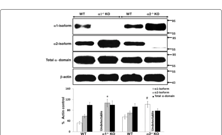
Figure 3Relative expression of the catalytic alpha (α) domain of AMPK in whole kidney cortex obtained from α1−/−and α2−/− knockout (KO). The relative expression of the α1 and α2 isoforms of AMPK, and of the total α(α1 + α2) domain, was evaluated in immunoblots of lysates of “snap frozen” samples of kidney cortex, obtained from α1−/−or α2−/−KO mice and their WT controls. Upper panel: Representative immunoblot. Molecular weight markers (in kDa) are shown on the right of each immunoblot. Lower panel: Densitometric quantitation of three immunoblots using β-actin as a loading control. * p < 0.01, α1−/−vs. WT mice, for α2 isoform expression; # p < 0.02, α2−/−vs. WT mice, for α1 isoform expression.
Cell viability
Cell viability was determined using the LIVE/DEAD Assay Kit purchased from Molecular Probes™ and used according to the manufacturer’s instructions. In brief, MPT cells were stained with ethidium homodimer-1 (EthD-1) and calcein AM. Live cells are identified by their ability to convert calcein AM, a non-fluorescent cell-permeant agent to calcein, an intensely fluorescent dye (excitation/emission wavelengths, ~495 nm/~515 nm) that is retained within live cells. Dead cells are identified by nuclear staining for EthD-1, which only enters cells with damaged plasma membranes and, upon binding to nucleic acids, undergoes a 40-fold enhancement of fluorescence (excitation/emission wavelengths, ~495 nm/ ~635 nm), thereby producing a bright fluorescence in dead cells. Since both dyes are essentially non-fluorescent before interacting with cells, background fluorescence is inherently low. Live and dead cells were quantitated using flow cytometry (FACScan, BD Biosciences), and data were analyzed using CELLQuestPro Version 3.3 (BD Biosciences). Cells were first analyzed by forward versus side scatter, and gated to remove debris, cell fragments, and cell aggregates. The proportion of live cells in each
sample was expressed as a percent of the total number of cells analyzed (10,000/sample).
Statistics
All data are presented as mean ± standard error (SE). Stu-dent’s t-test was used for comparing cell ATP levels and densitometry of immunoblots. The Bonferroni correction was applied when multiple comparisons were made. The viability of MPT cells cultured from KO versus WT mice and subjected to metabolic stress was compared by ANOVA for repeated measures using STATA® Data Ana-lysis and Statistical Software. All p values <0.05 were con-sidered statistically significant.
Results
Effect of metabolic stress on the viability of MPT cells from α1−/−and α2−/−versus WT mice
We determined the effect of graded ATP depletion, in-duced by exposing MPT cells to antimycin A and varying concentrations of dextrose, on cell viability, as assessed by flow cytometry. Cell viability was comparable in MPT cells from KO versus WT mice under unstressed control con-ditions (10 mM dextrose, no antimycin) (data not shown).
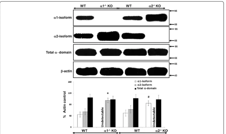
Figure 4Relative expression of the catalytic alpha (α) domain of AMPK in proximal tubular cells derived from α1−/−and α2−/− knockout (KO) mice. The relative expression of the α1 and α2 isoforms of AMPK, as well as of the total α domain (α1 + α2), was evaluated in immunoblots of lysates from confluent mouse proximal tubular (MPT) cells derived from the kidneys of either α1−/−or α2−/−KO mice, or their WT controls. Upper panel: Representative immunoblot. Molecular weight markers (in kDa) are shown on the right of each immunoblot. Lower panel: Densitometric quantitation of three immunoblots using β-actin as a loading control. * p < 0.02, α1−/−vs. WT mice, for α2 isoform expression; # p < 0.02, α2−/−vs. WT mice, for α1 isoform expression.
In the presence of antimycin, the percentage of viable MPT cells from AMPK KO and WT mice decreased pro-gressively as the concentration of dextrose was decreased (Figure 1). Nevertheless, at each dextrose concentration, the survival of MPT cells from α1−/− or α2−/− KO mice
was no different than that of MPT cells from WT controls (Figure 1).
Effect of metabolic stress on cytosolic ATP levels in MPT cells from α1−/−and α2−/−mice
Cytosolic ATP levels in control unstressed MPT cells in-cubated in 10 mM dextrose without antimycin for 4 hrs were no different for KO versus WT controls (21 ±8 nM/mg protein versus 23 ± 6 nM/mg protein for α1−/−
mice; and 26 ± 9 nM/mg protein versus 21 ± 7 nM/mg protein for α2−/− mice). Incubation of MPT cells in the
presence of antimycin for 4 hrs led to a reduction in ATP levels. The severity of ATP depletion was inversely proportional to the concentration of dextrose in the medium (Figure 2). However, the severity of ATP
depletion did not differ in MPT cells from KO versus WT mice at any dextrose concentration (Figure 2).
Relative expression of the catalytic alpha domains of AMPK in whole kidney cortex and in cultured proximal tubular cells obtained from α1−/−and α2−/−KO and WT mice
We used immunoblotting to determine the relative ex-pression of the α1 and α2 isoforms of AMPK, as well as total α domain expression (α1 + α2 isoforms), in lysates obtained from whole kidney cortex (Figure 3) and con-fluent monolayers of MPT cells (Figure 4), both derived from AMPK KO mice and their WT controls. As ex-pected, we detected no expression of the deleted α1 or α2 isoform in lysates from either whole kidney cortex (Figure 3) or MPT cell cultures (Figure 4) obtained from the α1−/−and α2−/−mice, respectively. Notably, however,
expression of the non-deleted α-isoform was markedly increased in kidney cortex and MPT cell cultures from AMPK KO mice when compared to their WT controls (Figures 3 and 4). As a result, total alpha domain
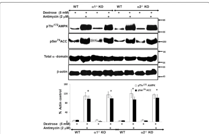
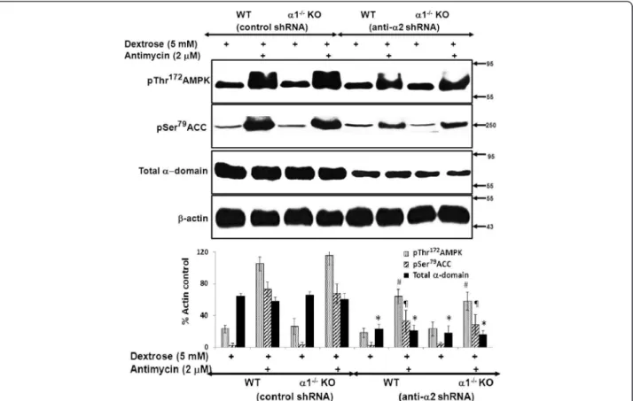
Figure 5Effect of metabolic stress on activation of the AMPK pathway in MPT cells from α1−/−and α2−/−KO mice. MPT cells obtained from α1−/−or α2−/−KO mice, or their WT controls, were subjected to metabolic stress by incubation in medium containing dextrose (5 mM) plus antimycin (2 μM) for 4 hrs. Control cells were incubated in dextrose in the absence of antimycin. Activation of the AMPK pathway was assessed by immunoblotting using antibodies that recognize either the phosphorylated (activated) form of AMPK or the phosphorylated (inhibited) form of acetyl coenzyme A carboxylase (ACC). Expression of total AMPK was also assessed. Upper panel: Representative immunoblot. Molecular weight markers (in kDa) are shown on the right of each immunoblot. Lower panel: Densitometric quantitation of three immunoblots using β-actin as a loading control. * p < 0.001, presence vs. absence of antimycin in MPT cells from α1−/−mice, α2−/−KO mice and their WT controls.
expression was comparable in kidney cortex and MPT cell cultures for both types of AMPK KO mouse versus its WT controls (Figures 3 and 4).
Effect of metabolic stress on activation of the AMPK pathway in MPT cells from α1−/−and α2−/−mice
We next compared the degree to which AMPK is activated by metabolic stress in MPT cells from AMPK KO versus WT mice. Metabolic stress was induced by treatment of cells with antimycin in the presence of 5 mM dextrose. Ac-tivation of AMPK was assessed by immunoblotting for phosphorylation of Thr172within the catalytic α domain of AMPK. Upon activation, AMPK phosphorylates a number of downstream targets [1,3-5]. As a further measure of AMPK activation, we evaluated the extent of phosphoryl-ation of ACC at Ser79, an event that inhibits ACC activity. We chose ACC, since it is one of the more thoroughly studied downstream targets of AMPK [1].
Treatment with antimycin increased phosphorylation of both AMPK and ACC to a comparable extent in
MPT cells from α1−/− and α2−/− mice versus their WT
controls (Figure 5). These data suggest that the identity of the alpha domain isoform does not influence the de-gree to which AMPK is activated by antimycin-induced metabolic stress, and that each isoform can substitute for the absence of the other.
Effect of pharmacologic inhibition of AMPK on stress-induced activation of AMPK in MPT cells from AMPK KO and WT mice
We examined the effect of compound C (CC), a pharma-cologic inhibitor of AMPK, on AMPK activity during metabolic stress in MPT cells from α2−/− and WT mice.
Antimycin increased the phosphorylation of both AMPK and its downstream target, ACC, in MPT cells from α2−/−
and WT mice (Figure 6). CC comparably reduced the stress-induced increase in phosphorylation of AMPK and ACC in MPT cells from α2−/−versus WT mice (Figure 6).
Similar results, in the absence and presence of CC, were obtained in MPT cells from α1−/−versus WT mice (data
not shown).
Figure 6Effect of pharmacologic inhibition of AMPK using compound C (CC) on the activity of the AMPK pathway in MPT cells from α2−/−KO and WT mice. MPT cells from α2−/−mice, or their WT control, were subjected to ATP depletion by incubation in medium containing dextrose (5 mM) and antimycin (2 μM), in the presence or absence of CC (20 μM) for 4 hrs. Activation of the AMPK pathway was assessed by immunoblotting, using antibodies that recognize either the phosphorylated (activated) form of AMPK, or the phosphorylated (inhibited) form of ACC. Expression of total alpha domain was also assessed. Upper panel: Representative immunoblot. Molecular weight markers (in kDa) are shown on the right of each immunoblot. Lower panel: Densitometric quantitation of three immunoblots using β-actin as a loading control. * p < 0.01, presence vs. absence of CC, for MPT cells from WT mice treated with antimycin; # p < 0.01, presence vs. absence of CC, for MPT cells from α2−/−mice treated with antimycin.
Effect of pharmacologic inhibition of AMPK on viability of metabolically stressed MPT cells from AMPK KO and WT mice
We next examined the effect of CC on MPT cell viability during metabolic stress. MPT cells from α2−/− mice and
their WT controls were subjected to graded ATP deple-tion (antimycin in the presence of varying concentra-tions of dextrose) for 16–18 hrs, in the presence of either CC (20 μM) or its vehicle. Control cells were incubated in dextrose without antimycin. In the absence of CC, metabolic stress induced a comparable decrease of viability in MPT cells from α2−/− versus WT mice
(Figure 7). Although inhibition of AMPK with CC re-duced MPT cell viability at all degrees of metabolic stress, the reduction in viability induced by CC was comparable in MPT cells from α2−/− versus WT mice
(Figure 7). Furthermore, regardless of the presence or absence of CC, ATP levels in metabolically stressed MPT cells did not differ for α2−/−versus WT mice (data
not shown). These data are consistent with those of im-munoblotting (Figures 5 and 6).
We performed similar experiments using MPT cells from α1−/− versus WT mice. In the absence of CC,
metabolic stress (antimycin plus 5 mM dextrose) re-duced cell viability to a similar extent in MPT cells from α1−/−versus WT mice (64 ± 7% and 59 ± 8% of control,
respectively). CC comparably exacerbated the stress-induced loss of cell viability in MPT cells from α1−/−
ver-sus WT mice (35 ± 6% and 38 ± 7% of control,
respectively). Thus, the effects of CC on the viability of stressed MPT cells was similar for α1−/−and α2−/−mice,
as compared to their WT controls.
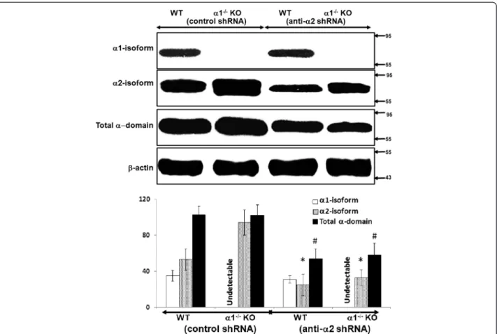
Effect of knockdown of the α2 isoform on expression and activation of the AMPK pathway in α1−/−and WT mice
MPT cells from α1−/−or WT mice were infected with
ei-ther control (scrambled) shRNA or shRNA designed to knock down expression of the α2 isoform of AMPK. In-fection with control shRNA did not alter expression of either the α2 isoform of AMPK or total α-domain AMPK in MPT cells from α1−/−and WT mice (Figure 8).
In contrast, infection with anti-α2 shRNA significantly reduced expression of the α2 isoform of AMPK as well as total alpha domain AMPK in MPT cells from both α1−/−and WT mice (Figure 8).
We next determined the effect of knockdown of the α2 isoform of AMPK on metabolic stress-induced acti-vation of the AMPK pathway. MPT cell monolayers from α1−/−and WT mice were infected with either
con-trol or anti-α2 shRNA. Following infection, MPT cells were incubated in dextrose (5 mM) in the absence or presence of antimycin. In cells infected with control shRNA, metabolic stress induced marked phosphoryl-ation of both AMPK and ACC (Figure 9). Infection with anti-α2 shRNA led to a marked but comparable reduc-tion in phosphorylareduc-tion of AMPK and ACC in MPT cells from α1−/−versus WT mice (Figure 9).
Figure 7Effect of pharmacologic inhibition of AMPK on viability of MPT cells from α2−/−KO and WT mice subjected to metabolic stress. A: MPT cells from α2−/−mice, or their WT control, were subjected to graded ATP depletion by incubation in the presence of antimycin (2 μM) and varying concentrations of dextrose for 16–18 hrs, in either the absence (solid lines) or the presence (dashed lines) of CC (20 μM). The percentage of viable cells remaining was determined by flow cytometry (n = 5). * p < 0.01, absence vs. presence of CC, for MPT cells from either α2−/−mice or their WT control (ANOVA for repeated measures). B: Representative flow cytometric analysis of metabolically stressed MPT cells from α2−/−mice incubated in the absence (left panels) or presence (right panels) of CC (20 μM). Cell viability was quantified by staining cells with calcein AM (x-axis) and ethidium homodimer-1 (Eth-H1) (y-axis). For each condition, 10,000 cells were analyzed, and the percentage of viable cells was calculated by dividing the number of calcein-positive and Eth-H1-negative cells by the total number of cells counted.
Effect of knockdown of the α2 isoform of AMPK on viability of metabolically stressed MPT cells from α1−/−KO mice
Finally, we examined the effect of knockdown of the α2 isoform of AMPK on the viability of metabolically stressed MPT cells. MPT cells from α1−/−and WT mice
were infected with either control shRNA or anti-α2 shRNA, and then incubated in the absence or presence of antimycin plus varying concentrations of dextrose. After 16–18 hrs, the percentage of viable cells was deter-mined by flow cytometry. As with pharmacologic inhib-ition of AMPK by CC, knockdown of the α2 domain comparably decreased the viability of metabolically stressed MPT cells from α1−/−versus WT mice (Figure 10).
Regardless whether cells were infected with control or anti-α2 shRNA, ATP levels did not differ in metabolically stressed MPT cells from α1−/−versus WT mice (data not
shown). Taken together, our studies on the effects of AMPK inhibition, accomplished either pharmacologically
or molecularly, demonstrate antimycin-induced metabolic stress comparably activates the α1 and α2 isoforms of AMPK, and that either isoform can substitute for the other in ameliorating stress-induced cell death.
Discussion
Increased activity of AMPK contributes to the pathogen-esis of a wide range of diseases, including diabetes melli-tus, the metabolic syndrome, neurodegenerative disorders, and cancer [24-30]. However, while an abundance of data indicates that AMPK is activated across multiple cell types during acute ischemic or toxic injury, the contribution by AMPK to cell survival during and following acute injury remains uncertain. Some studies have reported a cytopro-tective role for AMPK [31-35], while others have found that AMPK contributes to cell death [12,36-43]. The net effect of AMPK on cell survival likely depends upon mul-tiple variables, including cell lineage, the nature of the
Figure 8Effect of knockdown of the α2 isoform on expression of the α1 and α2 isoforms and of total alpha domain in MPT cells from α1−/−and WT mice. MPT cells from α1−/−mice, or their WT control, were infected with control (scrambled) shRNA or anti-α2 shRNA designed to knock down the expression of the α2 isoform of AMPK. The relative expression of the α1 and α2 isoforms of AMPK, as well as of the total α domain (α1 + α2), was evaluated in immunoblots of lysates from confluent MPT cells derived from the kidneys of α1−/−and α2−/−mice, and their WT controls. Upper panel: Representative immunoblot. Molecular weight markers (in kDa) are shown om the right of each immunoblot. Lower panel: Densitometric quantitation of three immunoblots using β-actin as a loading control. * p < 0.05, anti-α2 shRNA vs. control shRNA, for α2 isoform expression in MPT cells from WT and α1−/−mice; # p < 0.05, anti-α2 shRNA vs. control shRNA, for total alpha domain expression in MPT cells from WT and α1−/−mice.
toxic stimulus, and the specific pathways responsible for AMPK activation. In the kidney, few data are available, re-garding either the state of AMPK activity following ische-mic injury, or the role of AMPK in modulating renal tubular cell survival during metabolic stress.
Previously, we have shown that inhibition of AMPK, either pharmacologically (using CC) or via knockdown techniques, increased apoptosis of an immortalized MPT cell line subjected to metabolic stress [18]. These results suggested an anti-apoptotic role for AMPK in metabolically stressed kidney cells. In this study, we tested the hypothesis that primary cultures of MPT cells, derived from AMPK KO mice lacking either the α1 or α2 isoform of the catalytic domain, would be more sus-ceptible to apoptosis induced by metabolic stress than primary MPT cell cultures from their WT controls. To our surprise, stress-induced death was no more severe in MPT cells from KO mice compared MPT cells from the
WT controls (Figure 1). Moreover, while treatment of MPT cells with antimycin in the presence of decreasing concentrations of dextrose led to a progressive fall in cell ATP levels (Figure 2), the decrease in cell ATP levels at every degree of metabolic stress was comparable in KO versus WT mice (Figure 2).
Our data suggest that the lack of difference in suscep-tibility to stress-induced death by MPT cells from AMPK KO versus WT mice is related to a compensatory increase in the expression of the non-deleted alpha iso-form that occurs in the kidney cortex (Figure 3) and MPT cell cultures (Figure 4) from α1−/−and α2−/−mice.
Thus, expression of the α2 isoform of AMPK is up-regulated in the cortex (Figure 3) and primary MPT cell cultures (Figure 4) from the kidneys of α1−/−mice.
Simi-larly, expression of the α1 isoform is up-regulated in the cortex (Figure 3) and primary MPT cell cultures (Figure 4) from the kidneys of α2−/− mice. As a result, total
α-Figure 9Effect of knockdown of the α2 isoform of AMPK on the activity of the AMPK pathway in MPT cells from α1−/−and WT mice. MPT cells from α1−/−mice, or their WT control, were infected with control shRNA or anti-α2 shRNA designed to knock down the expression of the α2 isoform of AMPK. MPT cells were subjected to metabolic stress by incubation in medium containing dextrose (5 mM) plus antimycin (2 μM) for 4 hrs. Control cells were incubated in dextrose in the absence of antimycin. Activation of the AMPK pathway was assessed by immunoblotting using antibodies that recognize either the phosphorylated (activated) form of AMPK or the phosphorylated (inhibited) form of ACC. Expression of total AMPK was also assessed. Upper panel: Representative immunoblot. Molecular weight markers (in kDa) are shown om the right of each immunoblot. Lower panel: Densitometric quantitation of three immunoblots using β-actin as a loading control. * p < 0.01, anti-α2 shRNA vs. control shRNA, for total α domain expression in MPT cells from WT or α1−/−mice; # p < 0.02, anti-α2 shRNA vs. control shRNA, for phospho-AMPK expression in MPT cells from WT or α1−/−mice; p < 0.02, anti-α2 shRNA vs. control shRNA, for phospho-ACC expression in MPT cells from WT or α1−/−mice.
domain expression (α1 + α 2 isoforms) is comparable in AMPK KO versus WT mice.
We next examined the effect of metabolic stress on the phosphorylation, not only of the α-domain of AMPK, but also of ACC, an immediate downstream tar-get of AMPK that has been widely used as a marker of AMPK activity [1]. Interestingly, metabolic stress in-duced comparable degrees of phosphorylation of AMPK and ACC in MPT cells from α1−/−mice, α2−/−mice and
both of their WT controls (Figure 5). These findings sug-gest that equivalent expression of the total α-domain by KO and WT mice is matched by functional equivalence of AMPK activity.
We suggest that the lack of a difference in susceptibil-ity to antimycin-induced cell death by MPT cells derived from α1−/− and α2−/− mice versus their WT controls is
attributable to an adaptive equivalence in the amount and activity of the total alpha isoform of AMPK in MPT cells from the KO and WT mice. We further suggest that that each α isoform can substitute for the other in phosphorylation of downstream targets and in mediating the anti-apoptotic functions of AMPK. This interpret-ation is supported by studies in which we examined the effects of inhibiting AMPK in primary cultures of MPT cells from the AMPK KO and WT mice. Pharmaco-logical inhibition of AMPK (using CC) of MPT cells from AMPK KO and WT mice, reduced the antimycin-induced phosphorylation of AMPK and ACC (Figure 6),
and exacerbated the stress-induced death of MPT cells from the KO and WT mice (Figure 7). However, the extent to which CC inhibited AMPK phosphorylation (Figure 6), or worsened MPT cell death (Figure 7), was not different between MPT cells derived from the KO and WT mice. Similarly, inhibiting AMPK in MPT cells obtained from α1−/− mice and their WT controls by
knocking down the α2 isoform using shRNA, decreased antimycin-induced phosphorylation of AMPK and ACC (Figure 9), and exacerbated the amount of death of MPT cells (Figure 10) obtained from both the KO and WT mice to a comparable degree.
Our data show that while genetic deletion of either the α1 or α2 isoform of AMPK does not affect the response of MPT cells to metabolic stress, inhibition of AMPK duced by CC or molecular knockdown markedly in-creases the susceptibility of MPT cells from WT and KO mice to metabolic stress. It is likely that the compensa-tory increase in expression of the non-deleted α-isoform occurring in AMPK KO mice is due to increased protein synthesis. We speculate that, in the case of acute inhib-ition of AMPK, either by CC or molecular knockdown, there is insufficient time for a compensatory increase of protein synthesis of alpha isoforms to occur.
It is important to note that our results do not exclude the possibility that the various isoforms of AMPK may differ in role and function in different tissues [1,30]. To date, very little data exist on the consequences of genetic
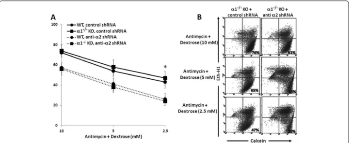
Figure 10Effect of knockdown the α2 isoform of AMPK on the viability of MPT cells from α1−/−KO and WT mice subjected to metabolic stress. A: MPT cells from α1−/−mice, or their WT control, were infected with control shRNA (solid lines) or anti-α2 shRNA designed to knock down the expression of the α2 isoform of AMPK (dashed lines). Cells were then subjected to graded ATP depletion by incubation for 16–18 hrs in the presence of antimycin (2 μM) and varying concentrations of dextrose. The percentage of viable cells remaining was determined by flow cytometry (n = 5). * p < 0.01, anti-α2 shRNA vs. control shRNA, for MPT cells from α1−/−or WT mice (ANOVA for repeated measures). B: Representative flow cytometric analysis of metabolically stressed MPT cells from α2−/−mice infected with anti-α2 shRNA (right panels) or control shRNA (left panels). Cell viability was quantified by staining cells with calcein AM (x-axis) and ethidium homodimer-1 (Eth-H1) (y-axis). For each condition, 10,000 cells were analyzed, and the percentage of viable cells was calculated by dividing the number of calcein-positive and Eth-H1-negative cells by the total number of cells counted.
deletion of one isoform on the expression and activity of the other isoform(s). Our results indicate that any at-tempt to link a particular phenotype (or lack of pheno-type) with the absence of one or other of the isoforms of AMPK should be done with caution, since expression and activity of the remaining isoform may be subject to an adaptive up-regulation.
Conclusions
Our study has several major conclusions. First, primary cultures of mouse proximal tubular (MPT) cells from both α1−/− and α2−/− mice demonstrate an adaptive
in-crease in expression of the intact α-domain isoform of AMPK, such that total α-domain expression is compar-able in KO versus WT mice. Second, the α1 and α2 iso-forms of AMPK are equally sensitive to metabolic stress, since exposure to antimycin led to comparable increases of AMPK activity in primary MPT cells from α1−/−and
α2−/− mice. Third, the α1 and α2 isoforms of AMPK
provide equivalent protection from stress-induced cell death, since primary MPT cells from α1−/− and α2−/−
mice versus their WT controls were equally susceptible to cell death from ATP depletion. Moreover, the use of compound C to inhibit the activity of the α1 or the α2 isoform in primary MPT cells derived from α2−/−and
α1−/−mice respectively, or the inhibition of the α2
iso-form in primary MPT cells from α1−/−mice (via
molecu-lar knockdown) both exacerbated loss of cell viability in response to ATP depletion to the same degree. Taken to-gether, these data suggest that the α1- and α2-isoforms of AMPK are not only similarly activated by ATP deple-tion, but also similarly effective in reducing cell death during metabolic stress. The adaptive up-regulation of the intact isoform of AMPK in KO mice is consistent with an overall critical role for AMPK in ameliorating apoptosis of proximal tubular cells in response to acute reductions of cellular ATP during ischemia.
Abbreviations
α:Alpha; β: Beta; γ: Gamma; ACC: Acetyl coenzyme A carboxylase; ADP: Adenine diphosphate; AICAR: N-(β-D-Ribofuranosyl)-5-aminoimidazole-4-carboxamide; AMPK: AMP-activated protein kinase; ATP: Adenine triphosphate; AMP: Adenine monohosphate; KO: Knockout; MPT: Mouse proximal tubule; shRNA: Short hairpin RNA.
Competing interests
The authors declare that they have no competing interest. Authors’ contributions
LZ, MT, and VP all contributed substantially to the generation of the data presented in this study. BV provided expert advice regarding the execution and interpretation of this study. WL and JSL contributed equally to the planning of studies and to writing of the manuscript. All authors read and approved the final manuscript.
Acknowledgements
W. Lieberthal was supported by a Veterans Affairs Merit Award and by awards from Dialysis Clinic, Inc. (DCI) (C-3176, C-2243 and C3351).
Author details
1Department of Medicine, Stony Brook University Medical Center, Stony
Brook, NY 11794, USA.2Northport Veterans Affairs Medical Center, Northport,
NY 11768, USA.3Inserm, U1016, Institut Cochin, Paris, France.4CNRS, UMR 8104, Paris, France.5Université Paris Descartes, Sorbonne Paris Cité, Paris,
France.6Department of Medicine, University of Illinois at Chicago, Chicago, IL
60612, USA.7Jesse Brown Veterans Affairs Medical Center, Chicago, IL 60612,
USA.
Received: 30 May 2013 Accepted: 6 November 2013 Published: 14 November 2013
References
1. Hardie DG, Ross FA, Hawley SA: AMPK: a nutrient and energy sensor that maintains energy homeostasis. Nat Rev Mol Cell Biol 2012, 13(4):251–262. 2. Steinberg GR, Kemp BE: AMPK in health and disease. Physiol Rev 2009,
89(3):1025–1078.
3. Hardie DG: Signal transduction: how cells sense energy. Nature 2011, 472(7342):176–177.
4. Bland ML, Birnbaum MJ: Cell biology. ADaPting to energetic stress. Science 2011, 332(6036):1387–1388.
5. Woods A, Johnstone SR, Dickerson K, Leiper FC, Fryer LG, Neumann D, et al: LKB1 is the upstream kinase in the AMP-activated protein kinase cascade. Curr Biol 2003, 13(22):2004–2008.
6. Xiao B, Sanders MJ, Underwood E, Heath R, Mayer FV, Carmena D, et al: Structure of mammalian AMPK and its regulation by ADP. Nature 2011, 472(7342):230–233.
7. Oakhill JS, Steel R, Chen ZP, Scott JW, Ling N, Tam S, et al: AMPK is a direct adenylate charge-regulated protein kinase. Science 2011, 332(6036):1433–1435.
8. Carling D: The AMPK-activated protein kinase cascade - a unifying system for energy control. Trends Biochem Sci 2004, 29:18–24.
9. Hardie DG: AMP-activated/SNF1 protein kinases: conserved guardians of cellular energy. Nat Rev Mol Cell Biol 2007, 8(10):774–785.
10. Hardie DG, Carling D: The AMP-activated protein kinase–fuel gauge of the mammalian cell? Eur J Biochem 1997, 246(2):259–273.
11. Hardie DG: AMP-activated protein kinase: the guardian of cardiac energy status. J Clin Invest 2004, 114(4):465–468.
12. Hallows KR, Mount PF, Pastor-Soler NM, Power DA: Role of the energy sensor AMP-activated protein kinase in renal physiology and disease. Am J Physiol Renal Physiol2010, 298:F1067–F1077.
13. Hue L, Beauloye C, Bertrand L, Horman S, Krause U, Marsin AS, et al: New targets of AMP-activated protein kinase. Biochem Soc Trans 2003, 31(Pt 1):213–215.
14. Hardie DG, Carling D, Carlson M: The AMP-activated/SNF1 protein kinase subfamily: metabolic sensors of the eukaryotic cell? Annu Rev Biochem 1998, 67:821–855.
15. Hardie DG, Pan DA: Regulation of fatty acid synthesis and oxidation by the AMP-activated protein kinase. Biochem Soc Trans 2002,
30(Pt 6):1064–1070.
16. Hay N, Sonenberg N: Upstream and downstream of mTOR. Genes Dev 2004, 18(16):1926–1945.
17. Gwinn DM, Shackelford DB, Egan DF, Mihaylova MM, Mery A, Vasquez DS, et al: AMPK phosphorylation of raptor mediates a metabolic checkpoint. Mol Cell2008, 30(2):214–226.
18. Lieberthal W, Zhang L, Patel VA, Levine JS: AMPK protects proximal tubular cells from stress-induced apoptosis by an ATP-independent mechanism: potential role of Akt activation. Am J Physiol Renal Physiol 2011, 301(6):F1177–F1192.
19. Viollet B, Andreelli F, Jorgensen SB, Perrin C, Geloen A, Flamez D, et al: The AMP-activated protein kinase alpha2 catalytic subunit controls whole-body insulin sensitivity. J Clin Invest 2003, 111(1):91–98. 20. Jorgensen SB, Viollet B, Andreelli F, Frosig C, Birk JB, Schjerling P, et al:
Knockout of the alpha2 but not alpha1 5’-AMP-activated protein kinase isoform abolishes 5-aminoimidazole-4-carboxamide-1-beta-4-ribofuranosidebut not contraction-induced glucose uptake in skeletal muscle. J Biol Chem 2004, 279(2):1070–1079.
21. Kroshian VM, Sheridan AM, Lieberthal W: Functional and cytoskeletal changes induced by sublethal injury in proximal tubular epithelial cells. Am J Physiol1994, 266(1 Pt 2):F21–F30.
22. Lieberthal W, Menza SA, Levine JS: Graded ATP depletion can cause necrosis or apoptosis of cultured mouse proximal tubular cells. Am J Physiol1998, 274(2 Pt 2):F315–F327.
23. Sinha D, Wang Z, Ruchalski KL, Levine JS, Krishnan S, Lieberthal W, et al: Lithium activates the Wnt and phosphatidylinositol 3-kinase Akt signaling pathways to promote cell survival in the absence of soluble survival factors. Am J Physiol Renal Physiol 2005, 288(4):F703–F713. 24. Gruzman A, Babai G, Sasson S: Adenosine monophosphate-activated
protein kinase (AMPK) as a New target for antidiabetic drugs: a review on metabolic, pharmacological and chemical considerations. Rev Diabet Stud2009, 6(1):13–36.
25. Mori H, Inoki K, Masutani K, Wakabayashi Y, Komai K, Nakagawa R, et al: The mTOR pathway is highly activated in diabetic nephropathy and rapamycin has a strong therapeutic potential. Biochem Biophys Res Commun2009, 384(4):471–475.
26. Luo Z, Saha AK, Xiang X, Ruderman NB: AMPK, the metabolic syndrome and cancer. Trends Pharmacol Sci 2005, 26(2):69–76.
27. Kola B, Boscaro M, Rutter GA, Grossman AB, Korbonits M: Expanding role of AMPK in endocrinology. Trends Endocrinol Metab 2006, 17(5):205–215. 28. Musi N, Goodyear LJ: Targeting the AMP-activated protein kinase for the
treatment of type 2 diabetes. Curr Drug Targets Immune Endocr Metabol Disord2002, 2(2):119–127.
29. Kato K, Ogura T, Kishimoto A, Minegishi Y, Nakajima N, Miyazaki M, et al: Critical roles of AMP-activated protein kinase in constitutive tolerance of cancer cells to nutrient deprivation and tumor formation. Oncogene 2002, 21(39):6082–6090.
30. Arad M, Seidman CE, Seidman JG: AMP-activated protein kinase in the heart: role during health and disease. Circ Res 2007, 100(4):474–488. 31. Russell RR 3rd, Li J, Coven DL, Pypaert M, Zechner C, Palmeri M, et al:
AMP-activated protein kinase mediates ischemic glucose uptake and prevents postischemic cardiac dysfunction, apoptosis, and injury. J Clin Invest 2004, 114(4):495–503.
32. Spector NL, Yarden Y, Smith B, Lyass L, Trusk P, Pry K, et al: Activation of AMP-activated protein kinase by human EGF receptor 2/EGF receptor tyrosine kinase inhibitor protects cardiac cells. Proc Natl Acad Sci U S A 2007, 104(25):10607–10612.
33. Shibata R, Sato K, Pimentel DR, Takemura Y, Kihara S, Ohashi K, et al: Adiponectin protects against myocardial ischemia-reperfusion injury through AMPK- and COX-2-dependent mechanisms. Nat Med 2005, 11(10):1096–1103.
34. Terai K, Hiramoto Y, Masaki M, Sugiyama S, Kuroda T, Hori M, et al: AMP-activated protein kinase protects cardiomyocytes against hypoxic injury through attenuation of endoplasmic reticulum stress. Mol Cell Biol 2005, 25(21):9554–9575.
35. Peralta C, Bartrons R, Serafin A, Blazquez C, Guzman M, Prats N, et al: Adenosine monophosphate-activated protein kinase mediates the protective effects of ischemic preconditioning on hepatic ischemia-reperfusion injury in the rat. Hepatology 2001, 34(6):1164–1173. 36. Li J, Zeng Z, Viollet B, Ronnett GV, McCullough LD: Neuroprotective effects
of adenosine monophosphate-activated protein kinase inhibition and gene deletion in stroke. Stroke 2007, 38(11):2992–2999.
37. McCullough LD, Zeng Z, Li H, Landree LE, McFadden J, Ronnett GV: Pharmacological inhibition of AMP-activated protein kinase provides neuroprotection in stroke. J Biol Chem 2005, 280(21):20493–20502. 38. Cao C, Lu S, Kivlin R, Wallin B, Card E, Bagdasarian A, et al: AMP-activated
protein kinase contributes to UV- and H2O2-induced apoptosis in human skin keratinocytes. J Biol Chem 2008, 283(43):28897–28908. 39. Memmott RM, Gills JJ, Hollingshead M, Powers MC, Chen Z, Kemp B, et al:
Phosphatidylinositol ether lipid analogues induce AMP-activated protein kinase-dependent death in LKB1-mutant non small cell lung cancer cells. Cancer Res2008, 68(2):580–588.
40. Okoshi R, Ozaki T, Yamamoto H, Ando K, Koida N, Ono S, et al: Activation of AMP-activated protein kinase induces p53-dependent apoptotic cell death in response to energetic stress. J Biol Chem 2008, 283(7):3979–3987. 41. Shaw MM, Gurr WK, McCrimmon RJ, Schorderet DF, Sherwin RS:
5’AMP-activated protein kinase alpha deficiency enhances stress-induced apoptosis in BHK and PC12 cells. J Cell Mol Med 2007, 11(2):286–298.
42. Tzatsos A, Tsichlis PN: Energy depletion inhibits phosphatidylinositol 3-kinase/Akt signaling and induces apoptosis via AMP-activated protein kinase-dependent phosphorylation of IRS-1 at Ser-794. J Biol Chem 2007, 282(25):18069–18082.
43. Dagon Y, Avraham Y, Berry EM: AMPK activation regulates apoptosis, adipogenesis, and lipolysis by eIF2alpha in adipocytes. Biochem Biophys Res Commun2006, 340(1):43–47.
doi:10.1186/1471-2369-14-251
Cite this article as: Lieberthal et al.: Susceptibility to ATP depletion of primary proximal tubular cell cultures derived from mice lacking either the α1 or the α2 isoform of the catalytic domain of AMPK. BMC Nephrology2013 14:251.
Submit your next manuscript to BioMed Central and take full advantage of:
• Convenient online submission • Thorough peer review
• No space constraints or color figure charges • Immediate publication on acceptance
• Inclusion in PubMed, CAS, Scopus and Google Scholar • Research which is freely available for redistribution
Submit your manuscript at www.biomedcentral.com/submit