HAL Id: hal-00738632
https://hal.archives-ouvertes.fr/hal-00738632
Submitted on 21 May 2020
HAL is a multi-disciplinary open access
archive for the deposit and dissemination of
sci-entific research documents, whether they are
pub-lished or not. The documents may come from
teaching and research institutions in France or
abroad, or from public or private research centers.
L’archive ouverte pluridisciplinaire HAL, est
destinée au dépôt et à la diffusion de documents
scientifiques de niveau recherche, publiés ou non,
émanant des établissements d’enseignement et de
recherche français ou étrangers, des laboratoires
publics ou privés.
Toxins and Ion transfers
Julien Barbier, Evelyne Benoit, Nicolas Gilles, Daniel Ladant, Marie-France
Martin-Eauclaire, César Mattei, Jordi Molgó, Michel R. Popoff, Denis Servent
To cite this version:
Julien Barbier, Evelyne Benoit, Nicolas Gilles, Daniel Ladant, Marie-France Martin-Eauclaire, et al..
Toxins and Ion transfers. SFET Publications, Gif-sur-Yvette, France, pp.123, 2011. �hal-00738632�
Collection
Rencontres en Toxinologie
Collection
Meetings on Toxinology
T
T
T
o
o
o
x
x
x
i
i
i
n
n
n
e
e
e
s
s
s
e
e
e
t
t
t
T
T
T
r
r
r
a
a
a
n
n
n
s
s
s
f
f
f
e
e
e
r
r
r
t
t
t
s
s
s
i
i
i
o
o
o
n
n
n
i
i
i
q
q
q
u
u
u
e
e
e
s
s
s
0. 0 5.0 10 .0 15.0 20 .0 25.030 .0 35.040 .0 45.0 50. 0 55.0 60 .0 65.0 0 50 1 00 1 50 2 00 2 50 3 00 3 50 mAU min All A B C D E F G H I J K L M N O P 1 2 3 4 5 6 7 8 9 10 11 12 13 14 15 16 17 1 8 19 20 21 22 23 24Crude venom
Venom fractionation
High throughput screening & hit identification
ACSKKWEYCIVPILGFVYCCPGLICGPFVCV
Sequence determination
Is
o
la
ti
o
n
of
bi
o
ac
ti
ve
pe
p
ti
d
es
T
T
To
o
ox
x
xi
i
in
n
ns
s
s
a
a
an
n
nd
d
d
I
I
Io
o
on
n
n
t
t
tr
r
ra
a
an
n
ns
s
sf
f
fe
e
er
r
rs
s
s
Comité d’édition – Editorial committee :
Julien BARBIER, Evelyne BENOIT, Nicolas GILLES, Daniel LADANT, Marie-France
MARTIN-EAUCLAIRE, César MATTEI, Jordi MOLGÓ, Michel R. POPOFF, Denis SERVENT
Société Française pour l'Etude des Toxines
Illustration de couverture – Cover picture :
Application d'une analyse à haut débit pour Nav1.7, basée sur la méthode FLIPR, pour la découverte d'inhibiteurs de
canaux Na+. L'identification et la séquence obtenue pour MrVIB du venin brut de Conus marmoreus sont illustrées ici.
De Richard LEWIS, Ching-I Anderson WANG, Sébastien DUTERTRE et Irina VETTER (ce volume).
Application of a FLIPR-based high throughput assay for Nav1.7 for the discovery of Na+channel inhibitors. Illustrated is
the identification and sequence obtained for MrVIB identified in the crude venom of Conus marmoreus. From Richard LEWIS, Ching-I Anderson WANG, Sébastien DUTERTRE and Irina VETTER (this volume).
Collection
Rencontres en Toxinologie
Collection
Meetings on Toxinology
La collection « Rencontres en Toxinologie » est publiée à l’occasion des colloques annuels
« Rencontres en Toxinologie » organisés par la Société Française pour l’Etude des Toxines
(SFET). Les ouvrages imprimés parus de 2001 à 2007 ont été édités par Elsevier (Paris,
France) puis la Librairie Lavoisier (Cachan, France). Depuis 2008, ils sont édités par la SFET et
diffusés sur le site
http://www.sfet.asso.fr
, en libre accès pour les auteurs et les lecteurs.
The series « Rencontres en Toxinologie » is published on the occasion of the annual Meetings
on Toxinology organized by the French Society of Toxinology (SFET). The printed books of the
series, from 2001 to 2007, were edited by Elsevier (Paris, France) and then the Librairie
Lavoisier (Cachan, France). Since 2008, they are edited by the SFET and are available on-line
on the site
http://www.sfet.asso.fr
, with free access for authors and readers.
Titres parus – Previous titles
Explorer, exploiter les toxines et maîtriser les organismes producteurs
Cassian Bon, Françoise Goudey-Perrière, Bernard Poulain, Simone Puiseux-Dao Elsevier, Paris, 2001
ISBN : 2-84299-359-4
Toxines et recherches biomédicales
Françoise Goudey-Perrière, Cassian Bon, Simone Puiseux-Dao, Martin-Pierre Sauviat Elsevier, Paris, 2002
ISBN : 2-84299-445-0
Toxinogenèse – Biosynthèse, ingénierie, polymorphisme et neutralisation des toxines Françoise Goudey-Perrière, Cassian Bon, André Ménez, Simone Puiseux-Dao
Elsevier, Paris, 2003 ISBN : 2-84299-481-7 Envenimations, intoxinations
Françoise Goudey-Perrière, Evelyne Benoit, Simone Puiseux-Dao, Cassian Bon Librairie Lavoisier, Cachan, 2004
ISBN : 2-7430-0749-4 Toxines et douleur
Cassian Bon, Françoise Goudey-Perrière, Max Goyffon, Martin-Pierre Sauviat Librairie Lavoisier, Cachan, 2005
ISBN : 2-7430-0849-0 Toxines et cancer
Françoise Goudey-Perrière, Evelyne Benoit, Max Goyffon, Pascale Marchot Librairie Lavoisier, Cachan, 2006
ISBN : 2-7430-0958-6
Toxines émergentes : nouveaux risques
Françoise Goudey-Perrière, Evelyne Benoit, Pascale Marchot, Michel R. Popoff Librairie Lavoisier, Cachan, 2007
ISBN : 978-2-7430-1037-9
Toxines et fonctions cholinergiques neuronales et non neuronales
Evelyne Benoit, Françoise Goudey-Perrière, Pascale Marchot, Denis Servent Publications de la SFET, Châtenay-Malabry, France, 2008
Epub on http://www.sfet.asso.fr - ISSN 1760-6004 Toxines et Signalisation - Toxins and Signalling
Evelyne Benoit, Françoise Goudey-Perrière, Pascale Marchot, Denis Servent Publications de la SFET – SFET Editions, Châtenay-Malabry, France, 2009
Epub on http://www.sfet.asso.fr - ISSN 1760-6004
Avancées et nouvelles technologies en Toxinologie - Advances and new technologies in Toxinology Julien Barbier, Evelyne Benoit, Pascale Marchot, César Mattéi, Denis Servent
Publications de la SFET – SFET Editions, Gif-sur-Yvette, France, 2010 Epub on http://www.sfet.asso.fr - ISSN 1760-6004
Cet ouvrage est publié à l’occasion du colloque « 19
èmesRencontres en Toxinologie », organisé
par la Société Française pour l’Etude des Toxines (SFET) les 28 et 29 novembre 2011 à Paris.
This book is published on the occasion of the 19
thMeeting on Toxinology, organized by the
French Society of Toxinology (SFET) on November 28
thand 29
th, 2011, in Paris.
Le comité d’organisation est constitué de – The organizing committee is constituted of :
Julien Barbier, Evelyne Benoit, Nathalie Hatchi, Daniel Ladant, Michel R. Popoff & Denis Servent.Le comité scientifique est constitué de – The scientific committee is constituted of :
Julien Barbier, Evelyne Benoit, Nicolas Gilles, Max Goyffon, Daniel Ladant, Pascale Marchot, Marie-France Martin-Eauclaire, César Mattéi, Jordi Molgó, Michel R. Popoff & Denis Servent.
Le comité de rédaction est constitué de – The redaction committee is constituted of :
Julien Barbier, Adriana Rolim Campos Barros, Evelyne Benoit, Nicolas Gilles, Max Goyffon, Marie-France Martin-Eauclaire, César Mattéi, Jordi Molgó, Michel R. Popoff & Denis Servent.
Sommaire - Content
Pages
Toxines et canaux ioniques – Toxins and ion channels
Animal toxins targeting voltage-activated sodium (NaV1.9) channelsFrank BOSMANS 7-10
Overview of the small voltage-gated K+ channels blockers from Androctonus venoms
Marie-France MARTIN-EAUCLAIRE, Pierre E.BOUGIS 11-13
Toxicity of sea anemone toxins related to their pharmacological activities on ion channels
Sylvie DIOCHOT, Emmanuel DEVAL, Jacques NOEL, Laszlo BERESS, Michel LAZDUNSKI, Eric LINGUEGLIA
15-27
Tools for studying peptide toxin modulation of voltage-gated sodium channels
Stefan H. HEINEMANN, Enrico LEIPOLD 29-37
An overview of the ion channel modulation and neurocellular disorders induced by ciguatoxins
César MATTEI, Jordi MOLGÓ, Evelyne BENOIT
39-42
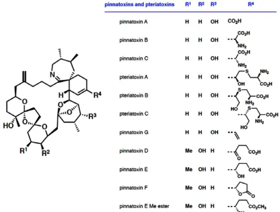
Pinnatoxins : an emergent family of marine phycotoxins targeting nicotinic acetylcholine receptors with high affinity
Rómulo ARÁOZ, Denis SERVENT, Jordi MOLGÓ, Bogdan I. IORGA,
Carole FRUCHART-GAILLARD, Evelyne BENOIT, Zhenhua GU, Craig STIVALA, Armen ZAKARIAN
43-47
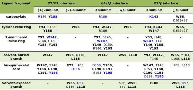
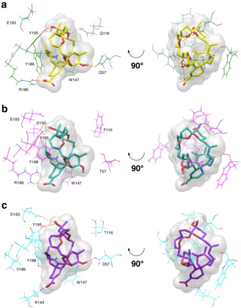
Insights into the interaction of pinnatoxin A with nicotinic acetylcholine receptors using molecular modeling
Rómulo ARÁOZ, Armen ZAKARIAN, Jordi MOLGÓ, Bogdan I. IORGA
49-54
Toxines formant des pores – Pore-forming toxins
VacA from Helicobacter pylori : journey and action mechanism in epithelial cells
Vittorio RICCI, Patrice BOQUET 55-60
Known and unknown mitochondrial targeting signals
Joachim RASSOW 61-66
Clostridium perfringens epsilon toxin : a fascinating toxin
Michel R. POPOFF 67-71
Binding partners of protective antigen from Bacillus anthracis share certain common motives
Christoph BEITZINGER, Angelika KRONHARDT, Roland BENZ
73-79
Ways for partial and total inhibition of staphylococcal bicomponent leucotoxins Gilles PREVOST, Mira TAWK, Mauro DALLA SERRA, Bernard POULAIN, Sarah CIANFERANI, Benoît-Joseph LAVENTIE, Emmanuel JOVER
81-88
The cholesterol-dependent cytolysins : molecular mechanism to vaccine development
Rodney TWETEN 89-94
Heat-stable enterotoxin b produced by Escherichia coli induces apoptosis in rat intestinal epithelial cells
H. Claudia SYED, J. Daniel DUBREUIL
95-97
On the mode of entry of clostridial neurotoxins into the cytosol of nerve terminals Paolo BOLOGNESE, Fulvio BORDIN, Cesare MONTECUCCO, Marco PIRAZZINI,
Ornella ROSSETTO, Clifford C. SHONE
99-101
Pages
Toxines comme outils et thérapeutiques – Toxins as tools and therapeutics
Tethering peptide toxins for neurocircuitry, cell-based therapies and drug discovery
Ines IBAÑEZ-TALLON 103-110
New aspects on membrane translocation of the pore-forming Clostridium botulinum C2 toxin
Eva KAISER, Katharina ERNST, Claudia KROLL, Natalie BÖHM, Holger BARTH
111-113
Ion channel toxins for drug discovery and development
Richard LEWIS, Ching-I Anderson WANG, Sébastien DUTERTRE, Irina VETTER 115-120
G protein-coupled receptors, an unexploited family of animal toxins targets: exploration of green mamba venom for novel ligands on adrenoceptors
Arhamatoulaye MAÏGA, Gilles MOURIER, Loic QUINTON, Céline ROUGET, Philippe LLUEL, Stefano PALEA, Denis SERVENT, Nicolas GILLES
121-125
Anti-tumor snake venoms peptides
Sameh SARRAY, Raoudha ZOUARI, Jed JEBALI, Ines LIMAM, Amine BAZAA,
Maram MORJANE, Zeineb ABDELKAFI, Olfa ZIRI, Najet SRAIRI, Salma DAOUED, Jose LUIS, Mohamed EL AYEB,Naziha MARRAKCHI
127-132
Effect of Dinoponera quadriceps venom on chemical-induced seizures models in mice Kamila LOPES, Emiliano RIOS, Rodrigo DANTAS, Camila LIMA, Maria LINHARES,
Alba TORRES, Ramon MENEZES, Yves QUINET, Alexandre HAVT, Marta FONTELES, Alice MARTINS
133-136
Effect of L-amino acid oxidase isolated from Bothrops marajoensis snake venom on the epimastigote forms of Trypanosoma cruzi
Ticiana PEREIRA, Rodrigo DANTAS, Alba TORRES, Clarissa MELLO, Danya LIMA, Marcus Felipe COSTA, Marcos TOYAMA, Maria de Fátima OLIVEIRA, Helena MONTEIRO, Alice MARTINS
137-140
Divers – Miscellaneous
Neurotoxicity of Staphylococcus aureus leucotoxins : interaction with the store operated calcium entry complex in central and sensory neurons
Emmanuel JOVER, Benoît-Joseph LAVENTIE, Mira TAWK, Bernard POULAIN, Gilles PREVOST
141-145
Atypical profile of paralytic shellfish poisoning toxins in clams from the Gulf of Gabes (Southern Tunisia)
Riadh MARROUCHI, Evelyne BENOIT, Jean Pierre LECAER, Jordi MOLGÓ,Riadh KHARRAT
147-150
Etude toxico-cinétique et biologique du venin de scorpion Androctonus mauretanicus chez le lapin
Fatima CHGOURY, Naoual OUKKACHE, Nadia EL GNAOUI, Hakima BENOMAR, Rachid SAÏLE, Noreddine GHALIM
151-154
Ion imbalance, tissue damage and inflammatory response induced by kaliotoxin Amina LADJEL-MENDIL, Nesrine SIFI, Marie-France MARTIN-EAUCLAIRE,
Fatima LARABA-DJEBARI
155-156
Cytotoxic and antioxidant activities of scorpion venom on cell lines
Djelila HAMMOUDI-TRIKI, Fatima LARABA-DJEBARI 157-159
Zn2+ : a required ion for biological and enzymatic activities of procoagulant
metalloproteinase (CCSV-MPase) isolated from Cerastes cerastes venom
Fatah CHERIFI, Jean-Claude ROUSSELLE, Abdelkader NAMANE, Fatima LARABA-DJEBARI
161-164
Preliminary characterization of the most dangerous snake venoms of Morocco
Naoual OUKKACHE, Balkiss BOUHAOUALA-ZAHAR, Noreddine GHALIM 165-172
A monitoring study of repetitive surgical oocyte harvest in Xenopus laevis
Rencontres en Toxinologie – Meeting on Toxinology, 2011 Editions de la SFET – SFET Editions
Accès libre en ligne sur le site – Free access on line on the site : http://www.sfet.asso.fr
Animal toxins targeting voltage-activated sodium
(Na
V1.9) channels
Frank BOSMANS
Molecular Physiology and Biophysics Section, Porter Neuroscience Research Center, National Institute of Neurological Disorders and Stroke, National Institutes of Health, Bethesda, MD 20892, USA
Tel : +1-301-594-6760 ; E-mail : [email protected]
Abstract
Voltage-activated sodium (NaV) channels are crucial for initiating and transmitting action
potentials, an ability that places them amongst the most widely targeted ion channels by drugs and animal venoms. An increasing number of toxins isolated from animal venom have been shown to interfere with the voltage-driven activation process of NaV channels, possibly by
interacting with one or more of their voltage-sensors. This mini-review summarizes our recent work on identifying novel animal toxin receptor sites within NaV channel voltage-sensors and
illustrates how chimeric approaches can be used to uncover molecules that interact with NaV1.9,
an enigmatic NaV channel involved in nociception.
Les toxines animales ciblant les canaux sodium (Na
V1.9) activés par
le potentiel
Les canaux sodium dépendants du potentiel (ou canaux NaV) sont cruciaux pour initier et
transmettre des potentiels d'action, une capacité qui les place parmi les canaux les plus ciblés par les médicaments et les venins animaux. Il a été démontré qu’un nombre croissant de toxines isolées de venins animaux interfèrent avec le processus d'activation des canaux NaV, peut-être en
interagissant avec un ou plusieurs de leurs senseurs de potentiel. Cette mini-revue résume notre travail récent sur l'identification des sites de nouvelles toxines animales se fixant sur les senseurs de potentiel des canaux NaV et démontre comment des approches chimériques peuvent être
utilisées pour découvrir des molécules qui influent sur NaV1.9, un canal NaV énigmatique impliqué
dans la sensation de douleur.
Keywords : Animal toxin, pain, sodium channel, voltage-sensor.
Na
Vchannel S3b-S4 paddle motifs
The NaV channel (Goldin et al., 2000) pore-forming subunit consists of four connected domains (I-IV) (Catterall, 2000), each having six transmembrane segments (S1-S6) (Figure 1a). These similar entities consist of a voltage-sensor (S1-S4) and a portion of the structure that forms the sodium ion selective pore in the membrane (S5-S6). The pore can open or close when all four voltage-sensors move in response to changes in membrane voltage. It is thought that each of the four voltage-sensors activates in response to changes in membrane voltage, however, those in domains I-III are most important for channel opening, whereas the one in domain IV plays a distinctive role in inactivating the channel (Cha et al., 1999; Sheets et al., 1999; Horn et al., 2000; Sheets et al., 2000; Chanda and Bezanilla, 2002; Bosmans et al., 2008; Campos et al., 2008).
Despite their physiological significance, structural information on NaV channels lags when compared to the structurally similar voltage-activated K+ (Kv) channels, where recent data has revealed structural features important for channel function. More specifically, studies on Kv channel voltage-sensors have identified an S3b-S4 helix-turn-helix motif, the voltage-sensor paddle, which moves at the protein-lipid interface and drives activation of the voltage-sensors which, in turn, opens the pore (Figure 1b) (Jiang et al., 2003; Alabi et al., 2007; Long et al., 2007; Chakrapani et al., 2008; Swartz, 2008). Besides its vital role in channel gating, the paddle motif is also an important pharmacological target in Kv channels, as spider toxins that partition into membranes interact with this region to inhibit channel opening (Swartz and MacKinnon, 1997b, a; Li-Smerin and Swartz, 1998, 2000; Lee et al., 2003; Phillips et al., 2005; Alabi et al., 2007; Swartz, 2007). Recently, we have shown that distinct paddle motifs also exist in each of the four voltage-sensors of NaV channels and that they can be transplanted into the four-fold symmetric Kv channel to study them in isolation (Bosmans et al., 2008; Milescu et al., 2009). Furthermore, we demonstrated that each of the four paddle motifs is capable of interacting with toxins from tarantulas and scorpions and that multiple paddle motifs are often targeted by a single toxin. For example, it was shown that the tarantula toxin ProTx-II (Middleton et al., 2002) can interact with the voltage-sensor in domain I, II and IV, whereas a related tarantula toxin, PaurTx3, only interacts with domain II (Bosmans et al., 2008). It is also interesting that the profiles of toxin-paddle interactions vary for
Animal toxins targeting NaV1.9 8
different subtypes of NaV channels. This chimeric approach was recently applied to NaV1.9, a relatively unknown NaV channel isoform involved in pain perception.
Figure 1. Cartoon representing top views of a NaV channel (a) and a KV
channel (b). The central Na+ or K+ selective pore is surrounded by the four
voltage-sensors in the four domains (delineated by dotted line). In the NaV
channel, the paddles (color) are not identical whereas in the KV channel, the
paddles (grey) are identical.
Figure 1. Figure représentant des vues de dessus d'un canal NaV (a) et un
canal potassium (KV) (b). Le pore central sélectif pour Na+ ou K+ est entouré
par les quatre senseurs de potentieldans les quatre domaines (délimités par la
ligne en pointillés). Dans le canal NaV, les palettes (couleur) ne sont pas
identiques alors que dans le canal KV, les palettes (en gris) sont identiques.
Animal toxin pharmacology of Na
V1.9
NaV channel expression is tissue-specific across different species. NaV1.7 is mainly expressed in sensory and sympathetic neurons in the peripheral nervous system. NaV1.8 and NaV1.9 are sensory neuron-specific channels that are normally found within small-diameter dorsal root (DRG) and trigeminal ganglia but not in the central nervous system neurons (Momin and Wood, 2008). Selective knockout of NaV1.7 expression in mouse nociceptors leads to a loss of acute mechanosensory and inflammatory pain (Nassar et al., 2004). Also, various human heritable pain disorders such as erythermalgia and paroxysmal extreme pain disorder map to mutations in SCN9A, the gene encoding NaV1.7 (Dib-Hajj et al., 2008). Studies of NaV1.8 in mice have revealed a role for this channel in inflammatory pain, neuropathic pain and noxious stimuli response (Joshi et al., 2006; Dong et al., 2007). NaV1.9 knockout mice have a largely absent inflammatory hyperalgesia in response to inflammatory mediators (Priest et al., 2005; Amaya et al., 2006). Although knockout mice are an extremely valuable tool to reveal the physiological role of NaV channels, the occurrence of genetic compensatory mechanisms might mask vital functional information. Therefore, the discovery of pharmacological tools that evoke a specific response from these channels is essential for elucidating their physiological function. To achieve this goal, heterologous expression and characterization of NaV1.7, NaV1.8, and NaV1.9 in oocytes or mammalian cells is of great importance.
Although NaV1.9 plays a key role in nociception, fundamental questions about its function and pharmacology remain unanswered because previous attempts to express this channel in heterologous systems have been unsuccessful (Blum et al., 2002; Dib-Hajj et al., 2002). In addition, studying NaV1.9-mediated currents in native DRG neurons is technically challenging because only a fraction of isolated neurons produces a measurable amount of these currents and other NaV channels activate over a similar voltage range. We circumvented heterologous expression obstacles by identifying and transplanting paddle motifs from the putative voltage-sensors of NaV1.9 into four-fold symmetric Kv channels and investigated the function of NaV1.9 voltage-sensors in channel gating and in forming toxin receptors (Bosmans et al., 2011).
By taking advantage of the portable nature of paddle motifs within voltage-sensing domains (Alabi et al., 2007; Bosmans et al., 2008; Milescu et al., 2009), we showed that these structural motifs also exist in each of the four NaV1.9 voltage-sensors, and that they can be transplanted into Kv channels to be studied in isolation. Our results revealed that each of the NaV1.9 paddle motifs can sense changes in membrane voltage and drive Kv channel voltage-sensor activation, similar to what was found for canonical NaV channels (Bosmans et al., 2008). Since the pharmacological sensitivities of NaV1.9 remain unexplored, we exploited these paddle constructs to search for toxins that might interact with NaV1.9 channels. To this end, we screened eighteen toxins from tarantula, scorpion and sea anemone venom against four NaV1.9 paddle constructs and observed six toxins that potently inhibit one or more of the chimeras (AaHII, ProTx-I, TsVII, GrTx-SIA, HaTx, BomIV). In addition, we discovered that the NaV1.9 paddle motifs from all four domains can interact with toxins from animal venom.
The two most interesting toxins that emerged from our screens were the scorpion toxin TsVII and the tarantula toxin ProTx-I, both of which interact strongly with NaV1.9 paddle motifs and potently facilitate the
slowly activating and inactivating NaV1.9-mediated current in rat DRG neurons (Cummins et al., 1999; Maruyama et al., 2004; Priest et al., 2005; Coste et al., 2007; Ostman et al., 2008). In addition to targeting NaV1.9, TsVII and ProTx-I have very different actions on NaV1.8, the other TTX-resistant NaV channel present in these sensory neurons. For example, TsVII produces a dramatic facilitation of NaV1.9-mediated currents while only modestly inhibiting NaV1.8, showing that the scorpion toxin can discriminate between these two TTX-resistant NaV channels. Conversely, ProTx-I causes both a pronounced potentiation of NaV1.9-mediated currents and a robust inhibition of NaV1.8 (Figure 2), making this toxin a formidable tool to discriminate the currents generated by these two channel isoforms in DRG neurons. Taken together, our results suggest that NaV1.9 channels possess functional voltage-sensors that interact with scorpion and tarantula toxins, features that are shared with canonical NaV channel isoforms. Furthermore, our screening with a wide range of toxins demonstrate that NaV1.9 has different pharmacological sensitivities than other NaV channel isoforms, a property that may be exploited for drug design.
Figure 2. Effect of ProTx-I on native NaV1.9- and NaV1.8-mediated currents in rat
DRG neurons. (a) Voltage protocol used to elicit both NaV1.9 (first depolarization
to -55 mV) and NaV1.8-mediated currents (second depolarization to 0 mV) in a
rat DRG neuron. (b) Evoked currents under control conditions (black) and after
addition of 100 nM ProTx-I (red for NaV1.9, green for NaV1.8). NaV1.9-mediated
currents are potentiated whereas NaV1.8-mediated currents are inhibited. Time
scales for both currents are indicated in the X-axis. Experiment by Michelino Puopolo, Harvard Medical School (Bosmans et al., 2011).
Figure 2. Effet de ProTx-I sur les courants natifs via NaV1.9 et NaV1.8 dans les
neurones DRG de rat. (a) Protocole de potentiel utilisé pour activer des
courants via NaV1.9 (première dépolarisation à -55 mV) et NaV1.8 (deuxième
dépolarisation à 0 mV) dans un neurone de DRG. (b) Courants évoqués dans des conditions de contrôle (noir) et après addition de 100 nM de ProTx-I (rouge
pour NaV1.9, vert pour NaV1.8). Les courants via NaV1.9 sont potentialisés mais
ceux via NaV1.8 sont inhibés. Les échelles de temps pour les deux courants sont
indiquées dans l’axe des abscisses. Expérience menée par Michelino Puopolo, Harvard Medical School (Bosmans et al., 2011).
Acknowledgements. The work summarized here was carried out by Frank Bosmans in the lab of Dr Kenton J Swartz
(NINDS-NIH, USA) and by Michelino Puopulo in the lab of Dr Bruce P Bean (Harvard Medical School, USA) with support from Dr Marie-France Martin-Eauclaire (CRN2M-Marseille, Marie-France).
References
Alabi AA, Bahamonde MI, Jung HJ, Kim JI, Swartz KJ (2007) Portability of paddle motif function and pharmacology in voltage sensors. Nature 450: 370-375
Amaya F, Wang H, Costigan M, Allchorne AJ, Hatcher JP, Egerton J, Stean T, Morisset V, Grose D, Gunthorpe MJ, Chessell IP, Tate S, Green PJ, Woolf CJ (2006) The voltage-gated sodium channel Na(v)1.9 is an effector of peripheral inflammatory pain hypersensitivity. J Neurosci 26: 12852-12860
Blum R, Kafitz KW, Konnerth A (2002) Neurotrophin-evoked depolarization requires the sodium channel Na(V)1.9. Nature 419: 687-693
Bosmans F, Martin-Eauclaire MF, Swartz KJ (2008) Deconstructing voltage sensor function and pharmacology in sodium channels. Nature 456: 202-208
Bosmans F, Puopolo M, Martin-Eauclaire MF, Bean BP, Swartz KJ (2011) Functional properties and toxin pharmacology of a dorsal root ganglion sodium channel viewed through its voltage sensors. J Gen Physiol 138: 59-72
Animal toxins targeting NaV1.9 10
Campos FV, Chanda B, Beirao PS, Bezanilla F (2008) Alpha-scorpion toxin impairs a conformational change that leads to fast inactivation of muscle sodium channels. J Gen Physiol 132: 251-263
Catterall WA (2000) From ionic currents to molecular mechanisms: the structure and function of voltage-gated sodium channels. Neuron 26: 13-25
Cha A, Ruben PC, George AL, Jr., Fujimoto E, Bezanilla F (1999) Voltage sensors in domains III and IV, but not I and II, are immobilized by Na+ channel fast inactivation. Neuron 22: 73-87
Chakrapani S, Cuello LG, Cortes DM, Perozo E (2008) Structural dynamics of an isolated voltage-sensor domain in a lipid bilayer. Structure 16: 398-409
Chanda B, Bezanilla F (2002) Tracking voltage-dependent conformational changes in skeletal muscle sodium channel during activation. J Gen Physiol 120: 629-645
Coste B, Crest M, Delmas P (2007) Pharmacological dissection and distribution of NaN/Nav1.9, T-type Ca2 + currents, and
mechanically activated cation currents in different populations of DRG neurons. J Gen Physiol 129: 57-77
Cummins TR, Dib-Hajj SD, Black JA, Akopian AN, Wood JN, Waxman SG (1999) A novel persistent tetrodotoxin-resistant sodium current in SNS-null and wild-type small primary sensory neurons. J Neurosci 19: RC43
Dib-Hajj S, Black JA, Cummins TR, Waxman SG (2002) NaN/Nav1.9: a sodium channel with unique properties. Trends Neurosci 25: 253-259
Dib-Hajj SD, Yang Y, Waxman SG (2008) Genetics and molecular pathophysiology of Na(v)1.7-related pain syndromes. Adv
Genet 63: 85-110
Dong XW, Goregoaker S, Engler H, Zhou X, Mark L, Crona J, Terry R, Hunter J, Priestley T (2007) Small interfering RNA-mediated selective knockdown of Na(V)1.8 tetrodotoxin-resistant sodium channel reverses mechanical allodynia in neuropathic rats. Neuroscience 146: 812-821
Goldin AL, Barchi RL, Caldwell JH, Hofmann F, Howe JR, Hunter JC, Kallen RG, Mandel G, Meisler MH, Netter YB, Noda M, Tamkun MM, Waxman SG, Wood JN, Catterall WA (2000) Nomenclature of voltage-gated sodium channels. Neuron 28: 365-368
Horn R, Ding S, Gruber HJ (2000) Immobilizing the moving parts of voltage-gated ion channels. J Gen Physiol 116: 461-476
Jiang Y, Lee A, Chen J, Ruta V, Cadene M, Chait BT, MacKinnon R (2003) X-ray structure of a voltage-dependent K+ channel.
Nature 423: 33-41
Joshi SK, Mikusa JP, Hernandez G, Baker S, Shieh CC, Neelands T, Zhang XF, Niforatos W, Kage K, Han P, Krafte D, Faltynek C, Sullivan JP, Jarvis MF, Honore P (2006) Involvement of the TTX-resistant sodium channel Nav1.8 in inflammatory and neuropathic, but not post-operative, pain states. Pain 123: 75-82
Lee HC, Wang JM, Swartz KJ (2003) Interaction between extracellular Hanatoxin and the resting conformation of the voltage-sensor paddle in Kv channels. Neuron 40: 527-536
Li-Smerin Y, Swartz KJ (1998) Gating modifier toxins reveal a conserved structural motif in voltage-gated Ca2 + and K+
channels. Proc Natl Acad Sci U S A 95: 8585-8589
Li-Smerin Y, Swartz KJ (2000) Localization and molecular determinants of the Hanatoxin receptors on the voltage-sensing domains of a K(+) channel. J Gen Physiol 115: 673-684
Long SB, Tao X, Campbell EB, MacKinnon R (2007) Atomic structure of a voltage-dependent K+ channel in a lipid
membrane-like environment. Nature 450: 376-382
Maruyama H, Yamamoto M, Matsutomi T, Zheng T, Nakata Y, Wood JN, Ogata N (2004) Electrophysiological characterization of
the tetrodotoxin-resistant Na+ channel, Na(v)1.9, in mouse dorsal root ganglion neurons. Pflugers Arch 449: 76-87
Middleton RE, Warren VA, Kraus RL, Hwang JC, Liu CJ, Dai G, Brochu RM, Kohler MG, Gao YD, Garsky VM, Bogusky MJ, Mehl JT, Cohen CJ, Smith MM (2002) Two tarantula peptides inhibit activation of multiple sodium channels. Biochemistry 41: 14734-14747
Milescu M, Bosmans F, Lee S, Alabi AA, Kim JI, Swartz KJ (2009) Interactions between lipids and voltage sensor paddles detected with tarantula toxins. Nat Struct Mol Biol 16: 1080-1085
Momin A, Wood JN (2008) Sensory neuron voltage-gated sodium channels as analgesic drug targets. Curr Opin Neurobiol 18: 383-388
Nassar MA, Stirling LC, Forlani G, Baker MD, Matthews EA, Dickenson AH, Wood JN (2004) Nociceptor-specific gene deletion reveals a major role for Nav1.7 (PN1) in acute and inflammatory pain. Proc Natl Acad Sci U S A 101: 12706-12711
Ostman JA, Nassar MA, Wood JN, Baker MD (2008) GTP up-regulated persistent Na+ current and enhanced nociceptor
excitability require NaV1.9. J Physiol 586: 1077-1087
Phillips LR, Milescu M, Li-Smerin Y, Mindell JA, Kim JI, Swartz KJ (2005) Voltage-sensor activation with a tarantula toxin as cargo. Nature 436: 857-860
Priest BT, Murphy BA, Lindia JA, Diaz C, Abbadie C, Ritter AM, Liberator P, Iyer LM, Kash SF, Kohler MG, Kaczorowski GJ, MacIntyre DE, Martin WJ (2005) Contribution of the tetrodotoxin-resistant voltage-gated sodium channel NaV1.9 to sensory transmission and nociceptive behavior. Proc Natl Acad Sci USA 102: 9382-9387
Sheets MF, Kyle JW, Hanck DA (2000) The role of the putative inactivation lid in sodium channel gating current immobilization.
J Gen Physiol 115: 609-620
Sheets MF, Kyle JW, Kallen RG, Hanck DA (1999) The Na channel voltage sensor associated with inactivation is localized to the external charged residues of domain IV, S4. Biophys J 77: 747-757
Swartz KJ (2007) Tarantula toxins interacting with voltage sensors in potassium channels. Toxicon 49: 213-230 Swartz KJ (2008) Sensing voltage across lipid membranes. Nature 456: 891-897
Swartz KJ, MacKinnon R (1997a) Hanatoxin modifies the gating of a voltage-dependent K+ channel through multiple binding
sites. Neuron 18: 665-673
Swartz KJ, MacKinnon R (1997b) Mapping the receptor site for hanatoxin, a gating modifier of voltage-dependent K+ channels.
Rencontres en Toxinologie – Meeting on Toxinology, 2011 Editions de la SFET – SFET Editions
Accès libre en ligne sur le site – Free access on line on the site : http://www.sfet.asso.fr
Overview of the small voltage-gated K
+channels
blockers from Androctonus venoms
Marie-France MARTIN-EAUCLAIRE
*, Pierre E.
BOUGIS
CNRS UMR6231, CRN2M, IFR11 Institut Jean Roche, Université de la Méditerranée, Faculté de Médecine secteur Nord, CS80011, Bd Pierre Dramard, F-13344 Marseille cedex 15, France
* Corresponding author ; Tel : +33 (0) 491698914 ; Fax : +33 (0) 491698839 ; E-mail : [email protected]
Abstract
Scorpion toxins have been used extensively to study the pharmacology of K+ channels, as well as
to decipher their pore topology, and an increasing amount of molecular data is still being published on this subject. During the last two decades, Androctonus venoms have provided several structurally distinct families of peptides exhibiting different K+-channel-blocking function.
We have largely participated to their purification, chemical, pharmacological and immunological characterization. In this article, we summarize our contribution to the current knowledge on these toxin/channel interactions.
Vue d’ensemble des petits peptides des venins d’Androctonus
capables de bloquer des canaux K
+activés par le potentiel
Les toxines de scorpion ont été considérablement utilisées pour étudier la pharmacologie des canaux K+ ainsi que pour décrypter la topologie du pore et une quantité croissante de résultats
moléculaires continue d’être publiée sur ce sujet. Au cours des deux dernières décades, les venins d’Androctonus ont fourni plusieurs familles de peptides distincts structurellement et capables de bloquer la fonction de différent types de canaux K+. Nous avons largement contribué
à leur purification et à leur caractérisation chimique, pharmacologique et immunologique. Dans cet article, nous résumons notre contribution aux connaissances actuelles des interactions toxine/canal K+.
Keywords : Potassium channels, scorpion toxins.
Introduction
K+ channel blockers’ toxins (KTx) from scorpion venoms are short peptides, which typically contain between 30-40 amino-acid residues cross-linked by 3-4 disulphide bridges forming compact and resistant molecules. They block K+ channels from the extracellular side by binding to the outer vestibule of K+ channels and in most cases insert a Lys side chain into the channel pore (Park and Miller, 1992). They are often present at low concentrations in the venoms (from 0.01 to 1% by weight). Usually, they have almost no toxic effects in mice by subcutaneous (s.c.) injection. However, they could be very toxic when injected intracerebroventricularly (i.c.v.) into the brain. Based on sequence identity and cysteine pairing, they have been classified into four families, the and -KTx (Tytgat et al., 1999) and today more than 120 KTx ranging from 23 up to 64 amino acids are sequenced. Most of their structures exhibit the characteristic fold called Cystein-Stabilized-Helix (CSH) motif, constituted by one -helix and two or three -strands, in which two disulfide bridges covalently link a segment of the -helix with one strand of the sheet structure, except for -KTxs which are formed by two parallel α-helices linked by two disulfide bridges. The -KTx family is constituted of the shortest peptides having diverse specific blocking activities against voltage-gated (KV) and calcium activated (KCa) channels. Longest peptides, with 45-68 amino acid residues reticulated by three disulfide bridges, have been later characterized and classified as the -KTx family. They have two structural and functional domains: an -helix in the N-terminal with cytolytic and antimicrobial activity, as well as a tightly folded C-terminal region with the CSH motif and K+ channel-blocking activities (Diego-García et al., 2008). At last, the -KTx family was described as specific for hERG channel (Corona et al., 2002).
This overview will summarize our main original contributions to the subject, in particular those obtained by the Androctonus mauretanicus venom analysis.
The smallest toxins in Androctonus mauretanicus venom: P01 (-KTx8) and P05
(-KTx5)
Small voltage-gated K+ channels blockers from Androctonus venoms 12
the so-called apamin-sensitive SKCa channel, as well as PO1 (-KTx 8.1, 3177 Da) which is much less active on the SKCa (300pM). They belong to the -KTx5.x's family (as Leiurotoxin1 or Scyllatoxin from the venom of the scorpion Leiurus quinquestriatus Hebraeus). Structure-function studies have shown that a highly positively charged region (in particular two Arg residues) of the -helix exposed to the solvent is involved in binding to the receptor, as it is for the bee venom apamin (Sabatier et al., 1993).
The Kaliotoxin subfamily (-KTx3 subfamily)
The KTX structure-function relationship studies were first performed using synthetic analogs such as KTX(1-37),
KTX(1-37)-am ide and shortened peptides including KTX(27-37), KTX(25-32) and KTX(1-11) (Romi et al., 1993). KTX(27-37)
and KTX(25-32) but not KTX(1-11) competed with 125I-KTX for its receptor on rat brain synaptosomes and act as antagonists of KTX. This demonstrated for the first time that the C-terminal region, particularly the -sheet of the toxin, was involved in the interaction with the receptor and the channel blockade. KTX was further widely used by different international groups for probing the vestibule topology from the lymphocyte KV1.3 channel. Synthetic KTX analogs have been used in combination with site-directed mutagenesis of the KV1.3 channel to identify pairs of residues in the toxin and channel which interact specifically. It was found that the side chain of the Lys 27 residue enters deeply into the pore and interacts with the Asp 402 residue of each channel subunit (Aiyar et al., 1995). Then, the KTX binding to a chimeric K+ channel (KcsA-K
V1.3) was investigated using solid-state nuclear magnetic resonance (ssNMR) (Lange et al., 2006). Significant chemical shift changes were observed for the KTX residues found in interaction with the channel, reflecting conformational changes involving -sheet contacts between the first and third -strand. For the channel, chemical-shift changes were seen also for channel residues in the KTX-binding region in both the pore helix and the selectivity filter. The ssNMR data directly show that Asp 64 in the KcsA-KV1.3 vestibule represents an important interaction site for KTX. Large chemical-shift changes were seen for Gly 77, Tyr 78 and Gly 79 in the selectivity filter. Also, chemical-shift changes were observed for the side chains of Glu 71 and Asp 80 that form carboxyl-carboxylate pairs on the backside of the filter (Lange et al., 2006).
The -KTx
15subfamily : K
V4.x and hERG blockers
The first member of the α-KTx15 subfamily characterized from Androctonus australis was Aa1 (3869 Da) (Pisciotta et al., 2000). At the primary sequence level, the toxin has an unusual N-terminal pyroglutamic acid. Aa1 totally blocked a fast IA-type K+ current from cerebellum granular cells. Shortly after, we have isolated a novel toxin from the venom of Androctonus mauretanicus, AmmTX3 (3827 Da), able to block a fast IA-type K+ current from striatal neurons in culture (Vacher et al., 2002). Then, several cDNAs encoding two Aa1 isoforms, AaTX1 (3867 Da) and AaTX2 (3853 Da) were identified by PCR amplification from a venom gland cDNA library of Androctonus australis (Legros et al., 2003) as well as in Androctonus amoreuxi (3769 Da). These toxins constitute the first members of the -KTx15 family (Figure 1). From a pharmacological point of view, the molecules shared the same target in rat brain. Autoradiograms demonstrated a heterogeneous distribution of 125I-toxin binding sites throughout the adult rat brain. High density of receptors was found in the striatum, hippocampus, superior colliculus and cerebellum (Vacher et al., 2001). The nature of the K+ channels blocked by the toxins was assessed by performing whole cell patch recording of the K+ currents of striatal neurons and of cerebellum granular cells in primary culture. In all cases, the AmmTX3 inactivates the transient A-current, but the sustained K+ current remains fully activated. Finally, analysis by electrophysiological recording of transient K+ currents in mammalian cells transfected with diverse cloned K+ channels showed that only the rapidly activating and inactivating KV4.1-mediated current was inhibited by AmmTX3 [with a 50% blocking concentration (IC50) of 105 nM]. The inhibition was less effective on KV4.2 and KV4.3 channels and the toxin did not affect other transient currents such as KV1.4 and KV3.4 (Vacher et al., 2006).
Further, we have reported that the -KTx15 peptides also show a significant hERG-blocking activity, like -KTx peptides. From a structural point of view, we have proposed that two separate functional surfaces, A and B, coexist on the molecule, and are responsible for two different K+-current-blocking functions (Huys et al., 2004). While KTxs interact with channels through their β-sheets, -KTxs modulate hERG through their -helix. A common "hot spot" with 2 basic residues (Arg18 and Lys19 in the -helix) confers hERG blockade activity to -KTx15 peptides (Abdel-Mottaleb et al., 2008).
Figure 1. Amino acid sequences of the Androctonus toxins from the -KTx15 family. *depicted from cDNAs
clones; Aa, Androctonus australis; Amm, Androctonus mauretanicus; Aam, Androctonus amoreuxi. Amino acids
in red in the -helix are crucial for hERG channel blockade and those in blue in the -sheet for KV channel
blockade.
Figure 1. Séquences d’acide aminés des toxines d’Androctonus de la famille -KTx15. *décrypté à partir
d’ADNcs; Aa, Androctonus australis; Amm, Androctonus mauretanicus; Aam, Androctonus amoreuxi. Les acides
aminés en rouge dans l’hélice- sont cruciaux pour le blocage des canaux hERG et ceux en bleu dans le
feuillet- pour le blocage des canaux KV.
(Average mass in Da)
Aa1
ZNETNKKCQGGSCASVCRRVIGVAAGKCINGRCVCYP 3851
AaTX1* ZIETNKKCQGGSCASVCRRVIGVAAGKCINGRCVCYP 3850
AaTX2* ZVETNKKCQGGSCASVCRRVIGVAAGKCINGRCVCYP 3839
AmmTX3 ZIETNKKCQGGSCASVC
RK
VIGVAAGKCINGRCVC
YP
3823
AamTX* ZVQTNKKCKGGSCASVCAKVIGVAAGKCINGRCVCYP 3722
Conclusion
Scorpion venom remains a proven resource for novel compound discovery, especially for the pharmacologists. A total of 209 -KTx amino acid sequences are now referenced in the UniProtKB data bank, but only some of these -KTx peptides have really been shown able to block K+ currents. For the other reported peptides there is often no direct evidence described for their function. While some of them represent new analogs of well-known families displaying unique selectivity or targeting, others exhibit novel structural features or activities. The majority of their action has been determined on the KV1.x subfamily or on the Ca2+-activated K+ channels (on SKCa-sensitive to the bee venom Apamin or on BKCa). The results that we obtained during the last two decades provide new insight into the possible targets of some -KTxs purified from different North African Androctonus venoms. In particular, we have greatly contributed to the -KTx15 family definition and pharmacological characterization. Moreover, KTX (-KTx3-1) was finally proved to be a wonderful tool, which helped us to depict the molecular mechanisms of interaction between K+ channels and peptide inhibitors and to demonstrate that the binding of K+ channel specific scorpion toxins does not take place only on the outer vestibule of the channel pore but also deeper into the selectivity filter. The binding involves a combination of hydrophobic, hydrogen bonding and electrostatic interactions, which induced significant structural rearrangements in both molecules. It was then proposed that structural flexibility of the K+ channel and the toxin represents an important determinant for the high specificity of toxin/K+ channel interactions (Lange et al., 2006).
Acknowledgements. We wish to thank Drs Abbas N., Bosmans F., Céard B., Legros C., Tytgat J. and Vacher H. for their
contribution in the purification, pharmacological or electrophysiological characterization and cloning and mutagenesis of the
Androctonus toxins blocking KV channels.
References
Abdel-Mottaleb Y, Corzo G, Martin-Eauclaire MF, Satake H, Céard B, Peigneur S, Nambaru P, Bougis PE, Possani LD, Tytgat J
(2008) A common "hot spot" confers hERG blockade activity to alpha-scorpion toxins affecting K+ channels. Biochem
Pharmacol 76: 805-815
Aiyar J, Withka JM, Rizzi JP, Singleton H, Andrews GC, Lin W, Boyd J, Hanson DC, Simon M, Dethlefs B (1995) Topology of the
pore-region of a K+ channel revealed by the NMR-derived structures of scorpion toxins. Neuron 15: 1169–1181
Corona M, Gurrola GB, Merino E, Restano-Cassulini R, Valdez-Cruz NA, Garcia B, Ramïrez-Dominguez ME, Coronas FV, Zamudio
FZ, Wanke E, Possani LD (2002) A large number of novel Ergtoxin-like genes and ERG K+ channels blocking peptides from
scorpions of the genus Centruroides. FEBS Lett 532: 121–126
Crest M, Jacquet G, Gola M, Zerrouk H, Benslimane A, Rochat H, Mansuelle P, Martin-Eauclaire MF (1992) Kaliotoxin, a novel peptidyl inhibitor of neuronal BK-type Ca(2+)-activated K(+) channels characterized from Androctonus mauretanicus
mauretanicus venom. J Biol Chem 267: 1640-1647
Diego-García E, Abdel-Mottaleb Y., Schwartz EF, de la Vega RC, Tytgat J, Possani LD (2008) Cytolytic and K+ channel blocking
activities of beta-KTx and scorpine-like peptides purified from scorpion venoms. Cell Mol Life Sci 65: 187-200
Huys I, Xu CQ, Wang CZ, Vacher H, Martin-Eauclaire MF, Chi CW, Tytgat J (2004) BmTx3, a scorpion toxin with two putative
functional faces separately active on A-type K+ and HERG currents. Biochem J 378: 745-752
Lange A, Giller K, Hornig S, Martin-Eauclaire MF, Pongs O, Becker S, Baldus M (2006) Toxin-induced conformational changes in a potassium channel revealed by solid-state nmr. Nature 440: 959–962
Legros C, Bougis PE, Martin-Eauclaire MF (2003) Characterisation of the genes encoding Aa1 isoforms from the scorpion
Androctonus australis. Toxicon 41: 115-119
Park CS, Miller C (1992) Interaction of charybdotoxin with permeant ions inside the pore of a K+ channel. Neuron 9: 307-313
Pisciotta M, Coronas FI, Bloch C, Prestipino G, Possani LD (2000) Fast K(+) currents from cerebellum granular cells are completely blocked by a peptide purified from Androctonus australis Garzoni scorpion venom. Biochim Biophys Acta 1468: 203-212
Sabatier JM, Zerrouk H, Darbon H, Mabrouk K, Benslimane A, Rochat H, Martin-Eauclaire MF, Van Rietschoten J (1993) P05, a new leiurotoxin I-like scorpion toxin: synthesis and structure-activity relationships of the alpha-amidated analog, a ligand of Ca(2+)-activated K(+) channels with increased affinity Biochemistry 32: 2763-2770
Tytgat J, Chandy KG, Garcia LM, Gutman GA, Martin-Eauclaire MF, Walt JJ, Possani LD (1999) A unified nomenclature for short chain peptides isolated from scorpion venoms: alpha-KTx molecular subfamilies. Trends Pharmacol Sci 20: 445–447 Vacher H, Romi-Lebrun R, Mourre C, Lebrun B, Kourrich S, Masméjean F, Nakajima T, Legros C, Crest M, Bougis PE,
Martin-Eauclaire MF (2001) A new class of scorpion toxin binding sites related to an A-type K+ channel: pharmacological
characterization and localization in rat brain. FEBS Lett 501: 31-36
Vacher H, Alami M, Crest M, Possani LD, Bougis PE, Martin-Eauclaire MF (2002) Expanding the scorpion toxin alpha-KTX 15 family with AmmTX3 from Androctonus mauretanicus. Eur J Biochem 269:6037-6041
Vacher H, Diochot S, Bougis PE, Martin-Eauclaire MF, Mourre C (2006) KV4 channels sensitive to BmTX3 in rat nervous system:
Rencontres en Toxinologie – Meeting on Toxinology, 2011 Editions de la SFET – SFET Editions
Accès libre en ligne sur le site – Free access on line on the site : http://www.sfet.asso.fr
Toxicity of sea anemone toxins related to their
pharmacological activities on ion channels
Sylvie DIOCHOT
1,2*, Emmanuel DEVAL
1,2, Jacques NOEL
1,2, Laszlo BERESS
3,
Michel LAZDUNSKI
1,2, Eric LINGUEGLIA
1,21 CNRS, Institut de Pharmacologie Moléculaire et Cellulaire, UMR 6097, 06560 Valbonne, France ; 2 Université de
Nice-Sophia Antipolis, UMR 6097, 06560 Valbonne, France ; 3 IPF Pharmaceuticals, Feodor Lynen Strasse 31,
Hanover 30625, Allemagne
* Corresponding author ; Tel : +33(0)4 9395 3422 ; Fax : +33(0)4 9395 7728 ; E-mail : [email protected]
Abstract
Sea anemone peptides have been isolated since more than 30 years, the majority being highly toxic for their natural preys, crustaceans, but also for mammals. These neurotoxins which may have also cardiotonic properties are activators of voltage-dependent Na+ channels. They
represent a structural group of four -fold peptides. Some of their basic and hydrophobic aminoacids are crucial determinants for their tissue and species selectivities. More recently, several groups of sea anemone peptides with different structures have been characterized with moderate toxicities on crustacean or mammals. They allowed the characterization of several subtypes of voltage-dependent K+ channels involved in autoimmune diseases, and of acid-sensing
ion channels (ASIC3) involved in pain processing. These last toxins could be promising tools for the design of new therapeutic molecules.
Toxicité des toxines d’anémones de mer en relation avec leur activité
pharmacologique sur les canaux ioniques
De nombreuses toxines peptidiques de venins d’anémones de mer ont été isolées depuis plus de 30 ans du fait de leur activité hautement toxique chez leurs proies naturelles, les crustacés, mais aussi chez les mammifères. Il s’agit de toxines neuro-excitatrices parfois cardio-stimulantes activant les canaux Na+ dépendants du potentiel. Elles forment un groupe structural homogène
de toxines ayant 4 feuillets dont la sélectivité inter-espèce et tissulaire semble dépendre de la présence de certains résidus d’acides aminés chargés ou hydrophobes. D’autres toxines, découvertes plus récemment, présentent des structures plus variables, avec des degrés de toxicité moindre chez les crustacés et mammifères. Ces dernières ont permis la caractérisation de sous-types de canaux K+ dépendants du potentiel dont certains sont impliqués dans les maladies
auto-immunes, et de certains canaux ASIC (canaux ioniques sensibles à l’acidité extracellulaire) impliqués dans la douleur. Ces dernières toxines sont des outils prometteurs pour la conception de nouvelles molécules thérapeutiques.
Keywords : Acid-sensing ion channels, KV channels, NaV channels, sea anemone, toxicity.
Introduction
1Animal venom components are usually called toxins, a term which evokes danger for living animals and humans, and are considered as bioharzardous material. But what is the definition of a toxin? Generally an animal toxin is defined as a natural component of the venom, able to disturb nerve, muscle or cardiac function, resulting in an effect which contributes to immobilize preys or to kill predators. A toxin acts at a very low concentration, with a highly specific activity on a cell membrane receptor. Very interestingly, evidences have been accumulated showing that some toxins can also be of a great utility as pharmacological tools, having low or no toxicity, and usable for therapeutic purposes (Koh and Kini, 2011; Miljanich, 2004; Mouhat et al., 2008).
Since more than 40 years, animal toxins have been isolated and characterized, first, due to a public health concern, to understand and treat the severe poisonings they are able to induce and which can be fatal in humans. Diverse degrees of toxicity are described such as cardiac arrhythmias, leading to cardiac arrest, neurological hyperexcitability, convulsions, hypertension, paralysis, blood perturbations (coagulation, hemolysis). Toxins (from spiders and scorpions) are also studied in agronomy, to discover new insecticides,
1Abbreviations: Acid Sensing Ion Channel (ASIC), Central Nervous System (CNS), Complete Freund’s Adjuvant (CFA),
Intra-CcerebroVentricular (i.c.v.), Intra-Cisternal (i.c.), Intra-Muscular (i.m.), Intra-Venous (i.v.), Molecular Weight (M. W.),
Peripheral Nervous System (PNS), Sea Anemone (SA), TetrodoToXin (TTX), Voltage-gated Na+ (Na
Sea anemone toxins targeting ion channels 16
because some for instance from spider and scorpions are very selective tools that provoke insect paralysis. Toxins can act on diverse specific targets including ion channels, cell receptors, and less specifically on membrane phospholipids. However, it has been also shown that venoms contain “non-toxic” toxins, which are not able to induce neither visible symptoms of toxicity nor lethality, when injected to mammals, insects or crustaceans. These “non-toxic” toxins are therefore particularly interesting tools to study the implication of the targeted receptors in diverse physiological functions and can sometimes lead to therapeutic applications.
Sea anemones belong to the phylum of Cnidaria, subdivided in three classes, Hydrozoa, Scyphozoa and Anthozoa. The last one includes around 6000 known species of sea anemones. The venom is present in specialized stinging organelles called nematocysts or cnidocysts, included in specialized cells, the cnidocytes. Nematocysts are distributed over the entire surface of the sea anemone body, which makes the venom difficult to extract. Usually, active components are extracted from the whole animal by using methanol/chloroform to obtain an aqueous extract which can be further separated by several steps of liquid chromatography (Beress et al., 1975b).
The best characterized toxins in sea anemones are: (i) a group of peptides (or small proteins; M. W. from 3,000 to 6,000 Da) acting on ion channels of excitable membranes and (ii) cytolysins (M. W. 15,000-20,000 Da) which are proteins that have been characterized by their hemolytic activity. Most of the sea anemone peptides have a preference for crustaceans, their natural prey, but due to a closed evolutionary context, they also act on insects (Bosmans and Tytgat, 2007). To a lesser extent, and often due to their high affinity for specific ionic channels, they are also toxic for mammals. This review focuses on the sea anemone toxins effective on crustaceans and mammals, and describes their different degree of toxicity in relation with their specific molecular targets among different families of ion channels.
Diversity of neurotoxic and/or cardiotoxic sea anemone peptides affecting
voltage-dependent Na
+channels
The most studied sea anemone (SA) toxins have been isolated more than 40 years ago based on their ability to induce severe neurotoxic and/or cardiotoxic effects on a variety of crustacean, insect or mammal nerve and muscle preparations (Beress and Beress, 1975a; Norton, 1991; Romey et al., 1976). These neurotoxins affect voltage-gated Na+ channels (Na
V) which are supporting action potential initiation. NaV channels are the molecular targets for toxins that can bind at six identified receptor sites, either by blocking the channel pore, either by modifying the gating properties of the channel (Catterall et al., 2007). SA toxins bind to the receptor site 3, located on the domain IVS5-S6 of the subunit of NaV channels which is also targeted by scorpion and funnel web spider toxins. They have allowed identification of subtypes of channels because they act with high affinities on different neuronal, muscular, or cardiac NaV. They slow-down the channel inactivation, allowing more Na+ entry into the cells and prolong action potential duration. Generally, the sea anemone toxins acting on NaV channels (SA-NaV toxins) have voltage-dependent effects because they bind with more affinity at polarized membrane potentials.
The SA-NaV toxins are polypeptides with molecular weights ranging between 3,000 and 5,000 Da that are cross-linked by 3 (or even more) disulfide bridges. Two different types have been described with distinct sequences and immunological properties. Type 1 toxins are isolated from Actiniidae (Anemonia sp, Anthopleura sp, Bunodosoma sp, Condylactis sp, see Table 1) and types 2 are isolated from Stichodactylidae families [Radianthus (Heteractis) sp, Stichodactyla sp; see Table 1; Bosmans and Tytgat, 2007]. The two types are also distinguished by their immunoreactivity, i.e. there is no antigenic cross-reactivity between them (Norton, 1991; Schweitz et al., 1985). Structurally, the two types of SA-NaV toxins contain four stranded anti-parallel -sheets connected by two loops. One toxin, halcurin, possesses structural properties common to both type 1 and 2 toxins indicating that all these toxins have probably evolved from the same ancestral gene (Ishida et al., 1997). The type 1 toxins display higher affinity for cardiac and skeletal muscle tetrodotoxin (TTX)-resistant NaV channels. In brief, resistance of NaV to TTX action is correlated to a high sensitivity to sea anemone toxins and vice versa.
Species selectivity can be observed for some toxins: AsI, AsIII, RpII, and ShI are inactive on NaV channels of mammalian system whereas others, such as AsV, AP-A and AP-B, are both active on crustacean and also very active on mammalian NaV channels (Kem et al., 1989; Lazdunski et al., 1986; Schweitz et al., 1981).
Activity of highly neurotoxic SA toxins (characterized by trembling of tail, fasciculations, salivation, difficulty in breathing and paralysis) is related to an effect on TTX-resistant and TTX-sensitive NaV channels (Norton et al., 1981; Romey et al., 1976; Schweitz et al., 1981). The most neurotoxic SA toxins active on mammals are found in the two structural groups: AsII, AsV, AP-B, BgII, BgIII (type 1), and RPI –V (type 2) (Table 1). The most potent toxins for crustaceans are ShI, AETXII, and AETXIII (Norton, 1991; Shiomi et al., 1997).
Neurotoxicity is sometime associated with cardiotoxicity resulting in paralysis after i.v. (mice), or i.m. (crustacean) administration, and can lead to death (convulsions, arrhythmia and ventricular fibrillation) using concentration as low as 2 µg/kg (i.c. in mammals, Table 1). The well known and characterized AsI, AsII and AsIII isolated from Anemonia sulcata are both active on crustacean inducing neurotoxic symptoms like tetanic contractions and paralysis in the crab after i.m. injections (Alsen, 1983). In mammal, cardiotoxic effects predominate after i.v. injection of AsI or AsII, compared to neurotoxic symptoms (Alsen, 1983). AsI and AsII (the most active) induce a significant cardiotonic (positive inotropic) effect (Alsen et al., 1978; Renaud et al., 1986) in different mammalian heart preparations. This effect is linked to the increased Na entry into cardiac cells coupled to a subsequent entry of Ca2+ through the Na/Ca exchanger. Cardiac effects are often characterized by a potent positive inotropic effect (case of AP-A at 0.1-1 µg/kg, i.v.) without any significant effect on heart or blood pressure, but at higher concentrations (>10 µg/kg, i.v.) they induce arrhythmia and
Table 1. Comparison of some sea anemone peptides structure, toxicities and targets.
Tableau 1. Comparaison des structures, propriétés toxiques et cibles de peptides d’anémones de mer.
Toxin
(origin: Genus species) M. W. (AA) Structure (PDB) classification Specie / tissue selectivity DLToxicity: 50 (µg/kg)
Target References
AeK
Actinia equina 3807(36)
2, 6C
SAK-I ? ? KV1 (Minagawa et al., 1998)
AETXI
Anemonia erythrea 4963(47)
46C
Type 1 Crust NL (i.v./mice) 2 (crab) NaV? (Shiomi et al., 1997) AETXII 6502(59) New struct, 10C Crust 0.5 (crab)
NL (i.v./mice) ? (Shiomi et al., 1997) AETXIII 6562(59) New Struct, 10C Crust 0.3 (crab)
NL (iv/mice) ? (Shiomi et al., 1997) AmI
Anthopleura maculata
2803(27) New ? 830 (crab) ? (Honma et al., 2005;
Honma and Shiomi, 2006) AmII 5128(45) 4,6C SAKIII ? NL (crab) paralytic KV? (Honma et al., 2005; Honma and Shiomi, 2006)
AmIII 5134(47) 46C
Type 1
Crust 70 (crab) NaV? (Honma et al., 2005) AsI (ATXI)
Anemonia sulcata 4834(46)
46C (1ATX)
Type 1 Cardiac, neuron Crust 236 (i.c./mice) 4 (crab) NaV? (Alsen et al., 1978; Beress and Beress, 1975a; Schweitz, 1984; Schweitz et al., 1981) AsII (ATXII) 4949(47) 46C
Type 1 mammals Crust& Cardiac, neuron
4 (crab)
2.5 (i.c./mice) NaV (Alsen et al., 1978; Beress and Beress, 1975a; Schweitz, 1984)
AsIII 2932(27) Crust
Neuron 300 (i.c./mice) 7 (crab) NaV 1975a; Schweitz et al., (Beress and Beress, 1981)
AsV 46C
Type 1 Crust, mammals Cardiac, neuron 2 (i.c./mice) <10 (crab) NaV (Schweitz et al., 1981)
AsKC2 6772(58) 2, 1, 6C
SAKII
KV1.2 (Schweitz et al., 1995)
AsKS 3834(36) 2, 6C
SAK-I KV1.2 Schweitz et al., 1995; (Diochot et al., 1998; Yeung et al., 2005)
BDS-I 4715(43) 4,6C (1BDS)
SAKIII KV3 (Diochot et al., 1998; Yeung et al., 2005) AP-A (AxI)
Anthopleura xanthogrammica
5132(49) 46C (1AHL)
Type 1 Crust, mammals Cardiac 5 (i.c./mice) 22 (crab) 66-400 (i.p./mice)
NaV
TTXr Shibata et al., 1976) (Schweitz, 1984; AP-B (AxII) 5268(49) 46C (1APF)
Type 1 Crust, mammals Neuron 0.2 (i.c./mice) 78 (crab) NaV (Norton, 1978; Norton et al., 1981)
AXPI-I (58) 2, 1, 6C
SAKII (Minagawa et al., 1997)
AP-C (APE2-1)
Anthopl. elegantissima 4877(47)
46C
Type 1 Crust, mammals Cardiac 1 (crab) TTXr NaV (Bruhn et al., 2001; Norton, 1978) APETx1 4552(42) 4,6C (1WQK)
SAKIII Cardiac HERG (Diochot et al., 2003)
APETx2 4558(42) 4,6C (1WXN)
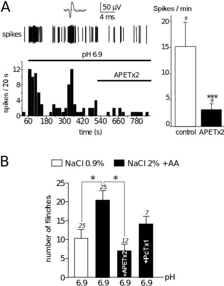
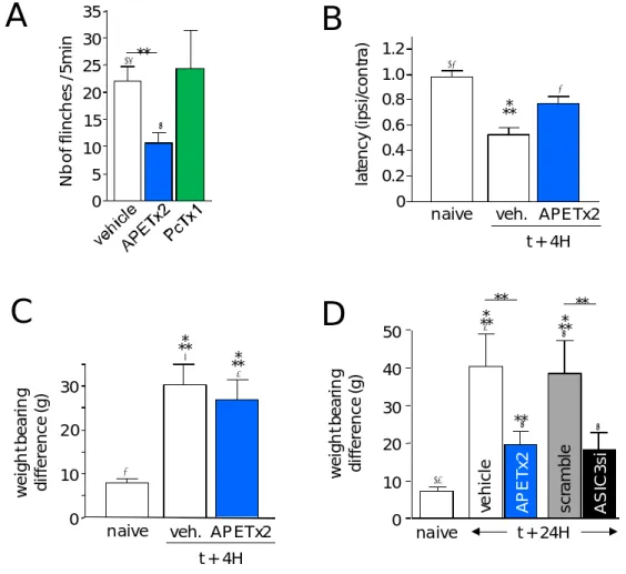
SAKIII Neuron NaASIC3 V1.8
(Blanchard et al., 2011; Diochot et al., 2004) BgII
Bunodosoma granulifera 5072(48)
46C
Type 1 mammals Insects, Cardiac, neuron
0.4 (i.c./mice) NaV (Bosmans et al., 2002; Goudet et al., 2001; Loret et al., 1994)
BgK 4275(37) 2, 6C
SAK-I Crust, mammals 4.5 (i.c./mice) KV1 (Aneiros Cotton et al., 1997) et al., 1993; BcIV Bunodosoma caissarum 4669(41) 4,6C SAKIII NL >2000 (crab) paralytic ? (Oliveira et al., 2006) CgII
Condylactis gigantea Type 1 46C Crust 0,2 (crab) (Salgado and Kem, 1992)
SGI 5395(50) 46C
Type 1 Crust NL (i.v./mice) 14 (crab) (Schweitz et al., 1981)
GiganTxI (48) 46C Type 2 ? NL >1000(crab) paralytic NL>1000 (i.c./mice) EGF activity (Shiomi et al., 2003) Halcurin
Halcurias sp 5080(47) Type 1 & 2 46C Crust NL >1000(mice) 6 (crab) (Ishida et al., 1997) Rp-I Radianthus (Heteractis)paumotensis 46C Type 2 Crust, mammals
Muscle & neuron 1.5 (i.c./mice) 36 (crab) NaV (Schweitz et al., 1985)
RpIII 5330(48) 46C
Type 2 Crust, mammals Muscle & neuron 2 (i.c. /mice) 10 (crab) NaV (Schweitz et al., 1985) ShI
Stichodactyla helianthus 5137(48)
46C (1SHI)
Type 2 NL (mice) Crust 0.3 (crab) >15000 (i.p./mice)
NaV (Kem et al., 1989; Salgado and Kem, 1992)
ShK 4055(35) 2, 6C (1ROO)
SAK-I Crust, mammals More toxic than BgK? KKVV1, 3
(Pennington et al., 1995; Yan et al., 2005)
ShPI 6110(55) 2, 1, 6C
SAKII ? ? ? (Antuch et al., 1993)
SHTXI, SHTXII
Stichodactyla haddonii 3059(28)
2, 6C
SAK-I Crust (NL) NL, paralytic 1000 (crab) KV1 (Honma et al., 2008) SHTXIII 7035(62) 2, 1, 6C
SAKII Crust (NL) >1000 (crab) NL, paralytic KV? (Honma et al., 2008)
SHTXIV 5229(48) 46C
Type 2 Crust 93 (crab) NaV? (Honma et al., 2008) References listed here are not exhaustive but are examples of some structural, pharmacological characterization of toxins and their toxicity studies; Sea anemone genus and species are indicated at the head of each group of toxin from a same SA. Abbreviations used are: Crust: crustacean, NL: