HAL Id: inserm-00851471
https://www.hal.inserm.fr/inserm-00851471
Submitted on 14 Aug 2013
HAL is a multi-disciplinary open access
archive for the deposit and dissemination of sci-entific research documents, whether they are pub-lished or not. The documents may come from teaching and research institutions in France or abroad, or from public or private research centers.
L’archive ouverte pluridisciplinaire HAL, est destinée au dépôt et à la diffusion de documents scientifiques de niveau recherche, publiés ou non, émanant des établissements d’enseignement et de recherche français ou étrangers, des laboratoires publics ou privés.
New methodological strategies in haematology using
cell-derived microvesicles
Eduardo Angles-Cano
To cite this version:
Eduardo Angles-Cano. New methodological strategies in haematology using cell-derived microvesicles: New methodological strategies in haematology. Nuevas Metodologias en el Estudio de Enfermedades Hematologicas, Apr 2013, Mazatlan, Mexico. pp.S38-S41. �inserm-00851471�
Programa educativo
New methodological strategies in haematology using cell-derived
microvesicles
Eduardo Angles-Cano, M.D., Sc.D.
Inserm UMR_S765 « Thrombosis: pathophysiology and new therapies»
Faculty of Pharmaceutical and Biological Sciences, Paris Descartes University
Correspondence to:
Eduardo ANGLES-CANO, M.D., Sc. D. Inserm U765
Faculté de Sciences Pharmaceutiques et Biologiques 4, Avenue de l’Observatoire, Cedex 75270
75006 Paris, France www.nietoeditores.com.mx
Microvesicles as messengers of cell and tissue dys-function
C
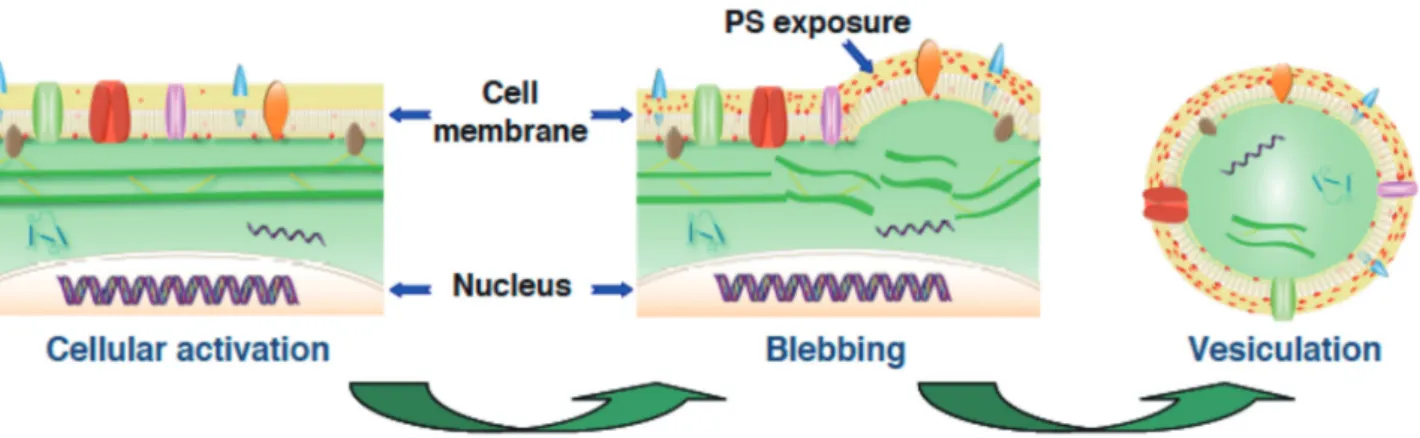
ellular microvesicles are membrane nanometric vesicles, 0.1-1 µm in size, released into body fluids by activated cells or during apoptosis in a variety of pathological conditions [1-4]. Characteristically, cell activation disturbs phospholipids transport and cytoskele-ton membrane connexions resulting in phosphatidylserine exposure, membrane blebbing and vesiculation (Fig. 1).The most well known cellular MVs are those of pla-telet, leukocyte, erythrocyte and endothelial cell origin found in circulating blood [5]. A number of studies have demonstrated that stimulation of these cells is followed by the characteristic features of cell activation: increased levels of cytoplasmic calcium associated to translocation of phosphatidylserine from the inner to the outer leaflet of the membrane and activation of calpains that, by clea-ving cytoskeletal filaments, facilitate MVs shedding [6]. The increase in intracellular Ca2+ concentration induces a
disordered state in the phospholipids asymmetry of quies-cent cells that is normally maintained by the concerted activity of transporter proteins [7, 8]: the ATP-dependent inward- and outward-directed transporters, flippases
(ami-nophospholipid translocase) and floppases (including the ATP-binding cassette transporter ABCA1) respectively, and the Ca2+-dependent scramblases that facilitate
bidi-rectional movement between the 2 membrane leaflets. The rate of phosphatidylserine translocation has been found to be sensitive to the altered expression of ABCA1 [9].
Microvesicles reflect the state of the parent cell
Besides phosphatidylserine microvesicles convey surface identity antigens and contain biomolecules that allow their identification and functional characterization [10]. These membrane glycroproteins and cytoplasmic compo-nents include the coagulation trigger tissue factor (TF), fibrinolytic enzymes, growth factors and their receptors, inflammatory mediators (cytokines, chemokines) and even mRNA or miRNA. Microvesicles may thus be considered as messengers of cell and tissue damage [10] and their presence in the circulation and other body fluids constitutes a signature of cell activity or dysfunction [11]. For these reasons, MVs have been proposed as pathogenic markers, key players of the haemostatic response [12]. At present, the most solidly established applied research on MVs is their procoagulant activity as a determinant of thrombosis risk in various clinical conditions [4, 13-15]. Evidence obtained is however largely associational and the main clinical application of MVs analysis has been the simple correlation of their levels with various disease states including cardiovascular diseases, diabetes, autoimmune diseases, inflammatory processes, sepsis and cancer.
Vascular ischemic accidents are the result of an occlu-sive thrombus formed in situ or of an embolus. Although a defect in fibrinolysis and/or localised proteolysis [16] is certainly implicated, we do not have at present a re-liable methodology to accurately assess the fibrinolytic activity in the intravascular space. Recent data strongly
New methodological strategies in haematology using cell-derived microvesicles
suggest that cellular MVs may be an important source of fibrinolytic and proteolytic activity in circulating blood [17]. For instance, MVs bearing plasminogen activators transform plasminogen into plasmin at their membrane and participate in a new fibrinolytic cross-talk mechanism that was only recently evidenced [18]. (see Angles-Cano E & Plawinski A, corresponding chapter in this volume).
Microvesicles: a dynamic pool of bioactive effectors
Other than the procoagulant factors and their distinctive glycoproteins, MVs may also carry bioactive components (membrane receptors, cytokines, transcription factors, mRNA) that are veritable indicators of the state of activa-tion of the parental cell, thus constituting a disseminated dynamic pool of bioactive effectors or messengers, as do-cumented by several in vitro studies [17 25, 19 22, 20 17]. Some of these MV components may exert local functions or be transferred to other cells: local fibrinolytic and pro-teolytic activities induced by uPA and metalloproteinases; transfert of GPIIb/IIIa from platelet MVs to leukocytes; transfert of mRNA, transfert of chemokin receptor; trans-fert of TF from leukocytes MVs to platelets, delivery of infectious agents into cells (human immuno deficiency virus, prions) [21] and transfert of oncogens from glioma MVs to naïve cells. Using proteomic approaches (two-dimensional electrophoresis and mass spectrophotometry), the number of proteins being identified in MVs of several origins has importantly expanded [22 24, 23 36, 24 34].
Microparticle’s identity unveil activated or suffering cells
Membrane glycoproteins distinctive of the parental cells are present on circulating MVs allowing thereby
identifi-cation of their cellular origin. Antibodies directed against these cell-specific antigenic determinants are used for this purpose in flow cytometry or antibody capture assays. An increase in the number of distinct MVs is now considered as an indicator of platelet, endothelial or leucocyte acti-vation [25 42, 26 16].
Identification of MVs constitutes therefore a solid advantage to determination of their sole number and re-presents a robust parameter when associated to thrombotic, systemic or inflammatory diseases. Furthermore, identifi-cation of MVs of practically any cell origin in plasma or other biological fluids (cerebrospinal fluid –CSF-, tears, exudates etc) would become possible if antibodies directed against cell-specific antigenic determinants were available. Their detection would certainly be considered as a direct message of tissue specific activation or damage. For instan-ce, the hypothesis that tumour-derived TF-positive MVs in plasma contribute to cancer-associated thrombosis is based on the finding of these MVs in patients with solid tumours and venous thrombosis. Clotting tests using tumour cell samples suggest that cancer cells are a potential source of circulating TF-positive MVs [27]. More specific informa-tion could be obtained if MVs of tumour origin (solid or leukemic) could be used as early messenger of relapse.
In recent years, tumour MVs have evolved as potential biomarkers. Indeed, tumour cells are able to constitutively release large amounts of MVs bearing tumour specic an-tigens [28]. These MVs may found into the bloodstream and other bodily fluids. Microvesicles released by malig-nant cancer cells can transfer various messages to target cells and may be critical to disease progression [29]. For example, solid tumours that are difficult to reach and de-Figure 1. Scheme of cell activation, phosphatidylserine (PS) exposure, membrane blebbing and release of microvesicles (vesiculation).
tect may reveal their presence by releasing MVs, and the presence of tumour-derived MVs in biological fuids may also be useful for detecting metastases.
In hematologic malignancies the study of MVs is gaining increased interest. For instance B-Cell-derived MVs from chronic lymphocytic leukaemia (CLL) express separate phenotypes during leukemic disease progression and underscores the important role of MVs in activa-tion of the tumour microenvironment [30]. TF-bearing promyelocytic-derived MVs in acute promyelocytic leu-kemia have been identified using and antibody to CD33. These MVs decreased the coagulation time and induced thrombin generation, thus indicating that the procoagulant state in acute promyelocytic leukemia is partially due to the TF-dependent procoagulant properties of circulating promyelocytic-derived MVs [31, 32]. Furthermore, procoagulant myeloblast-derived MVs were recently des-cribed in acute myeloblastic leukaemia [33]. Myeloblas origin was defined by cytofluorimetry using antibodies to CD117, CD13 and CD34. These MVs were highly pro-coagulant as determined with a thrombin generation test. The release of MVs by mature B cell tumours in childhook leukaemias may be related to the cellular activation status or to the activity of the leukaemia cell type [34].
In a proteomic study of mature B-cell neoplasms with B-cell hyperlymphocytosis, including CLL, small cell lymphoma from hairy cell leukemia or splenic lympho-ma with villous lymphocytes, CD148 was identified on lymphocyte MVs. The presence of this marker in MVs excludes the diagnosis of CLL and allows mantle cell lymphoma diagnosis to be suspected [35].
In summary, the generation of leukaemia/lymphoma cell MVs constitute a new tool for diagnosis and clinical/ therapeutic follow-up.
REFERENCES
1 Chironi GN, Boulanger CM, Simon A, Dignat-George F, Freys-sinet JM, Tedgui A. Endothelial microparticles in diseases. Cell
Tissue Res. 2009; 335: 143-51.
2 Sellam J, Proulle V, Jungel A, Ittah M, Miceli Richard C, Gottenberg JE, Toti F, Benessiano J, Gay S, Freyssinet JM, Mariette X. Increased levels of circulating microparticles in primary Sjogren's syndrome, systemic lupus erythematosus and rheumatoid arthritis and relation with disease activity.
Arthritis Res Ther. 2009; 11: R156.
3 Lynch SF, Ludlam CA. Plasma microparticles and vascular disorders. Br J Haematol. 2007; 137: 36-48.
4 Daniel L, Dou L, Berland Y, Lesavre P, Mecarelli-Halbwachs L, Dignat-George F. Circulating microparticles in renal diseases.
Nephrol Dial Transplant. 2008; 23: 2129-32.
5 Morel O, Toti F, Hugel B, Bakouboula B, Camoin-Jau L, Dignat-George F, Freyssinet JM. Procoagulant microparticles: disrupting the vascular homeostasis equation? Arterioscler
Thromb Vasc Biol. 2006; 26: 2594-604.
6 Pasquet JM, Dachary-Prigent J, Nurden AT. Calcium influx is a determining factor of calpain activation and microparticle formation in platelets. Eur J Biochem. 1996; 239: 647-54. 7 Bevers EM, Comfurius P, Dekkers DW, Zwaal RF. Lipid
trans-location across the plasma membrane of mammalian cells.
Biochim Biophys Acta. 1999; 1439: 317-30.
8 Daleke DL. Regulation of transbilayer plasma membrane phospholipid asymmetry. J Lipid Res. 2003; 44: 233-42. 9 Combes V, Coltel N, Alibert M, van Eck M, Raymond C,
Juhan-Vague I, Grau GE, Chimini G. ABCA1 gene deletion protects against cerebral malaria: potential pathogenic role of micropar-ticles in neuropathology. Am J Pathol. 2005; 166: 295-302. 10 Morel O, Toti F, Hugel B, Freyssinet JM. Cellular microparticles:
a disseminated storage pool of bioactive vascular effectors.
Curr Opin Hematol. 2004; 11: 156-64.
11 Mause SF, Weber C. Microparticles: protagonists of a novel communication network for intercellular information exchange.
Circ Res. 2010; 107: 1047-57.
12 Freyssinet JM. Cellular microparticles: what are they bad or good for? J Thromb Haemost. 2003; 1: 1655-62.
13 Piccin A, Murphy WG, Smith OP. Circulating microparticles: pathophysiology and clinical implications. Blood Rev. 2007; 21: 157-71.
14 Morel O, Toti F, Hugel B. Procoagulant microparticles: disrup-ting the vascular homeostasis equation? Arterioscler Thromb
Vasc Biol. 2006; 26: 2594-604.
15 Al-Massarani G, Vacher-Coponat H, Paul P, Widemann A, Arnaud L, Loundou A, Robert S, Berland Y, Dignat-George F, Camoin-Jau L. Impact of immunosuppressive treatment on endothelial biomarkers after kidney transplantation. Am J
Transplant. 2008; 8: 2360-7.
16 Meltzer ME, Doggen CJ, de Groot PG, Rosendaal FR, Lisman T. Reduced plasma fibrinolytic capacity as a potential risk factor for a first myocardial infarction in young men. Br J Haematol. 2009; 145: 121-7.
17 Lacroix R, Sabatier F, Mialhe A, Basire A, Pannell R, Borghi H, Robert S, Lamy E, Plawinski L, Camoin-Jau L, Gurewich V, Angles-Cano E, Dignat-George F. Activation of plasminogen into plasmin at the surface of endothelial microparticles: a me-chanism that modulates angiogenic properties of endothelial progenitor cells in vitro. Blood. 2007; 110: 2432-9.
18 Dejouvencel T, Doeuvre L, Lacroix R, Plawinski L, Dignat-George F, Lijnen HR, Angles-Cano E. Fibrinolytic cross-talk: a new mechanism for plasmin formation. Blood. 2010; 115: 2048-56.
19 Graves LE, Ariztia EV, Navari JR, Matzel HJ, Stack MS, Fis-hman DA. Proinvasive properties of ovarian cancer ascites-derived membrane vesicles. Cancer Res. 2004; 64: 7045-9. 20 Taraboletti G, D'Ascenzo S, Borsotti P, Giavazzi R, Pavan A,
Dolo V. Shedding of the matrix metalloproteinases MMP-2, MMP-9, and MT1-MMP as membrane vesicle-associated components by endothelial cells. Am J Pathol. 2002; 160: 673-80.
New methodological strategies in haematology using cell-derived microvesicles
21 Simak J, Holada K, D'Agnillo F, Janota J, Vostal JG. Cellular prion protein is expressed on endothelial cells and is released during apoptosis on membrane microparticles found in human plasma. Transfusion. 2002; 42: 334-42.
22 Janowska-Wieczorek A, Wysoczynski M, Kijowski J, Marquez-Curtis L, Machalinski B, Ratajczak J, Ratajczak MZ. Microvesi-cles derived from activated platelets induce metastasis and angiogenesis in lung cancer. Int J Cancer. 2005; 113: 752-60. 23 Plow EF, Pluskota E. It’s not size, it’s substance. Blood. 2007;
110: 2224-5.
24 Oh J, Takahashi R, Kondo S, Mizoguchi A, Adachi E, Sasahara RM, Nishimura S, Imamura Y, Kitayama H, Alexander DB, Ide C, Horan TP, Arakawa T, Yoshida H, Nishikawa S, Itoh Y, Seiki M, Itohara S, Takahashi C, Noda M. The membrane-anchored MMP inhibitor RECK is a key regulator of extracellular matrix integrity and angiogenesis. Cell. 2001; 107: 789-800. 25 Simak J, Gelderman MP, Yu H, Wright V, Baird AE. Circulating
endothelial microparticles in acute ischemic stroke: a link to severity, lesion volume and outcome. J Thromb Haemost. 2006; 4: 1296-302.
26 Distler JHW, Jüngel A, Huber LC, Seemayer CA, Reich CF, 3rd, Gay RE, Michel BA, Fontana A, Gay S, Pisetsky DS, Distler O. The induction of matrix metalloproteinase and cytokine expression in synovial fibroblasts stimulated with immune cell microparticles. Proc Natl Acad Sci U S A. 2005; 102: 2892-7. 27 Langer F, Spath B, Haubold K, Holstein K, Marx G, Wierecky
J, Brummendorf TH, Dierlamm J, Bokemeyer C, Eifrig B. Tissue factor procoagulant activity of plasma microparticles in patients with cancer-associated disseminated intravascular coagulation. Ann Hematol. 2008; 87: 451-7.
28 Giusti I, D'Ascenzo S, Dolo V. Microvesicles as potential ovarian cancer biomarkers. BioMed Research International. 2013; 2013.
29 Falanga A, Tartari CJ, Marchetti M. Microparticles in tumor progression. Thromb Res. 2012; 129 Suppl 1: S132-6. 30 Ghosh AK, Secreto CR, Knox TR, Ding W, Mukhopadhyay D,
Kay NE. Circulating microvesicles in B-cell chronic lymphocytic leukemia can stimulate marrow stromal cells: implications for disease progression. Blood. 2010; 115: 1755-64.
31 Ma G, Liu F, Lv L, Gao Y, Su Y. Increased promyelocytic-derived microparticles: a novel potential factor for coagulopathy in acute promyelocytic leukemia. Ann Hematol. 2013.
32 Kwaan HC, Rego EM. Role of microparticles in the hemostatic dysfunction in acute promyelocytic leukemia. Semin Thromb
Hemost. 2010; 36: 917-24.
33 Van Aalderen MC, Trappenburg MC, Van Schilfgaarde M, Molenaar PJ, Ten Cate H, Terpstra WE, Leyte A. Procoagulant myeloblast-derived microparticles in AML patients: changes in numbers and thrombin generation potential during chemothe-rapy. J Thromb Haemost. 2011; 9: 223-6.
34 Savasan S, Buyukavci M, Buck S, Ravindranath Y. Leukae-mia/lymphoma cell microparticles in childhood mature B cell neoplasms. J Clin Pathol. 2004; 57: 651-3.
35 Miguet L, Bechade G, Fornecker L, Zink E, Felden C, Gervais C, Herbrecht R, Van Dorsselaer A, Mauvieux L, Sanglier-Cianferani S. Proteomic analysis of malignant B-cell derived microparticles reveals CD148 as a potentially useful antigenic biomarker for mantle cell lymphoma diagnosis. J Proteome