CDK control of mitotic progression in Saccharomyces cerevisiae
by Rami S. Rahal B.Sc. Biochemistry McMaster University, 2002
SUBMITTED TO THE DEPARTMENT OF BIOLOGY IN PARTIAL FULFILLMENT OF THE REQUIREMENTS FOR THE DEGREE OF
DOCTOR OF PHILOSOPHY IN BIOLOGY AT THE
MASSACHUSETTS INSTITUTE OF TECHNOLOG
JUNE 2008
1MAY 2 9
2008
© 2008 Rami S. Rahal. All rights reserved.
The author hereby grants to MIT permission to reproduce and distribute publicly paper and electronic copies of this thesis document in whole or in part.
Signature of the Author:
Department of Biology May 23, 2008 Certified by:_ *Angelika Amon Professor of Biology Thesis Supervisor Accepted by: Stephen P. Bell Professor of Biology Chair, Committee for Graduate Students
CDK control of mitotic progression in Saccharomyces cerevisiae
by Rami S. Rahal
Submitted to the Department of Biology on May 23, 2008 in partial fulfillment of the requirements for the Degree of Doctor of Philosophy in Biology
Abstract
Mitotic cyclin-dependent kinases (CDKs) are best known for their essential functions in triggering entry into M-phase, where they have established roles in nuclear envelope breakdown, chromosome condensation, and Golgi fragmentation. Whether mitotic CDKs have essential functions within mitosis is unclear. The work presented in this thesis indicates that mitotic CDKs, Clbl/2-CDKs or
Clb2/3-CDKs, have two essential roles in mitotic progression. First, mitotic CDK activity was found to be required for the timely degradation of Securin/Pds1 at the metaphase-anaphase transition, demonstrating a requirement for mitotic CDKs in APC/C-Cdc20 activation in vivo. Second, mitotic CDK activity was necessary for the elongation of the mitotic spindle in anaphase. This requirement for mitotic CDKs is independent of cohesin removal and thus constitutes a novel role for CDK activity in mitotic progression. The dual roles of CDK in initiating loss of sister-chromatid cohesion and spindle elongation suggest a mechanism by which cells can couple and coordinate these events during mitosis. In addition, these results support a model in which different CDK activity threshold requirements help establish the order of events within in M phase, thereby ensuring cells initiate chromosome segregation only after they accumulate enough CDK activity to condense their chromosomes and to build a mitotic spindle.
Thesis Supervisor: Angelika Amon Title: Professor of Biology
Acknowledgments
I would like to express my deep gratitude to Angelika for her support during the last 5 years. I would also like to thank all members of the Amon lab, past and present, for creating such a
wonderful environment. I am especially grateful for having met Rosella Visintin, Frank Stegmeier, and Anupama Seshan in Angelika's lab.
I would like to thank Phil Sharp, Mike Yaffe and Frank Solomon for their support, encouragement, and thoughtful advice. I am greatly indebted to Frank for allowing me to barge into his office at all times of the day and for always being there to listen.
I am very grateful for having met Dr. Alexander Ball at McMaster University. Working in Dr. Ball's lab as an undergraduate was a life-changing experience and I could not have come to MIT without his help and generosity.
Finally, I am forever indebted to my family for their support and encouragement. My education has been their sole priority and they have done more for me than I could have ever imagined.
Table of Contents
A bstract ... ... 2
D edication ... ... 3
A cknow ledgem ents ... ... 4
T able of C ontents ... ... 5
Chapter I: Introduction ... 7
T he C ell C ycle ... ... 8
The Saccharomyces cerevisiae Cell Division Cycle ... .... 12
G p h ase ... 12 G 1/S ... ... 15 S p h ase ... ... 15 G 2 ... ... 16 M ito sis ... 16 M etap hase ... ... 17
The Metaphase-Anaphase Transition ... ... 18
The Anaphase Promoting Complex/Cyclosome (APC/C) ... 21
Mitotic regulation of APC/C-Cdc20... 25
Loss of sister-chromatid cohesion and chromosome segregation ... 29
A naphase/Telophase ... ... 30
Exit from M itosis ... ... 32
The CdcFourteen Early Anaphase Release (FEAR) Network ... 33
The Mitotic Exit Network (MEN) ... ... 34
C ytokin esis ... ... 37
Chromosome segregation in higher eukaryotes ... ... 38
Regulation of APC/C-Cdc20 activation by mitotic inhibitors ... 38
Regulation of APC/C-Cdc20 activation by mitotic timers ... 39
Regulation of APC/C-Cdc20 activity by ubiquitylation and de-ubiquitylation ... 40
Separase auto-cleavage ... ... 4 1 Mitotic CDKs act like Securins and Separases act like CDK inhibitors ... 41
Step-wise loss of arm and centromere cohesion in mitosis ... 42
T hesis Sum m ary ... ... 43
Chapter II: Mitotic CDKs control the metaphase-anaphase transition and trigger spindle elongation ... 57
S u m m ary ... ... 5 8 In trodu ction ... ... 59
R esu lts ... ... 6 2 Nuclear division is impaired in cells lacking Clbl/2-CDK activity. ... 62
The mitotic progression defect of clblA clb2-VI cells is independent of inhibitory Tyrl9 phosphorylation on Cdc28 ... .... 67
Clbl/2-CDK activity is required during metaphase to promote anaphase ... 67
Clbl/2-CDK activity is needed for Securin degradation ... 77
clblA clb2- VI cells are defective in spindle elongation in the absence of PDS1 ... 85
Inactivation of cohesin does not suppress the spindle elongation defect of clblA clb2- VI cells ... ....86
Clbl/2-CDK activity is required for anaphase spindle elongation ... 97 Clb2/3-CDK activity is required for Securin/Pds1 degradation and
anaphase spindle elongation ... 105
D iscu ssio n ... ... 1 14 Clb-CDK activity is required for APC/C-Cdc20 activation in vivo ... 114
Mitotic CDK activity is required for spindle elongation during anaphase ... 115
A model for how the rise in CDK activity during early mitosis establishes order to mitotic progression ... 129
M aterials and M ethods ... ... 133
R eferen ces ... 137
Chapter III: Components of the CdcFourteen Early Anaphase Release (FEAR) Network physically interact ... 143
S um m ary ... ... 144
In trod u ction ... 14 5 R esu lts ... 14 9 The FEAR network components Slk19, Espl, Cdc5 and Cdcl4 physically interact ... 149
The physical interactions of FEAR network components are not cell cycle regulated ... 155
The physical interactions of FEAR network components are not interdependent ... ... 161
The Polo-box Domain of Cdc5 is sufficient to bind Cdcl4 ... 167
The N-terminus of Cdcl4 is sufficient to bind to Cdc5's PBD ... 173
D iscussion ... 179
Are the observed physical interactions among FEAR network components relevant to FEAR network activation? ... . ... 180
Does Cdc5 bind directly to Cdcl4? ... 180
M aterial and m ethods ... ... 183
R eferences ... 187
Chapter IV: Discussion and Future Directions ... 193
K ey C onclusions ... ... ... 194
D iscussion O verview ... ... 195
Mitotic Regulation of APC/C-Cdc20 ... 196
Future Experim ents ... 201
Some unanswered questions about the APC/C-Cdc20 complex ... 204
Mitotic CDK activity is required for spindle elongation ... 207
CDK activity threshold requirements help establish order during mitotic progression ...210
Future Experim ents ... ... 2 12 Signaling in the Cdc Fourteen Early Anaphase Release (FEAR) Network ... 213
Some unanswered questions about the FEAR network ... 218
Concluding remarks on the FEAR network ... 221
C oncluding R em arks ... ... 222
Appendix I: The Replication Fork Block Protein Fobl Functions as a Negative Regulator of the FEAR Network ... ... 227
Appendix II: APC/C-Cdhl-mediated degradation of the Polo kinase Cdc5 promotes the return of Cdcl4 into the nucleolus... 243
Chapter I:
Introduction
Cell division is a fundamental biological process by which a single cell transforms into two identical cells. Biologists have long been fascinated by the inherent complexity of cell division and mystified by its high fidelity. How is it that a cell, a collection of water, protein, lipid, and sugar can make as
complex a decision as whether or not to divide? Upon deciding to divide, how does a cell ensure that the processes required for cell division - growth, genome duplication, genome segregation, and daughter cell separation occur in the appropriate order? It is specifically the ordering of events in the cellular milieu that is the most intriguing part of cell division. Finally, how does the cell complete these tasks so well? Cell division is seemingly effortless -just watch any budding yeast do it. Yet, exquisite controls must be present to ensure that errors are not made during genome duplication and its subsequent partitioning. For yeast and human cells, errors in chromosome segregation are deleterious.
The Cell Cycle
Work over the last 100 years has revealed that the eukaryotic cell division program has a nearly universal architecture consisting of four phases: Gap 1 (G1), Synthesis (S), Gap 2 (G2), and Mitosis (M) (Figure 1). Growth and the commitment to cell division occur in G 1, DNA synthesis and the
tethering of duplicated sister-chromatids occur in S phase, further growth and preparation for division occur in G2, and sister-chromatid separation and cell division occur in M phase .
Importantly, cells return to a G 1 state after completing M phase, hence the division program is cyclical.
Gap2:
Prepare to divide chromosomes
Mitosis:
Divide chromosomesSynthesis:
Copy chromosomes 0 Centrosome Kinetochore Linkages between sister-chromatids Mitotic spindleGapl:
Prepare to copy chromosomes
Figure 1: The Cell Division Cycle
The cell cycle is divided into four phases. During GI phase, the cell coordinates its growth with entry into the cell cycle. In many systems, centrosome duplication is initiated near the G1/S transition. During S phase, the genome is duplicated and sister-chromatids are bound together by protein linkages. During G2, cells continue their preparation for M phase and the division of their cytoplasm. During M, centrosomes separate and organize a microtubule spindle. The mitotic spindle attaches to sister-chromatids via their kinetochores and drives their separation in anaphase.
The progression of cells through the cell cycle is controlled by the activity of cyclin-dependent kinases (CDK) and their activating cyclin partners1,2. Cyclin-CDK complexes are the principal regulators of all known eukaryotic cell cycles, helping to control both progression through each cell cycle phase and the transition from one phase to the next. Phase-specific cyclins allow CDKs to have distinct functions during G1, S, G2, and M phase. In addition, the orderly fluctuations in cyclin abundance during the cell cycle help establish its directionality (S phase before G2). Cyclin levels oscillate due to waves of synthesis and rapid proteolysis .
Our current understanding of the cell cycle is a synthesis, based on the work of numerous investigators who studied the cell cycle in flies, frog eggs extracts, clam embryos, human cancer cells, and yeast cells. Studies on budding yeast cell division, in particular, have contributed significantly to our understanding of genes that control cell cycle progression. Budding yeast's genetic tractability and our ability to synchronize its divisions have been instrumental in making this organism an essential contributor to cell cycle research.
My thesis work focused on how chromosome segregation is regulated in budding yeast and the roles mitotic CDKs played in this process. The majority of this thesis will therefore describe cell cycle events as they occur in budding yeast. However, the regulatory circuits controlling cell division are highly conserved, and in many cases, function identically in yeast and vertebrate cells. For my introduction, I will briefly review our understanding of how yeast cyclin-CDK complexes control progression from GI to M phase and then discuss in detail our current understanding of the chromosome segregation machinery.
The Saccharomyces cerevisiae Cell Division Cycle
The cell division cycle of budding yeast is driven by a single CDK, CDC28, and 9 cyclin genes,
CLN1-3 and CLBI-6 3. CLNI-3 encode GI cyclins that coordinate growth and the commitment to
cell division, while CLB1-6 encode B-type cyclins that have overlapping functions in S phase and mitosis (Figure 2). Clb5/Clb6-CDK activity is important for the timely initiation of S phase4'5, while
Clb(1,2,3,4)-CDK activity is important for spindle morphogenesis and progression through mitosis 6-8. A great deal of functional redundancy is present among the cyclins since no single gene is
essential. For example, all single and pair-wise deletions of the G1 cyclins are viable, but only that triple mutant is inviable 9.
G1 phase
The progression of yeast cells through GI is regulated by multiple inputs, including nutrient
availability and the presence of a mating partner 3,10. The G1 phase is also where the coordination of cell growth and cell division occurs ". Although this process remains poorly understood, it is known to control the translation of Cln3, which form the earliest acting cyclin-CDK complex 12,13. Cln3-CDK complexes promote the transcription of Clnl and Cln2 by phosphorylating and triggering the nuclear export of Whi5, a transcriptional repressor bound to the promoters of Clni and Cln2 14,15. The
accumulation of Clnl/2 cyclins is important for a number of GI phase events, including bud formation, polarity establishment, the duplication of spindle pole bodies (yeast centrosomes), and, most importantly, for the activation of the S phase cyclins, Clb5 and Clb6 3.
F'IL~~ Id I~F~T% ~IEJti3I4-LJJN
Clb5/6-CDK
S
M
Cb/-CDK
3-CDK
Clnl/2/3-CDK
GI/SI
Metaphase/Anaphase Mitotic Exit
I
I
GI S Metaphase Anaphase G1
Cell Cycle Phase Figure 2: Cyclin-CDK activity during the budding yeast cell cycle
(A) Budding yeast cyclins are restricted to distinct phases ofthe cell cycle. Cln(l-3)-CDK activity accumulates in GI while Clb5/6-CDK activity accumulates in S phase. Budding yeast lack a canonical G2 phase. During late S and early M phase, Clb3/4-CDKs are the first to accumulate and are quickly fllowed by Clbl/2-CDKs.
(B) Oscillations in cyclin-CDK activity promote cell cycle progression. Cln(l-3)-CDK activity accumulates in GI, triggering the degradation of the Clb5/6-CDK inhibitor Sicl. The rise in Clb5/6-CDK activity triggers entry into S phase and DNA synthesis. Clb3/4-CDKs
accumulate in late S/early M and trigger the separation ofyeast SPBs (mitotic entry). Clbl/2-CDK activity accumulates shortly afer Clb3/4-Clbl/2-CDK activity and contributes to SPB
separation. Collectively, Clb(1-4)-CDKs preserve the mitotic state by repressing the transcription ofGl cyclins and preventing origin re-firing. At the metaphase-anaphase transition, the entire pool ofS phase Clbs and halfthe pool ofmitotic Clbs are degraded. Exit from mitosis occurs when mitotic Clb-CDK activity (red) is completely eliminated, permitting Cln cyclin accumulation and a return to the GI state.
G1/S
Clb5 and Clb6 are present in G1, but Clb5/6-CDK complexes are inactivate because they are bound by Sicl, a potent CDK inhibitor that is specific to B-type cyclin-CDK complexes 16. Clnl/2-CDK dependent phosphorylation of Sicl generates a "phospho-degron" that is recognized by an E3 ubiquitin ligase complex, SCF, in association with the specificity factor Cdc4 16-21. SCF-Cdc4
mediated ubiquitylation of Sicl 1 triggers its destruction by the 26S proteosome. The essential function of the G 1 cyclins appears to be the activation of Clb5/6-CDKs since SIC1 deletion or CLB5 over-expression can rescue the lethality of cinlA cln2A cln3A cells 4,19,22
S phase
Sic Il degradation at GI/S liberates active Clb5/6-CDKs, which along with other S phase kinases, promote replication origin firing by phosphorylating pre-replicative complexes (pre-RC) and other replication factors23. Although Clb5 and Clb6 are the canonical S phase cyclins, DNA replication
can still occur in clb5A clb6A cells due the remaining 4 B-type cyclins, Clbl-4'6. Collectively,
Clb-CDKs not only trigger DNA synthesis, they also ensure that it only occurs once per cell cycle by limiting origin firing during M phase 23.24
During S phase, protein based linkages are established between the duplicated sister chromatids to ensure they are properly segregated in anaphase. These linkages are mediated by the ring-shaped cohesin complex, which in budding yeast is composed of four subunits: Sccl/Mcdl, Scc3, Smcl, Smc3 25.26. The loading of cohesin complexes onto sister chromatids is thought to occur
concomitantly with their replication during S phase, although this process remains poorly understood
27. It is unclear how the cohesin complex promotes sister chromatid cohesion, but its ring like
G2
In most organisms, cells are unable to enter mitosis immediately after S phase because of inhibitory tyrosine phosphorylation of CDK, which is catalyzed by the Wee 1 family of kinases and opposed by the Cdc25 family of phosphatases 28,29. In G2, Tyr phosphorylation of CDK prevents the
accumulation of mitotic CDK activity, delaying mitotic entry, and allows cells time to continue their growth and preparation for cell division 1,28. Tyr phosphorylation of CDK also blocks mitotic entry in response to DNA damage to permit repair 30-32. Expression of a mutant CDK that can no longer be tyrosine phosphorylated causes premature mitotic entry in S. pombe33 or human cells34,35, but not in budding yeast36-39. Tyrosine phosphorylation of CDK does occur in budding yeast, but its primary function is to delay the cell cycle under specific conditions, such as those that trigger the
morphogenesis checkpoint (which responds to perturbations of the actin cytoskeleton)40. Therefore,
budding yeast lack a canonical G2 phase and do not undergo a typical G2/M transition.
Mitosis
In animal and plant cells, mitosis has distinct sub-phases that have defined cytological and molecular markers. Prophase is characterized by chromosome condensation and spindle assembly;
prometaphase by nuclear envelope breakdown and kinetochore capture by microtubules; metaphase by the congression of oriented sister-chromatids to the cell equator; anaphase by the separation of sister-chromatids; and telophase by chromosomes de-condensation and nuclear envelope
reformation'. Budding yeast lack all these hallmarks due to their small chromosomes, the
constitutive attachment of kinetochores to microtubules, and the lack of nuclear envelope breakdown during fungal mitoses. In the absence of clear cytological markers for early M phase, entry into mitosis and entry into metaphase are indistinguishable events in budding yeast. Mitotic entry is best defined in this organism by the separation of the duplicated spindle pole bodies (yeast centrosomes)
because this event requires mitotic Clb-CDK activity ',7'4 1. Therefore, I will begin my review of M phase by starting in metaphase and describing how sister-chromatids are properly aligned prior to the separation.
Metaphase
Faithful chromosome segregation during mitosis requires sister-chromatids to be properly aligned on the mitotic spindle prior to cohesion loss. Sister-chromatids are aligned in metaphase by the
microtubule based mitotic spindle, which attaches to individual chromatids via the kinetochore. Microtubule attachment to kinetochores proceeds through a random "search and capture"
mechanism, which does not guarantee proper sister-chromatid orientations42-44. Sister-chromatids are
"co-oriented" when their kinetochores are attached to microtubules emanating from the same spindle pole body, but "bi-oriented" when attached to microtubules emanating from opposite spindle pole bodies43. Every pair of sister-chromatids must be bi-oriented prior to anaphase onset. During early
mitosis, dedicated surveillance mechanisms exist to monitor and correct improper sister-chromatid orientations.
Cells determine if sister-chromatids are properly oriented on the spindle by monitoring the status of tension at sister-kinetochores. Tension is generated across bi-oriented sister-kinetochores when cohesin complexes linking sister-chromatids resist pulling forces exerted by the microtubules
emanating from opposite spindle poles 43,45-47. Co-oriented sister-chromatids, or those pairs in which
only one sister is attached to a microtubule, are unable to generate tension. Kinetochore-microtubule attachments that fail to create tension are severed by the Aurora family kinase Ipll 45,47-49 The recursive destabilization of improper kinetochore-microtubule attachments by Ipll ensures all sister-chromatids establish tension-generating microtubule attachments. What targets Ipll to tension-less
kinetochores and the mechanism(s) of tension sensing are not well understood. In budding yeast, tension sensing requires that Shugoshin family member Sgo 150
The Metaphase-Anaphase Transition
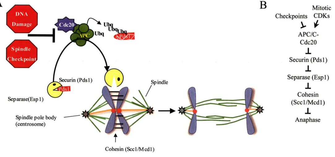
Cells initiate chromosome segregation at the metaphase-anaphase transition by removing the cohesin complexes holding sister chromatids together. At the onset of anaphase, the Scc l/Mcdl subunit of the cohesin complex is cleaved by the protease Separase (Espl 1 in budding yeast), which unlinks the sister-chromatids and allows them to be pulled to opposite poles of the cell (Figure3) 51-54. Prior to
anaphase, Separase is inactivated by the binding of Securin (Pdsl in budding yeast) 51,55-57 Securin/Pdsl has two opposing functions in Separase/Espl regulation: an activating one by promoting the nuclear import of Separase/Espl 58,59 and an inhibitory one by binding to
Separase/Espl 51. At the metaphase-anaphase transition, Securin/Pds 1 is targeted for proteosomal degradation by an E3 ubiquitin ligase complex, the Anaphase Promoting Complex/Cyclosome (APC/C), and its specificity factor Cdc20 51,57,60,61 (Figure 3).
B
A
UbqS~pD'j
(Pdsl) pindle Separase(Esp 1) Spindle pole I (centrosome)9
SN
D
aIa(Y
Cohesin (Sccl/Mcdl)
Figure 3: The metaphase -anaphase transition.
(A) Diagram of the chromosome segregation machinery during the metaphase-anaphase transition. Activation of APC/C-Cdc20 triggers Securin/Pdsl degradation, activating Separase/Esp 1. Separase/Esp 1 is a protease that cleaves the Sccl/Mcdl subunit of the cohesin
complexes tethering sister-chromatids together. The dissolution of sister-chromatid cohesion in metaphase is required for proper anaphase entry.
(B) Genetic architecture of the pathway that leads to loss of sister-chromatid cohesion.
Mitotic
Checkpoints CDKs
APC/C-Cdc20
1.
Securin (Pdsl)
Separase (Espl)
I
Cohesin
(Sccl/Mcdl)
Anaphase
mThe Anaphase Promoting Complex/Cyclosome (APC/C)
My thesis work uncovered a role for mitotic CDKs in APC/C-Cdc20 activation at the metaphase-anaphase transition. As a result, I will first introduce what we know about the structure and function of the APC/C complex and then review the regulatory pathways that control its activation during the metaphase-anaphase transition.
Architecture of the APC/C
The APC/C is a large ~1.5 mDa E3 ubiquitin ligase complex that is composed of at least 13 subunits in budding yeast, eight of which are essential '1,62-,66. APC/C's catalytic core is composed of Apcl 1, a RING-finger protein, and Apc2, a cullin protein, though Apcl I alone is sufficient for ubiquitin ligase activity in vitro 67-69. In vivo, APC/C activity is restricted to distinct cell cycle phases by its
association with two specificity factors, Cdc20 and Cdh 1 60
Cdc20 and Cdhi
Cdc20 and Cdhl are highly conserved WD40-domain containing proteins that enable APC/C to recognize distinct substrates during each cell cycle phase 6-.70 Budding yeast Cdc20 is essential and directs APC/C ubiquitylation during early mitosis, while Cdhl is not essential and directs APC/C ubiquitylation during mitotic exit/G161,70-73. At the metaphase-anaphase transition, APC/C-Cdc20 targets Securin/Pdsl, an inhibitor of anaphase entry, and Clb5, a potent-antagonist of APC/C-Cdh 1,
for degradation (other Clbs are also targeted)5 " . Securin/Pdsl and Clb5 are considered the
essential substrates for APC/C-Cdc20 since deletion of PDS1 and CLB5 rescues the inviability of
cdc20A cells74. APC/C-Cdhl ubiquitylates several mitotic determinants, including Clb
cyclins6',70, 7'.73.
Cdhl is not essential in budding yeast because CDK activity can be antagonized during mitotic exit/GI by the Clb-CDK inhibitor Sic 16170
IHlow APC/C-Cdc20 and APC/C-Cdhl recognize their targets is not completely clear, though this often requires short sequence elements in the substrate called destruction-boxes (D-boxes) and
KEN-boxes'5. The D-box consensus sequence, RxxLxxxN (x is any amino acid)7", is found in many APC/C-Cdc20 substrates and some APC/C-CdhI1 substrates75. The KEN box consensus sequence,
KEN77, is primarily found in APC/C-Cdhl substrates75.
The Toczyski lab has been able to demonstrate that Securin/Pds1 destruction and mitotic-CDK inactivation is indeed the essential function of the yeast APC/C by rendering it non-essential 78
Remarkably, over-expression of the CDK inhibitor Sic 1 and deletion of two APC/C-Cdc20
substrates, Securin/Pdsl and Clb5, allows yeast to tolerate loss of every essential APC/C subunit78.
The Toczyski lab then used this strain to build an "architectural map" of the APC/C by determining the inter- dependencies among the subunits for complex formation. This analysis revealed that the APC/C is composed of two sub-complexes that are bridged by the Apcl subunit (Figure 4). The
"catalytic core" sub-complex contains Apc2, Apc 11, and a small protein involved in substrate recruitment, Doc 179. The other sub-complex contains three essential, highly conserved,
tetratricopeptide repeat (TPR) subunits, Cdc23, Cdc27, and Cdcl6, and requires Apc4 and Apc5 to bind to Apc 179. The non-essential subunits, Cdc26, Swml, and Apc9 associate with the Cdc23-Cdc27-Cdc16 sub-complex79.
Figure 4: Architecture of APC/C
The APC/C is composed of two sub-complexes: one containing
Apcl 1, Apc2, and Docl (green) and another containing Cdcl6, Cdc23, Cdc27, Apc4, and Apc5 (yellow). The two sub-complexes are bridged by Apcl (orange). The three non-essential APC/C subunits (dashed lines), Cdc26, Apc9, and Swml, are associated with the Cdcl6-Cdc23-Cdc27 sub-complex. The position of Cdc20 or Cdhl in the complex is not known.
Cryo-EM analysis indicates that the stoichiometry of individual subunits in the APC/C is variable, with most proteins, except Apcl, having multiple copies8o. Cryo-EM analysis has also revealed that
the structure of the APC/C is nearly identical in budding yeast, fission yeast, Xenopus, and humans 80-83 _ in all cases the APC/C forms an asymmetric, roughly triangular shaped structure with a rigid
outer shell and a large internal cavity 60
Mitotic regulation of APC/C-Cdc20
The activation of the APC/C-Cdc20 at the metaphase-anaphase transition is an irreversible event that commits cells to genome segregation and is therefore tightly regulated. Although multiple
mechanisms contribute to APC/C-Cdc20 regulation, all primarily affect the interaction between Cdc20 and the APC/C. Surveillance mechanisms, such as the DNA damage checkpoint and the spindle assembly checkpoint, inhibit APC/C-Cdc20 activation. In addition, a number of mitotic kinases and phosphatases are able to regulate the APC/C-Cdc20 complex in vitro. All three forms of regulation prevent the untimely onset of anaphase by protecting Securin/Pds from proteolysis, thereby preventing Separase/Espl activation and sister-chromatid separation. Below, I will briefly review how these mechanisms collaborate to control the APC/C-Cdc20 complex and anaphase entry. It is important to note, however, that our current knowledge of these regulatory circuits cannot explain how the activation of the APC/C-Cdc20 complex is restricted to the metaphase-anaphase transition.
The DNA damage checkpoint
The DNA damage checkpoint in budding yeast halts cell cycle progression by overlapping
mechanisms that affect both the metaphase-anaphase transition and the mitosis-G1 transition (mitotic exit)84
. The most upstream kinases in the DNA damage response are Mec 1 and Tell, which are considered orthologues of mammalian ATR and ATM, respectively85. Signaling through the Mec 1
branch of the pathway will be discussed here because it is known to target the metaphase-anaphase transition.
In response to double stranded breaks, Mecl activates checkpoint signaling through two effector kinases that function in parallel: Chkl and Rad53 (similar to mammalian Chk2) 85. Chkl blocks the metaphase-anaphase transition by phosphorylating Securin/Pdsl, which is thought to protect it from ubiquitylation by APC/C-Cdc20 84,86. Consistent with this finding, mutation of Chkl
phosphorylation sites in Securin/Pds1 prevents its checkpoint-induced stabilization and increases the sensitivity of cells to DNA damage 86
The Rad53 branch of the DNA damage checkpoint is not known to directly target the metaphase-anaphase transition. Its primary function is to preserve the mitotic state by antagonize pathways that promote mitotic exit 84,87. However, it has been reported that Rad53 can interfere with the
metaphase-anaphase transition by blocking the interaction between Cdc20 and Securin/Pdsl1 88
Cdc20 and Securin/Pds 1 can be co-immunoprecipited from cycling cells, but not irradiated cells88.
This inhibition of the Cdc20-Securin/Pdsl interaction requires Mecl and Rad53, but not Chkl 88 How Rad53 regulates Cdc20-Securin/Pdsl binding is not understood.
Finally, it has also been reported that Mec 1 can activate cAMP-dependent protein kinase (PKA) signaling to inhibit APC/C-Cdc20 activity 89. In response to DNA damage, PKA, in a Mecl
dependent manner, phosphorylates Cdc20 on two consensus PKA phosphorylation sites. Cdc20 phosphorylation by PKA is thought to inhibit Cdc20 function and may block its interaction with substrates 89. Consistent with this hypothesis, expression of a mutant Cdc20 that can no longer be phosphorylated by PKA enhances the checkpoint defect of chkl mutants 89
The spindle assembly checkpoint
The spindle assembly checkpoint is a highly conserved surveillance mechanism that monitors the attachment of kinetochores to microtubules of the mitotic spindle43'90. Early in mitosis, the Aurora
kinase Ipll triggers the checkpoint by severing kinetochore-microtubule attachments that fail to generate tension 45,48,49. The resulting unattached kinetochores recruits spindle assembly checkpoint
factors and triggers checkpoint activation. The spindle assembly checkpoint can also be triggered by spindle poisons that disrupt microtubule polymerization 90-92. Under these conditions, the checkpoint
prevents precocious anaphase entry and is essential for cell viability91-94
Activation of the spindle assembly checkpoint in budding yeast requires two kinases, Mpsl and Bubl, and the checkpoint proteins Madl, Mad2, Mad3, and Bub390. Checkpoint signaling is initiated
at unattached kinetochores, which recruit several checkpoint factors, including Madl and Mad2 43,90
Unattached kinetochores allow Mad2 to undergo a conformational change that allows is it to bind and sequester Cdc20 90,95. Although the mechanisms of checkpoint activation and amplification are not completely understood, it is clear that the checkpoint halts cell cycle progression by interfering with APC/C-Cdc20 activation 96,97. Furthermore, the essential function of the spindle checkpoint is to
stabilize the APC/C-Cdc20 substrate Securin/Pds since pdslA cells fail to arrest in metaphase upon checkpoint activation 55,57,94
Phosphorylation of APC/C-Cdc20O by mitotic kinases
Cdc20 activity must be carefully controlled during the cell cycle since unrestrained APC/C-Cdc20 activity is lethal 98,99. However, the simultaneous disruption of both the DNA damage
checkpoint and the spindle assembly checkpoint, which negatively regulate APC/C-Cdc20 function, does not affect cell cycle progression nor does it allow precocious anaphase entry 100. Therefore,
other mechanisms must exist to restrict APC/C-Cdc20 activity to a narrow window of the cell cycle. Timely activation of APC/C-Cdc20 is not due to the regulated accumulation of its subunits since their levels do not fluctuate and because Cdc20 accumulation precedes APC/C-Cdc20 activation
72,101. Among the remaining (known) mechanisms of regulation, mitotic phosphorylation of core
APC/C subunits and Cdc20 is considered to be most important 60,101
Mitotic phosphorylation of APC/C-Cdc20 has been speculated to activate the complex at the metaphase-anaphase transition because in humans'0 2, frogs'0 3, clams'0 4, and budding yeast'05,
APC/C-Cdc20 phosphorylation correlates with its activation 60. Numerous mitotic kinases,
especially mitotic CDKs, are able to regulate APC/C-Cdc20 activity in vitro 89,102,105-118. However, it has been difficult to determine the functional significance of APC/C-Cdc20 phosphorylation because the large number of phosphorylation sites, 51 in human cells, has precluded mutational analysis 102
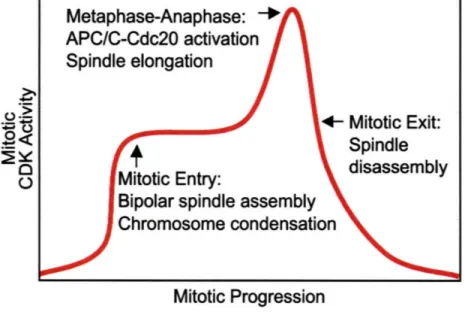
A requirement for mitotic CDK activity at the metaphase-anaphase transition is also difficult to demonstrate because mitotic CDK activity is required for mitotic entry and its elimination during mitosis triggers mitotic exit.
The only in vivo evidence for CDK regulation of APC/C-Cdc20 comes from studies on the budding yeast APC/C-Cdc20 complex0 51 . Rudner and Murray (2000) demonstrated that Clb2-Cdc28
complexes phosphorylate multiple sites in the APC/C subunits Cdcl6, Cdc23, and Cdc27 in vitro. In total, 12 CDK phosphorylation sites were found in these 3 proteins: 6 sites in Cdcl6, 1 site in Cdc23, and 5 sites in Cdc27. Importantly, mutation of these 12 phosphorylation sites to alanine abolished the in vivo phosphorylation of Cdc23, Cdc27, and Cdcl6105.
Rudner and Murray also examined cell cycle progression in strains carrying these APC/C phospho-mutants, which I will refer to as apc/c-12A mutants. Surprisingly, apc/c-12A mutant cells exhibited
only a 10-15 minute delay in metaphase and were completely viable10 5. The subtlety of this
phenotype suggests that either mitotic CDKs are not required for APC/C-Cdc20 activation in vivo or that there are additional CDK phosphorylation sites in other APC/C-Cdc20 components.
My thesis work examined the role of mitotic cyclins in triggering the metaphase-anaphase transition. I simultaneously eliminated Clbl-CDK and Clb2-CDK activities by deleting CLBI and using a temperature-sensitive allele of CLB2, clb2-VI. Elimination of Clb 1/2-CDK activity led to defects in the degradation of the APC/C-Cdc20 substrate Securin/Pds 1, suggesting the APC/C-Cdc20 complex is not fully activated in these cells. I also found that Clbl/2-CDK activity was required during the metaphase-anaphase transition, since Clb1/2-CDK inactivation in metaphase was sufficient to block anaphase entry. Consistent with these findings, over-expression of Cdc20 was able to partially rescue the Securin/Pds 1 degradation defect of clblA clb2- VI mutants. Furthermore, I found that mutation of the CDK consensus sites in Cdc20 does not affect its function in a wild-type background, but is lethal in apc/c-12A mutants (though I have not been able to determine if these sites are
phosphorylated in vivo). Therefore, mitotic CDKs seem to activate the APC/C-Cdc20 complex through overlapping mechanisms that target the core APC/C complex as well as Cdc20. Either mechanism appears to be sufficient for the metaphase-anaphase transition.
Loss ofsister-chromatid cohesion and chromosome segregation
As previously mentioned, destruction of Securin/Pdsl at the metaphase-anaphase transition is required for Separase/Espl activation and subsequent loss of sister-chromatid cohesion. Cleavage of the cohesin subunit Sccl/Mcdl by Separase/Espl is necessary for loss of sister-chromatid cohesion since mutation of the cleavage sites blocks sister-separation 53. In addition, cohesin inactivation is
sufficient for loss of sister-chromatid cohesion since temperature-sensitive alleles of Sccl/Mcdl can bypass the metaphase arrest induced by APC/C-Cdc20 or Separse/Espl inactivation 52,53,119,120
In an elegant series of experiments, Uhlmann et al. were able to show that Sccl/Mcdl cleavage is indeed sufficient for loss of sister chromatid cohesion by using TEV protease to trigger loss of sister chromatid cohesion54. They incorporated a TEV protease cleavage site within Sccl/Mcdl and then expressed TEV protease during a metaphase arrest induced by inactivation of APC/C-Cdc20. Remarkably, induction of TEV protease was sufficient to not only abolished sister chromatid
chromatid cohesion, but also sufficient for nuclear division and spindle elongation5 4. This work thus
suggested the spindle elongation in anaphase was a secondary consequence of loss of sister
chromatid cohesion and that the essential function of the metaphase-anaphase transition machinery (APC/C-Cdc20, Separase, Clb-CDKs) is to dissolve the linkages holding sister chromatids together. Under this model, the spindle is poised to elongate in metaphase, but it is held back only by the cohesins on chromosomes.
My thesis work on the function of Clb-CDKs, however, argues for a requirement for Clbl/2-CDK activity beyond APC/C-Cdc20 activation. I found that Clb-CDK activity is essential to trigger spindle elongation following cohesion loss. In addition, the precocious spindle elongation of cohesin mutants is suppressed by inactivation of Clbl/2-CDKs. How Clb-CDKs promote spindle elongation is not understood.
Anaphase/Telophase
As cells enter anaphase, they have nearly completed the cell division cycle and must now trigger a return to G1. During this period, cells must finalize the division of their genomes, prepare to exit
mitosis, and then complete the cytokinesis program. Importantly, cells must couple and coordinate these events to ensure that both mother and daughter cells receive a full genome complement prior to their separation (cytokinesis). Budding yeast cells have an additional hurdle to face since they must thread the mitotic spindle and its associated chromosomes through the bud neck before mitotic exit. Below I will briefly review what is known about spindle elongation and then discuss how the mitotic exit program eliminates mitotic-CDK activity and returns cells to the G 1 state.
Spindle elongation in Anaphase
In budding yeast, several genes have been implicated in promoting spindle assembly and elongation, including four kinesin related proteins. How these motor proteins and microtubule associated proteins cooperate to regulate anaphase A, when chromosomes are pulled to spindle poles, and anaphase B, when spindle poles separate, is not well understood. Below, I will discuss regulators of anaphase B since this phase is better understood.
Bipolar spindle assembly in yeast is known to require two motors of the BimC family: CIN8 and KIP1121. Although neither gene is essential, cin8A kiplA cells fail to establish a proper bipolar spindle following spindle pole body duplication'22
,123. During anaphase, Cin8 and Kipl are thought to crosslink microtubule plus ends at the spindle midzone and to promote microtubule sliding 121,124
These functions of Cin8 and Kipl may be important for spindle elongation in anaphase. 121,124 The
roles of Cin8 and Kip 1 in spindle assembly are antagonized by Kar3, a minus-end directed motor, since the spindle defects of cin8A kiplA cells can be partially rescued by deletion of KAR3 125.126 However, the antagonism of Cin8 and Kip 1 function by Kar3 is not absolute, since these motors seem to have some common roles in spindle organization 126
Stu2 is an essential microtubule plus end tracking protein of the XMAP215/DIS 1 family that promotes the dynamicity of microtubules'2 7130. Inhibition of Stu2 disrupts kinetochore microtubule
turnover and causes kinetochore microtubule pausing28' 130. In addition, Stu2 is the only protein known to be specifically required for anaphase spindle elongation'29. The functions of Stu2 seem to
be antagonized by Kip3, a plus end directed motor and microtubule depolymerase, because the deletion of KIP3 suppresses the spindle elongation defect of temperature sensitive stu2-10
mutants'29. In anaphase, Stu2 likely cooperates with Cin8 to promote spindle elongation because the
simultaneous overexpression of these two proteins is sufficient to trigger spindle elongation in S phase •'
Exit from Mitosis
During the metaphase-anaphase transition, a wave of Clb destruction is initiated by APC/C-Cdc20 that targets the entire pool of Clb5 and approximately half of the pool of Clb2 for degradation 132
However, to exit from mitosis, cells must eliminate all mitotic CDK activity and reverse many of the phosphorylation events carried out by mitotic-CDKs. In budding yeast, the protein phosphatase Cdcl4 triggers exit from mitosis by dephosphorylating CDK substrates, by triggering cyclin destruction, and by stabilizing mitotic-CDK inhibitors'33. Cdcl4 triggers cyclin destruction by
dephosphorylating Cdhl, allowing it to complex with APC/C and target B-type cyclins for proteolysisl34,135. In addition, Cdcl 14 dephosphorylates the Clb-CDK inhibitor Sicl, protecting it
from ubiquitylation, and it dephosphorylates a transcription factor for Sic 1, Swi5, permitting its nuclear entry 136
Cdcl4 has important functions in chromosome segregation and spindle organization that are
sequences, such as the rDNA locus'3 7'38, and modulating spindle midzone assembly 139,140. Cdcl4
also contributes to spindle stabilization in anaphase by decreasing microtubule dynamicity41.
Cdcl4's multiples roles in chromosome segregation, spindle organization, and Clb-CDK inactivation require it to be finely controlled during the cell cycle. The activity of Cdc 14 is regulated at the level of its association with an inhibitor, Cfil/Netl, which sequesters Cdc 14 in the nucleolus during G 1, S,
G2, and early M phase 142-145. In anaphase, two networks, the Cdc Fourteen Early Anaphase Release
(FEAR) network and the Mitotic Exit Network (MEN), coordinately act to release Cdcl4 from the nucleolus, thereby enabling Cdcl4 access to its targets (Figure 5)133. The FEAR network functions
in early anaphase and is important for efficient cell cycle exit, while the MEN acts in late anaphase and is essential for exit from mitosis 33
The CdcFourteen Early Anaphase Release (FEAR) Network
The FEAR network is not well understood, but is known to include Separase/Espl, the Polo-like kinase Cdc5, the kinetochore protein Slkl9, two proteins of unknown function, Spol2 and Bnsl, and the replication fork blocking protein Fobl 1 46,147. Recently, Clb 1l/2-CDKs and protein phosphatase
2A (PP2A) have been implicated in activation of the FEAR network 148-151. Epistasis analysis
suggests that the FEAR network consists of at least two parallel branches (Figure 5)152. Spol2, Bnsl,
and Fobl function in one branch of the pathway, while Separase/Espl and Slk19 act in another 152
The Polo-like kinase Cdc5 acts downstream of, or in parallel to, the Espl-Slkl9 branch"52. The
position of Cdc5 in the FEAR network has been difficult to determine due to its parallel role in MEN activation.
The FEAR network induced release of Cdc 14 requires phosphorylation of Cfi 1/Net 1, which is considered to be Clbl/2-CDK dependent 148. The current model posits that Clbl/2-CDKs are
unable to trigger Cdcl4 release during early mitosis due PP2A dependent dephosphorylation of Cfil/Netl' 50. In anaphase, Cdcl4 release is triggered by the destruction of Securin/Pdsl, which
liberates Separase/Espl and allows it to sequester PP2A away from Cfil/Net115 0. Clbl/2-CDKs
then, in a Spol2 and Cdc5-dependent manner, phosphorylate Cfil/Netl and allow transient Cdcl4 disassociation148"150. Why the release of Cdcl4 is non-sustainable in the absence of MEN activity is unknown.
The Mitotic Exit Network (MEN)
The MEN is a well-characterized Ras-like signaling pathway with the GTPase Teml acting at or near the top of the network (Figure 5) 1
33,153,154. The GTPase activity of Teml is thought to be regulated by
a putative guanine nucleotide exchange factor (GEF), Lte1' 55,15 6 157,158, and a two-component GTPase
activating complex (GAP) complex, Bub2-Bfal 93,94,157,159-168. Activation of Teml during anaphase
leads to the activation of two downstream kinases: Cdcl 5 and Dbf2 169-173. Although it is not clear
how Teml activates Cdcl5, Cdcl5 is known to phosphorylate and activate Dbf2 in vitro 174. The
activation of Dbf2 in vitro and in vivo requires the Dbf2-associated factor, Mob1174-176. Lastly, the Polo-like kinase, Cdc5, is a positive regulator of Cdcl4 release, functioning to inhibit the Bub2-Bfal complex 161,177,178. How the MEN triggers dissociation of Cdcl4 from Cfil/Netl is not known.
The activation of Tem I in anaphase is not well understood, but it appears to be coupled to the entry of the daughter-bound SPB into the bud153ls5 7,165. All MEN components that function downstream of
TemlI are localized to the cytoplasmic face of the SPB destined to enter the bud'57, 1,6 5 72,173,179-183
Ltel, however, is restricted to the cortex of the bud 157,158,65. MEN signaling is thought to become
activated when the elongating mitotic spindle threads the SPB and associated chromosomes through the bud neck, bringing Teml 1 in proximity to its putative exchange factor, Lte 1.
MEN
= POIN
I/
/Ltel
(GEF).
Bub2-Bfal
Tem(
GA
P )Temi
(GTPase)
4
Cdc15
...
(Kinase)
Dbf2-Mobl
(Kinase)
NON-SUSTAINABLE
CDC 14 RELEASE
4
'VtiI
Figure 5: The Cdc Fourteen Early Anaphase Release (FEAR) network and the Mitotic Exit Network (MEN) Cdcl4 release occurs in two phases during anaphase. The FEAR network function early in anaphase, when the spindle
is between 3-7jim, and initiates a non-sustainable release of Cdcl4. The Cdcl4 released by the FEAR network
dephosphorylates Cdcl5 and elevates its kinase activity. The MEN becomes activated when the spindle fully elongates (7-10tm) and delivers the SPB carrying Teml-Cdcl5-Dbf2-Mobl into the bud. MEN activity is required to maintain Cdcl4 in its released state.
sP
LKI
0
=J
· · · ·
Budding yeast, unlike fission yeast or vertebrate cells, determine the site of cell separation prior to assembly of the mitotic spindle. As a result, they must actively position and pull the mitotic spindle through the bud neck to guarantee that a full genome complement is delivered into the bud receives. Cytokinesis is coordinated with mitotic exit in budding yeast by the spindle position checkpoint, which ensures that the mitotic exit network (MEN) is activated only after the spindle enters the daughter bud 184. When spindles are mis-positioned in anaphase, the kinase Kin4 inactivates MEN signaling in a Bub2-Bfal-dependent manner, preventing Cdcl4 release 185,186. By delaying mitotic
exit, this checkpoint ensures cells have sufficient cells time to correctly align their mis-positioned spindles prior to mother-bud separation.
Cytokinesis
Cytokinesis is the last event in the cell cycle and its regulation in budding yeast remains poorly understood. The physical separation of the mother and daughter cells requires the coordinated action
of two pathways that function at the bud neck: 1) constriction of an acto-myosin ring and 2) formation of a primary septum' 87. Acto-myosin ring assembly begins early in the cell cycle by the
hierarchical recruitment of many factors, including septins, myosins, and formins and is completed in late anaphase by the formation of an F-actin ring 187. In animal cells, type II myosin controls the
constriction of the acto-myosin ring, but in budding yeast this may not be the case 1'T. Paradoxically, mutations in the motor domain of yeast type II Myosin, Myol, cause cytokinesis defects, but deletion of the entire Myol motor domain has no effect 87,•88. Therefore, the mechanisms driving acto-myosin ring contraction in budding yeast remain to be elucidated.
The primary septum of budding yeast is a chitin rich disk the separates the mother and daughter cells following acto-myosin ring constriction. Septum formation is controlled both spatially and
temporally during the cell cycle and requires the delivery of secretory vesicles to the bud neck'"9.
Here, the coordinated secretion of chitin synthases, hydrolytic enzymes, and other cell wall re-modeling enzymes helps to properly divide mother and daughter cell walls'89.
Chromosome segregation in higher eukaryotes
The machinery controlling chromosome segregation is remarkably well conserved across eukaryotes. In fact, every essential regulator of chromosome segregation in budding yeast has orthologues, with the same function, in flies, frogs, mice, and humans. The APC/C-Cdc20 complex, the cohesin complex, Separase, Securin, and mitotic cyclin-CDK activity have nearly identical functions in human cells as they do in yeast. The primary differences between the chromosome segregation machinery in yeast and human cells lies in the way it is regulated. Below I will highlight five modifications that differentiate the control of chromosomes segregation in vertebrate cells and budding yeast.
1. Regulation of APC/C-Cdc20 activation by mitotic inhibitors
In vertebrate cells, the protein Emil (Early Mitotic Inhibitor 1) functions as a negative regulator of APC/C during S phase, G2, and possibly mitosis 190. The precise function of Emil in APC/C regulation is unclear at this time because different groups have ascribed different functions to this regulator. Emil was originally reported to function as an inhibitor of APC/C-Cdc20 during early mitosis 190. Emi's accumulation is cell cycle regulated in Xenopus and human cells, accumulating during S phase and G2 and disappearing during mitotic entry 190,191. In cycling frog egg extracts, addition of Emil stabilizes APC/C-Cdc20 substrates and inhibits cyclin B ubiquitylationl90 Conversely, Emil depletion accelerates cyclin B degradation and blocks mitotic entry 190. These
results (and others) were interpreted to suggest that Emil is a potent inhibitor of APC/C-Cdc20 that functioned during early mitosis, before spindle checkpoint activation, to protect early mitotic cyclins from degradation190-192
Recently, however, two groups have re-examined Emil's role in cell cycle progression and both conclude that Emil functions primarily in S phase and G2 to limit APC/C-Cdhl activity193'"9 4
Treatment of cells with Emil-siRNAs prevents mitotic entry and triggers genome re-replication' 93"' 94
These effects were due Emi 1 's antagonism of APC/C-Cdhl, since co-depletion of Cdhl, but not Cdc20, stabilized the vertebrate replication licensing factor geminin and allowed mitotic cyclin accumulation'93,194
Intriguingly, Emil regulates APC/C activity as a "pseudosubstrate inhibitor. 95'" Although Emil is
not a substrate of the APC/C, it contains a degron motif present in many APC/C substrates, a
destruction box (D box), that enables it to bind independently to either APC/C or Cdh 1195. Emil also contains another region that is able to block APC/C's ubiquitin ligase activity, independently of D-box binding'95.
2. Regulation of APC/C-Cdc20 activation by mitotic timers
Analysis of mammalian cells depleted of spindle assembly checkpoint factors and kinetochore proteins has suggested the existence of a mitotic "timer" that functions independently of the spindle assembly checkpoint'96. Meraldi, et al, observed that siRNA-mediated knockdown of the spindle
assembly checkpoint proteins Madl and Mad2 had differential effects on the overall timing of mitosis, although each treatment abolished the spindle assembly checkpoint' 96. Knockdown of Madl had little effect on the time in took cells to initiate Anaphase A after chromosome congression, whereas knockdown of Mad2 reduced the Anaphase A onset to time to half that of control cells. Similar effects were observed upon depletion of BubRI, but not Mad3 or Bub1'96. The effects of BubRl and Mad2 co-depletion were not additive, suggesting these factors control mitotic timing through a common mechanism'96. Interestingly, the "timer" function of Mad2 and BubRl did not
depend on functional kinetochores, unlike their checkpoint roles'96. It is unclear how Mad2 and BubRI control of the duration of mitosis, but it likely occurs via APC/C-Cdc20 regulation.
3. Regulation of APC/C-Cdc20 activity by ubiquitylation and de-ubiquitylation
Spindle checkpoint activation arrests cells at the metaphase-anaphase transition by Mad2-dependent sequestration of Cdc204 3,9 0. However, upon bi-orientation of all sister-chromatids, it is unclear how checkpoint signaling is rapidly relieved to allow a switch-like transition into anaphase. Previous models have suggested that the Cdc20-Mad2 interaction might be short-lived, making checkpoint silencing a passive consequence of its satisfaction'97. However, recent evidence suggests that
disruption of the Cdc20-Mad2 interaction is an active process that, intriguingly, requires APC/C-dependent ubiquitylation of Cdc20 198,199
In an siRNA-screen for mammalian regulators of the spindle assembly checkpoint, Stegmeier, et al. found that the de-ubiquitylating enzyme USP44 was required to prevent precocious anaphase onset during unperturbed cell cycles, as well as upon spindle checkpoint activation'98. Simultaneously,
Reddy et al. found that addition of UbcH10, an APC/C E2, to an extract from checkpoint-arrested cells triggered Cdc20 ubiquitylation, dissociation of Cdc20 from Mad2, and degradation of APC/C-Cdc20 substrates 199. Furthermore, USP44 was found to antagonize UbcH 10 dependent dissociation
of Cdc20 and Mad2 in vitro19 9
Together these results propose a model for how cells control APC/C-Cdc20 activity during early mitosis. During pro-metaphase, unattached kinetochores catalyze conformational changes in Mad2 that allow it to tightly associated with Cdc20. Mitotically activated APC/C can inhibit this process by ubiquitylating Cdc20 and triggering its dissociation from Mad2. However, APC/C activity is restrained by USP44, which reinforces the checkpoint by de-ubiquitylation of Cdc20 and, potentially,
the APC/C-Cdc20 substrates Securin and Cyclin B. Upon bi-orientation of all sister-chromatid pairs, a reduction in spindle checkpoint signaling allows APC/C-dependent ubiquitylation of Cdc20 to drive its dissociation from Mad2.
3. Separase auto-cleavage
Examination of Separase levels in human cells revealed that Separase undergoes auto-catalyzed cleavage in anaphase200-20 2. Three Separase cleavage sites were identified that were conserved in
human, mouse, and Xenopus Separase200'202. Cleavage of Separase generates two fragments that
remain stably associated and, surprisingly, does not affect Separase activity200'20 2. However,
expression of a non-cleavable Separase by targeted mutagenesis of the Separase locus in human cells revealed a role for Separase cleavage in the G2/M transition2 03. The mitotic entry defects of non-cleavable Separase appear to be due stabilization of Weel and subsequent inhibition of mitotic CDK-activity203. Recently, Separase auto-cleavage has also been reported to regulate complex formation between Separase and PP2A, although the functional significance of this regulation remains elusive204. At present, it is unclear how Separase cleavage leads to Weel destabilization nor how
Separase auto-cleavage can occur early in the cell cycle, when the expression of its competitive inhibitor Securin is highest.
4. Mitotic CDKs act like Securins and Separases act like CDK inhibitors
Examination of Separase regulation in vertebrate cells has revealed it has a dually antagonistic relationship with mitotic cyclin B-CDK complexes20 5'206. In Xenopus egg extracts, stabilized cyclin
B 1 fragments are able to block anaphase entry without affecting the degradation of the APC/C-Cdc20 substrate Securin205. Cyclin B l-Cdkl phosphorylation of Separase was found to be necessary
for cyclin-B I1 dependent inhibition of anaphase entry20 5. Interestingly, however, Cdkl
phosphorylation of Separase is not sufficient for inhibition, as this also required subsequent cyclin 41
B1-Cdkl binding20 6. Cyclin B1-Cdk phosphorylation of Separase creates a docking site for cyclin
B 1, recruiting Cdkl to the complex2 06. Complex formation with Separase also inhibits Cdkl's kinase
activity, suggesting Separase can function as a CDK inhibitor206. In addition, cyclin B-Cdkl binding
to Separase is mutually exclusive to Securin binding and is independent of Separase's proteolytic activity206. Therefore, vertebrate Separase is dually antagonized in metaphase by canonical Securin and by mitotic CDKs, which by virtue of their binding and inhibition of Separase, function like Securins20 7. In support of this model, Securin null cells that also express non-phosphorylatable Separase preciously entered anaphase during spindle checkpoint activation208. These cells did not exhibit premature anaphase entry during an unperturbed mitosis, suggesting that additional mechanisms exist to restrain Separase activation208.
5. Step-wise loss of arm and centromere cohesion in mitosis
Loss of sister chromatid cohesion in vertebrate cells, unlike budding yeast, is thought to occur in a "step-wise" manner with arm cohesion removal occurring in prophase/pro-metaphase and
centromeric cohesion removal occurring only during the metaphase-anaphase transition209. The removal of arm cohesion requires phosphorylation of the cohesin subunit SA2, Polo-like kinasel (Plkl), Aurora B kinase, condensin I, and the conserved protein Wap120 9-22 0. However, it does not require cleavage of Sccl by Separase since expression of non-cleavable Sccl does not block loss of
arm cohesion (non-cleavable Scc 1 does block loss of centromeric cohesion)209,210. Centromeric
cohesion appears to be protected from this "prophase pathway" by Sgo, a member of the Shugoshin family of cohesion protectors221224. It is important to note that arm cohesin removal is only apparent
after spindle poison induced metaphase arrest and it is presently unclear how the removal of arm and centromeric cohesion are coordinated during unperturbed mitoses225. Furthermore, the functional
Thesis Summary
How cells couple and coordinate events during chromosome segregation in M-phase is not well understood. The work presented in this thesis suggests that cells couple loss of sister-chromatid cohesion with spindle elongation by placing both events under the control of mitotic CDKs. Clbl/2-CDK or Clb2/3-Clbl/2-CDK activity was found to be required for the in vivo activation of the APC/C-Cdc20 complex, an E3 ubiquitin ligase that triggers the metaphase-anaphase transition. In a second step, mitotic CDK activity was found to be required for spindle elongation. This second role of mitotic CDK activity is independent of cohesion loss and therefore constitutes a novel function for CDK in mitosis. Finally, a model is proposed in which different CDK threshold requirements help establish the order of consecutive mitotic events.
References
1. MORGAN, D. O. The cell cycle: Principles of control (New Science Press; Sinauer Associates, London Sunderland, MA, 2007).
2. MIELE, L. The biology of cyclins and cyclin-dependent protein kinases: An introduction. Methods Mol Biol 285, 3-21 (2004).
3. ANDREWS, B. & MEASDAY, V. The cyclin family of budding yeast: Abundant use of a good idea. Trends Genet 14, 66-72 (1998).
4. EPSTEIN, C. B. & CROSS, F. R. Genes that can bypass the cln requirement for saccharomyces cerevisiae cell cycle start. Mol Cell Biol 14, 2041-7 (1994).
5. SCHWOB, E. & NASMYTH, K. Clb5 and clb6, a new pair of b cyclins involved in DNA replication in saccharomyces cerevisiae. Genes Dev 7, 1160-75 (1993).
6. SURANA, U. et al. The role of cdc28 and cyclins during mitosis in the budding yeast s. Cerevisiae. Cell 65, 145-61 (1991).
7. FITCH, I. et al. Characterization of four b-type cyclin genes of the budding yeast saccharomyces cerevisiae. Mol Biol Cell 3, 805-18 (1992).
8. RICHARDSON, H., LEW, D. J., HENZE, M., SUGIMOTO, K. & REED, S. I. Cyclin-b homologs in saccharomyces cerevisiae function in s phase and in g2. Genes Dev 6, 2021-34 (1992).
9. RICHARDSON, H. E., WITTENBERG, C., CROSS, F. & REED, S. I. An essential gl function for
cyclin-like proteins in yeast. Cell 59, 1127-33 (1989).
10. MENDENHALL, M. D. & HODGE, A. E. Regulation of cdc28 cyclin-dependent protein kinase activity during the cell cycle of the yeast saccharomyces cerevisiae. Microbiol Mol Biol Rev 62, 1191-243 (1998).
11. POLYMENIS, M. & SCHMIDT, E. V. Coordination of cell growth with cell division. Curr Opin Genet Dev 9, 76-80 (1999).
12. LEVINE, K., HUANG, K. & CROSS, F. R. Saccharomyces cerevisiae gl cyclins differ in their intrinsic functional specificities. Mol Cell Biol 16, 6794-803 (1996).
13. TYERS, M., TOKIWA, G., NASH, R. & FUTCHER, B. The cln3-cdc28 kinase complex of s. Cerevisiae is regulated by proteolysis and phosphorylation. Embo J 11, 1773-84 (1992). 14. COSTANZO, M. et al. Cdk activity antagonizes whi5, an inhibitor of gl/s transcription in yeast.
Cell 117, 899-913 (2004).
15. DE BRUIN, R. A., MCDONALD, W. H., KALASHNIKOVA, T. I., YATES, J., 3RD & WITTENBERG,
C. Cln3 activates gl-specific transcription via phosphorylation of the sbf bound repressor whi5. Cell 117, 887-98 (2004).
16. SCHWOB, E., BoHM, T., MENDENHALL, M. D. & NASMYTH, K. The b-type cyclin kinase
inhibitor p40sicl controls the gl to s transition in s. Cerevisiae. Cell 79, 233-44 (1994). 17. BAI, C. et al. Skpl connects cell cycle regulators to the ubiquitin proteolysis machinery
through a novel motif, the f-box. Cell 86, 263-74 (1996).
18. FELDMAN, R. M., CORRELL, C. C., KAPLAN, K. B. & DESHAIES, R. J. A complex of cdc4p,
skplp, and cdc53p/cullin catalyzes ubiquitination of the phosphorylated cdk inhibitor sic lp. Cell 91, 221-30 (1997).
19. SCHNEIDER, B. L., YANG, Q. H. & FUTCHER, A. B. Linkage of replication to start by the cdk inhibitor sicl. Science 272, 560-2 (1996).
20. SKOWYRA, D., CRAIG, K. L., TYERS, M., ELLEDGE, S. J. & HARPER, J. W. F-box proteins are
receptors that recruit phosphorylated substrates to the scfubiquitin-ligase complex. Cell 91,