Conceptual Study for Tissue-Regenerative
Biodegradable Magnesium Implant Integrated
with Nitric Oxide-Releasing Nanofibers
The MIT Faculty has made this article openly available. Please share how this access benefits you. Your story matters.Citation Jeon, Jin-Kyung et al. "Conceptual Study for Tissue-Regenerative Biodegradable Magnesium Implant Integrated with Nitric Oxide-Releasing Nanofibers." Metals and Materials International 25, 4 (January 2018): 1098–1107 © 2019 The Korean Institute of Metals and Materials
As Published https://doi.org/10.1007/s12540-018-00232-9
Publisher Springer Science and Business Media LLC
Citable link https://hdl.handle.net/1721.1/128430
Terms of Use Article is made available in accordance with the publisher's policy and may be subject to US copyright law. Please refer to the publisher's site for terms of use.
A Conceptual Study for Tissue-Regenerative Biodegradable Magnesium
Implant Integrated with Nitric Oxide-Releasing Nanofibers
Jin-Kyung Jeon1‡, Hyunseon Seo1‡, Jimin Park1,2, Soo Ji Son3, Yeong Rim Kim4, Eun Shil Kim1, Jong Woong Park5, Woong-Gyo Jung5, Hojeong Jeon1,6, Yu-Chan Kim1, Hyun-Kwang Seok1,6, Jae Ho Shin3,4*, Myoung-Ryul Ok1*
1
Center for Biomaterials, Korea Institute of Science and Technology (KIST), 5, Hwarang-ro 14-gil, Seongbuk-gu, Seoul, 02792, Republic of Korea
2
Department of Material Science and Engineering, Massachusetts Institute of Technology, Cambridge, Massachusetts, 02139, USA
3
Department of Chemistry, Kwangwoon University, Seoul, 01897, Republic of Korea
4
Medical Sensor∙Biomaterial Research Center, Kwangwoon University, Seoul, 01897, Republic of Korea
5
Department of Orthopedic Surgery, School of Medicine, Korea University, Seoul, 02841, Republic of Korea
6
Division of Bio-Medical Science and Technology, KIST School, Korea University of Science and Technology, Seoul, 02792, Republic of Korea
* Corresponding Authors: jhshin@kw.ac.kr and omr2da@kist.re.kr. ‡ These authors contributed equally to this work
Abstract
The excessive initial corrosion rate of Mg is a critical limitation in the clinical application of biodegradable Mg implants because the device loses its fixation strength before the fractured bone heals. This study suggests a new approach to overcome this hurdle by accelerating tissue regeneration instead of delaying the implant biodegradation. As angiogenesis is an essential process in early bone regeneration, a Mg implant coated with electrospun nanofibers containing nitric oxide (NO), which physiologically promotes angiogenesis, is designed. The integrated device enables adjustable amounts of NO to be stored on the NO donor-conjugated nanofiber coating, stably delivered, and released to the fractured bone tissue near the implanted sites. An in vitro corrosion test reveals no adverse effect of the released NO on the corrosion behavior of the Mg implant. Simultanously, the optimal concentration level of NO released from the implant significantly enhances tube network formation of human umbilical vein endothelial cells (HUVECs) without any cytotoxicity problem. This indicates that angiogenesis can be accelerated by combining NO-releasing nanofibers with a Mg implant. With its proven feasibility, the proposed approach could be a novel solution for the initial stability problem of biodegradable Mg implants, leading to successful bone fixation.
Keywords
1. INTRODUCTION
The rapid growth in the size of the elderly population, the so-called age of aging, has led to a tremendous increase in the demand for orthopedic implants. In the past decades, metals possessing excellent mechanical properties together with chemical stability such as titanium alloys, cobalt-based alloys, and stainless steels have been selected as materials for hard tissue replacement or fixation[1]. Because of their ambivalent biochemical stability, however, the high-risk but unavoidable secondary surgical process to remove implants after they have finished their missions has been raised as a severe drawback. Owing to the biodegradability and osteogenic properties, magnesium (Mg) alloys have recently drawn considerable attention as optimal materials for next-generation orthopedic implants[2-7]. Numerous in-vitro and in-vivo studies have reported that Mg-based implants can gradually be degraded or absorbed after insertion without any toxicity-related side effects[8-10]. Moreover, Mg ions were shown to enhance the growth of new bone tissue[10-14].
However, Mg-based fixation devices present a crucial limitation in the form of an excessive initial biodegradation rate. The mechanism whereby Mg undergoes degradation in the human body is a corrosion reaction, which is especially accelerated in body fluid containing abundant chloride ions[2,15]. In general, the corrosion rate reaches its maximum within several days after implantation and dramatically decreases to stabilize at a lower level in approximately one week. The stabilization is a consequence of the naturally formed protection layers consisting of degradation products, the concentration of which are maintained in this range until complete disappearance[3-8]. Although the main advantage of Mg implants is the absolute decomposition, they are primarily designed to maintain a sufficiently high mechanical strength until the fractured bone heals. As the implanted Mg devices gradually lose their fixation strength due to the decrease in their size, it is necessary
to minimize their corrosion rate such that the biological healing rate can compensate for the loss in fixation strength. Thus, many researchers have focused on enhancing the initial corrosion resistance of Mg implants. Some progress in controlling the corrosion behavior has been achieved by polymer/ceramic encapsulation and by optimization of the allo y composition, manufacturing process, and crystal structures of the Mg alloy[5,7,9,16,17]. Nonetheless, Mg corrosion still remains a critical challenge for practical applications. This is because polymer/ceramic coatings offer poor protection as a result of their porous/loose structure and the Mg alloy design is inevitably limited by the intrinsic properties of the materials[2].
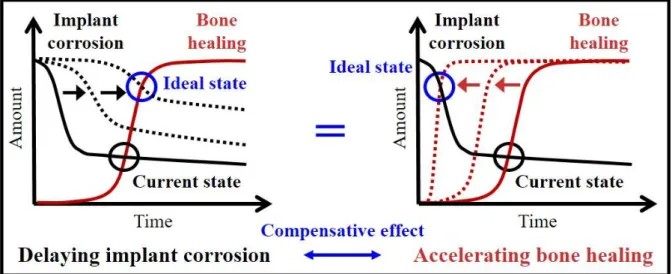
Herein, we propose a different approach to overcome the current problem presented by Mg-based biodegradable implants, i.e., accelerating the bone-healing process instead of reducing the initial corrosion rate (Figure 1). The promotion of bone regeneration would compensate for the rapid initial corrosion of the given Mg implants because this could also fulfill the mission of Mg implants to support the bone until it is restored. Among the complicated but highly systematic steps whereby tissue is regenerated, angiogenesis, which is the formation of new blood vessels from pre-existing ones, is one of the vital physiological actions. Notably, as for the regeneration of hard tissues, at an early stage of fractured bone healing, angiogenesis plays a crucial role in the mineralization and the resultant process of forming new bone tissue[18-22].
Recently, reactive oxygen species (ROS) has emerged as a potential therapeutic with its revealed vital roles in diverse biological responsiveness[23-27]. In particular, ROS has drawn considerable attention for its wound healing-related efficacy as a mediator of angiogenesis[28-31]. Thus, efforts to utilize ROS for biomedical applications were actively directed at introducing diverse ROS generation strategies such as natural release from donor materials[23,32-34], external electric/photonic stimuli[35-37], and electrochemical
reaction[38]. Indeed, we previously reported the promotion of angiogenesis induced by electrochemically generated hydrogen peroxide (H2O2), one of the typical endogenous
ROS[38]. It was confirmed that the appropriate concentration of H2O2 directly correlated with
enhancing angiogenesis and that an electrochemical system using Mg corrosion integrated with biochemically stable metallic implants could spontaneously generate H2O2. However, as
there is no practically applicable solid alloy to functioning as an anode when coupled to Mg, Mg-based biodegradable implants require a new ROS generating system.
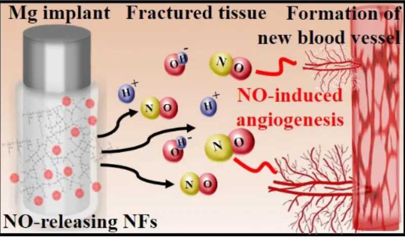
This prompted our design of a tissue-regenerative and biodegradable implant consisting of Mg alloy coated with nitric oxide (NO)-releasing nanofibers. NO, a common form of ROS existing in vivo, was chosen in this study with the aim of endowing a tissue-regenerative function by stimulating angiogenesis. Even though the physiological targets of NO as vital therapeutic factors have been well-revealed in their own way[23,29-31], their practical use in biomedical applications has not been sufficiently developed due to the difficulties associated with establishing an appropriate storage/delivery system in vivo. Macromolecular NO-releasing scaffolds are known to be difficult to deliver to the target inside the human body[33,39,40]. Small carriers such as nanoparticles conjugated with low molecular weight NO donors were suggested for targeted delivery, but they can only store minimal amounts of NO, and even that is unstable, leading to limited application[32,41]. On the other hand, our system can stably carry sufficient and adjustable amounts of NO by synthesizing NO donors-conjugated nanofibers. Then, the fabricated NO-releasing nanofibers can simply be coated onto the surface of the Mg alloy using electrospinning, regardless of the shape or size of the implant. The integrated device composed of the Mg alloy implant of commercialized composition (Mg-5 wt%Ca-1 wt%Zn) covered with NO-releasing nanofibers can directly deliver NO to the lesion, and promotes tube network formation of human umbilical vein endothelial cells (HUVECs), which refers to angiogenesis (Figure 2).
However, as the key concept of the NO-releasing nanofibers coating is to integrate tissue-regenerative function to a given Mg implant, it should be clarified in vitro that the coating does not cause any side effect such as ecceleration of Mg corrosion or cytotoxicity originated from NO or its carrier material. Such concerns were resolved by performing materials characterization and systematic in vitro cell studies, thereby verifying the feasibility of our conceptual design.
2. EXPERIMENTAL PROCEDURE
2.1. Preparation of Mg-5 wt%Ca-1 wt%Zn alloy rods.
Commercial pure Mg (99.98%) ingots, Ca (99.6%) metal grains, and Zn (99.99%) metal grains were melted in a stainless steel crucible in the furnace, in which the Ar gas was flowed under a vacuum of 5 10-2 torr, at 790 °C for 120 min, and the molten alloy was stirred for 5 min. Then, the alloy was poured into a stainless steel mold (cylinderical rod, 2 6 mm), which had been preheated to 500 °C, and cooled in the furnace. The prepared Mg-5 wt%Ca-1 wt%Zn alloy rods were washed with acetone and ethanol, respectively, before NO-releasing nanofibers coating.
2.2. Synthesis of NO-releasing nanofibers.
An aminoalkoxysilane solution was prepared by dissolution of 5.0 mmol of methyl aminopropyltriethoxysilane (MAP3) in 5.0 mL of methanol in the presence of sodium methoxide (5.0 mmol; an equimolar amount corresponding to the secondary amine content of the silane precursor). The solution was placed in an in-house NO reactor and flushed with argon (Ar) for 10 min, followed by a series of three charge/discharge cycles with Ar (10 atm, 3 10 min) to remove oxygen from the solution. The reaction vessel was then charged with
NO to 10 atm and sealed for three days at room temperature while stirring. Prior to removal of the N-diazeniumdiolate (NO donor)-modified MAP3 solution, unreacted NO was purged from the chamber with Ar gas for 10 min. N-Diazeniumdiolate-modified MAP3 solution was vacuum sealed and stored in a freezer at –20 °C until use.
Subsequently, 30.0 mmol of methylmethacrylate (MMA), 10.0 mmol of hexylmethacrylate (HMA), and 10.0 mmol of (trimethoxysilylpropyl)methacrylate (SiMA) were added to 30.0 mL of toluene with stirring, and then the mixed solution was heated to 80 °C. The polymerization reaction of the methacrylate mixture was initiated by 0.1 mmol of azobisisobutyronitrile (AIBN; dissolved in 2.5 mL of methanol) and reacted for 12 h. Upon completion of the reaction, the toluene was vaporized by vacuum distillation at 60 °C and vacuum dried at room temperature. The residual monomers and AIBN were washed copiously with hexane three times and the product was vacuum dried again. The synthesized poly(MMA-co-HMA-co-SiMA) was stored in vacuum packaging until use.
Next, 0.3 g of poly(MMA-co-HMA-co-SiMA) was dissolved in 0.7 g of acetone/dimethylformamide (3:1 in volume). Then, 143 μL of methyltrimethoxysilane (MTMOS; 1 mmol) and 250 μL of N-diazeniumdiolate-modified MAP3 (0.25 mmol corresponding to 25 mol.%, balance MTMOS) were added to 1.0 g of poly(MMA-co-HMA-co-SiMA) solution. Finally, 19.0 mg of aluminum acetylacetonate dissolved in 400 μL of acetone/dimethylformamide (3:1 in volume) and 30 μL H2O were consecutively added to the
solution of poly(MMA-co-HMA-co-SiMA), MTMOS, and N-diazeniumdiolate-modified MAP3. The mixture was then stirred for 1 h at 4 °C to minimize any thermal decomposition of the N-diazeniumdiolate NO donors. After stirring, the mixed solution was electrospun immediately.
The nanofibers for NO storage/delivery were prepared by an electrospinning system (NT-ESS-300, NTSee Co.; Gwangju, Korea) and Havard PHD 2000 syringe pump (Holliston,
MA, USA). The mixed solution prepared above was loaded in a plastic syringe and flowed through a 25-G needle. The electrospinning conditions were as follows: the volume flow rate was 10 μL/min, the applied voltage was 17.5 kV, the tip to collector distance was 15 cm, and the electrospinning time was 30 min. After stabilization in air for 30 min, the NO storage/delivery nanofiber webs were vacuum-sealed and stored in a freezer at –20 °C.
2.3. SEM observation.
The surface morphology of the NO-releasing nanofibers was measured using FE-SEM (Field emission scanning electron microscopy, Inspect F50, FEI Company, Hillsboro, OR, USA).
2.4. Quantitative analysis of NO
The quantitative analysis of NO was performed with NO-releasing nanofibers on sheets of four different sizes: (a) 0.18 cm2, (b) 0.36 cm2, (c) 0.73 cm2, (d) 1.44 cm2, to evaluate four different concentrations of NO eluate. Each of the nanofiber sheets was immersed in 1 mL of endothelial basal media-2 (EBM-2) at 37 ± 0.5 °C for 5 min. After removing the nanofiber sheet, the eluate containing four different concentrations of NO was measured by Griess assay[42]. The concentration of NO was recorded spectroscopically at 540 nm with UV-vis spectroscopy.
2.5. Immersion test.
Three experimental groups were formed: bare Mg alloy rod, Mg alloy rod coated with NO-charged nanofibers, and Mg alloy rod coated with uncharged nanofibers. The immersion test was performed in HBSS (Hank’s Buffer Salt Solution, pH of 7.4) at 37 ± 0.5 °C for seven days. The HBSS was changed every 24 hours considering the diuretic effect
in the human body. The hydrogen gas generated by the corrosion reaction of samples was collected at the top of the funnel, and the volumes of hydrogen gas were measured to determine the amount of Mg alloy that had corroded.
2.6. Electrochemical measurement.
Electrochemical experiments were typically conducted using a three-electrode electrochemical cell system. An Ag/AgCl electrode (BASi Ag/AgCl/3 M NaCl) was utilized as reference electrode, and a platinum (Pt) electrode was used as counter electrode. The electrochemical tests were performed under HBSS at 37 °C using a potentiostat system (CHI 760C, CH Instruments, Inc.). The electrode potential was converted to the reversible hydrogen electrode (RHE) scale using the following equation: E(NHE) = E(Ag/AgCl) + 0.059 × pH + 0.197 V. The potential of bare Mg alloy rods vs. Pt, Mg alloy rods coated with NO-charged nanofibers vs. Pt, and Mg alloy rods coated with uncharged nanofibers vs. Pt were recorded to investigate the difference in corrosion behavior.
2.7. L929 cells culture and cytotoxicity assay.
Fibroblastic L929 cells were cultured in Dulbecco’s modified Eagle’s medium (DMEM, Welgene) supplemented with 10% fetal bovine serum (FBS, Welgene) and 1% penicillin-streptomycin (GIMCO™) at 37±0.5 °C under a 5% carbon dioxide (CO2)
humidified atmosphere. The passage of cells was carried out every two days. Cultured L929 cells were seeded in a 96-well cell culture plate at a density of 7 103 cells/100 µL of DMEM supplemented with 10% FBS and incubated for 24 h with the three different experimental groups (bare Mg alloy rods, Mg alloy rods coated with NO-charged/uncharged nanofibers). After incubation, the cell cytotoxicity was analyzed through the CCK-8 assay and the cell viability was investigated at 410 nm using a microplate reader (VersaMax,
Molecular Devices LLC). Cells incubated without a Mg alloy rod (control group) were set to have a viability of 100%, and the relative values of the experimental groups were calculated.
2.8. HUVECs culture and proliferation assay.
The effect of NO on cell growth was identified with fourth-passage HUVECs. HUVECs (C2517; Lonza) were cultured in endothelial growth medium-2 (EGM-2) (CC-3162; Lonza) at 37 °C under a 5% CO2 humidified atmosphere. Cultured HUVECs were seeded on 96 well plates (5000 cells/well). The proliferation test was performed with EGM-2 based eluted samples including different concentrations of NO by using nanofiber sheets of four different sizes. HUVECs cultured with only EGM-2 were set as the control. The cell proliferative activity was identified by using 4-[3-(4-iodophenyl)-2-(4-nitrophenyl)-2H-5-tetrazolio]-1,3-benzene disulfonate (WST-1). Briefly, EGM-2 was removed and WST-1 solution (10 µL of WST-1 reagent and 100 µL of culture medium) was placed at each time point (24 h, 48 h, and 72 h). After incubation for 2 h at 37 °C, the absorbance was recorded with a microplate reader at 450 nm.
2.9. Angiogenesis test using HUVECs tube formation assay.
Angiogenesis was identified with endothelial cell network formation in vitro. First, Matrigel® Growth Factor Reduced (GFR) (Corning®) was placed on a 24-well cell culture plate (289 µl of Matrigel® per well). After incubation of the Matrigel® for 30 min, positive control, negative control, and Mg control groups were used to compare with the four eluted samples based on EBM-2. In the case of the positive control, non-stimulated HUVECs from NO were cultured in EGM-2. The negative control was cultured in EBM-2 without any supplements. The experimental groups were cultured in NO and Mg eluate based on 1 mL EBM-2 at 37 °C to compare with the negative control. After 5 min of elution time, the
nanofiber sheets and Mg rods were removed from the eluate and HUVECs (4.0 105 cell/mL) were placed in each of the remaining 1 mL eluate samples. Then, the eluate including 300 µL (1.2 105 cell/well) of the cell was placed on the prepared Matrigel®. After incubation for 12 h, optimum cell network formation was identified with fluorescence microscopy. Living cells were marked by adding 300 μL of Calcein AM dye (6 μM Calcein AM in EBM-2) to the well plate including HUVECs. After 30 min, the different levels of HUVEC network formation on the positive control (EGM-2), negative control (EBM-2), and NO sample groups were confirmed by using fluorescence microscopy. The formed tube branches were quantified by ImageJ program.
2.10. Statistical analysis.
Current data are expressed as means standard deviation (SD) of measurements (*p < 0.05, **p < 0.01, and ***p < 0.001) and were analyzed using one-way ANOVA testing with Tukey’s post hoc multiple comparisons (GraphPad Prism 7).
3. RESULTS AND DISCUSSION
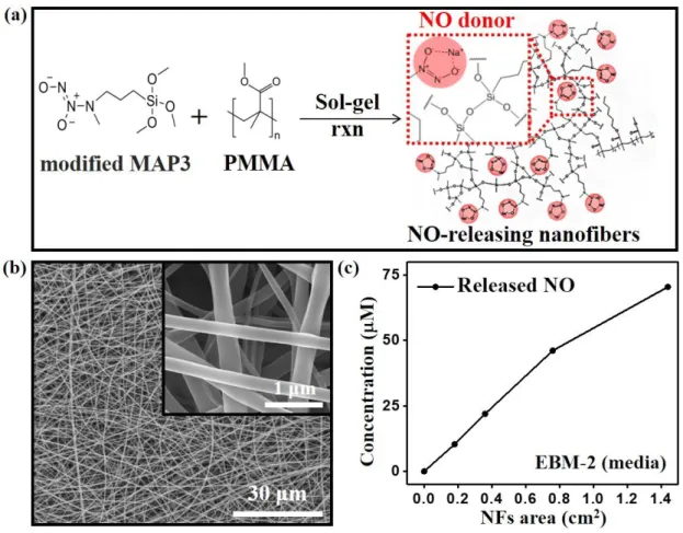
NO-releasing nanofibers were fabricated by electrospinning the polymer synthesized by a sol-gel reaction between poly(MMA-co-HMA-co-SiMA) and N-diazeniumdiloate (NO donor)-modified MAP3. NO remains stable after being electrospun onto the Mg alloy implant because of the formation of a resonance structure of diazeniumdiolates (Na-N-O-N-O, NO donor) and covalent bonding bridges with the siloxane groups (Si-O-Si, backbone silane) (Figure 3a). Figure 3b shows the microstructure of the electrospun NO-releasing nanofibers coated on the Mg alloy implant. The nanofibers are stacked in multi-layers, and the thickness of each fiber is below 500 nm. The highly porous structure of the nanofiber coating allows
body fluids to access the Mg implant, ensuring that the biodegradation property remains unhindered. Meanwhile, the NO-storage materials utilized in this study are non-degradable such that the integrated device composed of the Mg alloy rod and NO-releasing nanofiber coating layer, necessarily, cannot be fully biodegradable. However, the current NO-releasing coating/Mg implant system is an early version designed to determine the feasibility of studying the concept of tissue regenerative/biodegradable implants. Thus, further studies of biodegradable NO-releasing coating materials would be required before practical application would be possible.
The NO-releasing behavior of the nanofiber was examined in vitro by measuring its photo-absorption at 540 nm in endothelial basal media-2 (EBM-2) (Figure 3c)[42]. The result indicates that the concentration of released NO proportionally increases as the area of the sheet of nanofibers increases, releasing 50 nmol of NO from approximately 0.8 cm2 of the nanofiber sheet in 5 min. In principle, the amount of NO stored in a nanofiber sheet could be determined by controlling the quantity of conjugated NO donors. Instead, however, the NO concentration was controlled by changing the area of the nanofiber sheet with fixed NO-donating functional group content because the primary purpose of this study is to suggest the concept of tissue-regenerative Mg alloy implants.
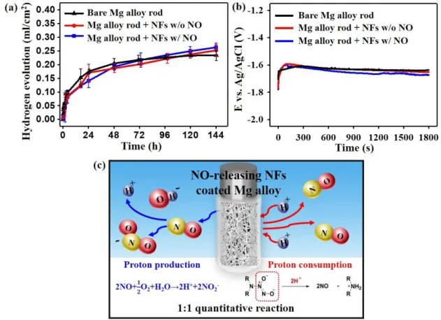
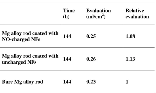
Investigating whether the released NO adversely affects the corrosion behavior of the Mg alloy implant is of extreme importance for evaluating the feasibility of our idea since the acceleration of corrosion would obviously be a critical obstacle for using the biodegradable implant system in practice. Thus, an immersion test in which measuring the cumulative amount of hydrogen gas was performed with a Mg alloy rod of commercialized composition (Mg-5 wt%Ca-1 wt%Zn) covered with NO-releasing nanofibers (Figure 4a). Although NO is a well-known corrosive gas, the result did not show any evidence of accelerated corrosion, contrary to our concern. The standard deviations of the hydrogen evolution in all three
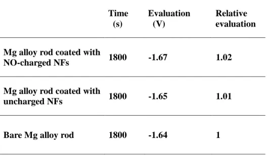
experimental groups are below 0.05 mL/cm2 (Table 1). The electrochemical analysis also exhibited similar trends, with the open circuit potential (OCP) values of all groups being within a 2% error range (Figure 4b and Table 2). A key reason for these results could be the balance between the production and consumption of protons during the NO-releasing process and its conversion reactions in aqueous solution (Figure 4c). When two NO molecules released from the nanofibers participate in the formation of more stable nitrogen oxides or their ions, two protons are generated in aqueous solution. The diazeniumdiolates (Na-N-O-N-O) in the nanofibers, meanwhile, require two protons to release two NO molecules[43]. Owing to these two counterbalanced reactions related to NO release, NO does not aggravate the corrosion properties of the overall NO-releasing coating/Mg implant system while providing tissue regenerative functions.
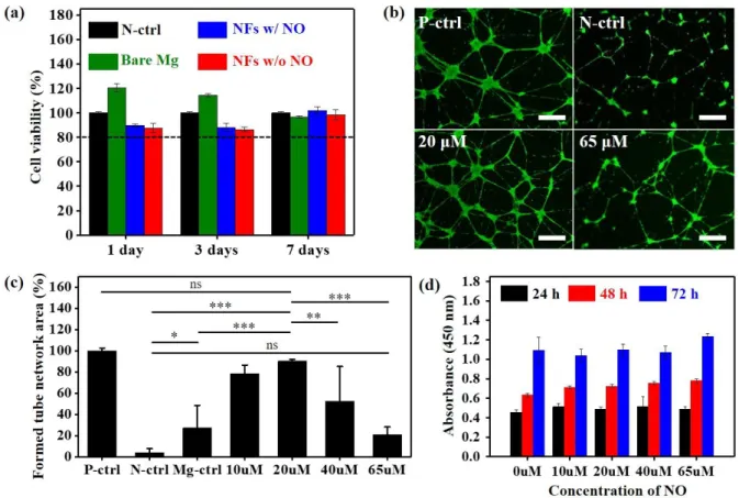
The cytotoxicity evaluation was performed with L929 cells by using a CCK-8 assay to verify the biocompatibility of the Mg alloy implant coated with NO-releasing nanofibers. Figure 5a shows that all constituent materials including the Mg alloy rod, NO-storage nanofibers, and released NO represent over 80 % of cell viability compared to the control, indicating negligible cytotoxicity. The extent to which angiogenesis induced by NO released from the nanofiber coating is promoted was analyzed by observing the network formation of HUVECs (Figure 5b). Based on vascular morphogenesis in the positive control, which was cultured in an endothelial growth medium-2 (EGM-2), the relative values of the network formation of HUVECs incubated for 12 h in EBM-2 with different NO concentrations were quantified (Figure 5c). We calculated the mesh areas of the formed tube network as an index of the progression of angiogenesis. The NO-stimulated HUVECs exhibited a significant enhancement in network formation until the NO concentration level reached 20 M in comparison with the negative control or bare Mg alloy control. This improved network formation of HUVECs reflects the efficacy of the released NO in promoting angiogenesis.
The bare Mg alloy control group also showed a somewhat meaningful enhancement that seemed to be consistent with the previously reported ability of Mg ions to improve the endothelial functions[44,45]. However, the HUVECs treated with 65 M of NO exhibited results comparable with that of the negative control and even recorded a lower level of network formation than the bare Mg alloy control, presumably becuase the toxicity exceeded the angiogenetic function at high NO concentrations[26]. A proliferation analysis of the HUVECs cultured in EGM-2 for three days, meanwhile, indicated consistently favorable effects of NO in all concentration ranges even up to 65M (Figure 5d). The numerous growth factors contained in EGM-2 seem to have a stronger impact on proliferation than NO. This is because the proliferation analysis required EGM-2, as the culture medium for the HUVECs, to survive for a few days, unlike the network formation experiment in which EBM-2 was used. In this regard, determination of the appropriate NO concentration in vivo for enhancing angiogenesis could be expected to be more complicated than the in-vitro optimization we investigated.
4. CONCLUSION
This study involved the design of a tissue regenerative and biodegradable metallic implant by incorporating an angiogenetic function induced by NO with biodegradable magnesium (Mg) alloy. NO can be controllably stored on the Mg alloy implant in the form of a NO donor-conjugated nanofiber coating. The nanofibers were synthesized via the sol-gel reaction of poly(MMA-co-HMA-co-SiMA) and N-diazeniumdiloate (NO donor)-modified MAP3, and can simply be coated onto the implant regardless of its shape or size by using electrospinning. The materials characterization and an in-vitro cell study served to prove the angiogenetic performance of the integrated device, a Mg alloy rod of commercialized composition (Mg-5 wt%Ca-1 wt%Zn) covered with NO-releasing nanofibers, thus showing
that it merits further in vivo investigation. We demonstrated 20 μM to be the optimal concentration level of released NO that promotes the tube network formation of HUVECs. At this level, NO neither worsens the Mg alloy corrosion rate nor the cell cytotoxicity. As the initial processes of bone healing strongly depend on the level of angiogenesis in the fractured tissues, NO-induced stimulation of angiogenesis would be best suitable for orthopedic application. Notably, in the case of Mg-based biodegradable implants that have constantly demanded a new solution to counteract their excessive degradation rate, the NO-induced acceleration of bone recovery can be a novel answer. We envision the extension of our approach of combining biodegradable implants with tissue regenerative functions provided by NO-stimulated angiogenesis to future clinical application as an improvement of current problematic biodegradable implant.
ACKNOWLEDGEMENT
This research was supported by a grant of the Ministry of Commerce, Industry, and Energy of the Korean Government (Project number: 10065241) and a grant from the Korea Institute of Science and Technology (2V05460, KIST-Korea University TRC program). This research was also supported by the Bio & Medical Technology Development Program of the National Research Foundation (NRF) funded by the Minister of Science, ICT & Future Planning (NRF-2015M3A9E2029186).
REFERENCES
1. Chen, Q. & Thouas, G. A. Metallic implant biomaterials. Mater. Sci. Eng. R 87, 1-57 (2015).
2. Gu, X. N., Li, S. S., Li, X. M. & Fan, Y. B. Magnesium based degradable biomaterials: A review. Front. Mater. Sci. 8, 200-218 (2014).
3. Witte, F.; Kaese, V.; Haferkamp, H.; Switzer, E.; Meyer-Lindenberg, A.; Wirth, C.; Windhagen, H. In vivo corrosion of four magnesium alloys and the associated bone response. Biomaterials 26, 3557-3563 (2005).
4. Staiger, M. P., Pietak, A. M., Huadmai, J. & Dias, G. Magnesium and its alloys as orthopedic biomaterials: a review. Biomaterials 27, 1728-1734 (2006).
5. Salahshoor, M. & Guo, Y. Biodegradable orthopedic magnesium-calcium (MgCa) alloys, processing, and corrosion performance. Materials 5, 135-155 (2012).
6. Waizy, H.; Seitz, J.-M.; Reifenrath, J.; Weizbauer, A.; Bach, F.-W.; Meyer-Lindenberg, A.; Denkena, B.; Windhagen, H. Biodegradable magnesium implants for orthopedic applications. J. Mater. Sci. 48, 39-50 (2013).
7. Atrens, A., Liu, M. & Abidin, N. I. Z. Corrosion mechanism applicable to biodegradable magnesium implants. Mater. Sci. Eng., B 176, 1609-1636 (2011).
8. Lee, J.-W.; Han, H.-S.; Han, K.-J.; Park, J.; Jeon, H.; Ok, M.-R.; Seok, H.-K.; Ahn, J.-P.; Lee, K. E.; Lee, D.-H.; Yang, S. J.; Cho, S. Y.; Cha, P. R.; Kwon, H.; Nam, T. H.; Han, J. H.; Rho, H. J.; Lee, K. S.; Kim, Y. C.; Mantovani, D. Long-term clinical study and multiscale analysis of in vivo biodegradation mechanism of Mg alloy. Proc. Natl. Acad. Sci. U. S. A. 113, 716-721 (2016).
9. Song, G. Control of biodegradation of biocompatable magnesium alloys. Corros. Sci. 49, 1696-1701 (2007).
10. Karus, T.; Fischerauer, S. F.; Hänzi, A. C.; Uggowitzer, P. J.; Löffler, J. F.; Weinberg, A. M. Magnesium alloys for temporary implants in osteosynthesis: in vivo studies of their degradation and interaction with bone. Acta Biomater. 8, 1230-1238 (2012).
11. Yoshizawa, S., Brown, A., Barchowsky, A. & Sfeir, C. Magnesium ion stimulation of bone marrow stromal cells enhances osteogenic activity, simulating the effect of magnesium alloy degradation. Acta Biomater. 10, 2834-2842 (2014).
12. Vormann, J. Magnesium: nutrition and metabolism. Mol. Aspects Med. 24, 27-37 (2003). 13. Zreiqat, H.; Howlett, C.; Zannettino, A.; Evans, P.; Schulze‐ Tanzil, G.; Knabe, C.; Shakibaei, M. Mechanisms of magnesium‐ stimulated adhesion of osteoblastic cells to commonly used orthopaedic implants. J. Biomed. Mater. Res., Part A 62, 175-184 (2002). 14. Yamasaki, Y.; Yoshida, Y.; Okazaki, M.; Shimazu, A.; Uchida, T.; Kubo, T.; Akagawa, Y.; Hamada, Y.; Takahashi, J.; Matsuura, N. Synthesis of functionally graded MgCO3 apatite accelerating osteoblast adhesion. J. Biomed. Mater. Res., Part A 62, 99-105 (2002).
15. Song, G. L. & Atrens, A. A. Corrosion mechanisms of magnesium alloys. Adv. Eng. Mater. 1, 11-33 (1999).
16. Cha, P. R.; Han, H. S.; Yang, G. F.; Kim, Y. C.; Hong, K. H.; Lee, S. C.; Jung, J. Y.; Ahn, J. P.; Kim, Y. Y.; Cho, S. Y.; Byun, J. Y.; Lee, K. S.; Yang, S. J.; Seok, H. K. Biodegradability engineering of biodegradable Mg alloys: tailoring the electrochemical properties and microstructure of constituent phases. Sci. Rep. 3, 2367; 10.1038/srep02367 (2013).
17. Yang, G. F.; Kim, Y. C.; Han, H. S.; Lee, G. C.; Seok, H. K.; Lee, J. C. In vitro dynamic degradation behavior of new magnesium alloy for orthopedic applications. J. Biomed. Mater. Res., Part B 103, 807 (2015).
18. Carano, R. A. & Filvaroff, E. H. Angiogenesis and bone repair. Drug Discovery Today 8, 980-989 (2003).
19. Gerber, H.-P.; Vu, T. H.; Ryan, A. M.; Kowalski, J.; Werb, Z.; Ferrara, N. VEGF couples hypertrophic cartilage remodeling, ossification and angiogenesis during endochondral bone formation. Nat. Med. 5, 623-628 (1999).
20. Street, J.; Bao, M.; Bunting, S.; Peale, F. V.; Ferrara, N.; Steinmetz, H.; Hoeffel, J.; Cleland, J. L.; Daugherty, A.; van Bruggen, N. Vascular endothelial growth factor stimulates bone repair by promoting angiogenesis and bone turnover. Proc. Natl. Acad. Sci. U. S. A. 99,
9656-9661 (2002).
21. Kanczler, J. & Oreffo, R. Osteogenesis and angiogenesis: the potential for engineering bone. Eur. Cells Mater. 15, 100-114 (2008).
22. Fang, T. D.; Salim, A.; Xia, W.; Nacamuli, R. P.; Guccione, S.; Song, H. M.; Carano, R. A.; Filvaroff, E. H.; Bednarski, M. D.; Giaccia, A. J. Angiogenesis is required for successful bone induction during distraction osteogenesis. J. Bone Miner. Res. 20, 1114-1124 (2005). 23. Carpenter, A. W. & Schoenfisch, M. H. Nitric oxide release: Part II. Therapeutic applications. Chem. Soc. Rev. 41, 3742-3752 (2012).
24. Ray, P. D., Huang, B.-W. & Tsuji, Y. Reactive oxygen species (ROS) homeostasis and redox regulation in cellular signaling. Cell. Signalling 24, 981-990 (2012).
25. Dickinson, B. C. & Chang, C. J. Chemistry and biology of reactive oxygen species in signaling or stress responses. Nat. Chem. Biol. 7, 504-511 (2011).
26. Winterbourn, C. C. Reconciling the chemistry and biology of reactive oxygen species. Nat. Chem. Biol. 4, 278-286 (2008).
27. Finkel, T. Signal transduction by reactive oxygen species. J. Cell Biology 194, 7-15 (2011).
28. Xia, C.; Meng, Q.; Liu, L.-Z.; Rojanasakul, Y.; Wang, X.-R.; Jiang, B.-H. Reactive oxygen species regulate angiogenesis and tumor growth through vascular endothelial growth factor. Cancer Res. 67, 10823-10830 (2007).
29. Zhang, R.; Wang, L.; Zhang, L.; Chen, J.; Zhu, Z.; Zhang, Z.; Chopp, M. Nitric oxide enhances angiogenesis via the synthesis of vascular endothelial growth factor and cGMP after stroke in the rat. Circ. Res. 92, 308-313 (2003).
30. Papapetropoulos, A., García-Cardeña, G., Madri, J. A. & Sessa, W. C. Nitric oxide production contributes to the angiogenic properties of vascular endothelial growth factor in human endothelial cells. J. Clin. Invest. 100, 3131-3139 (1997).
31. Diwan, A. D., Wang, M. X., Jang, D., Zhu, W. & Murrell, G. A. Nitric oxide modulates fracture healing. J. Bone Miner. Res. 15, 342-351 (2000).
32. Shin, J. H., Metzger, S. K. & Schoenfisch, M. H. Synthesis of nitric oxide-releasing silica nanoparticles. J. Am. Chem. Soc. 129, 4612-4619 (2007).
33. Robbins, M. E., Hopper, E. D. & Schoenfisch, M. H. Synthesis and Characterization of Nitric Oxide-Releasing Sol− Gel Microarrays. Langmuir 20, 10296-10302 (2004).
34. Miller, M. & Megson, I. Recent developments in nitric oxide donor drugs. Br. J. Pharmacol. 151, 305-321 (2007).
35. He, W.; Kim, H.-K.; Wamer, W. G.; Melka, D.; Callahan, J. H.; Yin, J.-J. Photogenerated charge carriers and reactive oxygen species in ZnO/Au hybrid nanostructures with enhanced photocatalytic and antibacterial activity. J. Am. Chem. Soc. 136, 750-757 (2013).
36. Macak, J., Schmidt-Stein, F. & Schmuki, P. Efficient oxygen reduction on layers of ordered TiO 2 nanotubes loaded with Au nanoparticles. Electrochem. Commun. 9, 1783-1787 (2007).
37. Li, Y., Zhang, W., Niu, J. & Chen, Y. Mechanism of photogenerated reactive oxygen species and correlation with the antibacterial properties of engineered metal-oxide nanoparticles. ACS nano 6, 5164-5173 (2012).
38. Park, J.; Du, P.; Jeon, J. K.; Jang, G. H.; Hwang, M. P.; Han, H. S.; Park, K.; Lee, K. H.; Lee, J. W.; Jeon, H.; Kim, Y. C.; Park, J. W.; Seok, H. K.; Ok, M.-R. Magnesium Corrosion Triggered Spontaneous Generation of H2O2 on Oxidized Titanium for Promoting Angiogenesis. Angew. Chem. Int. Ed. Engl. 54, 14753-14757 (2015).
39. Marxer, S. M., Rothrock, A. R., Nablo, B. J., Robbins, M. E. & Schoenfisch, M. H. Preparation of nitric oxide (NO)-releasing sol− gels for biomaterial applications. Chem. Mater. 15, 4193-4199 (2003).
Chem. Soc. Rev. 41, 3731-3741 (2012).
41. Saraiva, J., Marotta-Oliveira, S. S., Cicillini, S. A., Eloy, J. d. O. & Marchetti, J. M. Nanocarriers for nitric oxide delivery. J. Drug Delivery 2011, 936438 (2011).
42. Misko, T. P., Schilling, R., Salvemini, D., Moore, W. & Currie, M. A fluorometric assay for the measurement of nitrite in biological samples. Anal. Biochem. 214, 11-16 (1993). 43. Holland, R. J.; Klose, J. R.; Deschamps, J. R.; Cao, Z.; Keefer, L. K.; Saavedra, J. E. Direct Reaction of Amides with Nitric Oxide To Form Diazeniumdiolates. J. Org. Chem. 79, 9389-9393 (2014).
44. Maier, J. A., Bernardini, D., Rayssiguier, Y. & Mazur, A. High concentrations of magnesium modulate vascular endothelial cell behaviour in vitro. Biochim. Biophys. Acta, Mol. Basis Dis. 1689, 6-12 (2004).
45. Shechter, M.; Sharir, M.; Labrador, M. J. P.; Forrester, J.; Silver, B.; Merz, C. N. B. Oral magnesium therapy improves endothelial function in patients with coronary artery disease. Circulation 102, 2353-2358 (2000).
FIGURES
Figure 1. Schematic diagram of correlation between implant corrosion rate (black) and bone healing rate (red) showing the compensative effect of delaying the initial corrosion of the implant (left) and accelerating bone healing (right) on orthopedic functions.
Figure 2. Schematic illustration of Mg implant coated with NO-releasing nanofibers (NFs) and NO-induced angiogenesis in the fractured tissue near the implant.
Figure 3. (a) Synthetic process and (b) SEM images of NO-releasing nanofibers. (c) Quantitative analysis of NO released from nanofiber (NFs) sheets of four different sizes by the Griess assay. The NFs sheet was diluted in 1 mL EBM-2 at 37 ± 0.5 °C for 5 min.
Figure 4. Evaluation of corrosion behavior by measuring (a) hydrogen evolution and (b) open circuit potential (OCP) of bare Mg alloy rod (black), and Mg alloy rod coated with NO-charged (blue) and unNO-charged NFs (red). (c) Schematic illustration of mechanism of proton production and consumption reaction related to NO release.
Figure 5. (a) Viability of L929 cells cultured with bare Mg alloy rod (green), Mg alloy rod coated with NO-charged (blue), and uncharged NFs (red), compared to L929 cells cultured only in the medium (negative control, black). (b) Fluorescence confocal images of the tube network formation of HUVECs cultured in EGM (positive control), EBM (negative control), EBM with 20 µM of NO, and EBM with 65 µM of NO. Scale bar indicates 300 μm. (c) Quantitative analysis of the average capillary mesh area induced by the NO-stimulated HUVECs. Statistical significance was decided as * (p < 0.05), ** (p < 0.01), and *** (p < 0.001). (d) HUVECs proliferation with varying amounts of NO.
TABLES
Table 1. Volume of generated hydrogen gas after 144 h of immersion in HBSS of bare Mg alloy rod, Mg alloy rod coated with NO-charged NFs and with uncharged NFs. Standard deviation (SD) < 0.05. Time (h) Evaluation (ml/cm2) Relative evaluation
Mg alloy rod coated with
NO-charged NFs 144 0.25 1.08
Mg alloy rod coated with
uncharged NFs 144 0.26 1.13
Table 2. Open circuit potential of bare Mg alloy rod, Mg alloy rod coated with NO-charged NFs and with uncharged NFs at 1800 s.
Time (s) Evaluation (V) Relative evaluation
Mg alloy rod coated with
NO-charged NFs 1800 -1.67 1.02
Mg alloy rod coated with
uncharged NFs 1800 -1.65 1.01