ORIGINAL ARTICLE
The additional value of CT images interpretation
in the differential diagnosis of benign vs. malignant
primary bone lesions with 18F-FDG-PET/CT
K. Strobel&U. E. Exner&K. D. M. Stumpe&
T. F. Hany&B. Bode&K. Mende&P. Veit-Haibach&
G. K. von Schulthess&Juerg Hodler
Received: 6 May 2008 / Accepted: 12 June 2008 / Published online: 20 August 2008 # Springer-Verlag 2008
Abstract
Objective To evaluate the value of a dedicated interpreta-tion of the CT images in the differential diagnosis of benign vs. malignant primary bone lesions with 18fluorodeoxyglu-cose-positron emission tomography/computed tomography (18F-FDG-PET/CT).
Materials and methods In 50 consecutive patients (21 women, 29 men, mean age 36.9, age range 11–72) with suspected primary bone neoplasm conventional radiographs and 18F-FDG-PET/CT were performed. Differentiation of benign and malignant lesions was separately performed on conventional radiographs, PET alone (PET), and PET/CT with specific evaluation of the CT part. Histology served as the standard of reference in 46 cases, clinical, and imaging follow-up in four cases.
Results According to the standard of reference, conven-tional 17 lesions were benign and 33 malignant. Sensitivity, specificity, and accuracy in assessment of malignancy was
85%, 65% and 78% for conventional radiographs, 85%, 35% and 68% for PET alone and 91%, 77% and 86% for combined PET/CT. Median SUVmax was 3.5 for benign
lesions (range 1.6–8.0) and 5.7 (range 0.8–41.7) for malignant lesions.
In eight patients with bone lesions with high FDG-uptake (SUVmax≥2.5) dedicated CT interpretation led to the correct
diagnosis of a benign lesion (three fibrous dysplasias, two osteomyelitis, one aneurysmatic bone cyst, one fibrous cortical defect, 1 phosphaturic mesenchymal tumor). In four patients with lesions with low FDG-uptake (SUVmax<2.5)
dedicated CT interpretation led to the correct diagnosis of a malignant lesion (three chondrosarcomas and one leiomyo-sarcoma). Combined PET/CT was significantly more accu-rate in the differentiation of benign and malignant lesions than PET alone (p=.039). There was no significant differ-ence between PET/CT and conventional radiographs (p=.625).
Conclusion Dedicated interpretation of the CT part signif-icantly improved the performance of FDG-PET/CT in differentiation of benign and malignant primary bone lesions compared to PET alone. PET/CT more commonly differentiated benign from malignant primary bone lesions compared with conventional radiographs, but this differ-ence was not significant.
Keywords 18F-FDG-PET . Tumor imaging
Introduction
Currently, the workup of primary bone neoplasms includes conventional radiographs and typically magnetic resonance imaging (MRI) for local staging as well as bone
scintigra-K. Strobel (*)
:
K. D. M. Stumpe:
T. F. Hany:
K. Mende:
P. Veit-Haibach:
G. K. von SchulthessDivision of Nuclear Medicine, Department of Medical Radiology, University Hospital Zurich,
Raemistr. 100,
8091 Zurich, Switzerland e-mail: [email protected] U. E. Exner
Department of Orthopedic Surgery, University Hospital Balgrist, Zurich, Switzerland
B. Bode
Institute of Surgical Pathology, University Hospital Zurich, Zurich, Switzerland
J. Hodler
Department of Radiology, University Hospital Balgrist, Zurich, Switzerland
phy (BS) and computed tomography (CT) for general staging. If malignancy is suspected, bone biopsy has to be performed. Fluorodeoxyglucose (FDG)-PET and FDG-PET/CT are increasingly used for the differentiation of malignant and benign tumors in many organ systems [1–5]. However, the role of PET/(CT) in the evaluation of bone tumors is not well defined yet [6, 7]. Preliminary results showed that PET/(CT) may play an important role in biopsy guidance [8], grading [9, 10], staging [11], and therapy response assessment [12, 13]. Differentiation between benign and malignant primary bone lesions is crucial and has an important impact on therapy. FDG uptake measured by maximum standardized uptake value (SUVmax) is not reliable enough because of a considerable
overlap between FDG uptake of benign and malignant bone lesions. It is known that especially histiocytic or giant cell containing benign lesions can have FDG uptake >2.5 SUVmax [14]. Conventional PET scanners are
increasingly replaced by combined PET/CT. The CT part can be used for attenuation correction and anatomic correlation of FDG-positive lesions. In addition, a specific interpretation of the CT part of the PET/CT study may improve diagnostic performance [15]. The aim of this study was to evaluate the additional value of such an interpretation in the differential diagnosis of benign vs. malignant primary bone lesions.
Materials and methods Patients
Fifty consecutive patients (21 women, 29 male, mean age 36.9, age range 11–72) were prospectively included in this study. In all patients, a primary bone tumor was suspected because of clinical symptoms (pain, fracture; n=42) and/or imaging findings (n=8). In all patients, conventional radiographs and an 18F-FDG-PET/CT examination were performed. The time interval between the radiographs and PET/CT was <14 days in all cases. There was no therapeutic intervention between conventional and PET/ CT imaging.
The study was conducted in accordance with the guide-lines established by the local ethics committee.
PET/CT imaging protocol
All data were acquired on a combined PET/CT in-line system (Discovery LS or Discovery STE, GE Healthcare, Milwaukee, WI, USA).
Patients fasted for at least 4 h prior to scanning, which started approximately 60 min (median 58 min; range 52– 77 min) after the injection of 350–400 MBq of 18F-FDG.
All patients were tested for a normal glucose level before scanning. Patients with elevated glucose levels were rescheduled and scanned with normal glucose levels. No intravenous contrast agent was given. Initially, the CT scan was acquired starting from the level of the head using the following parameters: 40 mAs, 140 kV, 0.5 s/tube rotation, slice thickness 4.25 mm, scan length 867 mm, data-acquisition time 22.5 s. Breathhold CT in non-forced expiration position was performed. In the patients with primary tumors in the lower extremities, scanning of the lower legs was added.
Immediately following CT acquisition, a PET emission scan was acquired with an acquisition time of 3 min per bed position with a one-slice overlap in 2D mode (matrix 128× 128). The eight to nine bed positions starting from the head to the knees resulted in an acquisition time of approxi-mately 24–27 min. CT data were used for the attenuation correction, and the images were reconstructed using a conventional iterative algorithm (OSEM). The acquired images were viewed with a software providing multiplanar reformatted images of PET alone, CT alone and fused PET/ CT with linked cursors (Advantage Windows workstation, GE Healthcare, Milwaukee, WI, USA). PET/CT imaging was performed according to the published “procedure guideline for tumor imaging with 18F-FDG PET/CT 1.0” [16].
Standard of reference
Histology, obtained by image-guided (ultrasound or CT) or open biopsy or tumor resection served as the standard of reference in 46 cases. The histopathological examination were performed by a board certified pathologist (B.B). The tumor diagnoses were done according to the criteria of the World Health Organization and, if indicated, were con-firmed by the appropriate molecular methods [fluorescence in situ hybridization (FISH) and/or polymerase chain reaction (PCR)]. Imaging and clinical follow-up for at least 12 months (mean 24 months, range 12–36) was used as the standard of reference in the remaining four cases.
Interpretation of conventional radiographs
Conventional radiographs were analyzed by a radiologist (J.H.). The reader was blinded to the results of other imaging modalities and to the clinical history but aware about the suspicion of a bone tumor. Differentiation of benign and malignant lesions were based on the established criteria described by Lodwick and several other authors [17–19]. Signs of benignity were, for example, well-defined lesions, rim sclerosis, ground glass appearance. Signs of malignancy were, for example, ill-defined lesions, cortical destruction, malignant periosteal reactions.
PET/CT interpretation and measurement of SUVmax
Semiquantitative analysis of FDG uptake was performed by measuring the SUVmax. In our institution, SUV is corrected
for lean body mass. A personal scale (Tanita, model 2001; Tanita, Tokyo, Japan) with an integrated foot-to-foot bioelectric impedance analyser was used to determine the lean body mass (LBM) of the patients. The manufacturer supplied a model including gender, weight, height, and a
measured impedance value for determination of the percentage of body fat and for calculation of LBM. By using attenuation-corrected PET data, SUVmax was
calcu-lated with the following equation based on a freehand region of interest including the entire lesion on the fused PET/CT image: SUVmax(lbm)=(LBM–CFDG)/Dose where
LBM is measured in grams, CFDG is the concentration of 18
F-FDG in Becquerels per milliliter, and Dose is the injected dose measured in Becquerels.
Physiological 18F-FDG uptake and uptake caused by benign abnormalities for instance in muscles, brown fat, or pulmonary infiltrates were excluded from the analysis.
For the evaluation with PET alone, a SUVmaxcutoff of
2.5 was used for the differentiation of low FDG uptake (<2.5 max) versus high FDG-uptake (≥2.5 SUVmax).
Lesions with low FDG uptake were interpreted as benign and lesions with high FDG uptake as malignant The SUVmax measurements were performed by a nuclear
physician (K.D.M.S.), again, blinded to the results of the other imaging modalities and the clinical history but aware about the suspicion of a bone tumor. For the combined PET/CT evaluation, the CT part of the PET/CT study was separately analyzed by a reader with double board certification as a radiologist and a nuclear medicine
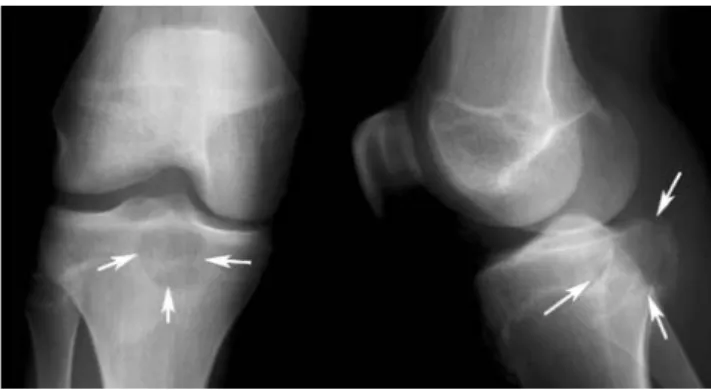
Fig. 1 Conventional X-ray images of the right knee of a 15-year-old boy showing an osteolysis (arrows) in the epiphysis of the right proximal tibia
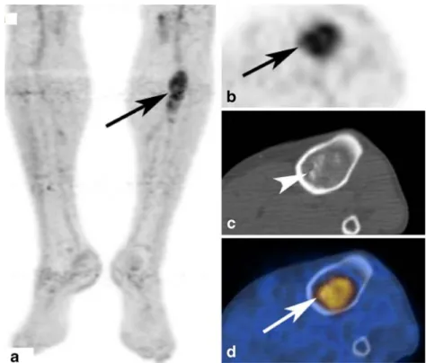
Fig. 2 (same patient as Fig.1) Moderate FDG-uptake (SUVmax
4.0) of the lesion on PET images (a, MIP). Axial PET (b) and fused PET/CT (d) images dem-onstrating that the tumor takes up FDG in the periphery with a FDG-negative centre. CT (c) shows a well-defined eccentric osteolysis without clear sclerotic rim. No calcifications are seen inside the lesion. So also in CT, this lesion was difficult to assess because clear signs of benignity like a sclerotic rim are missing. Biopsy and histological work-up confirmed the diagnosis of a chondroblastoma
physician (K.S.). He was also blinded to the results of the other imaging modalities and the clinical history. He was only aware that a bone tumor was suspected [20–22]. Signs of benignity were, for example, well-defined lesions, rim sclerosis, ground glass appearance. Signs of malignancy were, for example, ill-defined lesions, cortical destruction, malignant periosteal reactions. Bone and soft tissue window settings were used for the evaluation. In lesions with low uptake but aggressive CT appearance, including aggressive periosteal reactions and cortical destruction, the final PET/ CT interpretation was that of malignant lesion. In cases of PET-positive lesions, indicating malignancy with benign CT patterns such as a well-defined osteolysis with rim sclerosis, the final interpretation was that of a benign lesion.
Statistical analysis
Data were analysed using SPSS 15 for Windows (SPSS). Statistical significance was assessed with the sign test. p< 0.05 was considered to indicate a significant difference.
Results
Seventeen lesions were benign and 33 malignant. In the benign group, there were seven benign bone tumors (Figs.1,
2, 3 and 4), three fibrous dysplasias (Figs. 5 and 6), two osteomyelitis, one insertion tendinopathy, one stress fracture, one postoperative defect, one fibrous cortical defect, and one bone infarction. Of the 33 malignant lesions, there were 18 sarcomas (Fig. 7), six lymphomas, three metastases, one melanoma, one chordoma, one hemangioendothelioma, one eosinophilic granuloma, one malignant peripheral nerve sheath tumor, and one neuroendocrine tumor. Patient characteristics are summarized in Table1. Median SUVmax
of benign lesions was 3.5 (range 1.6–8.0) and 5.7 (range 0.8–41.7) for malignant lesions (Table 2). Sensitivity, specificity, accuracy, PPV, and NPV regarding the diagnosis of a malignant lesions was 85%, 65%, 78%, 82%, and 67% for CI, 85%, 35%, 68%, 72%, and 55% for PET alone and 91%, 77%, 86%, 88%, and 81% for combined PET/CT (Table3).
In eight patients with a SUVmax>2.5, the dedicated CT
interpretation led to the correct diagnosis of a benign lesion (three fibrous dysplasias, two osteomyelitis, one aneurys-mal bone cyst, one fibrous cortical defect, one phosphaturic mesenchymal tumor). In four patients with a SUVmax<2.5,
CT interpretation led to the correct diagnosis of a malignant lesion (three chondrosarcoma, one leiomyosarcoma). Com-bined PET/CT interpretation was significantly more accu-rate compared to PET alone (p=0.039). The diagnostic performance of PET/CT was not significantly different from conventional radiographs (p=0.63). Furthermore, no
statis-tically significant difference was found between PET alone and conventional radiographs (p=0.18).
Discussion
Although malignant bone lesions have generally higher FDG uptake than benign bone tumors, there is a consider-able overlap regarding the amount of FDG uptake. Our results confirm the findings of previously published studies that many benign lesions can have moderate to high FDG-uptake [14, 23]. This fact can lead to misinterpretation because incidentally detected benign FDG-positive bone lesions may mimic metastases if FDG-PET/CT is per-formed for staging of extra-skeletal malignancies [23,24]. Fibrous dysplasia is a good example where separate interpretation of CT images with the pathognomonic “ground glass” pattern and absence of bone destruction overrules the positive PET result and leads to the correct diagnosis of a “no-touch” benign lesion [25]. We found
Fig. 3 Conventional X-ray images of the left thigh in a 21-year-old male patient with a calcified lesion in the upper third of the diaphysis of the left tibia (arrows)
SUVmaxvalues>2.5 (range 2.9–8.0) in all four patients with
fibrous dysplasia. Aoki et al. published six cases with fibrous dysplasia, of which only two presented with a SUVmax>2.5 [14].
Low-grade chondrosarcomas are good examples in which interpretation of the CT part with the typical calcifications overrules a negative PET result and leads to the correct diagnosis of a malignant lesion. Three of our four chondrosarcomas had SUVmax values < 2.5 which
confirms the results of other authors that especially low-grade chondrosarcomas can be almost FDG-negative [26].
FDG uptake is not specific for the diagnosis of a malignant neoplasm. Traumatic, inflammatory, and infec-tious lesions like osteomyelitis can show significant FDG uptake as shown in experimental and clinical studies [27,
28]. We observed SUVmax of 5.2 and 6.9 in both of our
patients with biopsy-proven osteomyelitis.
F-18 FDG PET/CT has been employed for differentia-tion between malignant and benign fractures based on the SUVmax and based on medullary uptake, which is
charac-teristic for malignant fractures [29,30]. Fractures in two of our patients were caused by benign lesions, one was PET positive (SUVmax 3.9 in a patient with fibrous dysplasia)
and one PET negative (SUVmax 1.6 in a patient with a
stress fracture).
Our results underline that the CT part of the PET/CT study can add important information. Those evaluating PET/CT studies should be familiar with both the metabolic and morphologic features of bone tumors and tumor-like lesions.
Fig. 5 Conventional X-ray images (Fig. 5) of a 33-year-old female patient with an inhomogeneous ground-glass like lesion (arrows) in the left tibia
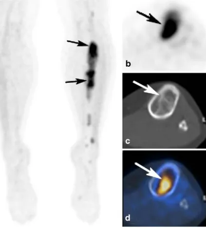
Fig. 4 (same patient as Fig.3) PET (MIP (a); axial PET (b); fused PET/CT images (d) with increased FDG-uptake (SUVmax.3.5) of the lesion
(arrows) indicating malignancy. CT (c) images demonstrating calcifications (arrowheads) without cortical destructions typical for an enchondroma, which was confirmed with biopsy
Similarly to previously published studies, our data indicate the difficulty to define a reliable cutoff value for the differentiation between benign and malignant lesions. Beside the previously described cutoff value of SUVmax=2.5, also
values of 2.0 or 3.0 do not provide sufficient accuracy [14,
31,32]. Since the SUV is a semiquantitative measurement, there are various calculation variants, and reproducibility suffers from influences such as blood glucose level, uptake time, and several others. Therefore, the use of additional criteria for diagnosing bone neoplasms is important [33]. Nevertheless, Dehdashti et al. have demonstrated that SUV measurements were more effective than subjective interpre-tation of FDG uptake in bone lesions [34]. We believe that a combined interpretation of metabolic information and mor-phologic information, both provided by a PET/CT examina-tion should be implemented.
Conventional radiographs remain the first imaging modality in the evaluation of suspected bone neoplasms. A final diagnosis can often be made based on radiographs, obviating additional imaging and biopsy. This is the case for fibrous dysplasia, Paget’s disease, and nonossifying fibroma. In equivocal cases and in aggressive tumors such as osteosarcoma, MR imaging is typically employed as the second imaging tool for grading and staging [35]. The importance of bone scintigraphy for the evaluation of bone tumors has decreased over the last years. However, this
Fig. 6 (same patient as Fig.5) FDG-PET (MIP (a); axial PET (b), fused PET/CT images (d) showing intense FDG-uptake (SUVmax.7.7) of the lesion
indi-cating malignancy. CT (c) dem-onstrating well-defined ground glass lesions without cortical destruction typical for fibrous dysplasia. Biopsy confirmed the diagnosis of fibrous dysplasia
Fig. 7 A 50 year-old female patient with a calcified lesion in the bone marrow of the left proximal humerus. PET (Fig. 7 a, b, d) images demonstrating low FDG-uptake (SUVmax2.3) in the lesion (arrows)
indicating benignity. CT (arrow, Fig. 7c) shows calcifications inside the lesion with cortical destructions (arrow, d) suspicious for chondrosarcoma. Histology showed a grade I–II chondrosarcoma
method still is valuable in staging of osteosarcoma. The accuracy of a bone scan can be increased by using SPECT and SPECT/CT [36,37].
Because combined FDG-PET/CT did not improve differentiation of bone lesions compared to conventional radiographs, it cannot be recommended for this indication. Another problem in clinical routine in most countries is the fact that PET/CT is only reimbursed for staging of confirmed malignant tumors but not for assessment of malignancy in equivocal cases. PET/CT has a potential role for the detection of transformation of a benign into a malignant bone tumor and of development into more aggressive patterns as observed in malignant lymphoma [38]. We observed no proven transformation in our patients, and studies with high numbers of patients with transforma-tion are missing because such malignant transformatransforma-tions are infrequent [39].
Our study has limitations. For the CT evaluation, only a low-dose CT part of the combined PET/CT study was available. Another approach would be to perform a thin-slice conventional “high-dose” CT centered on the primary bone lesions. This better CT quality may improve the
Table 1 Characteristics of 50 patients with benign and malignant bone lesions
Patient no. SUVmax Final diagnosis
1 5.2 Brodie abscess 2 2.5 Lymphoma 3 11.3 Osteosarcoma 4 7.4 Leiomyosarcoma 5 3.6 Neuroendocrine tumor 6 5.1 Ewing sarcoma 7 6.2 Lymphoma 8 1.6 Stress fracture 9 2.9 Fibrous dysplasia 10 6.9 Osteomyelitis
11 3.5 Phosphaturic mesenchymal tumor
12 1.4 Chondrosarcoma
13 8.0 Fibrous dysplasia
14 3.9 fibrous dysplasia with Pathologic fracture
15 9.0 Leiomyosarcoma
16 4.6 Fibrous cortical defect
17 9.0 Lymphoma
18 8.8 Malignant peripheral nerve sheath tumor 19 0.8 Eosinophilic granuloma 20 2.0 Osteochondroma 21 3.0 Hemangioendothelioma 22 4.0 Chondroblastoma 23 41.7 Lymphoma 24 14.9 Osteosarcoma 25 5.9 Lymphoma
26 3.7 Aneurysmatic bone cyst
27 3.1 Chondrosarcoma
28 1.4 leiomyosarcoma
29 4.7 Clear cell renal carcinoma metastasis 30 7.7 Fibrous dysplasia 31 2.2 Bone infarction 32 2.1 Insertion tendinopathy 33 2.2 Hemangioma 34 3.5 Enchondroma 35 1.3 Postoperative defect 36 2.2 Chondrosarcoma 37 8.7 Osteosarcoma 38 5.3 Osteosarcoma 39 5.7 Osteosarcoma 40 11.3 Lymphoma 41 3.5 Ewing sarcoma 42 10.9 NSCLC metastasis 43 3.0 Chordoma 44 10.7 NSCLC metastasis 45 12.0 Melanoma 46 5.0 Ewing sarcoma 47 7.8 Osteosarcoma 48 5.4 Ewing sarcoma 49 2.3 Chondrosarcoma 50 13.2 Osteosarcoma
NSCLC Nonsmall cell lung cancer, SUV conventionalized uptake value
Table 2 Polar plots showing the SUVmax of benign and malignant
bone lesions in 50 patients
Table 3 Performance of conventional X-rays (CI), PET alone (PET), and combined PET/CT (PET/CT) in the differentiation of benign vs. malignant primary bone lesions in 50 patients
Parameter CI (%) PET (%) PET/CT (%)
Sensitivity 85 85 91
Specificity 65 35 77
Accuracy 78 68 86
PPV 82 72 88
NPV 67 55 81
CI Conventional imaging, PPV positive predictive value, NPV negative predictive value
performance of the combined PET/CT. This study is intentionally limited to the assessment of the dignity of the primary lesion but does not assess additional informa-tion provided by the PET/CT such as grading and staging of a proven malignant tumor, or detection of multifocality, second primaries, or metastases. These aspects have been investigated before in other publications [7, 36, 40, 41]. Delayed images might help in the differentiation between benign and malignant bone lesions like those observed in soft tissue sarcomas [42]. Because of our busy schedule with approximately 20 PET/CT scans per day, we were not able to evaluate the additional value of delayed images.
In conclusion, dedicated interpretation of the CT part significantly improved the performance of FDG-PET/CT in differentiation of benign vs. malignant primary bone lesions compared to PET alone. PET/CT more commonly differ-entiated benign from malignant primary bone lesions compared with conventional radiographs, but this differ-ence was not significant.
References
1. Aquino SL, Kuester LB, Muse VV, Halpern EF, Fischman AJ. Accuracy of transmission CT and FDG-PET in the detection of small pulmonary nodules with integrated PET/CT. Eur J Nucl Med Mol Imaging 2006;33:692–96.
2. Yi CA, Lee KS, Kim BT, et al. Tissue characterization of solitary pulmonary nodule: comparative study between helical dynamic CT and integrated PET/CT. J Nucl Med 2006;47:443–50. 3. Sperti C, Pasquali C, Chierichetti F, Liessi G, Ferlin G, Pedrazzoli
S. Value of 18-fluorodeoxyglucose positron emission tomography in the management of patients with cystic tumors of the pancreas. Ann Surg 2001;234:675–80.
4. Pauls S, Buck AK, Halter G, et al. Performance of integrated FDG-PET/CT for differentiating benign and malignant lung lesions—results from a large prospective clinical trial. Mol Imaging Biol 2008;10:121–28.
5. von Schulthess GK, Steinert HC, Hany TF. Integrated PET/CT: current applications and future directions. Radiology 2006;238:405– 22.
6. Brenner W, Bohuslavizki KH, Eary JF. PET imaging of osteosarcoma. J Nucl Med 2003;44:930–42.
7. Aoki J, Endo K, Watanabe H, et al. FDG-PET for evaluating musculoskeletal tumors: a review. J Orthop Sci 2003;8:435–41. 8. Hain SF, O’Doherty MJ, Bingham J, Chinyama C, Smith MA.
Can FDG PET be used to successfully direct preoperative biopsy of soft tissue tumours? Nucl Med Commun 2003;24:1139–43. 9. Schulte M, Brecht-Krauss D, Heymer B, et al. Grading of tumors
and tumorlike lesions of bone: evaluation by FDG PET. J Nucl Med 2000;41:1695–701.
10. Folpe AL, Lyles RH, Sprouse JT, Conrad EU 3rd, Eary JF. (F-18) fluorodeoxyglucose positron emission tomography as a predictor of pathologic grade and other prognostic variables in bone and soft tissue sarcoma. Clin Cancer Res 2000;6:1279–87.
11. Tateishi U, Yamaguchi U, Seki K, Terauchi T, Arai Y, Kim EE. Bone and soft-tissue sarcoma: preoperative staging with fluorine 18 fluorodeoxyglucose PET/CT and conventional imaging. Radi-ology 2007;245:839–47.
12. Hawkins DS, Rajendran JG, Conrad EU 3rd, Bruckner JD, Eary JF. Evaluation of chemotherapy response in pediatric bone sarcomas by [F-18]-fluorodeoxy-D-glucose positron emission tomography. Cancer 2002;94:3277–84.
13. Schulte M, Brecht-Krauss D, Werner M, et al. Evaluation of neoadjuvant therapy response of osteogenic sarcoma using FDG PET. J Nucl Med 1999;40:1637–43.
14. Aoki J, Watanabe H, Shinozaki T, et al. FDG PET of primary benign and malignant bone tumors: conventionalized uptake value in 52 lesions. Radiology 2001;219:774–7.
15. Strobel K, Dummer R, Husarik DB, Perez Lago M, Hany TF, Steinert HC. High-risk melanoma: accuracy of FDG PET/CT with added CT morphologic information for detection of metastases. Radiology 2007;244:566–74.
16. Delbeke D, Coleman RE, Guiberteau MJ, et al. Procedure guideline for tumor imaging with 18F-FDG PET/CT 1.0. J Nucl Med 2006;47:885–95.
17. Lodwick GS, Wilson AJ, Farrell C, Virtama P, Dittrich F. Determining growth rates of focal lesions of bone from radio-graphs. Radiology 1980;134:577–83.
18. Freyschmidt J. [Conventionals and diagnostic strategies in diagnosis of bone tumors and tumor-simulating lesions]. Radiol-oge 1998;38:287–300.
19. Resnick D. Tumors and tumor-like lesions of bone: radiographic principles. In: Resnick D, Niwayama G (eds). Diagnosis of bone and joint disorders, 1988 ed. Philadelphia: W.B. Saunders Company; 1988. p. 3603–15.
20. Bloem JL, Taminiau AH, Eulderink F, Hermans J, Pauwels EK. Radiologic staging of primary bone sarcoma: MR imaging, scintigraphy, angiography, and CT correlated with pathologic examination. Radiology 1988;169:805–10.
21. Lukens JA, McLeod RA, Sim FH. Computed tomographic evaluation of primary osseous malignant neoplasms. AJR Am J Roentgenol 1982;139:45–8.
22. Andre M, Resnick D. Computed tomography. In: Resnick D, Niwayama G (eds). Diagnosis of bone and joint disorders. Philadelphia: W.B.Saunders Company; 1988. p. 143–203. 23. Goodin GS, Shulkin BL, Kaufman RA, McCarville MB. PET/CT
characterization of fibroosseous defects in children: 18F-FDG uptake can mimic metastatic disease. AJR Am J Roentgenol 2006;187:1124–8. 24. Strobel K, Bode B, Lardinois D, Exner U. PET-positive fibrous dysplasia—a potentially misleading incidental finding in a patient with intimal sarcoma of the pulmonary artery. Skeletal Radiol 2007;36(Suppl 1):S24–8.
25. Daffner RH, Kirks DR, Gehweiler JA Jr, Heaston DK. Computed tomography of fibrous dysplasia. AJR Am J Roentgenol 1982; 139:943–8.
26. Brenner W, Conrad EU, Eary JF. FDG PET imaging for grading and prediction of outcome in chondrosarcoma patients. Eur J Nucl Med Mol Imaging 2004;31:189–95.
27. Strobel K, Stumpe KD. PET/CT in Musculoskeletal Infection. Semin Musculoskelet Radiol 2007;11:353–64.
28. Wyss MT, Honer M, Spath N, et al. Influence of ceftriaxone treatment on FDG uptake–an in vivo [18F]-fluorodeoxyglucose imaging study in soft tissue infections in rats. Nucl Med Biol 2004;31:875–82. 29. Shin DS, Shon OJ, Byun SJ, Choi JH, Chun KA, Cho IH.
Differentiation between malignant and benign pathologic fractures with F-18-fluoro-2-deoxy-D-glucose positron emission tomogra-phy/computed tomography. Skeletal Radiol 2008;37:415–21. 30. Schmitz A, Risse JH, Textor J, et al. FDG-PET findings of
vertebral compression fractures in osteoporosis: preliminary results. Osteoporos Int 2002;13:755–61.
31. Kern KA, Brunetti A, Norton JA, et al. Metabolic imaging of human extremity musculoskeletal tumors by PET. J Nucl Med 1988;29:181–6. 32. Adler LP, Blair HF, Makley JT, et al. Noninvasive grading of musculoskeletal tumors using PET. J Nucl Med 1991;32:1508–12.
33. Keyes JW Jr. SUV: conventional uptake or silly useless value? J Nucl Med 1995;36:1836–9.
34. Dehdashti F, Siegel BA, Griffeth LK, et al. Benign versus malignant intraosseous lesions: discrimination by means of PET with 2-[F-18] fluoro-2-deoxy-D-glucose. Radiology 1996;200:243–7.
35. Adler LP, Blair HF, Makley JT, Pathria MN, Miraldi F. Comparison of PET with CT, MRI, and conventional scintigraphy in a benign and in a malignant soft tissue tumor. Orthopedics 1991;14:891–4. discussion 894–895.
36. Franzius C, Sciuk J, Daldrup-Link HE, Jurgens H, Schober O. FDG-PET for detection of osseous metastases from malignant primary bone tumours: comparison with bone scintigraphy. Eur J Nucl Med 2000;27:1305–11.
37. Horger M, Bares R. The role of single-photon emission computed tomography/computed tomography in benign and malignant bone disease. Semin Nucl Med 2006;36:286–94.
38. Bruzzi JF, Macapinlac H, Tsimberidou AM, et al. Detection of Richter’s transformation of chronic lymphocytic leukemia by PET/CT. J Nucl Med 2006;47:1267–73.
39. Yalniz E, Er T, Ozyilmaz F. Fibrous dysplasia of the spine with sarcomatous transformation: a case report and review of the literature. Eur Spine J 1995;4:372–4.
40. Eary JF, Conrad EU. PET imaging: update on sarcomas. Oncology (Williston Park) 2007;21:249–52.
41. Gerth HU, Juergens KU, Dirksen U, Gerss J, Schober O, Franzius C. Significant benefit of multimodal imaging: PET/CT compared with PET alone in staging and follow-up of patients with Ewing tumors. J Nucl Med 2007;48: 1932–9.
42. Lodge MA, Lucas JD, Marsden PK, Cronin BF, O’Doherty MJ, Smith MA. A PET study of 18FDG uptake in soft tissue masses. Eur J Nucl Med 1999;26:22–30.