HAL Id: hal-01984689
https://hal.sorbonne-universite.fr/hal-01984689
Submitted on 17 Jan 2019
HAL is a multi-disciplinary open access
archive for the deposit and dissemination of
sci-entific research documents, whether they are
pub-lished or not. The documents may come from
teaching and research institutions in France or
abroad, or from public or private research centers.
L’archive ouverte pluridisciplinaire HAL, est
destinée au dépôt et à la diffusion de documents
scientifiques de niveau recherche, publiés ou non,
émanant des établissements d’enseignement et de
recherche français ou étrangers, des laboratoires
publics ou privés.
Pluripotent Stem Cell-Based Drug Screening Reveals
Cardiac Glycosides as Modulators of Myotonic
Dystrophy Type 1
Yves Maury, Pauline Poydenot, Benjamin Brinon, Lea Lesueur, Jacqueline
Gide, Sylvain Roquevière, Julien Côme, Hélène Polvèche, Didier Auboeuf,
Jérome Alexandre Denis, et al.
To cite this version:
Yves Maury, Pauline Poydenot, Benjamin Brinon, Lea Lesueur, Jacqueline Gide, et al.. Pluripotent
Stem Cell-Based Drug Screening Reveals Cardiac Glycosides as Modulators of Myotonic Dystrophy
Type 1. iScience, Elsevier, 2019, 11, pp.258-271. �10.1016/j.isci.2018.12.019�. �hal-01984689�
Article
Pluripotent Stem Cell-Based Drug Screening
Reveals Cardiac Glycosides as Modulators of
Myotonic Dystrophy Type 1
Yves Maury,
Pauline Poydenot,
Benjamin
Brinon, ...,
Sandrine
Baghdoyan, Marc
Peschanski, Ce´cile
Martinat
cmartinat@istem.fr HIGHLIGHTS Myotonic dystrophy type 1 hPSCs were adapted for high content screeningFDA-approved cardiac glycosides normalize in vitro and in vivo DM1 biological markers
Cardiac glycosides synergize with the ERK pathway to normalize DM1 biomarkers This study emphasizes the value of human
pluripotent stem cells for drug discovery
Maury et al., iScience11, 258–
271
January 25, 2019ª 2018 The
Author(s).
https://doi.org/10.1016/ j.isci.2018.12.019
Article
Pluripotent Stem Cell-Based Drug Screening
Reveals Cardiac Glycosides as Modulators
of Myotonic Dystrophy Type 1
Yves Maury,
1Pauline Poydenot,
1Benjamin Brinon,
1Lea Lesueur,
2Jacqueline Gide,
1Sylvain Roquevie`re,
2Julien Coˆme,
1He´le`ne Polve`che,
3Didier Auboeuf,
3Je´rome Alexandre Denis,
2Genevie`ve Pietu,
2Denis Furling,
4Marc Lechuga,
1Sandrine Baghdoyan,
2Marc Peschanski,
1,2and Ce´cile Martinat
2,5,*
SUMMARY
There is currently no treatment for myotonic dystrophy type 1 (DM1), the most frequent myopathy of genetic origin. This progressive neuromuscular disease is caused by nuclear-retained RNAs containing expanded CUG repeats. These toxic RNAs alter the activities of RNA splicing factors, resulting in alter-native splicing misregulation. By combining human mutated pluripotent stem cells and phenotypic drug screening, we revealed that cardiac glycosides act as modulators for both upstream nuclear aggregations of DMPK mRNAs and several downstream alternative mRNA splicing defects. However, these occurred at different drug concentration ranges. Similar biological effects were recorded in a DM1 mouse model. At the mechanistic level, we demonstrated that this effect was calcium dependent and was synergic with inhibition of the ERK pathway. These results further underscore the value of stem-cell-based assays for drug discovery in monogenic diseases.
INTRODUCTION
Myotonic dystrophy type 1 (DM1) is the most common form of adult muscular dystrophy of genetic origin, with a prevalence of 1/8,000 worldwide, and remains a disease for which there is no treatment. Its multisystemic symptoms, which include myotonia, muscle wasting, cardiac conduction defects, insulin resistance, cataracts, and cognitive dysfunction, are linked to dysregulation and alternative splicing of RNA that affects hundreds of genes (Du et al., 2010; Nakamori et al., 2013; Turner and Hilton-Jones, 2014). The origin of this dysregulation lies in changes to the bioavailability of RNA-binding proteins, in particular MBNL1, which is sequestered in intranuclear ribonucleoprotein aggregates (called foci) and triggered by a CTG repeat expansion in the 30 UTR of the DMPK (dystrophic myotonia protein ki-nase) gene (Kanadia et al., 2003; Mahadevan et al., 1992). Accordingly, a major ongoing effort aims to generate therapeutic tools that are specifically engineered to target the upstream molecular substrate of the disease by modifying the pathological binding between the mutant mRNA and proteins (Thornton et al., 2017).
A complementary approach to drug discovery has recently been made possible by demonstrating that cells differentiated from pluripotent stem cell lines carrying the mutant DMPK gene—obtained as embryonic stem (ES) cell lines from preimplantation genetically diagnosed (PGD) embryos or via reprogramming induced pluripotent stem cell lines from patients—recapitulate major cellular and molecular hallmarks of the disease (Denis et al., 2013; Du et al., 2013; Gauthier et al., 2013; Marteyn et al., 2011; Ueki et al., 2017). These cell lines provide an endless supply of well-characterized biological resources that are amenable to unbiased phenotypic high-throughput drug screening. Over recent years, such an approach has been revealed to be instrumental in identifying candidate therapeutic compounds for other mono-genic diseases, such as familial dysautonomia, Huntington disease, Phelan-McDermid syndrome, and progeria (Charbord et al., 2013; Darville et al., 2016; Lee et al., 2012a).
In the current study, we used cells differentiated from an ES cell line, derived from a human embryo car-rying a DM1 mutation, to seek compounds that would affect foci and alternative splicing changes. This approach revealed the beneficial effects of US Food and Drug Administration (FDA)-approved cardiac glycosides both on DM1 biological markers and in an in vitro myogenic defect as a functional validation of our findings.
1CECS, I-STEM, AFM, 91100
Corbeil-Essonnes, France
2INSERM, UMR 861, UEVE,
ISTEM, AFM, 91100 Corbeil-Essonnes, France
3LBMC, 69007 Lyon, France 4Sorbonne Universite´s UPMC
Univ Paris 06, INSERM, Centre de Recherche en Myologie - UMRS974, Institut de Myologie, 75013 Paris, France 5Lead Contact *Correspondence: cmartinat@istem.fr https://doi.org/10.1016/j.isci. 2018.12.019
RESULTS
High-Throughput Drug Screening on DM1 Cells
The drug screening workflow involved two sequential steps: (1) a primary assay in which foci were quantified by automated cell imaging, (2) a dose-response exploration of hit compounds for their effects on both foci and selected DM1-specific alternative splicing defects using qRT-PCR. The entire screening process was conducted on mesodermal stem cells (DM1_MSCs), differentiated from an ES cell line derived from a PGD embryo carrying over 1,000 CUG repeats. Screening values were compared with non-treated DM1_MSCs and wild-type (WT)_MSCs that were differentiated from an unaffected ES cell line (Marteyn et al., 2011). Results were secondarily validated in other cell and animal models for the disease.
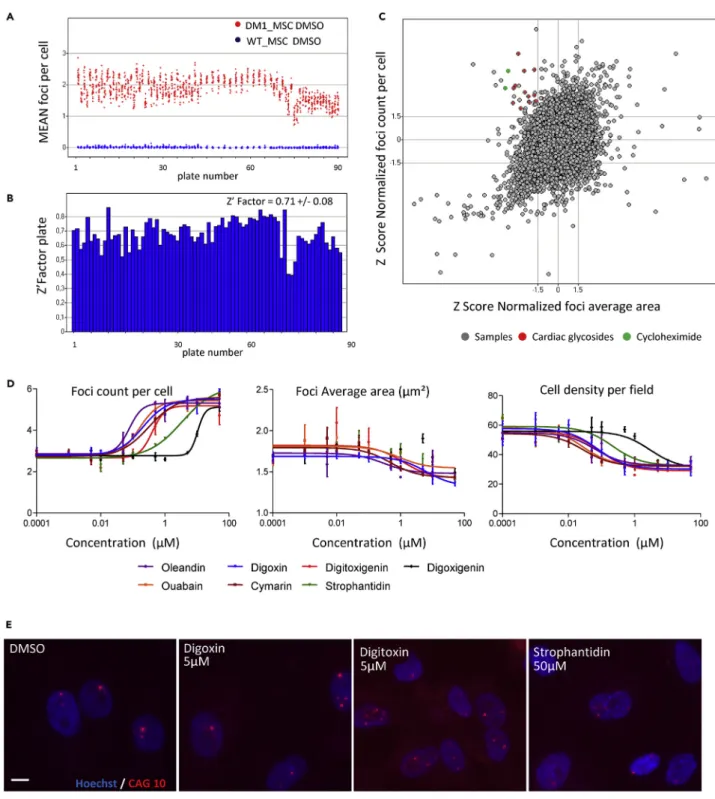
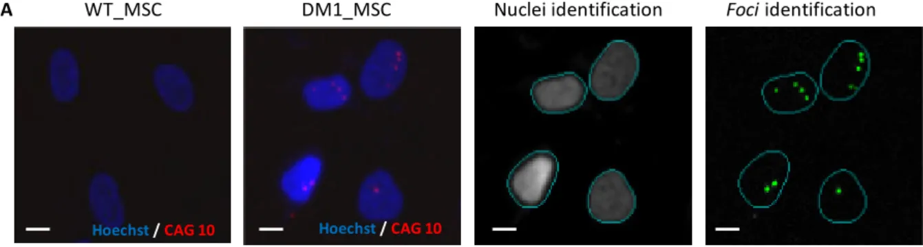
Nuclear foci were identified in DM1_MSCs using an automated RNA fluorescence in situ hybridization assay that was developed to detect expanded CUG repeats (Figure S1A). DM1_MSCs contained an average of 2.5 foci, whereas none were observed in control cells (Figure 1A). Results were confirmed as robust based on a high Z-factor, of 0.71, calculated for the assay (Figure 1B).
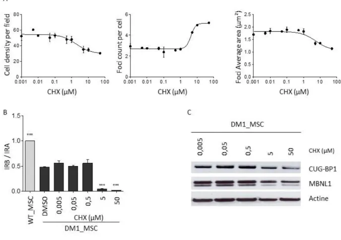
The primary drug screening was carried out by considering the number of intranuclear foci per cell using 12,089 compounds assayed at a concentration of 10mM, including a set of 1,120 FDA-approved drugs. Compounds were selected as candidates when the number of foci reached 1.5 standard deviations below the mean value for untreated DM1_MSCs, in the absence of unacceptable cytotoxicity (cutoff for cell survival set at 20% of control). This target was then revealed as non-informative, because none of the compounds that were tested demonstrated a capacity for decreasing the number of foci in the absence of unacceptable cell toxicity. Four hit compounds only emerged once these results were secondarily filtered by taking into account the average area of individual intranuclear foci. Their effects were characterized by an increased number of foci per nuclei in conjunction with their size reduction, which suggested a partial disaggregation of ribonucleoprotein inclusions. These compounds were cyclo-heximide (CHX) and three members of the cardiac glycoside family, namely, strophantidin, ouabain, and digoxigenin (Figures 1C andS1B). The first compound, CHX, was excluded from further analysis owing to its well-known activity as a protein synthesis inhibitor. In support of the hypothesis that the apparent reduction in size of the foci was due to a general decrease in cell protein content, we observed a decreased expression of MBNL1 and CUGBP1, as well as an exacerbation of defective alternative splicing associated with DM1 after treatment with CHX (Figure S2). The identification of three cardiac glycosides in the screening campaign suggested a common mechanism of action, and four additional members of this chemical family were therefore included in dose-response experiments, revealing similar dose-dependent effects: an increased number of foci per nucleus associated with a size reduction of each focus (Figures 1D and 1E).
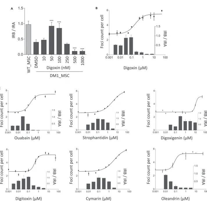
In parallel, a concomitant rescue of dysregulated alternative splicing was sought using the defective in-clusion of exon 11 in the insulin receptor (IR) gene that was described as a representative in DM1 (Savkur et al., 2001) (Figure 2A). The ratio of IRB (containing exon 11) to IRA (excluding exon 11) was used as a readout. Although the action of cardiac glycosides on the foci parameters was efficient at the micromolar range, it was associated with decreased cell viability, as well as an exacerbation of the DM1-related IR splicing defect, decreasing the ratio of IR transcripts. Conversely, all cardiac glycosides that were tested normalized the DM1-altered ratio of IR transcripts at concentrations ranging from 10 nM to 500 nM, de-pending on the chemical structure, in the absence of overt cytotoxicity and of any effect on foci (Figures 2B and 2C).
Digoxin Effect on Alternative mRNA Splicing
Digoxin, the best documented and most commonly prescribed medication in the cardiac glycoside family, was used as a representative to further characterize the therapeutic potential in DM1.
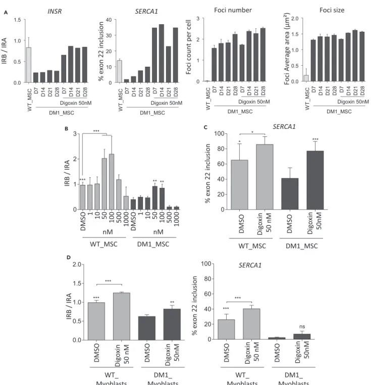
The long-term effect of this drug was demonstrated by chronic treatment of DM1_MSCs with 50 nM digoxin for 4 weeks in standard tissue culture T75 flasks, with analysis of one sample at each time point. This treat-ment induced a stable splicing switch of the IR exon 11 and SERCA1 exon 22 from a DM1 to a WT pattern (Figure 3A). This effect was observed in the absence of a concomitant effect of the treatment on the number and size of the foci. Other splice defects associated with DM1 were also shown to be normalized, including cTNT exon 5, CLCN-1 exon 7a in DM1_hES-derived MSCs, and NMDAR1 exon 5 in DM1_hES-derived neurons after a 48-hr treatment period (Figure S3).
Figure 1. Development of a High-Content Assay for Mutant DMPK Foci
(A) Mean number of detected foci per cell in untreated DM1-MSCs (red dots) and WT_MSCs (blue dots). Each dot represents one well in 90 screening plates.
(B) Determination of the Z0factor by using the number of detected foci per cell between DM1 and WT_MSCs for each plate, with a mean value of around
0.7 for the whole screening campaign.
(C) Scatterplot representation of the primary screening analysis based on the number of detected foci per cell and average area of foci after normalization in non-treated DM1_MSCs. Validated compounds are shown with colored dots.
(D) Dose-response analysis for number of foci, area of foci, and cell density in DM1_MSCs after 48 hr treatment with seven cardiac glycosides. Mean valuesG SD
are given (10 points in dose-response curve, each in triplicate).
(E) Representative images of mutant DMPK mRNA foci detected by RNA-fluorescence in situ hybridization in DM1_MSCs after treatment with three different
The ability of cardiac glycosides to modulate RNA processing independently of an effect on foci was confirmed by treating WT_MSCs with nanomolar digoxin concentrations. This also led to an increased in-clusion of exon 11 in IR and exon 22 in SERCA1 in these cells, although above control values in these cases (Figures 3B and 3C). Similar results were obtained with other cellular systems, namely, primary cultures of myoblasts and human embryonic stem cells (hES)-derived neurons (Figures 3D andS3C). In addition, there was no effect of digoxin, either on the level of expression or on the localization of MBNL1 and CUGBP1 in DM1 cells, the mislocalization and change in bioavailability of which are deemed responsible for the
A B
C
Figure 2. Digoxin Treatment in the Nanomolar Range Rescues Alternative IR and SERCA1 Splicing in DM1 Cells
(A) Dose-response analysis for alternative IR splicing in DM1_MSCs treated with digoxin for 48 hr. Relative expression level of the two isoforms of IR (IRA [-exon 11] and IRB [+exon 11]) were measured by quantitative real-time RT-PCR. Data represent the ratio of IRB/IRA and are indicated as mean G SD (n = 3). Data were analyzed with one way ANOVA, followed by a Dunnett post-hoc test. ***: p-value < 0.001.
(B) Superposition of the concentration-dependent effect of digoxin on the number of foci per cell (solid line, left axis) and alternative IR splicing (black bars, right axis) on DM1_MSCs treated for 48 hr.
(C) Dose-response analysis of six different cardiac glycosides on mutant DMPK foci (solid lines; n = 2; meanG SEM) and alternative IR splicing (bars; n = 2,
A
B C
D
Figure 3. Digoxin Treatment Rescues Alternative IR and SERCA1 Splicing in DM1 Cells
(A) Effect of chronic digoxin treatment on mutant DMPK foci and alternative splicing of IR and SERCA1. DM1_MSCs were continuously treated with 50 nM of digoxin over 4 weeks. Mutant DMPK foci and alternative splicing of IR and SERCA1 were analyzed every 7 days. Untreated WT_MSCs were used as the control. For alternative IR and SERCA1 splicing, one sample was analyzed at each time point. Relative expression levels of the two isoforms of IR (IRA [-exon 11] and IRB [+exon 11]) were measured by real-time quantitative RT-PCR. Data represent the ratio of IRB/IRA (n = 1 at each time point). Alternative IR splicing was analyzed using real-time quantitative RT-PCR. The percentage for inclusion of SERCA1 exon 22 was determined by RT-PCR, followed by analysis with a
2100 Bioanalyzer (Agilent). Quantification of each band was performed using 2100 Expert software. For mutant DMPK foci, data are presented as meanG SD
(n = 4).
(B) Concentration-dependent effect of digoxin on alternative IR splicing in WT and DM1_MSCs. Relative expression levels of the two isoforms of IRA [-exon 11] and IRB [+exon 11]) were measured by real-time quantitative RT-PCR. Data represent the ratio of IRB/IRA.
(C) Effect of digoxin treatment at 50 nM for 48 hr on the inclusion of exon 22 of SERCA1 in WT and DM1_MSCs, determined by RT-PCR, followed by analysis with a 2100 Bioanalyzer (Agilent).
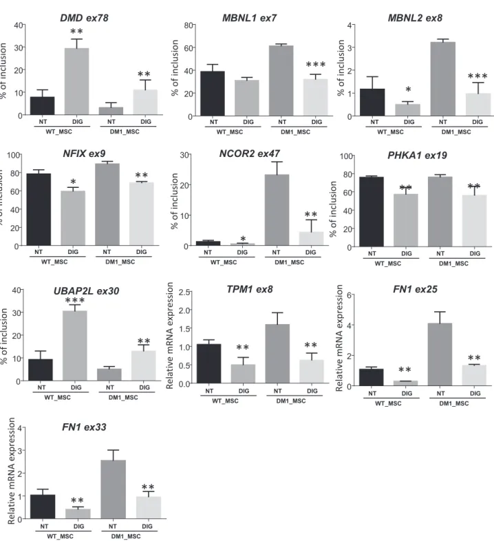
impairment of alternative mRNA splicing (Figures S4A–S4C and S4E). The consequences of digoxin treat-ment on alternative mRNA splicing were thus explored more comprehensively using deep RNA sequencing, comparing DM1_MSCs with or without treatment with 50 nM of the drug over a period of 2 days. Analysis of annotated splice junctions identified 44 modified splices with Deltac R 20 and 258 with 10% Delta c % 20 (Tables S1andS2). The comparison of this dataset with a recently published list of genes, which exhibited dysregulated alternative RNA splicing in muscle biopsies from patients with DM1 (Nakamori et al., 2013), revealed a beneficial effect of digoxin treatment on 10 different DM1-specific splicing defects as determined by qRT-PCR analysis (Figure 4).
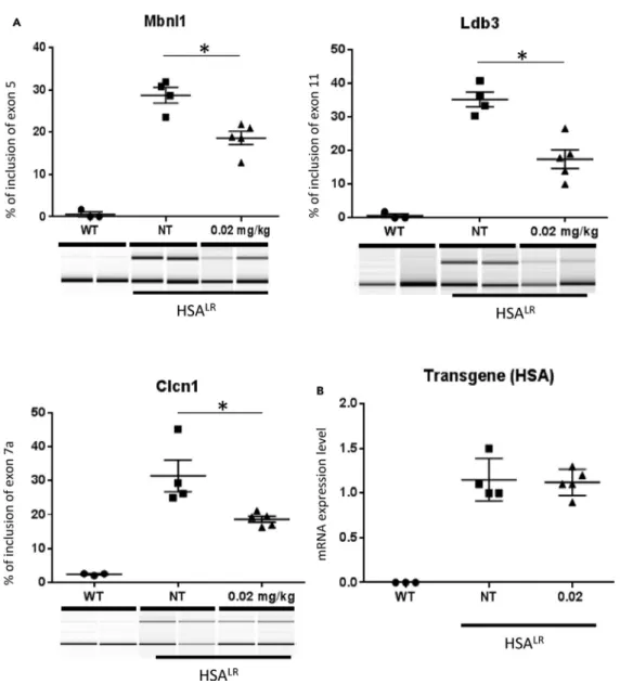
Finally, the effect of digoxin was evaluated in vivo, in the HSALRtransgenic DM1 mouse model, which expresses 220 CUG repeats in skeletal muscle and exhibits dysregulated alternative splicing (Mankodi et al., 2002). Digoxin was administrated daily by intraperitoneal injection for 7 days at two dosage regimens: 0.02mg/kg and 2 mg/kg/ d. Rescue of Mbnl1-, Ldb3-, and Clcn1-dysregulated RNA splicing was observed after digoxin treatment at the lower dosage (Figures 5A andS5), independent of any changes in expression of the HSA transgene (Figure 5B). Mbnl1 exon 5 inclusion, for instance, was recorded at 17.44%G 2.7% after digoxin treat-ment at 0.02mg/kg/d, down from 35.13%G 2.2% in untreated HSALRmice (FigureS5).
Molecular Mechanisms that Underlie the Effects of Cardiac Glycosides on Alternative RNA Splicing
A series of hypotheses were sequentially explored in a search for the molecular mechanisms underlying the potential therapeutic effects of cardiac glycosides.
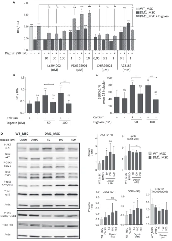
We first checked whether these effects were associated with a change in the intracellular concentration of calcium, which is the common outcome of treatments with cardiac glycosides. To this end, we used the calcium ionophore A23187, which increases intracellular calcium content (Dedkova et al., 2000). Treat-ment with the calcium ionophore A23187 had a similar effect on the IR splicing ratio as that produced by digoxin. A combination of digoxin and A23187 led to neither an additive nor a synergistic effect (Figure 6A), supporting the hypothesis of a calcium-dependent mechanism for digoxin effects on alternative mRNA splicing. This was further validated by the abolition of the drug effect on alternative IR and SERCA1 splicing when calcium was withdrawn from the cell culture medium (Figures 6B and 6C).
The next step in the analysis aimed at determining intracellular signaling pathways that may be triggered by the drug and that may affect alternative splicing. Cardiac glycosides are sodium-potassium pump inhib-itors that activate multiple cell signaling pathways, representing many other possible mechanisms by which these compounds might affect alternative splicing. Following treatment of DM1_MSCs with 50 or 100 nM digoxin for 48 hr, western blot analyses indicated a moderately increased phosphorylation of AKT and GSK3-b and a decreased activation of ERK (Figure 6D). A combination of digoxin with either LY294002 or CHIR99021, which specifically inhibit AKT and GSK3, respectively, attenuated the beneficial effect of digoxin on IR exon 11 inclusion (Figure 6A), indicating that these pathways contribute toward the effects of the drug. Conversely, inhibition of ERK phosphorylation by PD0325901 in DM1_MSC-normalized IR exon 11 inclusion in the absence of digoxin, and co-treatment with digoxin, resulted in an additive or synergistic effect, suggesting a cooperative mode of action (Figure 6A). Finally, two RNA-binding proteins that were previously shown to be modulated by cardiac glycosides (Anderson et al., 2012), namely, SRSF3 (SRp20) and TRA2B, were investigated. Western blot analysis revealed that their expression levels were not modified in treated DM1_MSCs (Figures S4D and S4E).
Functional Effects of Digoxin on Myogenic Differentiation
In an attempt to identify a functional correlate for these biological results, the digoxin effect was chal-lenged on DM1 primary human myoblast cells carrying 2000 CTG repeats and displaying defective myotube differentiation in vitro (Bigot et al., 2009; Thornell et al., 2009). Digoxin treatment at 50 nM for 7 days increased the number of myotubes that were formed, as quantified by both myosin heavy chain immunostaining (number of MF20-positive cells), and the number of nuclei per MF20-positive cell
Figure 3. Continued
(D) Effect of digoxin treatment on alternative IR and SERCA1 splicing in primary myoblasts isolated from healthy individuals and patients with DM1.
For all graphs, data are presented as meanG SD (n = 3) and analyzed with one-way ANOVA, followed by a Bonferroni post-hoc test. *p < 0.05, **p < 0.01,
(Figures 7A and 7B andS6). Calcium deprivation in the culture medium during myotube differentiation fully abolished the functional benefit of digoxin treatment (Figure 7C). Similar positive results on the myogenic process were obtained by applying a 50 nM digoxin pulse for the first two days of myoblast differentiation. This observation was further confirmed using other cardiac glycosides (Figure S7). In addition, qRT-PCR
Figure 4. Beneficial Effect of Digoxin Treatment in the Nanomolar Range on Other Alternative Splicing Associated with DM1
RT-PCR analysis of 10 forms of alternative splicing reported to be affected in patients with DM1 and rescued by digoxin treatment (50 nM for 48 hr) in WT and DM1_MSCs. The percentage for inclusion of isoform was determined by RT-PCR, followed by analysis with a 2100 Bioanalyzer (Agilent). Quantification of
each band was performed using 2100 Expert software. Data are presented as meanG SD (n = 3). Data were analyzed with one-way ANOVA, followed by a
analysis of alternate splicing variants revealed a normalization of MBNL1 exon 7, INSR exon 11, and DMD exon 78 in myotubes treated with 50 nM digoxin.
DISCUSSION
The main outcome of this study is the identification of the cardiac glycoside chemical family as constituting a potential therapeutic strategy for DM1. These compounds had the capacity to promote changes toward normalization in alternative mRNA splicing of genes affected by the DM1 mutation and myogenic fusion of affected myoblasts. These results were obtained using high-throughput drug screening, based on human pluripotent stem cell line derivatives that carried the mutant gene and recapitulated the cardinal cellular features of the disease. Similar experimental paradigms may be proposed for a vast array of monogenic diseases as these cellular models give access to relevant readouts.
Figure 5. Digoxin Treatment Partially Restores In Vivo Splicing Defects in a DM1 Mouse Model
(A) Analysis of Mbnl1, Ldb3, Clcn1 alternative splicing by RT-PCR in HSALRmice treated with intraperitoneal injection of
digoxin for 7 days (n = 3 for wild-type, n = 4 for mock-treated, and n = 5 for treated). RT-PCR was followed by analysis with a 2100 Bioanalyzer (Agilent). Quantification of each band was performed using 2100 Expert software. Data are presented
as meanG SEM. The p value was determined using the Mann-Whitney U test; *p < 0.05.
(B) Quantitative real-time qRT-PCR analysis of HSA transgene as a function of digoxin treatment. Data were normalized with the housekeeping gene 18S.
A
B C
D
Figure 6. Calcium-Dependent Effect of Digoxin on Alternative Splicing of IR and SERCA1
(A) Pharmacological approaches to analyze the combinatorial effect of AKT, GSK3, ERK, and calcium pathways with digoxin on alternative IR splicing. DM1_MSCs were treated for 48 hr, with or without 50 nM of digoxin, in combination with different concentrations of AKT inhibitor (LY29002), GSK3 inhibitor (CHIR99021), ERK inhibitor (PD0325901), or calcium ionophore (A23187). WT_MSCs were used as a positive control. Alternative IR splicing was quantified using qRT-PCR.
Data are presented as meanG SD (n = 3).
(B and C) Influence of calcium on the digoxin-mediated effect on alternative splicing of IR (B) and SERCA1 (C) in DM1_MSCs. Cells were maintained in medium with or without calcium and treated with digoxin (50 or 100 nM) for 48 hr.
DM1 is characterized by the accumulation of the mutant DMPK mRNA in cell nuclei, due to the presence of an abnormal tract of CTG triplet repeats in the 30UTR of the DMPK gene. This mutation provokes a major alteration in the bioavailability or activity of RNA-binding proteins, most particularly MBNL1 and CUGBP1, in part through the formation of intranuclearribonucleoprotein aggregates (Mankodi et al., 2001). This, in turn, induces changes in alternative RNA splicing in a number of genes that are physiological targets of those proteins. In the present study, cardiac glycosides were shown to affect these pathological mecha-nisms in different ways, depending upon their concentration. At high cytotoxic concentrations—in the micromolar range—they provoked a partial disaggregation of the inclusions. This, however, did not correlate with a normalization of the alternative RNA splicing activity, which rather deteriorated further as a consequence of cellular toxicity. Such a phenomenon was reminiscent of what was observed in a previous study in which the expression of MBNL1 was decreased in primary fibroblasts derived from patients with DM1 using a specific small interfering RNA (Childs-Disney et al., 2013).
The large variety of alternate splicing defects observed in DM1 are thought to be responsible for the multi-systemic nature of this disease, and some reports have already linked the alteration of single mRNA variants to specific disease phenotypes. For instance, the increased inclusion of CLC1 exon 7a is involved in the onset of myotonia, and repression of this exon inclusion using morpholino antisense oligonucleotide greatly reduced the myotonic grade observed in genetic DM1 mouse models (Wheeler et al., 2007). There-fore normalization of abnormal ratios of these transcripts, if obtained under treatment, is expected to be therapeutically relevant. The effects of cardiac glycosides do not, however, specifically target the causative molecular defect of the disease and the extent of the success of treatment for different disease phenotypes can only be partial. Whether the partial recovery of ratios might be therapeutically relevant will need to be explored in a clinical setting. Nevertheless, the positive effect of digoxin recorded in the present study on a myogenesis assay indicates that cardiac glycosides might have functional consequences. These com-pounds are still regularly prescribed for the management of atrial fibrillation and heart failure in the general population, although their clinical use has been steadily decreasing because of their narrow therapeutic window, which makes them a toxicity risk (Kanji and MacLean, 2012). Given that cardiac conduction abnor-malities are critical symptoms in DM1, careful consideration of the indication and cautious observation will be required for administration, as well as close monitoring of the side effects of cardiac glycosides in patients with DM1. However, there are already reports on several patients with DM1 exhibiting good tolerance to digoxin or digitoxin (Finsterer and Stollberger, 2012; LagoeiroJorge et al., 2012; Phillips and Harper, 1997; Schmitt and Schmidt, 1975). It is also notable that the narrow concentration range in which digoxin exerts a beneficial effect on DM1 phenotypes in this study is similar to tissue concentrations found in patients treated for heart failure (Hothi et al., 2014; Schoner and Scheiner-Bobis, 2007). The ultimate goal of a DM1 therapy is the correction of all affected genes. Targeting the causative mech-anisms of the pathology, at the level of either the DMPK gene mutation or by introducing competition to its mRNA binding to splicing factors such as MBNL1, would obviously be the most elegant therapy. This is be-ing actively pursued by a number of teams who have developed various approaches based on gene transfer, antisense oligonucleotides, or specific synthetic small molecules (Childs-Disney et al., 2012, 2013; Francois et al., 2011; Ketley et al., 2013; Laustriat et al., 2015; Lee et al., 2012b; Mulders et al., 2009; Nakamori et al., 2016; Pandey et al., 2015; Parkesh et al., 2012; Wheeler et al., 2012). Current results point to a parallel pharmacological approach, aiming at modifying alternative RNA splicing of as many DM1-affected genes as possible, without affecting the causative mechanisms of the disease. Such an approach would benefit from a combination of drugs. Indeed, cardiac glycosides are not an isolated case. Two small molecules have recently been clinically evaluated in DM1: the anti-myotonic mexiletine (Logigian et al., 2010) and, more recently, the anti-diabetic drug metformin (Laustriat et al., 2015). Interest-ingly, the mechanism of action for cardiac glyocosides differs from that for these previously mentioned
Figure 6. Continued
Medium without calcium was added 30 min before the digoxin (n = 3). Data were analyzed with one-way ANOVA followed by a Bonferroni post-hoc test. *p < 0.05, **p < 0.01, ***p < 0.001.
(D) Effect of different concentrations of digoxin on mitogen-activated protein kinase and mammalian target of rapamycin (mTOR) signaling pathways. Expression level of key modulators of Akt/mTOR signaling in DM1_MSCs treated with
different concentrations of digoxin for 48 hr, including Phospho(Ser473)-Akt, Phospho(Ser21/9)-GSK3a/b,
Phospho(Ser235-236)-ribosomal protein S6 (rpS6), Phospho (Thr202/Tyr204) ERK, and the corresponding total forms.
Antibodies used are liste inTable S3. Western blot analysis was quantified by using ImageJ software. Data are presented
A B
C
D
Figure 7. Digoxin Treatment Restores In Vitro Myogenic Defect in DM1
(A) Representative immunostaining for the myogenic marker Mf20 in WT and DM1 myoblasts differentiated for 6 days. DM1 myoblasts were treated with 50 nM of digoxin at day 1 of differentiation.
(B) Automated quantification of myogenic differentiation as determined by the measurement of the total area stained for Mf20 per field in WT-differentiated myoblasts and in DM1-differentiated myoblasts, treated or untreated with 50 nM digoxin, as well as the quantification of nucleus area per Mf20-positive myotube. All data were analyzed with one way ANOVA followed by a ‘‘Bonferroni’s Multiple Comparison Test’’ *p<0.01, ****p<0.0001.
(C) Calcium-dependent effect of digoxin on the in vitro myogenic impairment observed in DM1 myoblasts. Cells were differentiated in the presence or absence of calcium. The effect of digoxin treatment (50 nM for 7 days) on myogenesis was evaluated by automated quantification according to the total area stained for Mf20 per field and as the percentage of
the total area of nuclei identified within Mf20-positive myotubes. Data are presented as meanG SD (n = 3 independent
experiments, 3 replicates) and were analyzed with one-way ANOVA, followed by a ‘‘Bonferroni’s multiple comparison test’’; **p < 0.05, ***p < 0.001.
(D) Analysis of splice variants in WT and DM1 myoblasts differentiated for 8 days. Digoxin 50nM was applied as a chronic (7 days) or pulse (2 days) treatment. Percentage of exon inclusion was quantified by RT-PCR and Capillary Electrophoresis
using the Agilent bioanalyzer 2100. Data represent meanG SD (n=1 experiment – triplicates). All data were analyzed with
compounds. Cardiac glycosides modulate the Na+/K+ATPase protein complex upon binding and activate downstream signaling pathways via increased intracellular calcium concentrations (Fontana et al., 2013), which can independently induce changes in alternative mRNA splicing patterns (Razanau and Xie, 2013; Sharma and Lou, 2011; Sharma et al., 2014). In this case, the molecular mechanisms linking calcium concentration to the modulation of the splicing process may involve chromatin modifications, rather than RNA-binding proteins (Sharma et al., 2014). Thus the effects of metformin and cardiac glycosides do not rely on fully identical intracellular pathways. Therefore they may also modify various subsets of DM1-affected genes. It is thus tempting to speculate that a combination of several drugs may lead to ad-ditive or synergistic beneficial effects (Konieczny et al., 2017). From this perspective, it will also be interesting to analyze the effects of other drugs known to affect alternative RNA splicing, e.g., valproate, amyloride, or kinetin (Axelrod et al., 2011; Chang et al., 2011; Farrelly-Rosch et al., 2017), in the context of DM1 in vitro models. Calcium changes in treated cells may not, however, be the sole mechanism under-lying the effects of cardiac glycosides. In the present study, use of PD0325901, an inhibitor of the ERK pathway, appeared as yet another path for regulating alternative mRNA splicing, which may normalize a further subset of DM1-affected genes. DM1 myoblasts have been described to sustain abnormal ERK activity that might be responsible for the defective myogenic differentiation (Beffy et al., 2010). It would therefore be interesting to determine whether the digoxin effect on the functional in vitro myogenesis was triggered by a splicing modification or through an ERK1/2 inhibitory effect. In addition, our results also demonstrated that digoxin treatment led to an increased level of the inactive form of GSK3-b (phosphorylated at S9) in DM1_MSCs. Interestingly, inhibition of GSK3-b signaling with tideglusib is actually under clinical investigation in patients with DM1 (ClinicalTrials.govidentifier: NCT02858908). This development was based on different studies demonstrating that the active GSK3-b is elevated in skel-etal muscle biopsy samples from patients with DM1 and in skelskel-etal muscle of mice. Inhibition of GSK3-b signaling in both DM1 cell culture and mouse models reduced muscle weakness and myotonia in DM1 mice (Jones et al., 2012; Wei et al., 2018). In our study, the use of CHIR99021, a well-characterized GSK3-b inhibitor, did not improve IR exon 11 inclusion and minimized the digoxin effect when co-treatment was applied. Even if a combination of a cardiac glycoside with a GSK3-b inhibitor might theoretically allow the modification of a larger variety of exons and phenotypes, a combinatorial therapeutic strategy needs to be carefully evaluated, as one strategy can abolish the effect of the other.
The present study further emphasizes the value of pluripotent stem cell lines derived from mutant-gene-carrying donors as in vitro cell models for monogenic pathologies and drug discovery. Access to a never-ending supply of cells expressing a physiologically and pathologically relevant human genome is a first advantage over other cell models. The physiological relevance of our cellular model was also of particular importance here, because ratios of gene transcripts are highly dependent upon cell phenotypes. It is inter-esting to note that a previous study has already demonstrated the ability of cardiac glycosides to modulate alternative mRNA splicing of a large number of genes (Stoilov et al., 2008). However, using HEK cells as a model, this list did not include the 15 DM1-affected genes identified in the present study using mesodermal and neural derivatives of ES cells. In addition, the use of stem cell derivatives allows a comparison of popu-lations of cells displaying an exact same phenotype, which differ only in their genotype (e.g., DM1 versus WT). In summary, stem cell derivatives provide a very interesting platform for in vitro drug discovery, but results need to be confirmed using other relevant in vitro and in vivo models, as performed here, before identifying compounds that can be moved forward toward clinical application.
Limitations of the Study
In this study, we identified the capacity of the FDA-approved cardiac glycosides to normalize several alter-native mRNA splicing of genes affected by the causal mutation of DM1. Our results demonstrated that this effect is calcium dependent and can be synergic to the inhibition of the ERK signaling pathway. However, the exact molecular mechanisms by which cardiac glycosides can induce the normalization of these affected alternative splicing is still unclear. Our results suggest that the mechanisms of action of these compounds are not dependent on the two main splicing factors, known to be involved in the pathogenesis of DM1. Therefore additional experiments are needed to investigate the mechanisms by which cardiac glycosides normalize alternative splicing defects in DM1.
METHODS
SUPPLEMENTAL INFORMATION
Supplemental Information includes Transparent Methods, seven figure, and three tables and can be found with this article online athttps://doi.org/10.1016/j.isci.2018.12.019.
ACKNOWLEDGMENT
I-Stem is part of the Biotherapies Institute for Rare Diseases (BIRD), supported by the Association Franc¸ai-secontre les Myopathies (AFM-Te´le´thon). This project was supported by AFM and INSERM. This project was also funded by grants from ANR (ANR-16-CE17-0018), AFM, and the Laboratoired’Excellence Revive (Investissementd’Avenir; ANR-10-LABX-73). We thank Vincent Mouly (INSERM U 787, Paris), Nicolas Char-let (IGBMC, Illkirch), and Genevieve Gourdon (INSERM U781, Paris) for helpful contributions and critical re-views of the manuscript.
AUTHOR CONTRIBUTIONS
Conceptualization, Y.M., M.L., M.P., G.P., S.B., and C.M.; Methodology, Y.M., P.P., B.B., L.L., J.G., and C.M.; Investigation, Y.M., P.P., B.B., L.L., S.R., D.F., J.C., and C.M.; Formal Analysis, Y.M., M.L.,J.D., H.P., G.P., D.A., S.B., and C.M.; Writing – Original Draft, C.M., Y.M., and M.P.; Writing – Review and Editing, Y.M., S.B., L.L., and C.M.; Funding acquisition, C.M. and M.P.
DECLARATION OF INTERESTS
The authors declare no competing interests.Received: July 9, 2018 Revised: December 14, 2018 Accepted: December 20, 2018 Published: January 25, 2019
REFERENCES
Anderson, E.S., Lin, C.H., Xiao, X., Stoilov, P., Burge, C.B., and Black, D.L. (2012). Thecardiotonic steroid digitoxin regulates alternative splicing through depletion of the splicing factors SRSF3 and TRA2B. RNA 18, 1041–1049.
Axelrod, F.B., Liebes, L., Gold-Von Simson, G., Mendoza, S., Mull, J., Leyne, M., Norcliffe-Kaufmann, L., Norcliffe-Kaufmann, H., and Slaugenhaupt, S.A. (2011). Kinetin improves IKBKAP mRNA splicing in patients with familial dysautonomia. Pediatr. Res. 70, 480–483.
Beffy, P., Del Carratore, R., Masini, M., Furling, D., Puymirat, J., Masiello, P., and Simili, M. (2010). Altered signal transduction pathways and induction of autophagy in human myotonic dystrophy type 1 myoblasts. Int. J. Biochem. Cell Biol. 42, 1973–1983.
Bigot, A., Klein, A.F., Gasnier, E., Jacquemin, V., Ravassard, P., Butler-Browne, G., Mouly, V., and Furling, D. (2009). Large CTG repeats trigger p16-dependent premature senescence in myotonic dystrophy type 1 muscle precursor cells. Am. J. Pathol. 174, 1435–1442.
Chang, W.H., Liu, T.C., Yang, W.K., Lee, C.C., Lin, Y.H., Chen, T.Y., and Chang, J.G. (2011). Amiloride modulates alternative splicing in leukemic cells and resensitizes
Bcr-AblT315I mutant cells to imatinib. Cancer Res. 71, 383–392.
Charbord, J., Poydenot, P., Bonnefond, C., Feyeux, M., Casagrande, F., Brinon, B., Francelle,
L., Auregan, G., Guillermier, M., Cailleret, M., et al. (2013). High throughput screening for inhibitors of REST in neural derivatives of human embryonic stem cells reveals a chemical compound that promotes expression of neuronal genes. Stem Cells 31, 1816–1828.
Childs-Disney, J.L., Parkesh, R., Nakamori, M., Thornton, C.A., and Disney, M.D. (2012). Rational design of bioactive, modularly assembled aminoglycosides targeting the RNA that causes myotonic dystrophy type 1. ACS Chem. Biol. 7, 1984–1993.
Childs-Disney, J.L., Stepniak-Konieczna, E., Tran, T., Yildirim, I., Park, H., Chen, C.Z., Hoskins, J., Southall, N., Marugan, J.J., Patnaik, S., et al. (2013). Induction and reversal of myotonic dystrophy type 1 pre-mRNA splicing defects by small molecules. Nat. Commun. 4, 2044.
Darville, H., Poulet, A., Rodet-Amsellem, F., Chatrousse, L., Pernelle, J., Boissart, C., Heron, D., Nava, C., Perrier, A., Jarrige, M., et al. (2016). Human pluripotent stem cell-derived cortical neurons for high throughput medication screening in autism: a proof of concept study in SHANK3 haploinsufficiency syndrome. EBioMedicine 9, 293–305.
Dedkova, E.N., Sigova, A.A., and Zinchenko, V.P. (2000). Mechanism of action of calcium ionophores on intact cells: ionophore-resistant cells. Membr.Cell Biol. 13, 357–368.
Denis, J.A., Gauthier, M., Rachdi, L., Aubert, S., Giraud-Triboult, K., Poydenot, P., Benchoua, A.,
Champon, B., Maury, Y., Baldeschi, C., et al. (2013). mTOR-dependent proliferation defect in human ES-derived neural stem cells affected by myotonic dystrophy type 1. J. Cell Sci. 126, 1763–1772.
Du, H., Cline, M.S., Osborne, R.J., Tuttle, D.L., Clark, T.A., Donohue, J.P., Hall, M.P., Shiue, L., Swanson, M.S., Thornton, C.A., et al. (2010). Aberrant alternative splicing and extracellular matrix gene expression in mouse models of myotonic dystrophy. Nat. Struct. Mol. Biol. 17, 187–193.
Du, J., Campau, E., Soragni, E., Jespersen, C., and Gottesfeld, J.M. (2013). Length-dependent CTG.CAG triplet-repeat expansion in myotonic dystrophy patient-derived induced pluripotent stem cells. Hum. Mol. Genet. 22, 5276–5287.
Farrelly-Rosch, A., Lau, C.L., Patil, N., Turner, B.J., and Shabanpoor, F. (2017). Combination of valproic acid and morpholino splice-switching oligonucleotide produces improved outcomes in spinal muscular atrophy patient-derived fibroblasts. Neurochem.Int. 108, 213–221.
Finsterer, J., and Stollberger, C. (2012). Beneficial effect of digitoxin for heart failure from noncompaction in myotonic dystrophy 1. Int. J. Cardiol. 160, e50–e51.
Fontana, J.M., Burlaka, I., Khodus, G., Brismar, H., and Aperia, A. (2013). Calcium oscillations triggered by cardiotonic steroids. FEBS J. 280, 5450–5455.
Francois, V., Klein, A.F., Beley, C., Jollet, A., Lemercier, C., Garcia, L., and Furling, D. (2011). Selective silencing of mutated mRNAs in DM1 by using modified hU7-snRNAs. Nat. Struct. Mol. Biol. 18, 85–87.
Gauthier, M., Marteyn, A., Denis, J.A., Cailleret, M., Giraud-Triboult, K., Aubert, S., Lecuyer, C., Marie, J., Furling, D., Vernet, R., et al. (2013). A defective Krab-domain zinc-finger transcription factor contributes to altered myogenesis in myotonic dystrophy type 1. Hum. Mol. Genet. 22, 5188–5198.
Hothi, S.S., Chinnappa, S., and Tan, L.B. (2014). 200+ years of a misunderstood drug for treating chronic heart failure: digoxin, why and how should we continue using it? Int. J. Cardiol. 168, 645–647. Jones, K., Wei, C., Iakova, P., Bugiardini, E., Schneider-Gold, C., Meola, G., Woodgett, J., Killian, J., Timchenko, N.A., and Timchenko, L.T. (2012). GSK3beta mediates muscle pathology in myotonic dystrophy. J. Clin. Invest. 122, 4461–4472.
Kanadia, R.N., Johnstone, K.A., Mankodi, A., Lungu, C., Thornton, C.A., Esson, D., Timmers, A.M., Hauswirth, W.W., and Swanson, M.S. (2003). A muscleblind knockout model for myotonic dystrophy. Science 302, 1978–1980. Kanji, S., and MacLean, R.D. (2012). Cardiac glycoside toxicity: more than 200 years and counting. Crit. Care Clin. 28, 527–535. Ketley, A., Chen, C.Z., Li, X., Arya, S., Robinson, T.E., Granados-Riveron, J., Udosen, I., Morris, G.E., Holt, I., Furling, D., et al. (2013). High-content screening identifies small molecules that remove nuclear foci, affect MBNL distribution and CELF1 protein levels via a PKC-independent pathway in myotonic dystrophy cell lines. Hum. Mol. Genet. 23, 1551–1562.
Konieczny, P., Selma-Soriano, E., Rapisarda, A.S., Fernandez-Costa, J.M., Perez-Alonso, M., and Artero, R. (2017). Myotonicdystrophy: candidate small molecule therapeutics. Drug Discov. Today 22, 1740–1748.
Lagoeiro Jorge, B., Lanzieri, P., Braida do Carmo, F., Haffner, P.M., Lagoeiro Jorge, A., and Wolney de Andrade, M. (2012). Cardiomyopathy secondary to Steinert’s dystrophy. Insuficiencia Cardiaca 7, 143–147.
Laustriat, D., Gide, J., Barrault, L., Chautard, E., Benoit, C., Auboeuf, D., Boland, A., Battail, C., Artiguenave, F., Deleuze, J.F., et al. (2015). In vitro and in vivo modulation of alternative splicing by the biguanide metformin. Mol. Ther. Nucleic Acids 4, e262.
Lee, G., Ramirez, C.N., Kim, H., Zeltner, N., Liu, B., Radu, C., Bhinder, B., Kim, Y.J., Choi, I.Y., Mukherjee-Clavin, B., et al. (2012a). Large-scale screening using familial dysautonomia induced pluripotent stem cells identifies compounds that rescue IKBKAP expression. Nat. Biotechnol. 30, 1244–1248.
Lee, J.E., Bennett, C.F., and Cooper, T.A. (2012b). RNase H-mediated degradation of toxic RNA in myotonic dystrophy type 1. Proc. Natl. Acad. Sci. U S A 109, 4221–4226.
Logigian, E.L., Martens, W.B., Moxley, R.T.t., McDermott, M.P., Dilek, N., Wiegner, A.W.,
Pearson, A.T., Barbieri, C.A., Annis, C.L., Thornton, C.A., et al. (2010). Mexiletine is an effective antimyotonia treatment in myotonic dystrophy type 1. Neurology 74, 1441–1448. Mahadevan, M., Tsilfidis, C., Sabourin, L., Shutler, G., Amemiya, C., Jansen, G., Neville, C., Narang, M., Barcelo, J., O’Hoy, K., et al. (1992). Myotonic dystrophy mutation: an unstable CTG repeat in the 3’ untranslated region of the gene. Science 255, 1253–1255.
Mankodi, A., Takahashi, M.P., Jiang, H., Beck, C.L., Bowers, W.J., Moxley, R.T., Cannon, S.C., and Thornton, C.A. (2002). Expanded CUG repeats trigger aberrant splicing of ClC-1 chloride channel pre-mRNA and hyperexcitability of skeletal muscle in myotonic dystrophy. Mol. Cell 10, 35–44.
Mankodi, A., Urbinati, C.R., Yuan, Q.P., Moxley, R.T., Sansone, V., Krym, M., Henderson, D., Schalling, M., Swanson, M.S., and Thornton, C.A. (2001). Muscleblind localizes to nuclear foci of aberrant RNA in myotonic dystrophy types 1 and 2. Hum. Mol. Genet. 10, 2165–2170.
Marteyn, A., Maury, Y., Gauthier, M.M., Lecuyer, C., Vernet, R., Denis, J.A., Pietu, G., Peschanski, M., and Martinat, C. (2011). Mutant human embryonic stem cells reveal neurite and synapse formation defects in type 1 myotonic dystrophy. Cell Stem Cell 8, 434–444.
Mulders, S.A., van den Broek, W.J., Wheeler, T.M., Croes, H.J., van Kuik-Romeijn, P., de Kimpe, S.J., Furling, D., Platenburg, G.J., Gourdon, G., Thornton, C.A., et al. (2009). Triplet-repeat oligonucleotide-mediated reversal of RNA toxicity in myotonic dystrophy. Proc. Natl. Acad. Sci. U S A 106, 13915–13920.
Nakamori, M., Sobczak, K., Puwanant, A., Welle, S., Eichinger, K., Pandya, S., Dekdebrun, J., Heatwole, C.R., McDermott, M.P., Chen, T., et al. (2013). Splicing biomarkers of disease severity in myotonic dystrophy. Ann. Neurol. 74, 862–872. Nakamori, M., Taylor, K., Mochizuki, H., Sobczak, K., and Takahashi, M.P. (2016). Oral
administration of erythromycin decreases RNA toxicity in myotonic dystrophy. Ann. Clin. Transl. Neurol. 3, 42–54.
Pandey, S.K., Wheeler, T.M., Justice, S.L., Kim, A., Younis, H.S., Gattis, D., Jauvin, D., Puymirat, J., Swayze, E.E., Freier, S.M., et al. (2015). Identification and characterization of modified antisense oligonucleotides targeting DMPK in mice and nonhuman primates for the treatment of myotonic dystrophy type 1. J. Pharmacol. Exp. Ther. 355, 329–340.
Parkesh, R., Childs-Disney, J.L., Nakamori, M., Kumar, A., Wang, E., Wang, T., Hoskins, J., Tran, T., Housman, D., Thornton, C.A., et al. (2012). Design of a bioactive small molecule that targets the myotonic dystrophy type 1 RNA via an RNA motif-ligand database and chemical similarity searching. J. Am. Chem. Soc. 134, 4731–4742.
Phillips, M.F., and Harper, P.S. (1997). Cardiac disease in myotonic dystrophy. Cardiovasc. Res. 33, 13–22.
Razanau, A., and Xie, J. (2013). Emerging mechanisms and consequences of calcium
regulation of alternative splicing in neurons and endocrine cells. Cell.Mol. Life Sci. 70, 4527–4536. Savkur, R.S., Philips, A.V., and Cooper, T.A. (2001). Aberrant regulation of insulin receptor alternative splicing is associated with insulin resistance in myotonic dystrophy. Nat. Genet. 29, 40–47.
Schmitt, J., and Schmidt, C. (1975). [Steinert’s syndrome and the myocardium. Total gene expression by the myocardium]. J. Genet. Hum. 23, 59–64.
Schoner, W., and Scheiner-Bobis, G. (2007). Endogenous and exogenous cardiac glycosides and their mechanisms of action. Am. J. Cardiovasc. Drugs 7, 173–189.
Sharma, A., and Lou, H. (2011). Depolarization-mediated regulation of alternative splicing. Front. Neurosci. 5, 141.
Sharma, A., Nguyen, H., Geng, C., Hinman, M., Luo, G., and Lou, H. (2014). Calcium-mediated histone modifications regulate alternative splicing in cardyomyocytes. Proc. Natl. Acad. Sci. U S A 111, E4920–E4928.
Stoilov, P., Lin, C.H., Damoiseaux, R., Nikolic, J., and Black, D.L. (2008). A high-throughput screening strategy identifies cardiotonic steroids as alternative splicing modulators. Proc. Natl. Acad. Sci. U S A 105, 11218–11223.
Thornell, L.E., Lindstom, M., Renault, V., Klein, A., Mouly, V., Ansved, T., Butler-Browne, G., and Furling, D. (2009). Satellite cell dysfunction contributes to the progressive muscle atrophy in myotonic dystrophy type 1. Neuropathol. Appl. Neurobiol. 35, 603–613.
Thornton, C.A., Wang, E., and Carrell, E.M. (2017). Myotonic dystrophy: approach to therapy. Curr.Opin.Genet. Dev. 44, 135–140.
Turner, C., and Hilton-Jones, D. (2014). Myotonic dystrophy: diagnosis, management and new therapies. Curr.Opin. Neurol. 27, 599–606. Ueki, J., Nakamori, M., Nakamura, M., Nishikawa, M., Yoshida, Y., Tanaka, A., Morizane, A., Kamon, M., Araki, T., Takahashi, M.P., et al. (2017). Myotonic dystrophy type 1 patient-derived iPSCs for the investigation of CTG repeat instability. Sci. Rep. 7, 42522.
Wei, C., Stock, L., Valanejad, L., Zalewski, Z.A., Karns, R., Puymirat, J., Nelson, D., Witte, D., Woodgett, J., Timchenko, N.A., et al. (2018). Correction of GSK3beta at young age prevents muscle pathology in mice with myotonic dystrophy type 1. FASEB J. 32, 2073–2085. Wheeler, T.M., Leger, A.J., Pandey, S.K., MacLeod, A.R., Nakamori, M., Cheng, S.H., Wentworth, B.M., Bennett, C.F., and Thornton, C.A. (2012). Targeting nuclear RNA for in vivo correction of myotonic dystrophy. Nature 488, 111–115.
Wheeler, T.M., Lueck, J.D., Swanson, M.S., Dirksen, R.T., and Thornton, C.A. (2007). Correction of ClC-1 splicing eliminates chloride channelopathy and myotonia in mouse models of myotonic dystrophy. J. Clin. Invest. 117, 3952–3957.
ISCI, Volume
11
Supplemental Information
Pluripotent Stem Cell-Based Drug Screening
Reveals Cardiac Glycosides as Modulators
of Myotonic Dystrophy Type 1
Yves Maury, Pauline Poydenot, Benjamin Brinon, Lea Lesueur, Jacqueline Gide, Sylvain
Roquevière,
Julien
Côme,
Hélène
Polvèche,
Didier
Auboeuf,
Jérome
Alexandre
Denis, Geneviève Pietu, Denis Furling, Marc Lechuga, Sandrine Baghdoyan, Marc
Peschanski, and Cécile Martinat
Transparent methods
Cell culture
Human embryonic stem cells (hESCs) were maintained and differentiated into Mesenchymal Stem Cells
as previously described (Marteyn et al., 2011). Briefly, hES were manually dissociated and plated on
0.1% gelatin-coated dishes (Sigma) in Knockout DMEM medium optimized for ES cells (Invitrogen),
supplemented with 20% of Fetal Bovine Serum (FBS, Eurobio), 1mM L-glutamine (Invitrogen), 1%
non-essential amino-acid (Invitrogen), and 0,1% β-mercaptoethanol (Invitrogen). Medium was changed
every over two days. Confluent cells were passed with trypsin/EDTA 1X (Invitrogen) in new
gelatin-coated plates and plated at a density of 8.000/cm2. After three weeks of differentiation, MSC
phenotype was assessed by FACS analysis for CD44, Stro1, CD73 and endoglin expression (all from
ABCAM, 1/100). MSC cells amplification was performed as previously described (Marteyn et al., 2011).
Repeats size was estimated at 1.300 (Marteyn et al., 2011).
Control and DM1 patient myoblasts (2000 CTG repeats) provided by Denis Furling, were cultured in
DMEM/F-12 medium (Invitrogen) supplemented with 20% FBS (Eurobio) and 0.1% of penicillin–
streptomycin (Invitrogen). For differentiation, medium was changed to 2% FBS in 75% Alpha MEM
(Invitrogen) and 25% M199 medium (Invitrogen), supplemented with 1% penicillin–streptomycin and
10µg/mL insulin (Sigma). Medium was changed every 2 or 3 days.
Chemicals
Three different libraries were screened including 1.280 small molecules from the Prestwick library,
9.689 from ChemXinfinity and 1.280 from Lopac libray (Sigma-Aldrich). Individual small molecules,
Digitoxigenin, Ouabain, Strophanditin, Digoxigenin, Oleandin, Cymarin, Digoxin, Cycloheximide,
A23187, were purchased from Sigma-Aldrich, PD0325901 from Euromedex, LY294002 from OZYME,
CHIR99021 from Tocris.
Primary Screening
hESCs derived MSC were plated at 3.000 cells per well in 384-well plates (Corning) coated with 0.1%
gelatin. Cell seeding was performed using the automate Biocell 1800 (Agilent). Each compound was
tested at a final concentration of 10
M in 0.1% DMSO. Cells were treated with each compound in
duplicate for 48h. Sequential dilution of compounds and cell treatment was performed using the
automate BioCell 1800 (Agilent). Secondary validation tests were performed over a 10-point dilution
range in 96 or 384 well plate format. For experiments in calcium free conditions, prior to compound
addition, medium was changed to calcium free medium DMEM (Invitrogen) supplemented with 20%
of Fetal Bovine Serum, 1mM L-glutamine, 1% non-essential amino-acid, 0,1% β-mercaptoethanol and
400µM EDTA (Sigma-Aldrich).
Fluorescent In situ Hybridization (FISH)
All steps were performed using the plate washer CW-ELX405 (BioTek Instruments) combined with a
multidrop 384 (Thermofisher - Labsystem). Cells were fixed with PBS buffer containing 4% PFA
(Electron Microscopy Science,) for 20 minutes at room temperature and washed with 200µL of PBS.
Cells were then incubated overnight at 4 °C with 70% Ethanol. After PBS wash, cells were rehydrated
with a solution of PBS containing 5mM MgCl2 for 15-30 min and then sequentially incubated with
prehybridization buffer (50mM Phosphate buffer, 40% formamide, 2X SSC, 50 l/ well) for 15-30 min
and hybridization buffer (Prehybrydization buffer with 0,2% BSA and 7% Dextran, 50µl/well) containing
300ng/mL
of
the
(CAG)10-Cy5
probe
(5’Alexa
647-
TTCTTATTCTTCAgCAgCAgCAgCAgCAgCAgCAgCAgCAg3’) (Operon) overnight at 37°C. Washing steps
consisted in pre-warmed Washing buffer (Prehybrydization buffer + 0.2% BSA, 60µL/well) for 15 min
at RT followed by addition of washing buffer (80µL) and incubation for 30 min at 37°C. Cells were then
washed twice in PBS before nuclei counterstaining Hoechst 33258 (Invitrogen, 5µg/mL). Plates were
stored at 4°C until analysis.
Immunostaining
After fixation with PFA4% for 10’, Cells were incubated over night at 4°C with the following list of
primary antibodies : Mf20 (DSHB, 1/200), desmin (R&D system, 1/25), MBNL1 (Santa cruz, 1/1000),
Mb1a(4A8) (Provided by Dr. Glenn Morris). Appropriated Alexa fluorescent secondary antibodies
(1/1000, Invitrogen) and Hoechst (5µg/mL) were added for 1h30.
Image acquisition and analysis
Nuclear foci detection was performed using the Cellomics Array Scan VTI high content imaging system
(Thermofisher Scientist) with the 20X objective to automatically focus cell preparation. Images were
acquired in High Resolution camera mode on two channels: nuclei (channel 1, HOECHST XF93) and
DMPK foci (channel 2, Cy5 Sensitive XF110). Based on the Hoechst channels, a minimum of 250 nuclei
were identified using intensity thresholding, background correction and segmentation parameters.
Each nuclear region was then reported on the second channel (Cy5) to limit the foci detection to the
nuclear area. Foci were identified using a fixed intensity threshold, background correction and
segmentation parameters. To avoid unspecific signal quantification, foci detection was gated with
minimum and maximum area (0.1 µm² to 40µm²) or intensity (average intensity min 70, max 1300).
The effect of digoxin treatment on myogenic differentiation was automatically processed as previously
described (Polesskaya et al., 2013). Using 5X magnification, Hoechst staining was used for autofocus
and nuclei were identified applying intensity threshold, segmentation and background correction
parameters. In the same way, Mf20 were detected. The percent of the total detected nuclear area
within MF20 myotubes was used as the percent of positive cells and the total Mf20 area per field was
calculated.
Splicing analysis by RT-PCR
Total RNA was extracted using the RNeasy Micro/Mini kit (Qiagen) and reverse transcribed using
random hexamers and Superscript III Reverse Transcriptase kit (Invitrogen) according to the
manufacter’s protocol. For splicing analysis, PCR amplification of endogenous or exogenous genes was
carried out with recombinant Taq DNA polymerase (Invitrogen) and the primers listed in Table S1. The
amplification was performed using a first step at 94°C for 3 min followed by 35 cycles of 45 s at 94 °C,
30 s at 55 °C, 30 s at 72 °C, and with a final 10 min extension at 72 °C. The PCR products were analyzed
using DNA 1000 LabChip kit (Agilent) and quantified with the Bioanalyzer 2100.
Quantitative PCR reactions were carried out using a Chromo4 Real-Time System (Bio-Rad) with Syber
Green PCR Master Mix (Applied Biosystems). Detailed information on the primers sequences is
provided in TableS1. The 2−ΔΔCt method was used to determine the relative expression level of each
gene. Relative titration of INSR transcripts +/- exon 11, NMDAR1 transcripts +/- exon 5 and house
keeping gene 18S was monitored with TaqMan gene expression assays using the primers and MGB
probes described in TableS1 and TaqMan Gene Expression Master Mix (Applied Biosystem). This was
carried out using the Lightcycler 480 (Roche, France).
Protein extraction and western blot analyses
Western blots analyses were carried out as previously described(Denis et al., 2013; Gauthier et al.,
2013). Briefly, cells were lyzed in RIPA 1X buffer (Sigma) containing protease inhibitors (Sigma) and
phosphatases inhibitors (Roche). Nuclear and cytoplasmic fractions were obtained using the NE-PER
Nuclear and Cytoplasmic Extraction Kit (Thermo Scientific) according to manufacturer’s protocol.
Proteins were quantified by Pierce BCA Protein Assay kit (Pierce). Protein extracts (10 to 20µg) were
loaded on a 4–12% SDS-PAGE gradient (Nupage® Bis–Tris gels, Invitrogen) and transferred onto Gel
Transfer Stacks Nitrocellulose membranes (Invitrogen) using the iBlot Gel Transfer System (Invitrogen).
Membranes were then incubated overnight at 4°C with primary antibodies listed in Supplementary
Table 3. After hybridization of the peroxidase-conjugated secondary antibody, immunoreactive bands
were revealed
by using Amersham ECL Plus™ Western Blotting Detection Reagents (GE Healthcare).
Equal protein loading was verified
by the detection of Actin.
RNA sequencing library preparation and sequencing. Sequencing libraries were prepared according
to the Illumina TruSeq Stranded mRNA Sample Prep Kit (according to manufacturer’s protocol)
(Illumina, San Diego, CA). A 2 × 101 bp pairedend sequencing was performed on the HiSeq2000
instrument, using half a lane per sample, to produce on average 80 million read pairs per sample (160
million sequences) with an average insert length of 130 bp. Trimmomatic,52 Tophat2,53 Picard suite
(http://www.broadinstittute.github.io/picard), RNA-SeQC,54 and in-house metrics were used to
evaluate data quality.
RNA sequencing data analysis and identification of differential splicing events. Reads were aligned
using TopHat2 (v2.0.853). TopHat2 was run with the assistance of gene annotations (Illumina’s
iGenomes based on EnsEMBL r70), which means that the alignment was performed in three steps:
transcriptome mapping, genome mapping, and spliced mapping. The minimum and maximum intron
lengths were also reevaluated, respectively, to 30 and 1,200,000 to maximize the number of introns
detected. The mate inner distances were set to their corresponding values. Alignment files in bam
format were then filtered to removed poor mapping quality score (<10) and not primary alignments
and read pairs with one single read mapped were filtered using samtools (v0.1.855).
For the alternative splicing analysis, reads crossing the exon–exon junction (“junction reads”) were
extracted from the read alignment files to detect the exon skipping events. In order to avoid spurious
read alignments, we applied additional filters for the junction reads considered: no indels at the
junction site, no hard clipping, and a minimal overlap of 4 bp over the junction site. FasterDB gene and
exon annotations were used as a guide to detect known and new exon skipping events(Laustriat et al.,
2015). For each exon skipping event detected across all samples, junction reads corresponding to the
inclusion of the exon and junction reads corresponding to the exclusion of the exon were quantified.
The differential analysis was performed using KissDE, an R package developed as part of the KisSplice
post-processing workflow (Sacomoto et al., 2012). KissDE works on pairs of variants for which read
counts are available in each replicate of each condition and tests if a variant is enriched in one
condition. Counts are modeled using a negative binomial distribution. KissDE fits a generalized linear
model and tests for the effect of an interaction between the variant and the condition using a
likelihood ratio test with a 5% false discovery rate to control for multiple testing. A percent splicing
index (PSI or Ψ) value was then estimated for each sample as the ratio of inclusion junction reads to
the sum of inclusion and exclusion junction reads. As the datasets are paired, the difference of Ψ values
for each event (deltaPSI or ΔΨ) was calculated as the median of ΔΨ values for each replicate. A filter
was then applied on exon skipping events detected to select significant variants with an adjusted P
value ≤0.05 and ΔΨ value ≥10%. The RNA seq data are available in the Gene Expression Omnibus (GEO)
database (http://www.ncbi.nlm.nih.gov/geo) under the accession number GSE99944.
Treatment in mice
All the mouse procedures were done according to protocol approved by the Committee on Animal
Resources at the Centre d’Experimentation Fonctionnelle of Pitie-Salpetriere animal facility and under
appropriate biological containment. 2-3 months old homozygous HSA
LRtransgenic mice (strain 20b
that have been described previously (Mankodi et al., 2000) were treated with phosphate‐buffered
saline (PBS) or digoxin at the indicated dose and period by daily intraperitoneal injection. After the
treatments, mice were sacrificed, and the rectus femoris (quadriceps) muscle was obtained for splicing
analysis.
Statistical analysis
Screening data were analyzed using SpotFire software (Discngine). Data were processed using Graph
pad PRISM. Student’s t-test (ANOVA) was used to analyze two groups of data. For comparisons of more
than two groups, ‘Bonferroni’s multiple comparison test’ (one-way ANOVA) was used.
Denis, J.A., Gauthier, M., Rachdi, L., Aubert, S., Giraud-Triboult, K., Poydenot, P., Benchoua, A.,
Champon, B., Maury, Y., Baldeschi, C., et al. (2013). mTOR-dependent proliferation defect in human
ES-derived neural stem cells affected by myotonic dystrophy type 1. J Cell Sci 126, 1763-1772.
Gauthier, M., Marteyn, A., Denis, J.A., Cailleret, M., Giraud-Triboult, K., Aubert, S., Lecuyer, C., Marie,
J., Furling, D., Vernet, R., et al. (2013). A defective Krab-domain zinc-finger transcription factor
contributes to altered myogenesis in myotonic dystrophy type 1. Hum Mol Genet 22, 5188-5198.
Laustriat, D., Gide, J., Barrault, L., Chautard, E., Benoit, C., Auboeuf, D., Boland, A., Battail, C.,
Artiguenave, F., Deleuze, J.F., et al. (2015). In Vitro and In Vivo Modulation of Alternative Splicing by
the Biguanide Metformin. Molecular therapy Nucleic acids 4, e262.
Mankodi, A., Logigian, E., Callahan, L., McClain, C., White, R., Henderson, D., Krym, M., and Thornton,
C.A. (2000). Myotonic dystrophy in transgenic mice expressing an expanded CUG repeat. Science 289,
1769-1773.
Marteyn, A., Maury, Y., Gauthier, M.M., Lecuyer, C., Vernet, R., Denis, J.A., Pietu, G., Peschanski, M.,
and Martinat, C. (2011). Mutant human embryonic stem cells reveal neurite and synapse formation
defects in type 1 myotonic dystrophy. Cell Stem Cell 8, 434-444.
Polesskaya, A., Degerny, C., Pinna, G., Maury, Y., Kratassiouk, G., Mouly, V., Morozova, N., Kropp, J.,
Frandsen, N., and Harel-Bellan, A. (2013). Genome-wide exploration of miRNA function in
Sacomoto, G.A., Kielbassa, J., Chikhi, R., Uricaru, R., Antoniou, P., Sagot, M.F., Peterlongo, P., and
Lacroix, V. (2012). KISSPLICE: de-novo calling alternative splicing events from RNA-seq data. BMC
bioinformatics 13 Suppl 6, S5.
Figure S1: Global workflow for hit selection, Related to Figure 1
A- Representative images of WT and DM1 hESC derived MSC (WT_MSC and DM1_MSC) after Hoechst
staining and Fluorescent in situ hybridization for DMPK mRNA (WT_MSC) (Scale bare = 10µm).
B- Schematic representation of the cascade of experiments performed in this study.
Prestwick
1120
Chem X
9689
LOPAC
1280
12089 compounds
143 compounds
58 compounds
4 validated compounds
Cycloheximide
3 Cardiac glycosides
Primary
screening
(10
M)
Retest
(10
M)
EC50
Secondary
assay
DM1 associated
alternate splicing
WT_MSC
A
DM1_MSC
Hoechst/ CAG 10 Hoechst/ CAG 10Nuclei identification
Foci identification
Figure S2: Cycloheximide exacerbates DM1 associated phenotypes, Related to Figure 1
A- Dose response curves for effect of cycloheximide (CHX) on cell density, number and area of mutant
DMPK mRNA foci determined on DM1_MSC treated for 48h. Data represent mean ± SEM (n=2, 1
experiment). B- Quantification of IR alternate splicing in DM1_MSC treated with different
concentrations of CHX for 48h. Relative expression level of the two isoforms of IR (IRA (-exon 11) and
IRB (+ exon 11)) were measured by quantitative real time RT-PCR. WT_MSC was used as a positive
control. Data represented the ratio of IRB/IRA and are indicated as mean ± SD (n=2 experiments-3
replicates) and were analyzed with one way ANOVA followed by a Bonferroni post hoc test versus
DMSO treated cells. *: p-value < 0.05. **: p-value < 0.01. ***: p-value < 0.001. C- Western blot analysis
for expression of MBNL1 and CUGBP1 after treatment of DM1_MSC treated with different
concentrations of CHX for 48h.
-actin expression was used as an internal control of loading. D- Dose
response analysis for cell density determined by number of nuclei per field on DM1_MSC after 48h
treatment with seven cardiac glycosides. Data represent mean ± SEM (n=2).
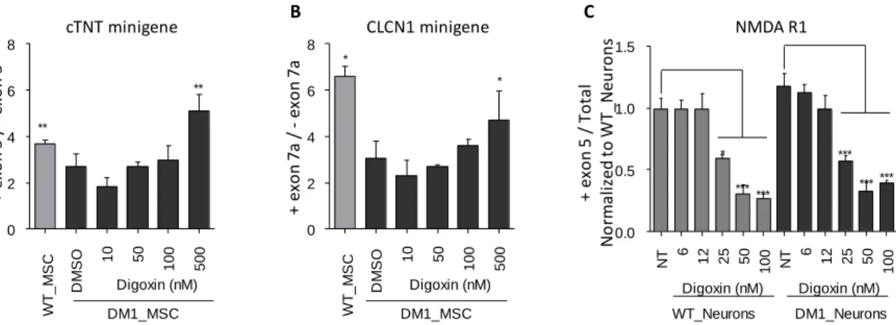
Figure S3: Beneficial effect of digoxin treatment on other splice defects associated to DM1, Related
to Figure 3
A-D. Dose-response analysis of digoxin treatment on alternate splicing of exon 5 of cTNT, exon 7a of
CLCN1 and exon 5 of NMDAR1. For cTNT and CLCN1, DM1_hESC MSC were transfected with a minigene
containing the exon of interest (n=3, 1 experiment). Alternate splicing of NMDAR1 as analyzed on WT
and DM1 hESC-derived neurons (n= 3, 1 experiment). Data represent mean ± SD and analyzed with
one way ANOVA followed by a Bonferroni post hoc test. *: value < 0.05. **: value < 0.01. ***:
p-value < 0.001.
A
B
C
cTNT minigene
CLCN1 minigene
NMDA R1
+
ex
o
n
5
/
-ex
o
n
5
+
ex
o
n
7
a
/
-ex
o
n
7a
+
ex
o
n
5
/
To
ta
l
No
rma
liz
ed
to
W
T_N
eu
ro
n
s
W T _ M S C D M S O 10 50 100 500 0 2 4 6 8 ** ** DM1_MSC Digoxin (nM) W T _ M S C D M S O 10 50 100 500 0 2 4 6 8 * * DM1_MSC Digoxin (nM) NT 6 12 25 50 100 NT 6 12 25 50 100 0.0 0.5 1.0 1.5 WT_Neurons DM1_Neurons * *** *** *** ****** Digoxin (nM) Digoxin (nM)Figure S4: Digoxin-mediated effect on DM1 alternate splicing is independent of MBNL1 and CUGBP1,
Related to Figure 3 and Figure 6
A
B
C
W
T_M
SC
DM1_MSC
50
100
500
D
MS
O
Actine
CUG-BP1
MBNL1
N
C N C N C N C N C
Actine
CUG-BP1
MBNL1
Lam A/C
W
T_M
SC
DM1_MSC
50
100
500
D
M
SO
D
H
o
ec
h
st
/
C
A
G
1
0
M
B
N
L1
WT_MSC DM1_MSC DM1_MSC 50 nM Digoxin DM1_MSC 100 nM Digoxin DM1_MSC 500 nM DigoxinSRp20
TRA2β
Actine
50
100
500
D
MS
O
DM1_MSC
SRP 20 DM S O 50 100 500 0 1 2 3 re la ti v e m R N A e x p re s s io n nM VUB03_DM1E
TRA2β
SRP20
TRA2 beta DM S O 50 100 500 0 1 2 3 4 * nM VUB03_DM1 re la ti v e m R N A e x p re s s io n Mbnl1 DM S O 50 100 500 0 2 4 6 ns nM VUB03_DM1 D M S O F c m R N A e x p re s s io nDMPK
MBNL1
CUG-BP1
CUGBP1 DM S O 50 100 500 0 1 2 3 nM VUB03_DM1 D M S O F c m R N A e x p re s s io n ** VUB03 DMPK DM S O 50 100 500 0 1 2 nM VUB03_DM1 D M S O F c m R N A e x p re s s io n nsR
el
at
iv
e
mR
NA
ex
p
res
si
o
n
DM1_MSC DM1_MSC DM1_MSC DM1_MSC DM1_MSCCUG-BP1 ratio Nucelar /Cyto
W T -M S C D M S O 5 0 n M 1 0 0 n M 5 0 0 n M 0 2 4 6 8 10 R a ti o C U G -B P 1 N u c le a r / C y to p la s n ic DM1_MSC ns ns MBNL1 ratio Nucelar /Cyto
W T -M S C D M S O 5 0 n M 1 0 0 n M 5 0 0 n M 0 2 4 6 R a ti o M B N L 1 N u c le a r / C y to p la s n ic ns ns DM1_MSC MBNL1 W T -M S C D M S O 5 0 n M 1 0 0 n M 5 0 0 n M 0.0 0.5 1.0 1.5 M B N L 1 /A c ti n ns DM1_MSC CUGBP1 W T -M S C D M S O 5 0 n M 1 0 0 n M 5 0 0 n M 0.0 0.5 1.0 1.5 C U G B P 1 /A c ti n ns DM1_MSC D M S O 50 1 0 0 5 0 0 0.0 0.5 1.0 S R P 2 0 / A c ti n SRP20 Digoxin (nM) * D M S O 50 1 0 0 5 0 0 0.0 0.4 0.8 1.2 T R A 2 b e ta / A c ti n TRA2 beta Digoxin (nM) ns ns