HAL Id: inserm-00555585
https://www.hal.inserm.fr/inserm-00555585
Submitted on 13 Jan 2011HAL is a multi-disciplinary open access archive for the deposit and dissemination of sci-entific research documents, whether they are pub-lished or not. The documents may come from teaching and research institutions in France or abroad, or from public or private research centers.
L’archive ouverte pluridisciplinaire HAL, est destinée au dépôt et à la diffusion de documents scientifiques de niveau recherche, publiés ou non, émanant des établissements d’enseignement et de recherche français ou étrangers, des laboratoires publics ou privés.
Transcription factor E4F1 is essential for epidermal stem
cell maintenance and skin homeostasis.
Matthieu Lacroix, Julie Caramel, Perrine Goguet-Rubio, Laetitia Linares,
Soline Estrach, Elodie Hatchi, Geneviève Rodier, Gwendaline Lledo, Carine de
Bettignies, Amélie Thépot, et al.
To cite this version:
Matthieu Lacroix, Julie Caramel, Perrine Goguet-Rubio, Laetitia Linares, Soline Estrach, et al.. Tran-scription factor E4F1 is essential for epidermal stem cell maintenance and skin homeostasis.. Pro-ceedings of the National Academy of Sciences of the United States of America , National Academy of Sciences, 2010, 107 (49), pp.21076-81. �10.1073/pnas.1010167107�. �inserm-00555585�
E4F1 is essential for epidermal stem cell maintenance and skin homeostasis
Matthieu Lacroix1, 3, 4, 10, &, Julie Caramel1, 3, 4, &, Perrine Goguet-Rubio2, 4, Laetitia Linares2, 4, Soline Estrach5, Elodie Hatchi1, 3, 4, Geneviève Rodier1, 3, 4, Gwendaline Lledo2, 4, Carine de Bettignies1, 2, Amélie Thépot6, Céline Deraison8, Karim Chébli1, 3, 4, Alain Hovnanian8,9, Pierre Hainaut6, Pierre Dubus7, Claude Sardet1, 3, 4, * and Laurent Le Cam1, 2, 3, 4, *.
1 Institut de Génétique Moléculaire de Montpellier UMR5535 CNRS, Montpellier 34293, France.
2 Institut de Recherche en Cancérologie de Montpellier, INSERM U896, Montpellier 34298, France.
3 Université Montpellier 2, Montpellier 34095, cedex 5, France. 4 Université Montpellier 1, Montpellier 34967, cedex 2, France. 5 INSERM U.634, Faculté de Medecine, Nice, 06107, France. 6 IARC, Lyon, 69008, France.
7 INSERM 33076, France.
8 INSERM, U563, Toulouse F-31300, France.
9 current address: CHU Necker-Enfants malades, Département de Génétique, 75015 Paris, France.
10 current address: INSERM, U563, Toulouse F-31300, France.
* Corresponding authors: claude.sardet@igmm.cnrs.fr (C.S.) and laurent.lecam@inserm.fr (L.L.C.)
& equal contribution
Running Title: Role of E4F1 in skin homeostasis
Abstract
Growing evidences suggest that the multifunctional protein E4F1 is involved in signalling pathways that play essential roles during normal development and tumorigenesis. Here, we have generated E4F1 conditional knock-out mice to address E4F1 functions in vivo in newborn and adult skin. E4F1 inactivation in the entire skin or in the basal compartment of the epidermis induces skin homeostasis defects, as evidenced by transient hyperplasia in the interfollicular epithelium and alteration of keratinocyte differentiation, followed by loss of cellularity in the epidermis and severe skin ulcerations. E4F1 depletion alters Epidermal Stem Cells (ESC) clonogenic activity ex vivo and ends in exhaustion of the ESC pool in vivo, indicating that the lesions observed in the E4F1 mutant skin result, at least partly, from cell autonomous alterations in ESC maintenance. Clonogenic potential of E4F1 KO ESC is rescued by Bmi1 over-expression or by Ink4a/Arf or p53 depletion. Skin phenotype of E4F1 KO mice is also delayed in animals with Ink4a/Arf and E4F1 compound gene deficiencies. Our data identify a novel regulatory axis essential for ESC-dependent skin homeostasis implicating E4F1 and the Bmi1-Arf-p53 pathway.
Abbreviations:
ESC, Epidermal Stem Cells; HF, Hair Follicle; 4OHT, 4-Hydroxy Tamoxifen; IFE, Interfollicular Epithelium; K5, K6, K10, K14 and K15, Keratin-5, 6, 10, 14 and 15 respectively; KO, Knock-Out; LRC, Label Retaining Cells; RERT , Cre-ERT2 knock-in mice;
/body Introduction:
E4F1 is an ubiquitously expressed transcription factor of the Gli-Kruppel family that was identified as a cellular target of the adenoviral oncoprotein E1A (1). While several cellular targets of E1A (E2F/pRB, CBP/p300, PCAF, CtBP, ATF/Creb etc…) have been extensively studied and recognized as central regulators of cell proliferation and survival, E4F1 biological functions remained poorly investigated. E4F1 is a multifunctional protein with transcriptional and atypical ubiquitin E3-ligase activities. E4F1-mediated ubiquitylation of p53 does not lead to proteasomal degradation but modulates p53 transcriptional activities involved in alternative cell fates: growth arrest or apoptosis (2). The notion that E4F1 plays an important role in the p53 pathway is reinforced by other reports showing that E4F1 not only directly interacts with p53 itself (2, 3) but also with regulators/effectors of this pathway, including p14ARF (4), the polycomb member Bmi1 (5), and the p53 target gene FHL2 (6). Nevertheless, E4F1 functions most likely extend beyond the regulation of p53. Thus, physical interactions between E4F1 and components of other oncogenic pathways have been reported, including RASSF1A (7), pRB (8), HMGA2 (9) and Smad4 (10).
Using a gene targeting approach in mouse, we previously showed that E4F1 constitutive inactivation results in embryonic lethality around the time of implantation. E4F1
KO blastocysts in culture exhibit mitotic defects including lagging chromosomes, chronic
activation of the mitotic checkpoint, and massive cell death (11). More recently, shRNA-mediated partial depletion of E4F1 was also shown to rescue hematopoietic stem cell (HSC) exhaustion in mouse resulting from inactivation of the polycomb member Bmi1 (5), suggesting an important role for E4F1 in HSC homeostasis.
To date, little information is currently available regarding the in vivo functions of E4F1 in adult tissues. Here, we generated several mouse strains to explore the roles of E4F1 in skin homeostasis.
Constant renewal of the Interfollicular Epithelium (IFE) and of Hair Follicles (HF) relies on the recruitment of epidermal stem cells (ESC) located in the basal layer of the IFE and in the bulge region of HF, respectively. ESC fuel the highly proliferative transit amplifying compartments (TAC) in the basal layer of the IFE and in the bulb of the HF. TAC cells then embark on differentiation programs to generate the spinous, granular and cornified layers in the IFE or the different lineages of the mature HF (12-14). Several essential molecular circuitries that orchestrate epidermal stem cells maintenance have been previously described, including p63-, BMP-, TGF-ß-, Wnt/ß-catenin-, and Notch-initiated signalling cascades (12, 15, 16).
Here, we show that inactivation of E4F1 in the entire skin or in the basal compartment of the epidermis results in severe epidermal defects both in neonates and in adult mice, revealing a yet unidentified regulatory axis essential for ESC-dependent skin homeostasis implicating E4F1 and the Bmi1-Ink4a/Arf-p53 pathway.
Results:
Inactivation of E4F1 induces transient hyperplasia in the epidermis followed by permanent loss of epidermal cells and severe skin ulcerations.
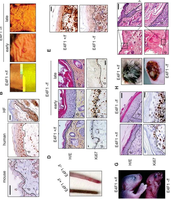
We first investigated E4F1 expression pattern in epidermis. Immunohistochemical analyses of murine and human skin sections showed nuclear expression of E4F1 in the basal and suprabasal layers of the interfollicular epithelium (IFE), as well as in the bulb and bulge regions of the hair follicle (HF) (Fig. 1A).
To analyse the role of E4F1 in skin homeostasis, we generated two mouse models with homozygote deletion of the E4F1 gene in the entire skin or in the epidermis only, using conditional (Cre-ERT2) and tissue specific (K5-Cre) Cre/Lox-P systems, respectively. In short, we first generated E4F1-/flox mice harboring E4F1 conditional knock-out allele (Fig. S1) and inter-crossed them with Cre-ERT2 KI/KI mice (RERT) which express tamoxifen-inducible Cre recombinase-ERT2 fusion protein under the control of the ubiquitously active RNA polII promoter, i.e. in all tissues (17). As expected, topical applications of 4-hydroxy-tamoxifen (4OHT) on the tail skin or on a shaved area of the back skin of adult E4F1-/flox; RERT animals, resulted in efficient Cre-mediated recombination of the E4F1 locus in the skin, as monitored on genomic DNA, mRNA and protein samples prepared from the treated area (Fig. S1).
Between 1 and 2 weeks after 4OHT-administration, E4F1-depleted back skin thickened, became wrinkled and ruffled. Those early lesions evolved 1 to 2 weeks later into severe skin ulcerative lesions (Fig. 1B). Strikingly, E4F1 inactivation in unshaved skin tail of
E4F1-/flox; RERT animals resulted in complete alopecia 6 weeks after 4OHT applications (Fig.
1C). Histological analyses revealed that skin thickening was the result of a massive hyperplasia of the epidermis with increased cellularity in the IFE and the infundibulum (Fig. 1D). Consistent with the observed hyperplasia, an abnormal proportion of epidermal cells
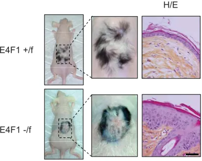
were proliferating in E4F1 KO skin, as indicated by increased immunostaining of skin sections for the proliferation marker Ki67 (Fig. 1D), and anti-BrdU staining performed on skin sections harvested shortly after injection of animals with BrdU (Fig. S2). This phenotype was skin autonomous since similar hyperplasia was recapitulated upon engraftment of E4F1 -/flox; RERT p1 neonatal back skin onto nude mice (Fig. S3). At later time points (3 to 4 weeks
after 4OHT administration), the initial hyperplasia of E4F1 KO; RERT skin was followed by broad disorganisation of the IFE and massive hyperkeratosis associated with partial or complete loss of cellularity in the IFE (Fig. 1D).
To further show that the observed phenotypes resulted from epidermis specific defects, we crossed E4F1-/flox mice with Keratin 5-Cre (K5-Cre) transgenic mice expressing Cre in the epidermal basal cell layer, from E15.5 onwards (18). E4F1-/flox;K5-Cre neonates
exhibited efficient E4F1 gene inactivation in the epidermis as shown by immunohistochemical staining and western-blot analyses with anti-E4F1 antibodies (Fig. 1E and S1). 3 to 4 days after birth, these animals exhibited epidermal hyperplasia, and died with acute symptoms of dehydration (Fig. 1F). As in E4F-/flox; RERT animals, this hyperplasia was
associated with hyperproliferation of epidermal cells, as shown by increased Ki67 staining (Fig. 1G). Consistent with increased proliferation, E4F1 KO epidermis also exhibited higher mitotic index, as illustrated by increased number of phospho histone H3 (Ser10, PHH3) positive basal cells (Fig. S5). Of note, we previously showed that E4F1 KO blastocysts also exhibited PHH3-positive cells (11), but in this case, cells were blocked in mitosis with abnormal mitotic figures. We did not observe abnormal mitotic figures in E4F1 KO epidermal basal cells, indicating that the increased mitotic index likely reflects over-proliferation of basal cells, rather than mitotic progression defects.
In order to bypass neonatal lethality of E4F1 KO; K5-Cre animals and investigate later phenotypes, we engrafted E4F-/flox; K5-Cre or control skins onto nude mice. 2 weeks after
engraftment, E4F1 KO; K5-Cre skins failed to regenerate a normal epidermis. Similarly to the late phenotype of E4F-/flox; RERT animals, E4F1 KO, K5-Cre engrafted skins exhibited
marked hypocellularity in the epidermis, hyperkeratosis and severe ulcerative lesions (Fig. 1H). Of note, hair follicles were also lacking in E4F1 KO, K5-Cre engrafted skins. Collectively, our data indicate that E4F1 inactivation results in transient hyperplasia but ultimately leads to permanent loss of epidermal cells in the IFE and HFs.
E4F1 inactivation results in expansion of the basal cells compartment of the epidermis and in abnormal keratinocyte differentiation in vivo.
We next assessed which epidermal cell populations were present in these hyperplasic lesions. In both 4OHT-treated E4F1-/flox; RERT and E4F1-/flox; K5-Cre epidermis, cells of the
hyperplasic IFE stained positive for the basal cell-specific keratin-14, integrin-α6, markers (Fig. 2A and S4). Expression of these markers extended to upper cellular layers, in contrast to normal IFE of control animals where their expression was restricted to the basal cell layer. In addition, E4F1-floxed IFE massively expressed keratin-6 (Fig. 2B and S4), which expression is normally restricted to sweat glands and HF in normal skin but can be expressed in the IFE undergoing abnormal proliferation or wound healing (19). Collectively, these analyses indicated that E4F1 inactivation in the IFE first resulted in massive expansion of keratinocytes with basal/TAC properties.
In addition to this massive expansion of basal cells, we noticed the absence of the suprabasal and granular layers as illustrated by the lack of expression of early (keratins 1 and 10), as well as late differentiation markers (involucrin, loricrin, filaggrin) in E4F1 KO epidermis in both E4F1flox; K5-Cre and E4F1flox; RERT models (Fig. 2B, S4). No significant
cell death was observed among highly proliferative cells of the IFE during the development of hyperplasia, as revealed by immunostaining of skin sections for the presence of activated
caspase 3- or TUNEL-positive cells. Therefore, it is likely that hyperproliferative keratinocytes present in hyperplasic lesions had finally undergone an abnormal differentiation program of stratification, rather than apoptosis, leading to the observed hyperkeratosis. These data indicate that E4F1 inactivation strongly perturbates both the proliferation and differentiation of epidermal lineages in vivo leading to a strong alteration of skin homeostasis.
E4F1 inactivation induces epidermal stem cell defects.
We next investigated the mechanisms by which E4F1 inactivation in the IFE led to a massive but transient expansion of the basal/TAC compartment, followed by its exhaustion. Several scenarios could explain this sequence of events. First this could reflect an increase in the intrinsic capacity of basal/TAC keratinocytes to proliferate. However, this is unlikely, since E4F1 depletion in freshly isolated populations of primary keratinocytes in culture did not recapitulate the massive over-proliferation observed in skin sections, as illustrated by FACScan analyses of BrdU/Propidium Iodide- (PI) or PHH3/PI-labelled keratinocytes isolated from E4F1-/flox; RERT or control p1 neonates (Fig. S5). Similar conclusions were
obtained in primary human keratinocytes upon shRNA-mediated depletion of endogenous human E4F1 (Fig. S5). These observations, together with the withering and transient aspect of the hypertrophy phenotypes led us to investigate an alternative hypothesis involving perturbations of the resident stem cells (ESC). Several aspects of E4F1 KO phenotypes are reminiscent of skin lesions observed in KO animals exhibiting stem cell defects. Hence, conditional perturbation in the epidermis of several key signalling molecules, including the small GTPase Rac1, Myc, Smad4, NFATC1, leads ESC to exit their normal microenvironment (“niche”) to enter into the TAC where they become transiently proliferating keratinocytes upon exposition to natural pro-mitogenic signals present in this amplification compartment (20-24). This results in transient hyperplasia followed by a
permanent exhaustion of the epidermal stem cell pool and loss of renewal capacity of the epithelium.
To address this scenario, we first performed standard in vitro clonogenic assays on feeder layers with total primary keratinocytes isolated from in vivo recombined hyperplasic regions of E4F1-/flox; RERT adult mice, as well as from E4F1-/flox; K5-Cre p1 neonates or
control littermates. After 10 to 15 days in culture, typical holoclones, corresponding to the long-term clonal outgrowth of epidermal cells with stem cell properties (25, 26) were detected in controls but not in E4F1 KO conditions in both models (Fig. 3A). Importantly, similar results were recapitulated when E4F1 inactivation was induced ex vivo by adding 4OHT in the culture medium of E4F1-/flox; RERT cells (Fig. 3A), showing thatthese defects were stem cell autonomous. Similarly, clonogenic assays performed with primary human keratinocytes transduced ex vivo with E4F1 shRNA constructs showed that partial E4F1 depletion resulted in a 3 fold reduction in the number of holoclones (Fig. 3B).
We next investigated whether E4F1 inactivation also resulted in exhaustion of the stem cell pool in vivo by analysing expression of various ESC markers. FACscan analyses of dissociated epidermis revealed that E4F1-/flox; K5-Cre neonatal epidermis contained fewer
ESC co-expressing the CD34 surface marker and high levels of α6-integrin (CD34+/ α6high) than control epidermis (8% +/- 2 vs 1,5% +/- 1, n=3) (Fig. 3C). Similarly, immunostainings performed on back skin sections or on whole mount of tail epidermis harvested from adult
E4F1-/flox; RERT animals 4 to 6 weeks following E4F1 inactivation, showed loss of
expression of the HF bulge markers CD34 and keratin-15 (Fig. 3D and S6).
To confirm these HF ESC defects in vivo, we crossed E4F1-/flox; RERT animals with Keratin15-EGFP (K15-GFP) transgenic mice expressing the GFP reporter under the control
of the keratin-15 promoter (27). In 7 to 12-week-old K15-GFP; E4F1+/flox; RERT control
complete loss of GFP-positive cells was observed 4 to 6 weeks after E4F1 inactivation in
E4F1 KO; RERT animals, confirming the exhaustion of the HF ESC (Fig. 3E). Finally, we
tracked HF bulge/ESC as BrdU Long Term Retaining Cells (LRC) using an in vivo labelling protocol that marks self-renewing and multi-potent epidermal cells (26, 28). E4F1-/flox; RERT
and E4F1+/flox; RERT neonates were injected with BrdU and after a chase period of three
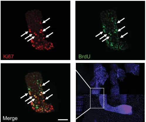
months, the poorly proliferative adult ESC were identified on whole mounts of tail epidermis, as BrdU-positive LRC. 2 to 3 weeks after E4F1 inactivation the LRC zone extended and an increased numbers of LRCs were co-labeled with Ki67 in E4F1 KO HF as compared to control HF (Fig. 3F early and S7), suggesting that E4F1 inactivation induced HF ESC to transiently proliferate. At later time points (6 weeks after 4OHT), whereas LRCs were still restricted to the bulge of control HF, BrdU staining finally disappeared in the E4F1 KO HF (Fig. 3F late), supporting the notion that the end result of E4F1 inactivation was an exhaustion of the ESC pool. Finally, consistent with the role of ESC in regenerating the various epithelial cell types of hairy skin after injury (26), we also observed that wound healing was strongly altered in E4F1 KO skin (figure S8). Altogether, these data indicate that
E4F1 is essential for ESC maintenance in vivo and ex vivo, and strongly suggest that the
hyperplasia and following loss of cellularity of E4F1 KO epidermis, are primarily due to cell autonomous perturbations of the ESC pool that aliments TAC compartments.
Down regulation of the Bmi1-Ink4a/Arf-p53 axis partly rescues ESC-clonogenic potential and skin lesions of E4F1 KO.
We next addressed the molecular pathway by which E4F1 could regulate ESC maintenance. E4F1 associates with the polycomb member Bmi1 and the tumor suppressors Arf, p53 and pRb (2-5, 8). Although there are evidences that these factors have independent functions, they define a well established functional cascade with Bmi1 triggering the
repression of the Ink4a/Arf locus whose products, p19Arf and p16Ink4a, act as potent inhibitors of the p53- and pRb-dependent activities, respectively (29). Several reports suggest that the negative regulation of Ink4a/Arf expression may be a central event in stem cell renewal in several tissues (30-33) including epidermis (34). Strikingly, we observed that E4F1 KO primary keratinocytes exhibited enhanced Ink4a and Arf mRNA levels compared to control cells (Fig. 4A), suggesting that the deregulation of the Ink4a/Arf locus might be involved in
E4F1 KO phenotype. Consistent with this hypothesis, clonogenic assays performed with
primary keratinocytes isolated from E4F1-/flox; RERT; Ink4a/Arf-/- compound mice showed that Ink4a/Arf inactivation partly restored long-term outgrowth of E4F1 KO ESCex-vivo (Fig.
4B, C). The number of viable clones growing in this condition was similar to that obtained from control skin.
Importantly, the in vivo skin phenotype of E4F1 KO mice was also delayed in animals with Ink4a/Arf and E4F1 compound gene deficiencies. Hence, the initial epidermal hyperplasia of 4OHT-treated E4F1-/flox; RERT; Ink4a/Arf-/- animals was almost undetectable at early time points following E4F1 inactivation (Fig. 4D, early), compared to control littermates with E4F1 KO only. Nevertheless, at later time points, these animals ended up to develop a moderate hyperplasia and hyperkeratosis (Fig. 4D, late).
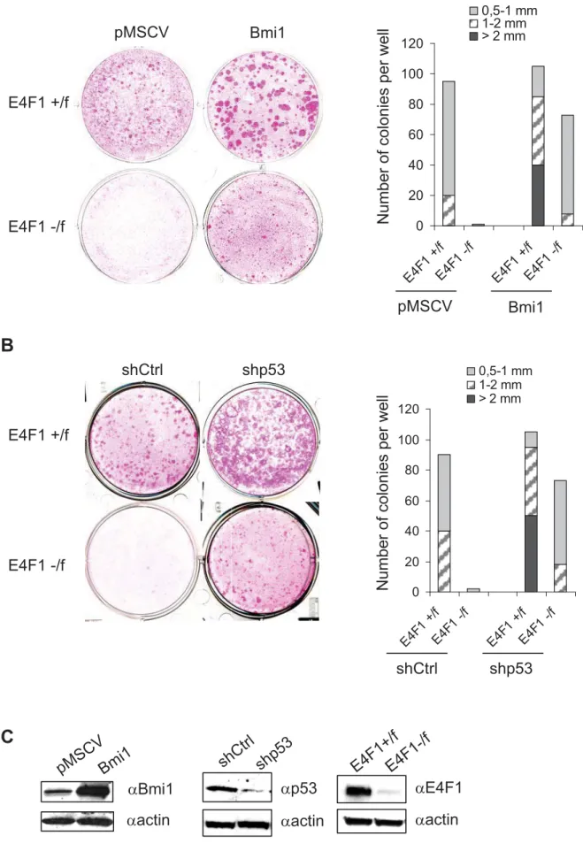
To provide further evidence for a role of the Ink4a/Arf–pRb/p53 cascades in E4F1 KO phenotype, we next tried to rescue the clonogenic potential of E4F1 KO ESC by targeting Bmi1, one of the main upstream regulator of the Ink4a/Arf locus. Transduction of keratinocytes by retroviruses expressing Bmi1 rescued long-term outgrowth and clonogenic potential of E4F1 KO cells (4OHT-treated E4F-/flox; RERT keratinocytes) (Fig. 5A and 5C).
To assess which Ink4a/Arf downstream targets might be involved, clonogenic assays were also performed upon shRNA-mediated (lentiviral vectors) depletion of either murine
clonal outgrowth of E4F1 KO keratinocytes, highlighting the role of the Bmi-Arf-p53 axis rather than the Bmi-p16-pRb axis in E4F1 KO skin phenotypes. Of note, E4F1 KO rescued clones that developed upon inactivation of Ink4a/Arf, depletion of p53 or overexpression of Bmi1, all remained significantly smaller in size, compared to those growing from control cells (Fig. 4B-C, 5A-B), suggesting that E4F1 might also impinge on other molecular circuitries that orchestrate ESC maintenance.
Altogether, our data provide the first evidence that E4F1 is essential for ESC-dependent skin homeostasis and identify a novel regulatory axis involved in this process implicating Bmi1 and the Arf-p53 pathway.
Discussion
In vivo functions of E4F1 remain poorly documented. Here, we created E4F1
conditional KO mice and reported that E4F1 inactivation in the epidermis led to neonatal lethality that resulted from skin homeostasis defects. We found that E4F1 depletion in E4F1
KO; K5-Cre neonates first led to a rapid thickening of the IFE associated with increased
numbers of highly proliferative keratinocytes in the basal cell layers (TAC compartment) of the IFE. Comparable results were obtained upon E4F1 inactivation in adult skin obtained by topical applications of 4OHT on the back skin of E4F1-/flox; RERT mice. In both E4F1 KO
models, hyperplasia was transient and followed by a severe disorganization and almost complete loss of viable epithelial cell layers in the IFE and HF. In addition, perturbations of epidermal differentiation were observed in E4F1 depleted epidermis, in both models.
Strikingly, E4F1 depletion in vitro, in freshly isolated populations of murine or human primary keratinocytes in culture, did not recapitulate the over-proliferation observed in skin sections, ruling out the possibility that the hyperplasia originates from an intrinsic increase in the proliferation capacity of E4F1 KO TAC keratinocytes. As described for other gene deficiencies in epidermis (20, 22), our data rather suggest that E4F1 KO phenotypes resulted from cell autonomous perturbations in resident stem cells. Indeed, E4F1 depletion in vivo or
ex vivo turned out to dramatically impair the clonogenic potential of both murine and human
epidermal cell populations. In addition, a strong reduction in the expression of various ESC markers was observed in vivo in E4F1 KO skins. Finally, BrdU labelling of LRC suggested that E4F1 KO HF stem cells first exited their normal location in the bulge region of the HFs to enter in a transient phase of proliferation, before disappearing. As predicted by this stem cell-oriented scenario, exhaustion of the resident stem cell pool in E4F1 KO skin ultimately led to a complete loss of cellularity in the IFE and alteration of wound healing.
Seeking for the pathway by which E4F1 could regulate ESC maintenance, we found that the clonogenic potential of E4F1 KO ESC was restored upon targeting of the Bmi1-Ink4a/Arf-p53 axis but not by pRb depletion, highlighting the role of the Bmi-Arf-p53 axis rather than the Bmi-p16-pRb axis in E4F1 KO skin phenotypes. Consistently, E4F1 inactivation correlated with increased Ink4a/ARF expression and our in vivo data showed that the development of hyperplasia was delayed and reduced in animals with Ink4a/Arf and E4F1 compound gene deficiencies. It remains unclear how this protein network impinges on ESC homeostasis and ultimately raises the question about the physiological functions of this Bmi1-Ink4a/Arf-p53 axis in ESC. Although poorly documented, it has been suggested that it might play its usual "gate-keeper" function in these cells, notably in response to genotoxic or developmental stresses, as illustrated in mice with dysfunctional telomeres (35, 36) or in p63
KO mice (34, 37). As described in other tissues, it is also formally possible that the
Bmi1-ARF-p53 protein network plays a role in the normal program of ESC self-renewal and maintenance. Hence, in absence of acute genotoxic stress, inactivation of p53 or Ink4a/Arf has been shown to impact on the self-renewal capacity of hematopoietic multipotent progenitors (38) and neural stem cells (39). Even more intriguing is the recent finding that p53 KO favours symmetric divisions and self-renewal capacity of mammary gland progenitor cells (40). The latter observation raises questions about similar roles of p53, and indirectly E4F1, in the epidermis where proper columnar stratification and tissue organization are driven, at least in part, by oriented, asymmetric cell divisions (41, 42).
Our data also highlight the complexity of E4F1 connections with the polycomb family member Bmi1. Indeed, others have shown that shRNA-mediated depletion of E4F1 can rescue premature senescence of Bmi1 KO hematopoietic stem cells and restore the viability of animals transplanted with those rescued cells, in a Ink4a/Arf- and p53-independent manner (5). Our data indicate that the genetic links between E4F1 and Bmi1 in stem cell maintenance
are complex and might differ from one tissue to the other since in epidermis, we clearly observed that Bmi1 ectopic overexpression partly rescued stem cell defects of E4F1 KO ESC. Of note, we currently do not rule out that E4F1 might also exert yet unidentified additional cellular functions independent from Bmi1 and the Arf-p53 axis. Supporting this notion, we found that targeting the Bmi1-Arf-p53 axis did not fully restore ESC clonal outgrowth and normal skin phenotype. Animals with Ink4a/Arf and E4F1 compound gene deficiencies ended up to develop a moderate hyperplasia and hyperkeratosis at late time points. Likewise, in clonogenic assays, E4F1 KO rescued clones that developed upon depletion of p53 or
Ink4a/Arf, or upon over-expression of Bmi1, all remained significantly smaller in size,
compared to those growing from control cells. Therefore, in addition to its connection with the Bmi1-Arf-p53 cascade, one could hypothesize that E4F1 might also impinge on other
hitherto described molecular circuitries that orchestrate epidermal stem cells maintenance,
including TGFß-, Wnt/ß-catenin-, or Notch-initiated signalling cascades (12-14). Interestingly, a physical interaction of E4F1 with Smad4 has recently been shown in BMP-mediated regulation of myogenic differentiation (10). Further studies will be required to evaluate whether this interaction is also implicated in epidermal homeostasis.
In conclusion, the roadblock to developing an E4F1 conditional KO mouse has been overcome and provides the first evidence, that E4F1, through its connection with the Bmi1 and the Ink4a/Arf-p53 axis, is an important player of stem cells-dependent skin homeostasis. This finding, together with previous reports showing that E4F1 is targeted by several oncoproteins and tumor suppressors, also raise an interesting question about E4F1 potential implication in the development/maintenance of cancer stem cells which presence is suspected in several cutaneous malignancies, including skin carcinoma and malignant melanoma.
Material and Methods:
Ethics Statement
All experiments involving mice were approved by Montpellier's ethic committee for animal warefare.
Generation of E4F1-/flox mice and experimental treatment of mice
Generation of the E4F1flox allele and of E4F1-/flox mice are detailed in supplementary
informations (SI). E4F1-/flox mice were crossed with Cre-ERT2 knock-in mice (RERT) (17) or
Keratin5-Cre transgenic mice (18) to obtain E4F1-/flox; RERTKI/KI animals, and E4F1-/flox;
K5-Cre animals, respectively. E4F1flox; RERT mice were also crossed with homozygous Keratin15-EGFP transgenic mice (Jackson lab) (27) to generate E4F1flox; RERTKI/KI;
K15-GFP animals and with Ink4a/Arf -/- mice (43) to generate E4F1flox; RERTKI/KI; Ink4a/Arf
-/-animals. E4F1 recombination in adult E4F1flox; RERT skin was induced by topical
applications of 4-hydroxy Tamoxifen (4OHT) (Sigma) (2mg dissolved in ethanol per application per day, 4 applications total), on the shaved back skin or the tail. For skin grafting experiments, dorsal skin from E4F1flox; RERT or E4F1flox; K5-Cre neonate donors
were transplanted onto athymic nude recipient mice (Charles River), as previously described (44). For visualisation of BrdU-label-retaining cells (LRC), ten days old mice were injected with BrdU (50mg/kg of body weight, Sigma) every 12 hours for a total of four injections. To achieve short-term labeling of cells undergoing DNA synthesis in vivo (for IHC experiments), BrdU (50mg/kg of body weight) was injected intra-peritoneally, 4 hours prior to euthanasia. Histochemistry, immunolabelling of skin sections and whole mounts
Immunohistochemistry of skin sections was performed as described in SI with the following primary antibodies: anti -E4F1 (B-21 rabbit polyclonal) generated by our laboratory, -Ki67 (#SP6, Neomarkers), keratin 6 (#SPM269, Abcam), keratin10 (#PRB159P, Covance),
-involucrin (#Sc15230, Santa Cruz), -CD34 (RAM34, BD Pharmingen), and -keratin 15 (#LHK15, Vector Laboratories). Immunohistofluorescence stainings with anti-α6-integrin (#GoH3, BD Biosciences) and anti-keratin 14 (#AF64, Covance) were performed on 10μm cryosections fixed for 10 minutes in 100% acetone. BrdU-positive cells were detected using anti-BrdU antibody (BD Biosciences). Whole mounts of tail epidermis and detection of LRC were prepared as described previously (28) and pictures were generated using a Zeiss 510 confocal microscope and presented as Z-stack projections after deconvolution with Imaris software.
Culture of primary keratinocytes and clonogenic assays
Murine primary keratinocytes were isolated from newborn skin after overnight treatment with dispase (5 mg/mL, Roche) or from adult back skin after trypsin-EDTA overnight treatment, and grown in free EMEM medium (Biowhittaker, Lonza), containing 10% calcium-free FCS (Sigma) and 10 µg/ml murine EGF (Roche). Cre-mediated recombination of E4F1 flox alleles were achieved by adding 4OHT (1 µM, Sigma) in the culture medium. Human primary keratinocytes were isolated from skin biopsies obtained after medical surgery and cultured in Green medium containing 1,2 mM calcium as previously described (45) and according to Helsinki Declaration. Clonogenic assays were perfomed as described in SI. Colonies were fixed with 4% PFA and stained with 1% Rhodamine B (Sigma). Quantitative analysis of the total number and size of clones was performed with ImageJ.
Retroviral and lentiviral particles productions and infections
Viral particles were produced as described in SI, with the following constructs: pMSCV-Bmi1 (29), pMKO vector encoding either control or anti human E4F1 shRNAs (sequences available upon request); pLKO1 encoding shRNAs directed against murine RB1 or p53 (Sigma mission shRNA clones, NM_011640.1-625s1c1), or control irrelevant sequence. Western blotting
Total cell extracts were prepared in 50 mM Tris-HCl, pH 7.4, 100 mM NaCl, 5 mM EDTA, 0,5% NP40, supplemented with protease inhibitors (Complete, Roche). Immunoblots were probed with primary antibodies directed against E4F1 (8), p53 (1C12, Cell signalling), Bmi1 (#F6, Millipore), β-actin (Sigma)
FACScan analyses
Freshly isolated cell suspensions were incubated with FITC-conjugated anti-CD34 (RAM34, BD Biosciences) and PE-Cy5-conjugated anti-α6-integrin primary antibodies for 30 minutes at 4°C. For cell cycle analyses, formalin-fixed cells were incubated with BrdU or anti-phospho-serine 10 histone H3 (6G3 #9706, Cell Signalling) and PI (Sigma) as previously described (2). Cells were analysed on a FACS Calibur (Becton Dickinson) and data were processed with Flowjo software (Treestar).
Acknowledgments
We are grateful to all members of the LLC and CS laboratories for helpful discussions and critical readings of the manuscript, to C. Jacquet, E. Jouffre and P. Cavelier for technical help in mouse handling and histological analyses. We thank the “Clinique de la souris, Strasbourg” for injection of E4F1 flox ES cells into blastocysts. We thank C. Blanpain and M. Van Lohuizen for reagents and advices, as well as, J. Jorcano, A. Ramirez, A. Gandarillas, M. Barbacid and M. Serrano for providing us with K5-Cre transgenic, RERT knock-in and
ink4a/Arf-/- mice. Microscopy imaging and histological analyses were performed on the MRI imaging core facility (Montpellier) and RHEM histology (Montpellier) facility, respectively. CS and JC are supported by the Agence Nationale pour la Recherche (ANR blanche), by the AICR foundation, by la Fondation pour la Recherche Médicale (Equipe labellisée 2007) and by institutional supports from CNRS. LLC is supported by the INSERM Avenir Program and the Association pour la Recherche contre le Cancer. ML and EH are supported by fellowships from ARC.
References:
1. Raychaudhuri P, Rooney R, & Nevins JR (1987) Identification of an E1A-inducible cellular factor that interacts with regulatory sequences within the adenovirus E4 promoter. Embo J 6(13):4073-4081.
2. Le Cam L, et al. (2006) E4F1 is an atypical ubiquitin ligase that modulates p53 effector functions independently of degradation. Cell. 127(4):775-788.
3. Sandy P, et al. (2000) p53 is involved in the p120E4F-mediated growth arrest.
Oncogene 19(2):188-199.
4. Rizos H, et al. (2003) Association of p14ARF with the p120E4F transcriptional repressor enhances cell cycle inhibition. J Biol Chem 278(7):4981-4989.
5. Chagraoui J, et al. (2006) E4F1: a novel candidate factor for mediating BMI1 function in primitive hematopoietic cells. Genes Dev. 20(15):2110-2120.
6. Paul C, et al. (2006) The LIM-only protein FHL2 is a negative regulator of E4F1.
Oncogene. 25(40):5475-5484. Epub 2006 May 5471.
7. Fenton SL, et al. (2004) Identification of the E1A-regulated transcription factor p120 E4F as an interacting partner of the RASSF1A candidate tumor suppressor gene.
Cancer Res 64(1):102-107.
8. Fajas L, et al. (2000) pRB binds to and modulates the transrepressing activity of the E1A-regulated transcription factor p120E4F. Proc Natl Acad Sci U S A 97(14):7738-7743.
9. Tessari MA, et al. (2003) Transcriptional activation of the cyclin A gene by the architectural transcription factor HMGA2. Mol Cell Biol 23(24):9104-9116.
10. Nojima J, et al. Dual roles of smad proteins in the conversion from myoblasts to osteoblastic cells by bone morphogenetic proteins. J Biol Chem 285(20):15577-15586. 11. Le Cam L, Lacroix M, Ciemerych MA, Sardet C, & Sicinski P (2004) The E4F
protein is required for mitotic progression during embryonic cell cycles. Mol Cell Biol 24(14):6467-6475.
12. Blanpain C & Fuchs E (2009) Epidermal homeostasis: a balancing act of stem cells in the skin. Nat Rev Mol Cell Biol 10(3):207-217.
13. Cotsarelis G (2006) Epithelial stem cells: a folliculocentric view. J Invest Dermatol 126(7):1459-1468.
14. Brouard M & Barrandon Y (2003) Controlling skin morphogenesis: hope and despair.
Curr Opin Biotechnol 14(5):520-525.
15. Dotto GP (2008) Notch tumor suppressor function. Oncogene 27(38):5115-5123. 16. Candi E, et al. (2008) p63 in epithelial development. Cell Mol Life Sci.
65(20):3126-3133.
17. Guerra C, et al. (2003) Tumor induction by an endogenous K-ras oncogene is highly dependent on cellular context. Cancer Cell. 4(2):111-120.
18. Ramirez A, et al. (2004) A keratin K5Cre transgenic line appropriate for tissue-specific or generalized Cre-mediated recombination. Genesis. 39(1):52-57.
19. Weiss RA, Eichner R, & Sun TT (1984) Monoclonal antibody analysis of keratin expression in epidermal diseases: a 48- and 56-kdalton keratin as molecular markers for hyperproliferative keratinocytes. J Cell Biol 98(4):1397-1406.
20. Benitah SA, Frye M, Glogauer M, & Watt FM (2005) Stem cell depletion through epidermal deletion of Rac1. Science. 309(5736):933-935.
21. Horsley V, Aliprantis AO, Polak L, Glimcher LH, & Fuchs E (2008) NFATc1 balances quiescence and proliferation of skin stem cells. Cell 132(2):299-310.
depletion of follicle stem cells. Mol Biol Cell 20(3):882-890.
23. Waikel RL, Kawachi Y, Waikel PA, Wang XJ, & Roop DR (2001) Deregulated expression of c-Myc depletes epidermal stem cells. Nat Genet 28(2):165-168.
24. Arnold I & Watt FM (2001) c-Myc activation in transgenic mouse epidermis results in mobilization of stem cells and differentiation of their progeny. Curr Biol 11(8):558-568.
25. Barrandon Y & Green H (1987) Three clonal types of keratinocyte with different capacities for multiplication. Proc Natl Acad Sci U S A 84(8):2302-2306.
26. Blanpain C, Lowry WE, Geoghegan A, Polak L, & Fuchs E (2004) Self-renewal, multipotency, and the existence of two cell populations within an epithelial stem cell niche. Cell 118(5):635-648.
27. Morris RJ, et al. (2004) Capturing and profiling adult hair follicle stem cells. Nat
Biotechnol 22(4):411-417.
28. Braun KM, et al. (2003) Manipulation of stem cell proliferation and lineage commitment: visualisation of label-retaining cells in wholemounts of mouse epidermis. Development 130(21):5241-5255.
29. Jacobs JJ, Kieboom K, Marino S, DePinho RA, & van Lohuizen M (1999) The oncogene and Polycomb-group gene bmi-1 regulates cell proliferation and senescence through the ink4a locus. Nature 397(6715):164-168.
30. Janzen V, et al. (2006) Stem-cell ageing modified by the cyclin-dependent kinase inhibitor p16INK4a. Nature 443(7110):421-426.
31. Krishnamurthy J, et al. (2006) p16INK4a induces an age-dependent decline in islet regenerative potential. Nature 443(7110):453-457.
32. Molofsky AV, et al. (2006) Increasing p16INK4a expression decreases forebrain progenitors and neurogenesis during ageing. Nature 443(7110):448-452.
33. Signer RA, Montecino-Rodriguez E, Witte ON, & Dorshkind K (2008) Aging and cancer resistance in lymphoid progenitors are linked processes conferred by p16Ink4a and Arf. Genes Dev 22(22):3115-3120.
34. Su X, et al. (2009) Rescue of key features of the p63-null epithelial phenotype by inactivation of Ink4a and Arf. EMBO J 28(13):1904-1915.
35. Martinez P, et al. (2009) Increased telomere fragility and fusions resulting from TRF1 deficiency lead to degenerative pathologies and increased cancer in mice. Genes Dev 23(17):2060-2075.
36. Flores I & Blasco MA (2009) A p53-dependent response limits epidermal stem cell functionality and organismal size in mice with short telomeres. PLoS One 4(3):e4934. 37. Su X, et al. (2009) TAp63 prevents premature aging by promoting adult stem cell
maintenance. Cell Stem Cell 5(1):64-75.
38. Akala OO, et al. (2008) Long-term haematopoietic reconstitution by Trp53-/-p16Ink4a-/-p19Arf-/- multipotent progenitors. Nature 453(7192):228-232.
39. Armesilla-Diaz A, et al. (2009) p53 regulates the self-renewal and differentiation of neural precursors. Neuroscience 158(4):1378-1389.
40. Cicalese A, et al. (2009) The tumor suppressor p53 regulates polarity of self-renewing divisions in mammary stem cells. Cell 138(6):1083-1095.
41. Clayton E, et al. (2007) A single type of progenitor cell maintains normal epidermis.
Nature 446(7132):185-189.
42. Lechler T & Fuchs E (2005) Asymmetric cell divisions promote stratification and differentiation of mammalian skin. Nature 437(7056):275-280.
43. Serrano M, et al. (1996) Role of the INK4a locus in tumor suppression and cell mortality. Cell. 85(1):27-37.
human epidermal cells onto athymic animals. J Invest Dermatol 91(4):315-318. 45. Bitoun E, et al. (2003) LEKTI proteolytic processing in human primary keratinocytes,
tissue distribution and defective expression in Netherton syndrome. Hum Mol Genet 12(19):2417-2430.
Figure legends:
Figure 1. E4F1 inactivation triggers transient hyperplasia of the epidermis followed by permanent loss of epidermal cells and severe skin ulcerations. (A) Immunohistochemical (IHC) analysis of E4F1 expression in human skin or murine back skin and hair follicle (HF). Scale bar = 40µm. The dotted line indicates the dermis-epidermis junction. d: dermis. (B) Macroscopic alterations of back skin of adult E4F1-/flox; RERT mice 7 (early) or 15 days (late)
after Cre-mediated E4F1 inactivation. (C) Representative picture of E4F1flox; RERT tails at
late time points (6 weeks) after 4OHT applications. (D) Hematoxylin and eosin (H/E) staining (upper panels) and Ki67 IHC analysis (lower panels) of dorsal skin sections prepared from 4OHT-treated E4F1-/flox; RERT adult mice at early and late time points. Left panels show
similar analyses performed on 4OHT-treated E4F1+/flox; RERT control animals. Scale bar =
20µm. (E) IHC analysis of dorsal skin sections from E4F1+/flox and E4F1-/flox; K5-Cre
neonates using anti-E4F1 antibody. Scale bar = 40µm. (G) Photograph of representative
E4F1+/flox and E4F1-/flox; K5-Cre neonates (P4) showing acute symptoms of dehydration. (F)
H/E staining (upper panels) and Ki67 IHC analysis (lower panels) of dorsal skin sections prepared from E4F1flox; K5-Cre P4 neonates. Scale bar = 20µm. (G) Back skin from
E4F1+/flox; K5 or E4F1-/flox; K5 P1 neonates engrafted onto recipient nude mice. Photographs
of representative engrafted skin, 2 weeks after engrafment and H/E stained sections. Scale bar = 40µm.
Figure 2. E4F1 inactivation results in expansion of the basal cells compartment of the epidermis and in abnormal keratinocyte differentiation in vivo. (A) E4F1 inactivation results in hyperproliferation of keratin14- and α6-integrin-positive basal/TAC cells. Immunostainings of dorsal skin sections from E4F1+/flox and E4F1-/flox; K5-Cre neonates with
anti-keratin 14 (K14) and anti-α6 integrin (α6) antibodies. (B) Immunohistochemical analysis of dorsal skin sections from E4F1+/flox and E4F1-/flox; K5-Cre neonates with anti-Keratin6
(K6), anti-Keratin10 (K10) or anti-involucrin (invol) antibodies. Scale bar = 40µm.
Figure 3. E4F1 inactivation results in epidermal stem cells exhaustion in vivo and ex vivo. (A) Clonogenic assays performed with primary murine keratinocytes prepared from back skin of E4F1flox; RERT adult mice 6 days after 4OHT applications (in vivo), or from E4F1flox; RERT neonatal skins and treated with 4OHT in culture medium, 24 hours after
plating (ex vivo). Assays were also performed with primary keratinocytes isolated from
E4F1flox; K5-Cre neonates (P1). Rhodamine B staining was performed after 15 days of
culture. Data shown are representative of experiments performed in duplicates and repeated at least 3 times. Scale bar = 1cm. (B) Clonogenic assays (left panel) performed with human primary keratinocytes transduced with retroviral vectors expressing shRNAs targeting either human E4F1 (shE4F1) or non relevant sequence (shCtrl). Quantification (right panel) of total number and size (diameter, in mm) of clones from 3 independent experiments. (C) Multiparameter FACScan analyses of primary keratinocytes isolated from E4F1flox; K5-Cre
P3 neonates after α6-integrin and CD34 immunostainings. The percentage of α6high/CD34+ epidermal stem cells is indicated (Average of 3 independent experiments +/-SD). (D) Immunohistochemical analyses of back skin sections from E4F1flox; RERT adult mice, 3
weeks after 4OHT applications with antibodies directed against the stem/bulge specific markers CD34 (upper panels) and keratin-15 (K15, lower panels). Arrows indicate K15- or CD34-positive stem cells located in the bulge region of hair follicles. Scale bar = 80µm. (E) Whole mounts of tail epidermis prepared from E4F1flox; RERT; Keratin15-GFP mice 4 weeks
after 4OHT applications. White arrows point K15-GFP+ ESC that locate in the bulge region of HF in control animals (E4F1+/flox) treated with 4OHT (left panels). DAPI staining was
performed to show HF morphology. Scale bar = 80 µm. (F) Analyses of Long term BrdU-Retaining Cells (LRC) in whole mounts of tail epidermis prepared from E4F1flox; RERT
16-week old mice at different time points after 4OHT applications. Anti-BrdU (green) and anti-Ki67 (Red) immunostainings of control and E4F1 -/flox; RERT HF 3 (early) or 6 (late) weeks
after E4F1 inactivation showing transient hyperproliferation followed by loss of LRC (white arrows). LRC locate in the bulge region of HF from control skin treated with 4OHT for 6 weeks (E4F1+/flox, left panels). Scale bar = 80µm.
Figure 4. Deletion of the Ink4a/Arf locus partially rescues E4F1 KO skin defects in vivo and ex vivo. (A) Quantitative analyses of Ink4a and Arf mRNA levels in E4F1flox; K5-Cre
freshly isolated primary keratinocytes. (B) Clonogenic assays performed with E4F1flox; RERT; Ink4a/Arf-/- or E4F1flox; RERT; Ink4a/Arf+/+ primary keratinocytes in presence of 4OHT. One representative experiment of 3 independent experiments performed in duplicates. Quantitative analyses of the number and size (diameter, in mm) of clones was performed after 15 days in culture. (C) High magnification photograph of representative clones. Scale bar = 0,5 mm. (D) H&E stainings of dorsal skin sections prepared from 4OHT-treated E4F1flox; RERT; Ink4a/Arf+/+ 12-week old mice (upper panels) or E4F1flox; RERT; Ink4a/Arf-/- mice
(lower panels). The early epidermal hyperplasia of 4OHT-treated E4F1-/flox; RERT; INK4a/ARF-/- animals was reproducibly less severe and delayed compared to animals lacking
E4F1 only (E4F1-/flox; RERT; INK4a/ARF+/+).
Figure 5. Down-regulation of the Bmi1-Arf-p53 axis rescues E4F1 KO ESC defects. (A) Primary keratinocytes isolated from E4F1flox; RERT neonates were transduced with control
(pMSCV) or Bmi1 encoding retroviruses (Bmi1) and used for clonogenic assays in presence of 4OHT. (B) Clonogenic assays performed with E4F1flox; RERT primary keratinocytes
transduced with control (shCtrl) or p53 (shp53) shRNAs encoding lentiviruses in presence of 4OHT. In A-B, photographs (upper panels) and quantifications (lower panels) of one representative experiment. 3 independent experiments were performed in duplicates. Quantitative analyses of the number and size (diameter, in mm) of clones were performed after 10 days. (C) Western blot analyses showing ectopic overexpression of Bmi1 (left panels) in rescued E4F1 KO clones, or efficient shRNA-mediated depletion of p53 (center panels) in rescued E4F1 KO clones (right panels).
Fig1 Lacroix et al H/E Ki67 B early late E4F1 +/f E4F1 -/f 4F 1 + /f E 4F 1 -/ f D G mouse human HF early late E4F1 +/f E4F1 -/f H/E Ki67 H E4F1 -/f E4F1 +/f E4F1 -/f E4F1 +/f E E4F1 +/f E4F1 -/f d d d d d d d d d d HF d d d d d d d
K14
D6
E4F1 +/f
E4F1 -/f
invol
Fig2 Lacroix et al
K10
K6
A
B
E4F1 +/f
E4F1 -/f
8% +/- 2% 1,5% +/- 1% 0 10 20 30 40 50 60 1-2 mm > 2 mm
C
D
E4F1 +/f
E4F1 -/f
CD34
K15
RERT
K5-Cre
E4F1 +/f
E4F1 -/f
+ 4OHT
in vivo
A
B
- 4OHT
+ 4OHT
ex vivo
shCtrl
shE4F1
Number
of colonies per well
shC trl shE 4F1
E4F1 -/f
E4F1 +/f
D6
CD34
Fig3 Lacroix et al
E
E4F1 +/f
F
E4F1 +/f E4F1 -/f
GFP
DAPI
early
late
E4F1 -/f
DAPI
E4F1 +/f
E4F1 -/f
Ink4a/Arf
-/-Ink4a/Arf +/+
E4F1 +/f
early
late
E4F1 -/f
D
Ink4a/Arf
-/-Ink4a/Arf +/+
B
0 10 20 30 40 50 60 70 80 90 E4F1 +/f E4F1 -/f E4F1 +/f E4F1 -/f 0,5-1 mm 1-2 mm > 2 mmInk4a/
Arf
-/-Ink4a/
Arf +/+
Number
of colonies per well
0,5-1 mm 1-2 mm > 2 mm 0 0,5 1 1,5 2 19
A
0 0,5 1 1,5 2Fold change
E4F1 +/f E4F1 -/fInk4a
Arf
Fig4 Lacroix et al
C
E4F1 -/f
E4F1 +/f
Ink4a/Arf
-/-Ink4a/Arf +/+
0 20 40 60 80 100 120 E4F1 +/f E4F 1-/f E4F1 +/f E4F 1 -/ f 0,5-1 mm 1-2 mm > 2 mm
A
B
DBmi1
pMSCV
Bmi1
Number
of colonies per well
Bmi1
pMSCV
E4F1 +/f
E4F1 -/f
pMSCVBmi1Dactin
C
Dp53
shCt rl shp53DE4F1
Dactin
E4F1+ /f E4F1 -/fDactin
0 20 40 60 80 100 120 E4F1 +/f E4F 1 -/f E4F1 +/f E4F1 -/f 0,5-1 mm 1-2 mm > 2 mmE4F1 +/f
E4F1 -/f
shCtrl
shp53
Number
of colonies per well
shCtrl
shp53
Supplementary informations: Lacroix M, Caramel J et al.
Supplementary Materials & methods
Generation of E4F1-/flox mice
The E4F1 targeting construct was generated by cloning 129/SvJ murine genomic E4F1 sequences (11) into the pDT915 targeting vector. Lox-P sites were introduced before exon 4 and after exon 14 as indicated in supplemental figure S1. A hygromycin-TK cassette flanked by two FRT sequences was also added after the polyA signal of the E4F1 gene. The targeting vector was linearized by NotI and electroporated in J1 ES cells (129/SvJ). The later were selected with hygromycin B (130μg/ml, Calbiochem) for a week before clonal amplification. Correct targeting of the E4F1 locus was assessed on genomic DNA prepared from hygromycin-resistant ES clones by southern blotting using probes corresponding to 5’ (400bp) and 3’ (360bp) flanking sequences of the E4F1 gene (Figure S1). Targeted ES cells were electroporated with a Flipase expressing vector (gift of Dr. Dymecki S.) and selected using Ganciclovir (4μM) to enrich for clones in which the hygromycin-TK cassette was deleted. Several E4F1+/flox ES clones displaying a normal karyotype, were subsequently injected into C57Bl/6 blastocysts. Chimeric mice were mated with C57Bl/6 mice for germline transmission of the E4F1 flox allele. Heterozygous mutant mice E4F1+/flox and E4F1+/- were interbred to generate E4F1-/flox animals. E4F1 recombination was assessed by immunoblotting or by quantitative PCR analysis on skin genomic DNA using primers specific for the E4F1 locus, as described in Supplemental Fig. S1C-E (primer A: 5’-GGCTGCTGCGTGGATTTC, B: 5’-GCTAGGTAGGGTAGGAGGCTGTCT, C: 5’-ACCGGCGTGTTCACTCAGAC, D: GCAGAACTGGCACACGTGG, E: TTCGGTATAGTGTTGAGG, F: 5’-AGGGGCTGGGCTACAATGG).
Genotyping
E4F1flox; RERT mice were genotyped by PCR on tail genomic DNA using the following
primers: i) E4F1 wild type (E4F1+) and conditional knock-out Flox alleles (E4F1flox): 5’-CCTTGAGCACGGAGGAGAGC-3’ and 5’-GCCCTAGCCTGCTC-TGCCATC-3’. ii) E4F1 constitutive knock-out allele (E4F1-): CACTGCCTTGGAGGACTTTG-3’ and CCTCTGTTCCACA-TACACTTCATTC-3’. iii) wild type and knock-in RERT alleles: GTCAGTACACATACAGACTT-3’, TGAGCGAACAGGGCGAA-3’ and 5’-TCCATGGAGCACCCAGTGAA-3’. iv) keratin5-Cre transgene: 5‘-AACATGCTTCATCGTCGG-3’ and 5‘-TTCGGATCATCAGCTACACC-3’. v)
keratin15-GFP transgene: CTACGGCGTGCAGTGCTTCAGC-3’ and
GGCGAGCTGCACGCTGCGTCCTC-3’. vi) Ink4a/Arf alleles: wild type ATGATGATGGGCAACGTTC-3’ and CAAATATCGCACGATGTC-3’; knock-out: 5’-CTATCAGGACATAGCGTTGG-3’ and 5’-AGTGAGAGTTTGGGGACAGAG-3’.
Histochemistry, immunolabelling of skin sections and whole mounts
Skin biopsies were either fixed in 4% neutral-buffered formalin (24h) and paraffin embedded, or frozen in Tissue-Tek OCT (Sakura) for cryosections. Paraffin-embedded tissues were sectioned and processed for immunohistochemistry (IHC) or hematoxylin and eosin (H/E) stainings. IHC was performed on 4μm sections using appropriate primary antibodies and the corresponding biotinylated secondary antibody coupled to streptavidin-peroxidase complex (ABC Vectastain kit, Vector Laboratories). Revelation was performed using the peroxidase substrate DAB (Vector Laboratories). All secondary antibodies were diluted at 1/200 for immunohistochemistry (DAKO) and 1/1000 for immunohistofluorescence (Alexa-Invitrogen). Apoptotic cells were detected using TUNEL staining kits (Roche) or by IHC with anti-caspase 3 antibody.
For cell cycle analyses, BrdU (10 mM, Sigma) was added in the culture medium for 24 hours. For clonogenic assays, 2000-10000 total primary keratinocytes per well were plated on a confluent feeder layer of inactivated J2-3T3 fibroblasts (3 hour-treatment with mitomycin C at 4µg/ml, Sigma) grown on collagen-I-coated 6-well plates, at 32°C, 8% CO2 (mouse keratinocytes) or at 37°C, 10% CO2 (human keratinocytes). Clonogenic assays were performed in calcium-free DMEM-HamF12 (3:1) (Invitrogen), containing 10% calcium-free FCS (Sigma), 4mM L-Glutamine, 110mg/l Na Pyruvate, 8 ng/ml Choleratoxine, 0.4 µg/ml Hydrocortisone, 5 µg/ml Insulin (Sigma), 10 µg/ml murine EGF.
Retroviral and lentiviral particles productions and infections
Retroviral and lentiviral particles were produced in 293T cells by transient transfection using Jet-PEI reagent (Ozyme) of gag/pol, env-VSV-G, and indicated viral constructs. 48 hrs after transfection, viral supernatants were harvested and added on primary keratinocytes overnight with polybrene at 8μg/ml (Sigma). Antibiotic selection of transduced primary murine keratinocytes was performed 48 hours later with puromycin (1,25 µg/mL, pLKO1), or blasticidin (5µg/mL, pMSCV).
Legends of Supplementary figures.
Figure S1. Generation and validation of E4F1 conditional knock-out mice. (A) Schematic representation of the unmodified (upper), the targeting vector (middle), and recombined E4F1
flox allele (lower). (B) Homologous recombination was verified on both ends by southern
blotting using 5' (left panel) and 3' (right panel) external probes. (C) The hygromycin-thymidine Kinase (Hygro-TK) resistance cassette was removed in vitro after a second round of electroporation of homologous recombinant ES clones using the Flpe recombinase (upper panel). Schematic representation of the floxed E4F1 allele after Cre-mediated recombination (lower panel). (D) Cre-mediated recombination of the E4F1flox allele in skin. Quantitative (left panel) or semi-quantitative (right panel) PCR analyses were performed on genomic DNA extracted from back skin of E4F1+/- or E4F1-/flox; RERT adult mice after topical application of
4OHT, or from back skin of E4F1+/- or E4F1-/flox; K5-Cre P1 neonates. The primer pairs used
for PCR amplifications are indicated by arrows in (C). Note that primers pair A/B generates a PCR amplicon only after Cre-mediated recombination. Normalisation for equal amount of genomic DNA in those assays was controlled with primers (E/F) located in the non-recombined region of the E4F1 locus. (E) Immunoblots showing E4F1 expression after Cre-mediated recombination in primary keratinocytes isolated from E4F1flox; RERT, and treated
with 4OHT, or from E4F1flox; K5 neonatal epidermis (right panel). ns: non-specific signal.
Figure S2. E4F1 inactivation results in hyperproliferation of epidermal cells. Immunofluorescent images of dorsal skin sections prepared from 4OHT-treated E4F1flox; RERT adult mice injected with BrdU (4h), immunostained with anti-BrdU (red) antibody and
co-stained with DAPI (blue). The dashed line indicates the basal membrane and interface between epidermis (top) and dermis (bottom). Scale bar = 50 µm. Genotype of the analysed mice is indicated. Histogram shows the quantification of the percentage of BrdU-positive
cells in a representative experiment.
Figure S3. E4F1 inactivation results in skin autonomous effects in the RERT model. Back skin from E4F1+/flox; RERT or E4F1-/flox; RERT P1 neonates was engrafted onto
recipient nude mice and treated with 4OHT. Photographs at low (left panels) or higher (middle panels) magnification of representative engrafted skin, 2 weeks after E4F1 inactivation. H/E stained sections of engrafted skins showing hyperplasia of E4F1 depleted epidermis (right panels). Scale bar = 40µm.
Figure S4. E4F1 inactivation results in expansion of the basal cells compartment of the epidermis and in abnormal keratinocyte differentiation. (A) E4F1 inactivation results in hyperproliferation of keratin14- and α6-integrin-positive basal/TAC cells. Immunostainings of dorsal skin cryosections from E4F1+/flox and E4F1-/flox; RERT mice with anti-keratin 14
(K14) and anti-α6 integrin (α6) antibodies. Scale bar = 40µm. (B) Immunohistochemical analysis of dorsal skin sections from E4F1+/flox and E4F1-/flox; RERT mice with anti-Keratin6
(K6), anti-Keratin10 (K10) or anti-involucrin (invol) antibodies. Scale bar = 40µm.
Figure S5. E4F1 inactivation does not enhance the short term proliferation of primary keratinocytes in culture. (A) E4F1 inactivation in vitro does not result in murine keratinocyte hyperproliferation. Cell cycle profiles of murine E4F1+/flox or E4F1-/flox; RERT
primary keratinocytes after 5 days of culture in presence of 4OHT. The percentage of cells in each phase of the cell cycle was determined by FACS scan analyses after Propidium Iodide (PI)/BrdU co-staining. (B) E4F1-depletion in vitro does not result in human keratinocyte hyperproliferation. PI/BrdU co-staining profiles of human primary keratinocytes transduced with retroviral vectors expressing either a shRNA targeting human E4F1 (shE4F1) or a
control shRNA (shCtrl). Western blot analysis (right panels) of total protein extracts showing efficient E4F1 depletion. (C) Anti-Phospho Histone H3 / PI labelling and FACScan analyses of murine E4F1+/flox or E4F1-/flox; RERT primary keratinocytes after 5 days of culture in
presence of 4OHT. Nocodazole treated cells (Noco, 10µM) were used as positive control. (D) Immunohistochemical analysis of dorsal skin sections from E4F1+/flox or E4F1-/flox; K5-Cre P1
neonates with anti-Phospho Histone H3 antibody. The number of PPH3 positive cells per field is indicated.
Figure S6. E4F1 inactivation results in loss of Keratin15 stem cell marker expression. Immunostaining of HF with anti-keratin15 (K15, red) in whole mounts of tail epidermis prepared from E4F1-/flox; RERT and control mice 6 weeks after 4OHT applications. Small
inset represents the same HFs after anti-integrinα6 (α6, green) and DAPI costaining (blue) to show general morphology. HF K15+ bulge region is indicated by a white bracket. Scale bar = 50 µm.
Figure S7. E4F1 inactivation triggers hyperproliferation of LRC. Analysis of BrdU-labelled LRC cells (green) and of proliferative cells (Ki67, Red) in whole mounts of tail epidermis prepared from E4F1-/flox; RERT mice 2 weeks after 4OHT applications. Lower
magnification image of the same HF after DAPI staining (blue) to show general morphology. BrdU and Ki67 double positive HF LRC stem cells are indicated by white arrows. Scale bar = 50 µm.
Figure S8. E4F1 inactivation alters wound healing.
A punch was made
on the back skin of E4F1+/flox and E4F1-/flox; RERT mice treated with 4OHT and its closure observed at the1
2
3
4
14
EcoRI Xho I Xho I EcoRI EcoRI12.5 Kb
8 Kb
Wild Type
2
3
4
14
FRT HYGRO-TK FRT Lox-P Lox-P Not I1
2
3
4
14
EcoRI Xho I Xho I EcoRI EcoRI Lox-P Xho I FRT HYGRO-TK FRT Lox-P EcoRI5.6 Kb
7.7 Kb
Flpe-mediated recombination
Targeting
vector
Modified locus
5 ’ext probe 3’ ext probe
XhoI
EcoRI
+/+ +/f
+/+ +/f
12,5 Kb 7,7 Kb 8 Kb 5,6 Kb Xho I EcoRIFigureS1
Lacroix et al
B
C
A
1
2
3
Lox-P FRTFloxed locus
1
2
3
4
14
FRT Lox-PModified locus
CRE Recombinase
Lox-P ATG ATG A D B C E FD-E4F1
E4F1 +/fE4F1 -/f ns E4F1 +/ -E4F1 -/ fD-E4F1
nsE
25
50
75
100
E4F1
+/-E4F1 -/f
+/- -/f
A/B
C/D
E/F
ncy (%)
D
0 10 20 30 40 50
FigureS2
Lacroix et al
E4F1 +/f
E4F1 -/f
BrdU
E4F 1+/f E4F 1-/fBrdU
positi
ve epidermal
cel
ls
(%)
Figure S3 Lacroix et al
E4F1 +/f
E4F1 -/f
K14
D6
E4F1 +/f
E4F1 -/f
invol
K10
K6
Figure S4 Lacroix et al
A
B
E4F1 +/f
E4F1 -/f
0% 20% 40% 60% 80% 100% G2-M S G1 0% 20% 40% 60% 80% 100% G2-M S G1