1
Ileal-J-pouch volvulus after restorative proctocolectomy
1
L. Corbière, MD 1, V. Desfourneaux, MD 1, A. Merdrignac, MD, PhD 2.
2
1 Department of hepatobiliaryand digestive surgery, 2 rue Henri Le Guilloux, CHU Rennes, Rennes,
3
France.
4
2 Department of hepatobiliaryand digestive surgery, Inserm, Institut NuMeCan (Nutrition Metabolism and
5
Cancer), University of Rennes, 2 rue Henri Le Guilloux, CHU Rennes, Rennes, France.
6
7
Corresponding author :8
Dr Aude Merdrignac9
Service de chirurgie hépatobiliaire et digestive
10
CHU Rennes
11
2 rue Henri Le Guilloux
12
35033 Rennes13
France14
Tel : +33 29928426515
Fax : +3329928412916
[email protected]17
18
Grant support: the study did not receive grant support.
19
Conflict of interest: none declared
20
Electronic Word count: 394 words
21
Author contribution: L. Corbière and A. Merdrignac contributed to the conception of the work, drafted the
22
work, gave final approval of the version to be published and agree to be accountable for all aspects of the
23
work in ensuring that questions related to the accuracy or integrity of any part of the work are appropriately
24
investigated and resolved.
25
V. Desfourneaux contributed to the conception of the work, revised it critically for important intellectual
26
content, gave final approval of the version to be published and agree to be accountable for all aspects of
27
the work in ensuring that questions related to the accuracy or integrity of any part of the work are
28
appropriately investigated and resolved.
29
30
Manuscript Click here to download Manuscript manuscript_ileal pouch
volvulus_JGS.docx
2
Case Presentation31
A 45-year-old woman presented with bowel obstruction. She underwent a restorative
32
proctocolectomy with ileal-J-pouch-anal anastomosis (IPAA) 3 years previously for „ulcerative colitis-like“
33
Crohn’s disease resistant to medical treatment. On physical examination, she had lower abdominal
34
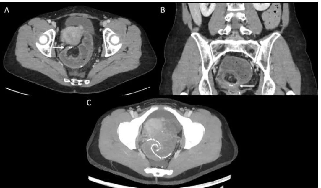
tenderness and acute intestinal obstruction. Abdominal computed tomography scan revealed small bowel
35
obstruction above a volvulus of the ileal-J-pouch (fig. 1 A and B). Axial maximum intensity projection
36
showed whirling of staple lines of the pouch (fig 1 C). Endoscopy confirmed a complete whirling of the
37
pouch but endoscopic detorsion failed and was complicated with pouch perforation. Surgical exploration
38
showed pouch ischemia without necrosis. Manual detorsion of the pouch, suture of the perforation and
39
pexy of the pouch were done. Suture was protected with an omental flap and a diverting stoma.
40
Opacification of the pouch and pouch endoscopy two months later showed normal aspect of the pouch.
41
Outcomes of the closure of stoma were uneventful.
42
Discussion
43
Ileal pouch volvulus is a rare complication, only 22 cases reported in a recent review, usually
44
diagnosed by computed tomography [1]. The delay of occurrence was late with a median time to volvulus
45
after IPAA of 36 months as in the present case. Secondary complications (i.e. ischemia, perforation)
46
occurred in 23% of cases and could lead to pouch excision [1]. Due to the small samples of patients,
47
highlighting risk factors for ileal pouch volvulus is not statistically possible. Absence of adherences
48
responsible of the mobility of the pouch is frequently described without abnormalities of pouch or
49
anastomosis technique. The majority of cases was published in the last 5 years. This fact possibly reflects
50
a large majority of laparoscopically performed IPAA resulting in less peritoneal adherences. The majority of
51
patients were female with larger and wider pelvis than men [1,2]. Some authors hypothesized that position
52
of the mesentery of the pouch could facilitate pouch volvulus. Recently, three cases described by Geers et
53
al. had IPAA constructed with mesentery positioned anteriorly [3]. Surgical management with pouch pexy
54
after endoscopic detorsion is the most frequent treatment. The case of a successful endoscopic detorsion
55
without surgical treatment is described in one patient free of relapse after 3-year follow-up [2]. Pouch pexy
56
consists in suturing both efferent and afferent limb of the pouch to the pelvic walls with interrupted
57
multifilament sutures [3]. This bilateral pouch pexy achieves a firm anchoring of the IPAA to avoid
58
recurrence.
59
3
References60
1. Jawoosh M, Haffar S, Deepak P, Meyers A, Lightner AL, Larson DW, Raffals LH, Murad MH, Buttar N,
61
Bazerbachi F. Volvulus of the ileal pouch–anal anastomosis: a meta-narrative systematic review of
62
frequency, diagnosis, and treatment outcomes. Gastroenterol Rep. 2019;7(6):403‑ 10.
63
2. Landisch RM, Knechtges PM, Otterson MF, Ludwig KA, Ridolfi TJ. Pouch Volvulus in Patients Having
64
Undergone Restorative Proctocolectomy for Ulcerative Colitis: A Case Series. Dis Colon Rectum.
65
2018;61(6):713‑ 8.
66
3. Geers J, Bislenghi G, D’Hoore A, Wolthuis AM. Surgical Management of an Ileal J-Pouch-Anal
67
Anastomosis Volvulus. Dis Colon Rectum. 2019;62(8):1014‑ 9.
68
4
Figure legend69
Figure 1: Abdominal computed tomography showed a whirl sign corresponding to the rotation of the
ileal-J-70
pouch around its axis (white arrows, A and B) confirmed by the rotation of staple lines in maximum intensity
71
projection (C).