HAL Id: inserm-01485521
https://www.hal.inserm.fr/inserm-01485521
Submitted on 8 Mar 2017HAL is a multi-disciplinary open access archive for the deposit and dissemination of sci-entific research documents, whether they are pub-lished or not. The documents may come from teaching and research institutions in France or abroad, or from public or private research centers.
L’archive ouverte pluridisciplinaire HAL, est destinée au dépôt et à la diffusion de documents scientifiques de niveau recherche, publiés ou non, émanant des établissements d’enseignement et de recherche français ou étrangers, des laboratoires publics ou privés.
Distributed under a Creative Commons Attribution - NonCommercial - NoDerivatives| 4.0 International License
Generation of human muscle fibers and satellite-like cells
from human pluripotent stem cells in vitro
Jérome Chal, Ziad Al Tanoury, Marie Hestin, Bénédicte Gobert, Suvi Aivio,
Aurore Hick, Thomas Cherrier, Alexander Nesmith, Kevin Parker, Olivier
Pourquié
To cite this version:
Jérome Chal, Ziad Al Tanoury, Marie Hestin, Bénédicte Gobert, Suvi Aivio, et al.. Generation of human muscle fibers and satellite-like cells from human pluripotent stem cells in vitro. Nature Protocols, Nature Publishing Group, 2016, 11 (10), pp.1833-50. �10.1038/nprot.2016.110�. �inserm-01485521�
***Final Author Version*** for Publication doi:10.1038/nprot.2016.110
at http://www.nature.com/nprot/journal/v11/n10/abs/nprot.2016.110.html
Generation of human muscle fibers and satellite-like cells from human
pluripotent stem cells in vitro
Authors: Jérome Chal1,2,3,4,#, Ziad Al Tanoury1,#, Marie Hestin2,3,4, Bénédicte Gobert1,
Suvi Aivio2,3,4, Aurore Hick5, Thomas Cherrier1, Alexander P. Nesmith6, Kevin K.
Parker6 & Olivier Pourquié1,2, 3,4,* Affiliations:
1
Institut de Génétique et de Biologie Moléculaire et Cellulaire (IGBMC), CNRS (UMR 7104), Inserm U964, Université de Strasbourg, Illkirch Graffenstaden, France.
2
Department of Pathology, Brigham and Women’s Hospital, 77 Avenue Louis Pasteur, Boston, Massachusetts, USA.
3
Department of Genetics, Harvard Medical School, 77 Avenue Louis Pasteur, Boston, Massachusetts, USA.
4
Harvard Stem Cell Institute. 77 Avenue Louis Pasteur, Boston, Massachusetts, USA.
5
Anagenesis Biotechnologies, Parc d’innovation, 650 Boulevard Gonthier d’Andernach 67400 Illkirch Graffenstaden, France.
6 Disease Biophysics Group, Wyss Institute for Biologically Inspired Engineering, School of Engineering and Applied Sciences, Harvard University, Cambridge, USA.
# equal contribution
* Contact Information: Olivier Pourquié, Ph.D.
Department of Genetics, Harvard Medical School and Department of Pathology, Brigham and Women’s Hospital, 77 Avenue Louis Pasteur, Boston, MA, USA.
email: pourquie@genetics.med.harvard.edu
Keywords: pluripotent stem cells, myogenesis, skeletal muscle, satellite cell, bioengineering, directed differentiation, embryonic stem cell, iPS
Abstract
Progress towards finding a cure for muscle diseases has been slow due to the absence of relevant cellular models and the lack of a reliable source of muscle progenitors for biomedical investigation. Here, we report an optimized serum-free differentiation protocol to efficiently produce striated, millimeter-long muscle fibers together with satellite-like cells from human pluripotent stem cells (PSCs) in vitro. By mimicking key signaling events leading to muscle formation in the embryo, in particular the dual modulation of Wnt and Bone morphogenetic pathway (BMP) signaling pathways, this
directed differentiation protocol does not require genetic modifications or cell sorting. Robust myogenesis can be achieved in vitro within 1 month by personnel experienced in human PSC culture. The differentiating culture can be subcultured to produce large amounts of myogenic progenitors amenable for numerous downstream applications. Beyond the study of myogenesis, this differentiation method offers an attractive platform to develop relevant in vitro models of muscle dystrophies, drug screening strategies, and to provide a source of cells for tissue engineering and cell therapy approaches.
INTRODUCTION
Until recently little progress has been made to differentiate human PSCs toward a myogenic fate. This in turn has hampered both the development of relevant human in
vitro models of muscle diseases and the assessment of cell therapy approaches. Here
we report an optimized directed differentiation protocol for in vitro production of mature muscle fibers and their associated progenitors from human PSCs. This is a detailed version of the protocol reported in Chal et al., 20151 with a number of improvements including the expansion and cryopreservation of the hPSC-derived myogenic progenitors. By mimicking the early signaling events occurring during paraxial mesoderm specification in the embryo - in particular the simultaneous activation of Wnt and inhibition of BMP signaling pathways - this protocol recapitulates the essential steps of skeletal myogenesis in vitro in less than 30 days. The resulting fibers show spontaneous contraction and provide a niche for associated Pax7-positive satellite -like cells. An additional subculturing step allows for the preparation of proliferative myogenic
populations that can be further amplified, cryopreserved or differentiated into muscle fibers. This protocol is readily amenable for multi-format in vitro cell assays and various downstream applications. All together, the recapitulation of myogenesis in a dish will be an invaluable tool for the muscle, stem cell, muscle physiology and pathology, and developmental biology communities for both basic and applied research.
Development of the Protocol
Directed differentiation methods aim at exposing PSCs to differentiation cues allowing the sequential recapitulation of key stages of paraxial mesoderm development and its differentiation into skeletal muscle. Early attempts to generate skeletal muscles used spontaneous differentiation of embryoid bodies, resulting in heterogeneous differentiation and low efficiency2,3. Progress in 2D- culture systems was slow until
recently when manipulations of key signaling pathways involved in paraxial mesoderm specification were incorporated to differentiation protocols. In the embryo, Wnt signaling is required for paraxial mesoderm induction4-7 (for review8). In embryos mutant for
Wnt3a or its targets T or Tbx6, ectopic neural tubes form instead of the paraxial mesoderm in the posterior part of the embryo7,9-11. In PSC culture, Wnt signaling
promotes mesodermal differentiation12-15 and several recent myogenic induction
protocols rely on early Wnt activation1,16-19. Wnt signaling also acts upstream of the Fibroblast growth factor (FGF) pathway in paraxial mesoderm precursors of the tail bud, by triggering expression of the Fgf8 ligand20. Later on, at the somite level, secreted Wnts from surrounding tissues are also critical for dermomyotome specification (for review21).
BMP signaling controls the medio-lateral identity of posterior mesoderm cells22-26. In amniote embryos, high BMP leads to the specification of more lateral tissues such as extraembryonic mesoderm, or lateral plate mesoderm (LPM), a tissue that contributes to the limbs and body wall mesenchyme and to the long bones of the limbs but which does not form skeletal muscle. Ectopic BMP signaling can divert cells fated to form paraxial mesoderm to a lateral plate fate in vivo22. Conversely BMP loss of function mutation
leads to expanded paraxial mesoderm domain27. In vivo, the paraxial mesoderm is
protected from BMP signaling produced by the LPM by the inhibitors Noggin and Chordin which are expressed by the notochord, the intermediate mesoderm and the dorsal somite24,25,28. In vitro, in absence of proper BMP signaling inhibition, the early paraxial mesoderm induced by Wnt signaling produces BMP4 and "drifts" by an autocrine effect to generate LPM derivatives (J.C., Z.A.T. and O.P., unpublished observation). At the heart of the protocol presented here lies the use of dual modulation
of the Wnt and BMP pathways to efficiently induce paraxial mesoderm from hPSCs. This is a prerequisite for the production of skeletal myogenic progenitors in large number. Other studies have shown that Wnt activation and BMP inhibition can produce
mesodermal fates from human PSCs, such as Intermediate mesoderm29 and
chondrogenic mesoderm30 which are closely related to the paraxial mesoderm. On the
other hand, activation of BMP signaling has been used to produce anterior LPM derivatives such as cardiovascular cell types31-34. While little is known about the culture
of early embryonic paraxial mesoderm from primary cultures35-38, myogenic progenitors
and skeletal myoblast cultures have been extensively studied in vitro39-43.
The protocol described here stems from the transposition of a serum-free protocol developed for adherent mouse ESC culture to human pluripotent cell cultures 1 (Figure1). At its core lays a dual modulation of Wnt and BMP pathways. It is based on our understanding of paraxial mesoderm development in vivo, where Wnt is activated to specify early paraxial mesoderm while BMP is inhibited to prevent the newly specified paraxial mesoderm cells to drift to a lateral plate fate22. In absence of BMP inhibition, Wnt activated- PSCs differentiate preferentially to LPM derivatives such as endothelial progenitors44. For the hPSC protocol, the first step of the mESC protocol aiming at producing epiblast stage cells, was removed as hPSCs are more related to mouse EpiSCs45. Fine-tuning of the factors' concentrations and exposure time was also necessary to obtain optimal paraxial mesoderm induction and subsequent skeletal myogenesis (Figure 2). Long term differentiated hPSC-culture (>1 month) are very dense, produce abundant extracellular matrix (ECM), but retain Pax7+ myogenic progenitors. We noticed that dissociation and replating of these differentiated cultures led to a significant enrichment in myogenic cells (Figure 3). This step allows to produce
in larger quantity more homogeneous myogenic cultures that are amenable for cryostorage or for diverse culture set-ups (Figure 4, 5).
Key advantages of the differentiation protocol reported here are the ease of implementation of a simple and robust 2D-culture system, higher yields, the production of both myogenic progenitors and mature fiber cell types, and a faster experimental timeline over existing protocols16,19,46-48. Unlike other protocols, this method does not
require genetic manipulation such as forced expression of transcription factors49-53, nor
resort on cell sorting strategies to purify the populations of interest17,54-57. Primary
myoblast cultures obtained from biopsies and myogenic cell lines do not usually generate mature fibers but produce shorter, irregularly shaped myotubes lacking striation. In contrast, the protocol presented here describes how to generate in 30 days, striated millimeter-long skeletal muscle fibers in vitro, thus providing the relevant cell types to study both embryonic development and mature muscle diseases.
Overview of the Procedure
The starting material can be standard hESC or hiPSC culture58-62(Figure 1a). Briefly,
cells are dissociated to single cells and seeded at low density on Matrigel-coated wells. Cells are left to recover in maintenance conditions until they form small irregular aggregates, at which time (Day 0) they are changed to a sequence of differentiation media (Figure 1b). This sequence aims at first generating induced paraxial mesoderm progenitors (iPAMs) corresponding to the presomitic mesoderm stage in vivo, and then promoting their differentiation into skeletal muscles within 30 days (Figures 1, 2). In addition to myofibers, the hPSC-derived culture retains myogenic progenitors including Pax7+ satellite-like cells (Figure 2). These progenitors can be expanded to generate myogenic subcultures that can in turn be cryostored, redifferentiated (Figure 3, 4) or used for various downstream applications (Figure 5).
This protocol allows for the production of both myogenic progenitors, satellite-like cells and myofibers, which enable novel hPSC-based model to study important fundamental and applied skeletal muscle biology questions (Table 1 ). The expansion of hPSC- myogenic cultures is compatible with various experimental designs (Figure 5). Below we discuss the potential applications the different cell types generated by the protocol can be used for.
Paraxial mesoderm. The protocol described here provides a novel, tractable, in vitro
system to study paraxial mesoderm formation (Figure 2). It is amenable for quantitative and real-time imaging and flow cytometry analysis. Important developmental biology questions related to paraxial mesoderm development can be investigated, including the specification of neuro-mesodermal progenitors, segmentation, somitogenesis, and lineage specification63,64. Combined with genetic manipulations, it allows to assess the
role of individual genes in normal and pathological conditions. Production of hPSC-derived myogenic progenitors may also be used as a more relevant and standardized source of cells for bioengineering approaches that have traditionally relied on cell lines and biopsies (Figure 5).
Paraxial mesoderm Myogenic progenitors Skeletal muscle fibers Myotubes Satellite-like cells Dev. Biology
in vitro model Cell fate decision Myogenesis Specification Niche &
Cell biology
Physiology Bioengineering Tissue chips Electrophysiology Coculture system Homeostasis
Pathology Disease modeling
Musculoskeletal system, Myopathies Gene correction
Regenerative
Table (1) : Overview of the possible applications pertaining to hPSC-derived myogenic
cultures. Columns represent distinct developmental /differentiation stages. Rows represent biomedical disciplines.
Myotubes and fibers. One of the main advantage of this protocol, is the production
from hPSCs of both myogenic progenitors and millimeter-long, striated, often spontaneously contracting, skeletal myofibers (Figure 2, 4). Primary differentiation or secondary differentiation after subculturing allows production of myofibers on demand. This culture system allows also for the study of skeletal myogenesis including important events difficult to visualize in vivo such as myoblast fusion, satellite cell formation, myofibrillogenesis or fiber type specification. hPSC -derived myogenic cultures are a valuable alternative to primary myogenic cultures and immortalized cell lines. It is amenable for scale-up, or miniaturization for drug screening65, and can be combined
with 3D bioengineered substrates (Figure 5a), tissue array/chip66,67 (Figure 5b) and
complex electrophysiology or optogenetic time-lapse platforms68-78. Miniaturization is necessary for drug screening strategies aiming at identifying compounds able to promote progenitors' proliferation or fibers' maturation79,80. Furthermore, co-culture with other relevant cell types could allow the study of important aspects of muscle biology, namely the neuromuscular junction (motoneurons)81-89, the myotendinous junction (tenoblasts)90,91, vascularization (endothelial cells)92-95 and metabolic coupling (adipocytes)96. Healthy cells can also be directly compared to cells harboring various
mutations. In vitro models of human muscular dystrophies involving forced expression of myogenic factors such as Pax3/7 or MyoD in patients' induced PSCs to induce myogenesis have been reported, but the physiological relevance of the induced fibers remains to be established97-102. The protocol presented here recapitulates the
developmental sequence of myogenic differentiation from hPSCs without introduction of exogenous genetic material and thus may provide more physiologically relevant models of these diseases. This may eventually allow the development of in vitro strategies for therapeutic approaches103-108. Muscle tissue engineering is a very active field, which
has so far essentially relied on primary cultures derived from human or animal biopsies, and immortalized cell lines. Such applications will benefit from an unlimited and
reproducible source of human myofibers and their progenitors, including satellite-like cells68,109-113.
Satellite-like cells. In adult muscle, quiescent stem cells (Pax7+ satellite cells) found in
very small numbers in close association with muscle fibers allow for muscle repair during regeneration114,115. Elegant mouse genetics experiments have shown that satellite cells represent the key population allowing muscle regeneration in vivo
116-118.The transplantation of human skeletal myoblasts was shown to be inefficient as
grafted cells massively die thus failing to engraft in the host119-122. In contrast, even
minute numbers of satellite cells efficiently contribute to muscle regeneration in injured or dystrophic mouse muscles123-128. Thus, satellite cells represent the ideal candidate
for cell therapy approaches aiming at reconstructing muscles. However, these cells are found in very limited number in adult muscles and they cannot be amplified in vitro as they lose their regenerative properties in culture123,129. Thus access to an unlimited
source of satellite cells differentiated from pluripotent cell cultures could open the possibility to develop cell therapy protocols for muscular dystrophies. We have shown that the long term Pax7+ cells produced in vitro from mouse ES cells become tightly associated to differentiating fibers, locating under their basal lamina as expected for satellite cells. These Pax7+ cells can give rise to both muscle fibers and Pax7+ satellite cells when grafted in vivo suggesting that the Pax7+ cells differentiated in vitro correspond to satellite cells1. The muscle fibers differentiated in vitro from mouse ES
cells exhibit characteristics of early post-natal fibers suggesting that the associated Pax7+ cells likely correspond to post-natal satellite cells. Remarkably, whereas adult satellite cells immediately become activated in vitro, losing the capacity to self-renew, immature satellite cells can still produce both Pax7+ satellite cells and myoblasts when cultured in vitro130. This property is shared by both mouse and human Pax7+ cells differentiated in vitro with our protocols. Furthermore, immature satellite cells were recently shown to exhibit remarkable regenerative properties which might be a significant advantage in the context of cell therapy protocols aiming at regenerating muscle130. We observed that the differentiation of human Pax7+ cells produced in vitro with our protocol is very similar to that of the mouse Pax7+ satellite-like cells and we anticipate that these cells also correspond to immature human satellite-like cells.
Further analysis of the human Pax7+ cells produced with this protocol will be necessary to compare them to recently characterized human muscle stem cells128,131,132. In particular, the assessment of their regenerative properties in vivo will be key to be considered as a potential source of cells for cell therapy approaches133. These cultures could also permit to study the development of human satellite cells, a process on which nothing is currently known.
Alternative Methodology
Distinct methodologies have been used to produce skeletal muscle cells from hPSCs. In most cases, differentiation strategies have been first optimized on mouse embryonic stem cells (ESCs) before transposition to human PSCs (reviewed in119,134-141). Direct conversion methods consist of overexpressing myogenic factors in a stem cell to force it
to differentiate into a myogenic progenitor, bypassing paraxial mesoderm specification142,143. The general concept of cell reprogramming by introduction of a
factor was first demonstrated with the ability of MyoD to convert fibroblasts into myoblasts144. The method was perfected over time by the use of different vectors,
including inducible systems to overexpress transcription factors- chiefly MyoD, Pax3 or Pax7 which can all elicit myogenic differentiation with various degrees of efficiency100,145-150. However, these methods require introduction of exogenous DNA into the cells, often via viral vectors, which is an issue for potential biomedical applications. Additionally, the molecular identity of the generated myogenic cell has not been investigated in great details. For MyoD conversion, myofiber maturation in vitro is usually improper due to incomplete myogenic activation and conversion143. The use of the upstream regulators, Pax3/7, although not restricted to the myogenic lineage151-155 allows for the production of in vitro myogenic progenitors146,150 that need to be further enriched by cell sorting. Recent work suggests that these induced myoblasts are able to generate striated fibers156. However, culture conditions are generally undefined,
aimed at generating other cell types found in the adult skeletal muscle, such as pericytes/mesoangioblasts which exhibit myogenic properties157-160.
Limitations of the protocol
Biological relevance. Very little is known about human paraxial mesoderm
development due to the difficulty to access early human embryos. The protocol described here can provide a window in the development of this key lineage that not only gives rise to skeletal muscle but also to the axial skeleton and the dermis of the back. Access to an unlimited supply of genetically defined myogenic cells will allow the development of many biomedical applications to study muscle physiology and pathology. One major critique is the intrinsically artificial nature of the culture system which is vastly different compared to an organism in its full complexity161. Another
relates to the differentiated cell types obtained in vitro, whose maturation is incomplete being closer to the early post-natal stage than to adult, which would be more desirable for relevant physiological and disease modeling investigations162. This might be
overcome with more integrated cell culture systems, such as cocultures or "on-a-chip" design aiming at interconnecting tissues in a physiologically relevant manner 76,81,88.
Research applications. Dense cell culture can render the optical analysis difficult if not
unfeasible. Furthermore, primary differentiation is done typically on plastic cell culture dishes that might not be compatible with high-end imaging. However, we also successfully differentiated human muscle fibers from iPS cells on Thermanox coverslips. Subculturing can also circumvent both issues by allowing the control of seeding densities and cell culture format, allowing more flexible experimental setups (Figure 5). It is also well known that various hPSCs exhibit different propensity to differentiate toward one specific germ layer and tissue163,164. As such, it is expected that the nature of the starting cell lines will be a source of variability that can lead to a different level of myogenic differentiation. We believe we provide a robust conceptual framework based on developmental principles that is amenable to most cell lines with limited adaptations. In our hands, we have routinely achieved efficient differentiation of
about ~8 independent lines including hiPS11a (HSCI, Harvard University), NCRM1 and NCRM5 (RUCDR, Rutgers University), H9 (WiCell, Madison, WI) and HUES1 (HSCI, Harvard University).
Biomedical applications. All cell material and reagents will need to meet safety and
regulations approval by Regulatory agencies if the cell populations are to be used in clinical trials and in a regenerative/cell therapy context165. However, we expect that the
first biomedical applications of PSCs differentiated in vitro will be in the domain of high throughput screening, in vitro disease modeling and cell toxicity assays65,76,166, for which
such approval is not required.
Experimental design
hPSCs lines. Quality of the starting hPSC line is essential (Figure 1a). Cell lines
should be karyotypically normal, pathogen -free and pluripotent. hPSC cell lines should come from recognized cell repositories. hPSCs can be maintained by a variety of methods58,59. Regular monitoring and characterization of hPSC culture is essential.
Large archive and working banks should be done at low passages.
Mycoplasma detection can be performed using PCR-based detection method such as the Venor GeM Mycoplasma Detection Kit. Complementary ways to assess the hPSC status include evaluation of colony growth, immunohistochemistry or Fluorescence-activated flow cytometry (FACS) analysis for stem cell markers expression, embryoid bodies formation and histological analysis of hPSCs-derived teratoma60. All cell lines used in this study come from recognized repositories and are mycoplasma-free.
Cell culture reagents. Key reagents, including the Knockout Serum
Replacement, chemical compounds, and recombinant proteins should be systematically tested. In particular, reagents' providers and production lots should be recorded. Due to variations in manufactured biologics, different lots should be tested in parallel and
benchmarked against previous references on side-by-side differentiations to ensure an optimal experimental outcome.
Culture format. The protocol was originally developed for standard 2D adherent
cell culture, in 24- to 6-well plates. While the step-by-step procedure provided below is optimized for a 12-well plate format, transposition to 24-, or 6- well plate formats is straightforward by respecting the corresponding surface ratio and volumes. It is possible also to further scale up or down by seeding on other types of plates (see for example167). We successfully differentiated cells in 96-well plate and in P10 cm dish
format.
Culture density. Both the cell seeding density and the culture's confluency at
Day 0 are critical for the success of differentiation. Seeding density should be within 15,000 and 45,000 cells /cm2 (it can be lower for larger surface area). After 1 to 2 days
of recovery in medium containing the anti-apoptotic ROCK inhibitor168, 10-25%
confluent homogenous cultures can be used for directed differentiation. Deviation from these guidelines leads usually to failure of differentiation either due to cell death or to overcrowding and culture detachment en masse.
Culture substrate. The default substrate is Matrigel (Corning; see also169) which
is compatible with robust cell attachment and myogenic differentiation. More defined substrates such as Gelatin, or specific recombinant laminin and fibronectin -coating have been used in the past for myogenic progenitors170-173, however we observed that
maturing myogenic culture were more susceptible to detach on those substrates.
Subculturing step. During differentiation, cultures become confluent within a
week. After 30-40 days (Figure 1c), they are very dense and tend to detach from the substrate as sheets of cells, possibly due to myofibers' spontaneous contractions. We found that replating and splitting of these original cultures leads to a significant enrichment in myogenic cells and to less dense cultures easier to image (Figure 3, 5). Passaging can be performed by combined mechanical and enzymatic dissociation of 10 to 50+ day-old culture with 25-45 days being the optimal window (Figure 3). Because of extensive extracellular ECM deposition, older cultures are harder to dissociate properly
without causing significant cell death. Optimal reseeding density corresponds to a splitting ratio of 1:4 to 1:8; or about 60-70,000 cells/cm2. Subculturing results in selection of myogenic progenitors while depleting unwanted differentiated contaminants and accumulated culture debris. While post-mitotic cells (myocytes and myofibers) are essentially lost by the subculture step, myogenic precursors are preserved and can be amplified with commercially-available growth media used for primary muscle cultures and cell lines (compare Figure 1c and 3d). For this purpose, we used the Skeletal Muscle Cell Growth Media-2 (Clonetics, Lonza; see Media composition). De novo myofibers can be produced again by differentiation of the subculture (Figure 3-5). Furthermore, confluent cultures left in proliferative media are fairly stable and retain a pool of Pax7-myogenic progenitors for months, which can be used later for downstream applications (Figure 3-5). The subculture is thus amenable for further passaging, cryostorage, and/or secondary differentiation (Figure 3).
Myogenic Differentiation media. Secondary differentiation can be achieved
either by transferring to "terminal differentiation" media such as horse serum containing media40, or serum-free media containing insulin and the N2 supplement57,174-176 (Figure
3-5).
Differentiation yield. Both the total number and the percentage of target cell
type should be quantified to assess protocol efficacy and reproducibility, and possible troubleshooting. The protocol was designed as a multistep protocol with a sequence of
defined media and developmental milestones. Proper differentiation should be
assessed regularly. This is particularly important when working with novel hPSC lines or several lines for side-by-side comparison. We favor immunohistochemistry for specific validated markers to evaluate differentiation efficiency (Figures 2, 4; Table 2). Paraxial mesoderm progenitors’ specification can be monitored with antibodies against T/ Brachyury, or Tbx610,177,178. It should be kept in mind that most markers are not entirely
specific to the target tissue along the whole developmental spectrum (see Table 2). We found that Msgn1179,180 is the most reliable and specific marker of paraxial mesoderm
progenitors. While, unfortunately, no validated Msgn1 antibody is currently available, Msgn1 expression can be monitored by RT-qPCR or using reporter cell lines (data not
shown). Like Tbx6, Msgn1 expression is expected to peak around day 4-5 of differentiation with this protocol, accounting for 70-90% of the cells. Myogenesis per se should be monitored starting on week 2 by following MyoD and Myogenin expression
181-183, while myotubes formation and fiber maturation can be assessed using skeletal
muscle proteins or isoforms, such as Myosin Heavy chains184 (Figure 2, Table 2). The
presence of satellite-like cells in long term culture can be evaluated with the expression of Pax7185,186 (Figure 2b, 4a,b). By 4 weeks (~d30) of differentiation, about 22% of
nuclei are Myogenin-positive while another 23% are Pax7-positive1. Upon subculturing
and 2 more weeks of differentiation of 3 independent cell lines, up to 40% of total nuclei were Myogenin -positive (Figure 5d). Mature skeletal myofiber density was ranging from 9,000 to 20,000 fibers per cm2 (Figure 5c and data not shown).
Level of expertise to implement the protocol. Prior experience with routine human
PSC culture and cellular phenotyping is valuable, especially to scale the culture up or down, and to adapt the protocol to specific applications. Familiarity with the concept of directed differentiation can be advantageous for protocol troubleshooting.
MATERIALS REAGENTS Cells
• Human Pluripotent Stem cells, we have successfully used the following: hiPSCs: hiPS11a (HSCI, Harvard University), NCRM1, NCRM5 (RUCDR, Rutgers University) hESCs: H9 (WiCell, Madison, WI), HUES1 (HSCI, Cambridge, MA)
CAUTION Cell line identity should be regularly verified and cultures tested for
mycoplasma contamination. Experiments involving human pluripotent stem cells must conform to relevant Institutional and National regulations, we gained ethical approval from IACUC /ESCRO 2014-06-02 CRITICAL Different cell lines may respond to
directed differentiation with variable level of myogenic differentation.
Media
• mTeSR1 (Stemcell Technologies, cat. no. 05851) • DMEM/F12 1:1 (Life Technologies, cat. no. 11320-082)
• Knockout Serum Replacement (KSR; Life Technologies, cat. no.10828-028)
CRITICAL Lot-to-lot variability may impair the differentiation protocol
• L-Glutamine- Penicillin-streptomycin (Gibco, cat. no. 10378-016)
• 2-Mercaptoethanol, 55mM (βME; Life Technologies, cat. no.21985-023)
• Non-essential amino acid solution [NEAA] (Thermo Fisher, cat. no. 11140-050) • Insulin-Transferrin-Selenium (ITS; Life Technologies, cat. no. 41400-045) • BSA (Sigma, cat. no. A7906)
• N-2 Supplement (Life Technologies, cat. no.17502-048) • Horse serum (Invitrogen, cat. no. 16050-130)
• SkGM-2 (SkGM; Lonza, cat. no. CC-3245)
• Cryostem Freezing medium (Stemgent, cat.no.01-0013-50)
Cell culture reagents
• Dulbecco’s Phosphate Buffered Saline (DPBS), calcium/magnesium free (Gibco, cat. no.14190-144)
•Tris Buffered Saline (TBS), 10X (Sigma, cat.no.T5912-1L) • Sterile cell culture–grade water (Invitrogen, cat. no. 10977-015) • Matrigel, hESC-qualified (MG; Corning, cat. no. 35277)
• Gelatin solution EmbryoMax 0.1% (wt/vol) (EMD Millipore, cat. no. es-006-b) • Trypsin EDTA 0.25 M, pH 8.0 (Life Technologies, cat. no. 25200-056)
• TrypLE Express (Invitrogen, cat. no. 12605010)
• Dispase (1U/mL) (Stem cell technologies, cat.no.07923) • Collagenase type I (Thermo Fisher, cat. no. 17100-017) • DMSO, Cell culture grade (Sigma , cat. no. D2650)
• Trypan Blue solution 0.4%, Cell culture grade (Sigma, cat.no.T8154-20ML)
Factors
• CHIR99021 (Tocris Bioscience, cat. no. 4423)
CRITICAL GSK3 beta inhibitors other than CHIR may not be as efficient
• LDN-193189 (Stemgent, cat. no. 04-0074)
CRITICAL BMP type I receptor inhibitors other than LDN may not be as efficient
• Y-27362 Dihydrochloride (Rocki; Tocris Bioscience, cat. no. 1254) • HGF, Recombinant Murine (PeproTech, cat. no. 315-23)
• FGF2, Recombinant Murine (PeproTech, cat. no. 450-33) • IGF1, Recombinant Murine (Peprotech, cat. no. 250-19)
• Paraformaldehyde (Electron Microscopy Sciences, cat. no. 15710)
CAUTION Paraformaldehyde must be handled under a chemical cabinet.
• Triton X-100 (Sigma-Aldrich, cat. no. T8787-250ML) • Tween-20 (Sigma-Aldrich, cat. no. P7949-500ML)
• Fetal Bovine Serum (FBS; Hyclone/GE, cat. no. SH30070.03) • Hoechst 33342 (Life Technologies, cat. no. H3570)
• Primary antibodies (see Table 1)
• Secondary antibodies, species specific anti-IgG(H+L) - Alexa fluor conjugated (Molecular probe)
• Isopropanol, 70% (VWR, cat. no. 89499-420)
• Venor GeM Mycoplasma Detection Kit, PCR-based (Minerva Biolabs, Sigma cat. no. MP0025-1KT)
● EQUIPMENT
• Cell culture dishes (Corning, cat. nos. 353001; 353002) • Cryovials (Nalgene, cat. no. 5000.0020)
• Needles, 25G (Becton Dickinson, cat. no. 305125) • Sterile cell scrapers (Celltreat, cat. no. 229306)
• Sterile plastic tubes, 15ml & 50mL (VWR, cat. nos. 89039-66; 8939-658) • Test tubes, 5mL round bottom, snap cap (Corning, cat. no.352063)
• Cell filter, Celltrics 30μl sterile and non-sterile (Partec, cat. no. 2326; 04-004-2316)
• Sterile plastic pipettes (Corning, cat. nos. 356543; 356551; 356525) • Filter 0.22μm (Pall, cat. no. 4652)
• Cell strainer 70μm (ThermoFisher, cat. no. 22363548)
• Pipettes Filtered tips (VWR, cat.nos. 89003-060; -056; -048; 89368-974) • Freezing container (Biocision, cat. no. BCS-405G)
• Filter for vacuum system 0.2μm hydrophobic (Pall, cat. no. 4251) • Pasteur pipettes (VWR, cat.no. 14673-010)
• Water purification system (EMD Millipore, cat.no. SYNSVR0WW) • Cell counter (Nexcelom Bioscience, cat. no. Auto2000)
• Pipette-aid Easypet 3 (Eppendorf, cat. no. 4430000018)
• Pipettes Research plus 4-pack (Eppendorf, cat. no. 022575442) • Centrifuge 5810 w rotor A-4-62 (Eppendorf, cat. no. 022627007)
• Biosafety cabinet HERAsafe KS Type KS12 (Thermo Scientific, cat. no. 51022482) •Vacuum filtering units, M-Vac (Argos Technologies; cat. no. VWR # 89129-568) • Incubator CO2 Incubator (Panasonic, cat. no. KM-CC17RUI)
• Digital Microscope EVOS XL Imaging System (Life Technologies, cat. no. AME3300) • Digital Fluorescence Microscope EVOS FL Imaging System (Life Technologies, cat. no. AMF4300)
• Picking station, Lynx Stereo Dynascopic Microscope (Vision Engineering, cat. no. Lynx)
• Freezer -80C, Ultra-low Temp Freezer (Panasonic,cat. no. MDF-U53VA) • Liquid nitrogen storage tank (Cryosafe, cat. no. T CAT 7PS )
● REAGENT SET UP
• Recombinant Factors stock solution (FGF2, HGF, IGF1) Resuspend the
lyophilisate in sterile cell culture–grade PBS supplemented with 0.1% BSA to desired stock concentration. Aliquot in 50µL volume and store at -20 °C for up to 6 months. FGF2 stock solution 10µg/ml, use at 20ng/ml; HGF stock concentration 100μg/ml, use at 10ng/ml; IGF stock concentration 10μg/ml, use at 2ng/ml.
• Chemical compounds stock solution (CHIRON99021, LDN-193189, Y27362)
Resuspend the lyophilisate in cell culture grade DMSO (CHIRON, LDN-193189) or H2O
(Y27362) to desired concentration. Aliquot in 50µL volume and store at −20 °C for up to 6 months. CHIR99021 stock concentration 10mM, use at 3μM; LDN-193189 stock concentration 1mM, use at 500nM; Y27362 (Rock inhibitor) stock concentration 10mM, use at 10μM.
• Matrigel and Matrigel -coated plates Thaw the stock solution overnight on ice at
4°C, and once it is in
liquid form keep it on ice and divide the solution into aliquots (lot-specific volume by manufacturer's recommendation) using cold tips and cold tubes. Store at -80°C. When needed, thaw aliquots on ice, dilute with 12ml ice cold DMEM/F12 resulting in 3mg/ml protein concentration and mix by pipetting. Add 1ml of diluted Matrigel solution per 10cm2 of multiwell plates or dishes to cover surface evenly. Keep on ice for at least 4h
and prior to use incubate at 37 °C for 30 min. Store Matrigel coated plates at 4°C for up to 2 weeks.
CRITICAL Matrigel solidifies above 4°C so ensure all the cell culture plastic is ice cold
during handling.
• mTeSR medium. Thaw 100mL bottle of 5x Supplement overnight at 4°C and add it to
the mTeSR- medium. Mix and divide into 40mL aliquots. Store at -20°C for up to 3 months. Upon use, thaw an aliquot at room temperature and keep at 4°C for up to 2 weeks.
• SkGM-2 medium. Supplement SkGM basal medium by adding the components of
SkGM Bulletkit. Mix and aliquot in 50mL tubes. Store at -20°C for up to 3 months. Upon use, thaw an aliquot and keep at 4°C for up to 2 weeks.
• Paraformaldehyde. Dilute the 16% glass sealed EM-grade Paraformaldehyde stock
in DPBS. Leftover 16% stock solution can be frozen for later use. 2-4% working solution should be prepared fresh extemporaneously.
CAUTION: Paraformaldehyde is toxic and must be manipulated under a chemical cabinet. Used solution should be disposed according to local institutional guidelines.
• TBST Buffer. Dilute the Tris-Buffered Saline (TBS) 10X buffer with ultrapure water,
and supplement with 0.1% (vol/vol) Tween-20. Store at room temperature (RT, 15-25°C) for up to 3 weeks.
• Blocking Reagent. Dilute the TBS 10X buffer with ultrapure water, and supplement
• Differentiation media.
Prepare cell media according to the table below and filter on 0.22µm mesh. Small compounds and recombinant proteins should be added extemporaneously but base media can be stored at 4 °C for up to 2 weeks.
Small compounds and recombinant proteins (in italic in the tables) should be added immediately before use.
Di-CL /Di-CLF Media
DK-HIFL Media
Composition Volume (250mL) Final concentration
DMEM/F12 209.5mL
KSR 37.5 mL 15% (vol/vol)
Non-essential amino
acids 2.5 mL 1% (vol/vol)
Penicillin / Streptomycin 0.5 mL 0.2% (vol/vol)
2-Mercaptoethanol 454 µL 0.1mM
Recombinant HGF 25 µL 10 ng/mL
Recombinant IGF-1 50 µL 2 ng/mL Recombinant FGF-2 500 µL 20 ng/mL
LDN-193189 125 µL 0.5 µM
DK-I / DK-HI Media
Composition Volume (250mL) Final concentration
DMEM/F12 209.5mL
KSR 37.5 mL 15% (vol/vol)
Non-essential amino
acids 2.5 mL 1% (vol/vol)
Penicillin / Streptomycin 0.5 mL 0.2% (vol/vol)
2-Mercaptoethanol 454 µL 0.1mM
Recombinant HGF 25 µL 10 ng/mL
Recombinant IGF-1 50 µL 2 ng/mL
Composition Volume (250mL) Final concentration
DMEM/F12 244.5 mL
ITS 2.5 mL 1% (vol/vol)
Non-essential amino
acids 2.5 mL 1% (vol/vol)
Penicillin / Streptomycin 0.5 mL 0.2% (vol/vol) (20 IU + 0.02
mg)/ml
CHIR-99021 75 µL 3 µM
LDN-193189 125 µL 0.5 µM
N2 Media
Composition Volume (250mL) Final concentration
DMEM/F12 242mL
ITS 2.5 mL 1% (vol/vol)
N2 supplement 2.5 mL 1% (vol/vol)
Penicillin / Streptomycin 0.5 mL 0.2% (vol/vol)
● PROCEDURE
Pluripotent cell culture preparation ● TIMING 5 - 7 d
1| Pluripotent cell culture maintenance and amplification. Culture hPSCs colonies on
35mm Matrigel coated dishes. Passage when culture reaches ~70% confluency (Figure 1a).
2| Prepare new 35mm dishes to passage colonies: pre incubate Matrigel coated dishes
at 37oC for 30min. Just prior to passaging replace Matrigel with 2mL of room
temperature mTeSR.
3| Passage cells either by the cutting method (option A) or by the Dispase method
(option B).
CRITICAL STEP While any hPSC lines can be passaged by cutting method, allowing
the selection of the optimal colonies, the Dispase method allows for easier bulk passaging, however it should be performed only on cultures with low overall
spontaneous differentiation. Some hPSC line are more prone to culture failure (cell death, differentiation) with Dispase method.
Option A- Cutting method
1. Using a 25 gauge needle tip, cut healthy hPSC colonies into small pieces
using an "eyepiece-less" stereo microscope (Lynx).
CRITICAL STEP Colony pieces should not be too small or too big, approximately 200 x
200 µm use the width of the needle as a size proxy (Figure 1a).
2. Scrape and collect colony pieces using a 20μL Pipette. Gently aspirate while
detaching the pieces from cell culture dish with the pipette tip. Option B- Dispase method
1. Discard medium, rince dish with PBS, add 1mL of dispase (1U/ml) to the dish
and incubate 5min at 37 °C, 5% CO2, for 5-7 min.
CRITICAL STEP Differentiated colonies should be scraped away beforehand.
2. Remove the dispase solution, rince dish twice with PBS, add 1ml of mTeSR
and detach the colonies using a cell scraper.
3. Triturate detached colonies to fragment them to suitable size. CRITICAL STEP
Colony pieces should not be too small or too big, approximately 200 x 200 µm, usually achieved by pipetting up and down 2-3 times using a 1000 µL pipette
4| Transfer 50-100 colony pieces/ fragments onto the new Matrigel-coated dishes (step
2) and distribute fragments evenly by gently rocking the plate back and forth, and side
to side (Figure 1a). Place in incubator at 37°C, 5% CO2.
Predifferentiation setup (Day-2) ● TIMING 2 d
5| Single-cell dissociation of PSCs culture. Change mTeSR medium every day until
hPSC cultures reach confluency of ~70%.
CRITICAL STEP Starting hPSC culture must consist of homogeneous and healthy
looking colonies.
6| Pre-treat cells with 10µM Rock inhibitor in mTeSR for at least 2 hours. 7| Prepare 12-well plates coated with Matrigel.
CRITICAL STEP This procedure can be easily performed with 24- and 6-well plate by
adapting volume and cell numbers to the surface ratio.
8| Rinse hPSC culture with DPBS without Calcium and Magnesium.
9| Add 100μl/cm2 of TrypLE Express and incubate at 37 °C, 5% CO2, for 5-7 min.
10| Observe cell dissociation under microscope and help mechanical dissociation by
tapping the plate.
Most of the colonies will dissociate to a suspension of single cells or small loose, irregular aggregates.
11| Collect cell suspension in a 15ml sterile tube and gently further triturate with a
1000µL pipette.
12| Add 10ml of DMEM/F12 to inactivate TrypLE Express. 13 | Centrifuge cells at 300g for 5min.
14| Remove supernatant and resuspend the pellet gently in 1ml of mTeSR + 10µM
Rocki.
15| Count the cells and use Trypan Blue or similar to assess cell viability.
16| Dilute cells in mTeSR + 10µM Rocki so as to plate them at 3 × 10⁴ cells per cm2 (
ie. 1.2 x10⁵ cells in 1.5mL of medium for 1 well of a 12-well plate).
17| Rinse the Matrigel-coated differentiation plates with PBS and plate cells at 3 ×
104 cells per cm2 in mTeSR + 10µM Rocki. (one p35 dish can typically produce enough
cells for a whole 12 -well plate)
CRITICAL STEP Even distribution of the cells is critical to create homogenous
differentiation conditions. Cells plated on 24-well plate and smaller tend to clump on the edges and in the center of the well if not properly distributed. Cell density and plating efficiency are crucial for the survival of the culture. Too sparse plating leads to
detachment of the colony within the first five days, and too dense culture will quickly become over-confluent and result in heterogeneous differentiation of the culture.
18| Incubate the plate at 37°C, 5% CO2, overnight.
19| Observe the cells and change mTeSR daily until hPSCs cultures reach a confluency
of ~15 -20%.
During Rocki treatement, cells at low density to adopt a spiky morphology. Cultures will have many small (30< cells) colonies which is optimal to start differentiation (usually after 1-2 days) (Figure 1c).
CRITICAL STEP Small colonies should be evenly distributed on the dish surface
optimal for optimal differentiation.
TROUBLESHOOTING
Directed Differentiation (Day 0- 40) ● TIMING 30 - 40 d
20|Day 0. Once cultures have reached 15-20% confluency, change the medium to DiCL
(Dmem-ITS-Chir-Ldn) medium to initiate differentiation. This is Day 0 of differentiation. Refresh media daily until Day 3 (Figure 1b,c).
21| Day3. Change medium to DiCLF (Dmem-ITS-Chir-Ldn-Fgf) medium and refresh
daily until Day 6.
Ideally, medium should be changed daily until Day 12 of differentiation. However, one day (weekend) can be omitted while respecting the different media sequence.
22| Day 6, change medium to D-KHIFL (Dmem-KSR-Hgf-Igf-Fgf-Ldn) and refresh daily
until Day 8.
CRITICAL STEP Significant cell death will be observed the first week, this is normal
selective action of the sequence of media.
23| Day 8, change medium to D-KI (Dmem-KSR-Igf) and refresh daily until Day 12. TROUBLESHOOTING
24| Day 12, change medium to D-KHI (Dmem-KSR-Hgf-Igf) and refresh every other day
thereafter (Figure 1b).
CRITICAL STEP Media change over the weekend can be avoided by doubling the
culture media volume beforehand.
25| Observe the cultures for myotube formation and change the differentiation medium
every other day. Myotubes are usually visible after 30 days of differentiation (Figure 1c, 2b)
CRITICAL STEP Differentiated cultures are delicate, as myotubes detach easily.
TROUBLESHOOTING
Culture characterization ● TIMING 2-4d
26| Wash twice with DPBS cultures obtained either through primary differentiation (
steps 20-25) or through differentiation of subcultures (steps 37-54)
27| Fix cultures plates with 250µL/cm2 of 2% (vol/vol) paraformaldehyde for 10 min at
RT or O/N at 4°C.
CRITICAL STEP Paraformaldehyde must be handled under a chemical cabinet. 28| Rinse cells with DPBS.
PAUSE POINT Fixed cells can be kept in DPBS at 4°C for at least 2 weeks before
proceeding with the staining.
29| Permeabilize the cells by adding 250µL/cm2 of TBST buffer, at RT, for 3 min.
Repeat three times.
30| Incubate cells in Blocking solution at 250µL/cm2 for 30min at RT.
31| Incubate cells in primary antibodies diluted in Blocking solution overnight at 4°C on
a shaker. See Table 1 for a list of antibodies we have successfully used.
CRITICAL STEP When working with novel/not validated antibodies, specificity and
optimal dilution of the antibody should be evaluated separately.
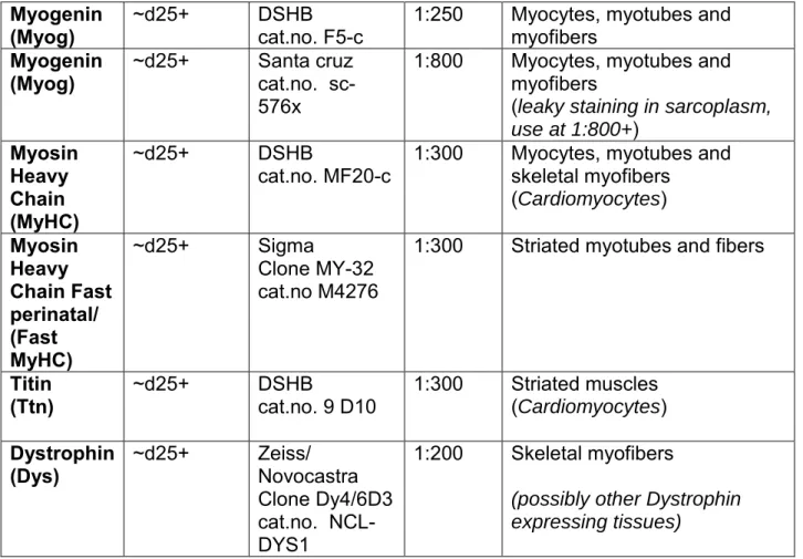
Marker Temporal
Expression Antibody Reference Working dilution Differentiation specificity Stage
(and other tissues marked)
T/
Brachyury ~ d2 R&D cat. no. AF2085 1:250 Early mesoderm (notochord) Tbx6 ~ d4 Abcam cat.no. ab38883 1:200 Early mesoderm, PSM Pax3 ~d5-15 DSHB
cat.no. Pax3-c 1:250 Anterior PSM, Dermomyotome (neural tube, neural crest)
MyoD ~d15+ Santa cruz
Clone 5.8A cat.no. sc-32738
1:200 Myoblasts, myotubes
Pax7 ~d25+ DSHB
Myogenin
(Myog) ~d25+ DSHB cat.no. F5-c 1:250 Myocytes, myotubes and myofibers
Myogenin
(Myog) ~d25+ Santa cruz cat.no. sc-576x
1:800 Myocytes, myotubes and myofibers
(leaky staining in sarcoplasm,
use at 1:800+) Myosin Heavy Chain (MyHC) ~d25+ DSHB
cat.no. MF20-c 1:300 Myocytes, myotubes and skeletal myofibers (Cardiomyocytes) Myosin Heavy Chain Fast perinatal/ (Fast MyHC) ~d25+ Sigma Clone MY-32 cat.no M4276
1:300 Striated myotubes and fibers
Titin
(Ttn) ~d25+ DSHB cat.no. 9 D10 1:300 Striated muscles (Cardiomyocytes)
Dystrophin
(Dys) ~d25+ Zeiss/ Novocastra Clone Dy4/6D3 cat.no. NCL-DYS1
1:200 Skeletal myofibers
(possibly other Dystrophin expressing tissues)
Table 2: List of markers used to characterize the differentiating hPSC-derived myogenic
cultures and corresponding antibodies for immunohistochemistry (Figures 2, 4, 5).
32| Wash cells 3 times with TBST for 5min.
33| Incubate cells in secondary antibodies conjugated with a fluorochrome and Hoechst
diluted in Blocking solution overnight at 4°C, on a shaker. CRITICAL STEP Protect
cells from light from this step onwards.
34| Wash cells 3 times with TBST for 5min. 35| Add 250µL/cm2 of DPBS to cells.
PAUSE POINT Store for up to 3 weeks for later analysis, light -protected at 4°C.
36| Image cells with a fluorescence microscope equipped with the appropriate
CRITICAL STEP Despite being fairly stable, staining analysis should be performed
within days to weeks to avoid signal fading and diffusion (Figures 2,4).
Culture expansion (Day 10+) ● TIMING 3-4 d
CRITICAL Culture can be split anytime between Day 12 to Day 100+ of culture.
Cultures 30 Days+ are a mix of myogenic progenitors (PAX7+, MYOD+ ), and post-mitotic myocytes, myotubes and fully differentiated myofibers (MYOG+, MyHC+, Titin+) as well as other cell types including fibroblasts, neuronal contaminants (Figure 1c, day40). For best results, starting differentiated hPSC culture can be prescreened for its
myogenic phenotype consisting in elongated bipolar cells organized in stream-like sheets of tissue (Figure 1c).
37| Passaging Differentiating cells and Myogenic progenitor culture expansion (Figure
3). Pre-treat cultures to be dissociated with 10µM Rock inhibitor in D-KHI medium for at
least 2 hours.
38 | Prepare multiwell plates or dishes coated with Matrigel for subculture.
39 | Rinse cell culture to be dissociated with DPBS without Calcium and Magnesium. 40| Add 250μl/cm2 of TrypLE Express and incubate at 37 °C, 5% CO2, for 5-7 min.
41|Fragment mechanically the dissociating culture using two 25 gauge needle as
scalpel, to promote further enzymatic dissociation.
42| Add an additional 250μl/cm2 of TrypLE Express and incubate again at 37 °C, 5%
CO2, for 5-7 min.
43|Collect the dissociated cells and transfer into a 50ml tube containing 20ml of DMEM
with 10% FBS.
44| Repeat step 40-43 until all the culture detached from the dish and composed of
dissociated cells and larger aggregates. Complete enzymatic incubation should not exceed 45 min.
CRITICAL STEP Dissociation needs to be thorough enough to result in single cell
suspension, but gentle enough not to reduce viability. Typically you should observe >80% of viability immediately after dissociation.
CRITICAL STEP If 2 rounds of TrypLE Express incubation and mechanical
fragmentation appears insufficient in dissociating the culture, TrypLE Express can be replaced by 0.25% Trypsin on the next round. Other enzyme mixes and concentrations have also been used successfully, including Dispase/Collagenase; Collagenase IV/ Trypsin-EDTA. Incubation time and trituration may have to be adjusted.
CAUTION Note that older cultures (50+ day old) cannot usually be fully dissociated and
45| After dissociation of the cultures, further triturate the suspension with a 10mL pipet
to break cell clumps.
TROUBLESHOOTING
46| Filter cell suspension on a 70µm cell strainer placed on a 50ml collection tube. 47| Centrifuge cells at 300g for 5 min.
48| Remove supernatant and resuspend the pellet gently in Skeletal muscle Growth
Medium (SkGM) medium supplemented with 10µM Rocki.
49| Count cells and use Trypan Blue or similar to assess cell viability
CRITICAL STEP Routinely, after dissociation with TrypLE/Trypsin, cell viability
decrease with the age of the dissociated cultures. From < 30 day-old culture, about 95% of cells are viable, while 85% from D30-D80 culture and 80% for D80-120 cultures.
50| Replate on Matrigel-coated wells at 60-70,000 cells/cm2 in SkGM + 10µM Rocki,
corresponding approximatively to a subculturing ratio is 1:4 -1:8 per surface area. (ie. ~2.5 x10⁵ cells in 1.5mL of medium for 1 well of a 12well plate). 1 p35 dish can typically
produce enough differentiated cells to cover half of a 12 -well plate.
CRITICAL STEP Cell density is critical and plating too few cells will prevent proliferation
of progenitor cells. Replated cells should reach confluency within 3 days in SkGM – media for optimal culture of progenitor cells.
51| Incubate the plate at 37°C with 5% CO2 overnight.
52| Observe cell attachment and recovery, change SkGM medium every second day,
until desired confluency is reached. Passage the culture which will now contain a high percentage of myogenic progenitors (Figure 3d-j, 4).
53| Expand the subculture (Figure 3). We recommend that stocks be cryopreserved at
the earliest passages, ideally at every passage (Box 1) (Figure 4e).
CRITICAL STEP Cells can be further subcultured, over 100 days for downstream
applications or kept in SkGM at high density without losing myogenic competency.
TROUBLESHOOTING
54| To further differentiate the cells to myofibers, change media to Horse serum (HS)
2% medium after 3-4 days in SkGM. As a serum-free alternative, culture can be differentiated in N2 media (Figure 3c, e, g-j.
CAUTION While both HS and N2 -based media promote myofibers formation. N2
media, being less rich, may not be able to support differentiated cultures over extended time period (>2 weeks) compared to HS-based medium.
Box 1 Culture cryopreservation ●TIMING 2 d
2. Resuspend the pellet in 1ml DMEM/F12. CRITICAL STEP Cultures older than 30
-days will have persisting cell clumps after dissociation that can be frozen together with single cells and when replated will give rise to myogenic cells. 3. Centrifuge cells at 300g for 5min, remove the supernatant and resuspend the
pellet in Cryostem freezing medium. Optimal freezing density is approximated by eye due to existence of small clumps after dissociation. CRITICAL STEP As a
guideline, one well of 6-well plate for a 30+ days culture can generate 5 x 1mL cryovials. Place cryovials in a freezing container at −80°C overnight.
4. Next day, transfer cryovials to liquid nitrogen storage tank for long term storage.
PAUSE POINT Frozen cryovials of cells can be kept indefinitely in liquid nitrogen END BOX
Box 2 Cryostock thaw ●TIMING 1 d
1. Transfer promptly frozen vials to a water bath set at 37°C until vials is partially thawed
2. Resuspend and transfer gently the vials content with 4ml DMEM/F12 in a 15mL collection tube.
3. Centrifuge cells at 300g for 5min, remove the supernatant and resuspend the pellet in SkGM. As a guideline for optimal seeding density, 1 vial as prepared in Box1 should be seeded one well of 24-well Matrigel coated plate. After 3 days, culture can be expanded again following steps 28-42
● TIMING
Steps 1–4, pluripotent cells preparation: 5-7 d Steps 5–19, predifferentiation setup: 2 d Steps 20–25, directed differentiation: 30-40d+
Steps 26–36, characterization of myogenic cultures: 2-4d Steps 37–54, culture expansion: 3-4d
Box1, culture cryopreservation: 2 d Box2, cryostock thaw: 1 d
● TROUBLESHOOTING
● Table 2 TROUBLESHOOTING TABLE
Steps Problem Possible reason(s) Solution(s) 19 Poor cell survival Single cell dissociation
was done by pipetting too forcefully or for too long.
Starting with healthy
undifferentiated hPSCs culture with optimal colonies size, before necrotic zone form at their centers, at ~70% confluence. Culture can be more resistant to cell death by treating them with the anti-apoptotic Rocki and seed dissociated cells in Rocki-containing mTeSR.
19 Poor cell
attachment Matrigel was not coated properly.
Starting hPSCs were grown on feeders.
Test Matrigel lot for routine hPSCs culture maintenance. Follow Matrigel coating
procedure properly ensuring it is manipulated at 4°C at all time.
Adapt hPSCs culture to
Matrigel +mTeSR maintenance conditions. It may take several passages, by manually
selecting undifferentiated colonies.
Increase the seeding cell number density.
20-23 Cell cultures
detach en masse Cells were plated at too high density. The differentiation protocol was started on too dense culture.
Optimize the seeding cell density and the recovery time before starting the
differentiation (variable cell line to cell line).
25 Poor myogenicity Cells do not respond to signals.
Signaling pathway modulators are not prepared correctly or are not active.
Start with healthy
undifferentiated hPSC culture. Compare side-by side
differentiation of several cell lines as differentiation efficiency varies from cell line to cell line. Prepare chemical compounds and recombinant proteins according to the procedure and reagent set-up to keep their activity.
Steps Problem Possible reason(s) Solution(s) 45 Poor cell dissociation
Cell culture is resistant to enzymatic dissociation
Culture is very dense and deposits of extracellular matrix block the activity of the enzyme.
Preincubate in DPBS for 10min, to
loosen up cell adhesion. Triturate the culture
mechanically with needles before and during enzymatic dissociation.
Extend the length and intensity of enzymatic incubation while pretreating the culture with Rocki
53 Poor myogenicity in
subcultures Proliferative contaminant cell populations invade the culture.
Dissociate more mature cultures that are less likely to contain proliferative contaminants.
● ANTICIPATED RESULTS
This protocol allows for the efficient production of expandable muscle progenitors from hPSCs that can be amplified, subcultured, cryopreserved, further differentiated into skeletal myofibers and characterized. The derivation of the muscle progenitors is achieved by a sequence of differentiation media with defined compositions that directs hPSCs to form paraxial mesoderm and further differentiate into myoblasts.
Primary differentiation
The differentiation reagent setup and the first week of differentiation are the most critical steps for successful myogenic differentiation. Following cell seeding, cells recover by reforming evenly distributed adherent small and irregular cell aggregates within 1-2 days in mTeSR + Rocki up to 15-20% confluence (Figure 1c). During the first week, the culture goes through several crisis phases associated with significant cell death, possibly due to the selective action of the sequence of differentiation media (Figure 1c). We usually observe that the addition of bFGF at day 3 leads to important cellular aggregates rearrangements in the culture. Surviving mesodermal progenitors with a mesenchymal phenotype proliferate and become confluent within a week (Figure1c). Differentiation into paraxial mesoderm can be monitored by the expression of Tbx6 around day 4-5 of differentiation, and Tbx6-positive population should account for about 80% of the total culture at day4 (Figure 2). Proper progression to an anterior PSM fate and dermomyotomal specification can be assessed by the activation of Pax3 expression starting around 1 week of differentiation until 3 weeks (Figure 2). Further myogenic commitment can be assessed by expression of the muscle regulatory factors in particular MyoD and Myogenin, as well as Pax7-positive progenitors. By 4 weeks of differentiation, both Myog+ and Pax7+ populations represent combined 40-50% of the total nuclei. Myotubes/myofibers will be also clearly visible by phase-contrast as early as 4 weeks (30 days+) and mature over additional 5 weeks+ of culture (Figure 3e, g-i). At this stage, fibers can be millimeter-long, exhibit striations, best evidenced by immunohistochemistry for sarcomeric proteins, such as Titin and Myosin heavy chain (Figure 2b). Maturity of the contractile apparatus is evidenced by spontaneous twitching. While the myogenic culture become very dense and tends to detach after 4 weeks of
culture, it still contains myogenic progenitors (Figure 1c). It is then advisable to proceed for analysis or to passage the culture for expansion (Figure 3). The expected Fast MyHC -positive fibers yield with this protocol is around 0.6-0.9 cell per hPSC cell seeded initially 1.
Subculture and secondary differentiation
The dissociation of the dense primary culture will eliminate dead cells, debris, ECM deposit, unwanted derivatives but also existing (postmitotic) myofibers (Figure 3d). About 80% of the single mononucleated cells isolated are viable. The replating in Skeletal Muscle Growth Media selectively promotes myogenic progenitors expansion. This leads to more homogeneous, less dense cultures of proliferating myogenic progenitors and eliminates lots of cell debris and unwanted derivatives that are byproduct of the differentiation (compare Figure 1c and 3d). With seeding at 60-70,000 cells/cm2 corresponding approximately to a subculture ratio of 1:4 to 1:8, culture
confluence is achieved again within few days forming a sheet of elongated bipolar myoblasts. We found that confluent subcultures can be maintained in SkGM media for an extended period of time without losing their myogenic potential when further passaged. Switch to K-HI, N2 media or alternatively Horse serum- based media will promote further myogenic differentiation and fusion of progenitors (Figure 3c). This can be achieved within a week, although muscle fibers continue to mature for 2 weeks, as evidenced by fiber size and myofibrills. Mature fibers from subcultures measure up to several millimeters in length with a width ranging from 5 to 20+μm (Figure 2b, 4, 5). As for the primary culture, myogenic differentiation can be monitored and cellular phenotype can be assessed by immunohistochemistry (Figure 4). After replating, up to 40% of total nuclei (from 3 independent cell lines) were Myog-positive and mature skeletal myofiber density was ranging from 9,000 to 20,000 fibers per cm2 (Figure 5c-d).
Acknowledgements
We thank all co-authors of the original article describing this technology for their initial support and contribution. We thank Getzabel Guevara for lab assistance. We thank Charlotte Fugier and Fanny Bousson for comments and feedbacks. We are grateful to Laurence Daheron and members of the Pourquié laboratory for comments. We thank Amélie Freismuth and Marion Humbert from the IGBMC cell culture service for hiPSC culture assistance. This work was supported by an advanced grant from the European Research Council to O.P., by the FP7 EU grant Plurimes (agreement no. 602423), by a strategic grant from the French Muscular Dystrophy Association (AFM) to O.P., and by the INGESTEM project (ANR).
Author contributions
J.C. and Z.A.T designed and performed experiments and protocol optimizations, analyzed data and coordinated the project. M.H and S.A carried out hPSCs differentiation and protocol optimization under the supervision of J.C. B.G. carried out some hPSCs optimization experiments under the supervision of Z.A.T. A.H. contributed to hiPS cell culture analysis and data interpretation. T.C. contributed to hiPSC culture differentiation and analysis. A.P.N. manufactured micropatterned substrates and performed the fiber structural analysis. K.K.P. provided expertise. O.P. supervised the overall project. J.C., Z.A.T and O.P. performed the final data analysis and wrote the manuscript.
Competing financial interests
The authors declare competing financial interests: details are available in the online version of the paper. The work described in this article is partially covered by patent application no. PCT/EP2012/066793 (publication no. WO2013030243 A1). O.P. and J.C. are co-founders and shareholders of Anagenesis Biotechnologies, a startup company specializing in production of muscle cells in vitro for cell therapy and drug screening.