HAL Id: hal-03192602
https://hal.sorbonne-universite.fr/hal-03192602
Submitted on 8 Apr 2021
HAL is a multi-disciplinary open access
archive for the deposit and dissemination of sci-entific research documents, whether they are pub-lished or not. The documents may come from teaching and research institutions in France or abroad, or from public or private research centers.
L’archive ouverte pluridisciplinaire HAL, est destinée au dépôt et à la diffusion de documents scientifiques de niveau recherche, publiés ou non, émanant des établissements d’enseignement et de recherche français ou étrangers, des laboratoires publics ou privés.
Coinfected With HIV and Chronic Hepatitis B Virus
From Clinical Outpatient Centers in France: Protocol
for an Ambispective, Longitudinal Cohort Study
Anders Boyd, Lorenza Dezanet, Raisha Kassime, Patrick Miailhes, Caroline
Lascoux-Combe, Julie Chas, Pierre-Marie Girard, Joël Gozlan, Fabien Zoulim,
Constance Delaugerre, et al.
To cite this version:
Anders Boyd, Lorenza Dezanet, Raisha Kassime, Patrick Miailhes, Caroline Lascoux-Combe, et al.. Subclinical and Clinical Outcomes in Patients Coinfected With HIV and Chronic Hepatitis B Virus From Clinical Outpatient Centers in France: Protocol for an Ambispective, Longitudinal Cohort Study. JMIR Research Protocols, JMIR publications, 2021, 10 (4), pp.e24731. �10.2196/24731�. �hal-03192602�
Original Paper
Subclinical and Clinical Outcomes in Patients Coinfected With
HIV and Chronic Hepatitis B Virus From Clinical Outpatient
Centers in France: Protocol for an Ambispective, Longitudinal
Cohort Study
Anders Boyd1, MPH, PhD; Lorenza N C Dezanet1, MD, PhD; Raisha Kassime1, MSc; Patrick Miailhes2, MD; Caroline Lascoux-Combe3, MD; Julie Chas4, MD; Pierre-Marie Girard1,5, MD, PhD; Joël Gozlan5,6, MD, PhD; Fabien Zoulim7, MD, PhD; Constance Delaugerre8, MD, PhD; Hayette Rougier9, MSc; Karine Lacombe1,5, MD, PhD
1
Sorbonne Université, INSERM, Institut Pierre Louis d'Épidémiologie et de Santé Publique, Paris, France
2Service de Maladies Infectieuses et Tropicales, Hôpital de la Croix-Rousse, Hospices Civils de Lyon, Lyon, France 3
Service de Maladies Infectieuses, Hôpital Saint-Louis, APHP, Paris, France 4Service de Maladies Infectieuses, Hôpital Tenon, APHP, Paris, France 5
Service de Maladies Infectieuses, Hôpital Saint-Antoine, APHP, Paris, France 6Centre de Recherche Saint-Antoine, Paris, France
7
Centre de Recherche sur le Cancer de Lyon, Unité 1052, INSERM, UMR 5286, CNRS, Lyon, France
8Laboratoire de Virologie, Hôpital Saint-Louis, APHP; Université de Paris, INSERM U944, Institut de Recherche Saint-Louis, Paris, France 9
Institut de Médecine et d’Epidémiologie Appliquée (IMEA), Paris, France
Corresponding Author:
Anders Boyd, MPH, PhD Sorbonne Université, INSERM
Institut Pierre Louis d'Épidémiologie et de Santé Publique Hôpital Saint-Antoine, SMIT
184 Rue du Faubourg St. Antoine Paris, 75012
France
Phone: 33 1 49 28 21 49
Email: [email protected]
Abstract
Background: Previous large-scale studies have examined the effect of chronic hepatitis B virus (HBV) infection on overall and
cause-specific mortality in individuals with HIV. However, few studies have collected data on the subclinical indicators of HBV that lead to these severe outcomes in the coinfected population.
Objective: In this study, we aim to describe the procedures of a cohort study extension aimed at assessing HBV-DNA replication,
serological markers of HBV (hepatitis B e antigen [HBeAg] and hepatitis B surface antigen), and liver fibrosis and how these subclinical outcomes relate to mortality in predominately tenofovir-treated, coinfected patients with HIV-HBV. We assessed the characteristics at cohort inclusion of those who participated in the cohort extension, as well as those who did not participate due to being lost to follow-up or death.
Methods: Patients with HIV and chronic HBV who completed follow-up in a prospective cohort study conducted in 4 outpatient
centers (Paris and Lyon, France; 2002-2011) were invited to participate in a cross-sectional visit from November 2016 to March 2018, during which a comprehensive evaluation of HIV- and HBV-related disease was undertaken. Virological and clinical data since the previous study visit were retrospectively collected.
Results: Of the 308 individuals enrolled in the cohort, 147 (47.7%) participated in the cross-sectional study. At this visit, most
participants were HBeAg negative (111/134, 82.8% with available data), had undetectable HBV DNA (124/132, 93.9% with available data), and were undergoing antiretroviral therapy containing tenofovir disoproxil fumarate or tenofovir alafenamide (114/147, 77.6%). There were no significant differences in characteristics at cohort inclusion between those who did and did not complete the cross-sectional visit, except for a lower proportion with an AIDS-defining illness (30/147, 20.5% vs 49/161, 30.4%,
respectively; P=.04). Of the 161 nonparticipating individuals, 42 (26.1%) died, 41 (25.4%) were lost to follow-up and known to be alive, and 78 (48.4%) were lost to follow-up with unknown vital status. Most differences in characteristics at cohort inclusion were observed between deceased individuals and those participating in the cross-sectional visit or those lost to follow-up. With this extension, the median follow-up time of the overall cohort is presently 9.2 years (IQR 3.4-14.6).
Conclusions: Extended follow-up of the French HIV-HBV cohort will provide important long-term data on the subclinical
trajectory of HBV disease in the coinfected population. The biases due to the relatively high rate of those lost to follow-up need to be assessed in future studies of this cohort.
International Registered Report Identifier (IRRID): DERR1-10.2196/24731
(JMIR Res Protoc 2021;10(4):e24731) doi: 10.2196/24731
KEYWORDS
liver cirrhosis; hepatitis B; viral load; longitudinal studies; immunosuppression
Introduction
BackgroundWith the advent of potent antiretroviral therapy (ART), the incidence of death related to AIDS has substantially declined among individuals with HIV, leading to new causes of mortality [1]. Liver-related death has been topmost among these recent causes, due in part to chronic hepatitis B virus (HBV) coinfection, and it has been a major concern over the past decade [2].
Because high levels of plasma HBV replication are associated with liver cirrhosis, hepatocellular carcinoma, liver-related death, and overall mortality [3], the suppression of circulating virus is the primary goal of therapy. The potent anti-HBV agent tenofovir (TFV), which is able to extensively suppress HBV replication and be easily incorporated in most ART regimens, has been available since 2002 [4]. Nevertheless, there are conflicting data regarding whether the risk of severe liver-related morbidity (ie, hepatic decompensation, portal hypertension, end-stage liver disease, and hepatocellular carcinoma) and mortality are indeed reduced in coinfected versus mono-infected patients despite the use of this agent [5-10].
To characterize the risk of severe liver-related morbidity and mortality over time in the coinfected population, an adequate evaluation of HBV-DNA viral replication, HBV serological battery, and liver fibrosis is needed. These parameters are important subclinical indicators of chronic HBV disease. Unfortunately, many hospital-based cohorts, including individuals with HIV, do not have regularly collected HBV virological and serological data, either due to structural or cost issues [11-13]. Furthermore, most HIV-HBV cohort studies presently include either retrospective data or small patient sizes, wherein less potent anti-HBV agents, such as lamivudine, were administered [14-16]. Therefore, there is a need for cohort studies to collect more exhaustive data on subclinical indicators of HBV in the HIV-HBV coinfected population, especially during TFV-containing ART.
It was in this context that our research group initiated the French HIV-HBV cohort, which followed coinfected patients between 2002 and 2011 [17]. We recently invited participants of the cohort to return for a study visit in 2017-2018 with the purpose of (1) performing a cross-sectional, comprehensive evaluation
of viral hepatitis and liver fibrosis and (2) retrieving biological and clinical data since their previous study visit in the cohort. The primary research aim of the cohort is to assess HBV-DNA replication, serological markers of HBV (hepatitis B e antigen [HBeAg] and hepatitis B surface antigen [HBsAg]), and liver fibrosis and assess how all 3 of these outcomes relate to mortality in predominately TFV-treated, coinfected patients with HIV-HBV.
Objectives
In this paper, we present a protocol of this cohort. First, we aimed to expand on the procedures of the extension of this cohort study. The individual characteristics of the overall cohort were described and compared between those who did and did not participate in the cross-sectional visit mentioned earlier. Second, to provide more accurate information on mortality, we described the procedures by which we obtained the vital status of individuals who were lost to follow-up and assessed the number of cohort participants lost to follow-up. The characteristics of individuals who continued follow-up, who were lost to follow-up, and who were deceased were then compared. Finally, we discussed the importance of these data and how they will be used in future research.
Methods
Study Design and Visits
The French HIV-HBV cohort is a closed, prospective, longitudinal cohort study that included patients initially from 7 academic hospital centers located in Paris and Lyon, France, and is aimed at evaluating the determinants of liver-related morbidity and mortality in individuals coinfected with HIV-HBV. The inclusion criteria were HIV-positive serology confirmed by western blot, HBsAg-positive serology for at least 6 months, a Karnofsky score ≥70, aged 18 years or older, and provided signed written informed consent. Exclusion criteria were acute HBV infection (ie, HBsAg-positive for less than 6 months); any severe physical, clinical, or mental condition preventing participation (as determined by a Karnofsky score
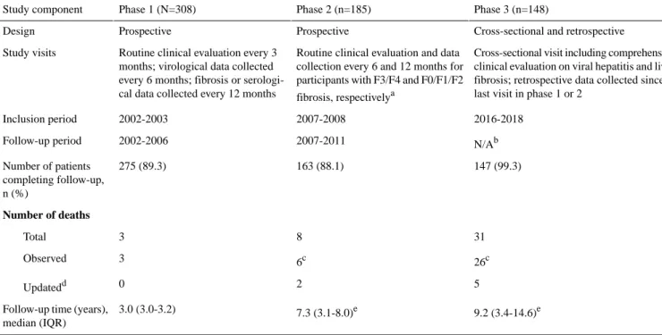
≤70 or from recommendations by the treating physician); not fulfilling all inclusion criteria; or refusal to participate. Patient recruitment and follow-up of the cohort occurred in 3 phases, with the first 2 phases previously described in detail [17]. Table 1shows the general characteristics of each study phase.
Table 1. General information at each phase of data collection.
Phase 3 (n=148) Phase 2 (n=185)
Phase 1 (N=308) Study component
Cross-sectional and retrospective Prospective
Prospective Design
Cross-sectional visit including comprehensive clinical evaluation on viral hepatitis and liver fibrosis; retrospective data collected since last visit in phase 1 or 2
Routine clinical evaluation and data collection every 6 and 12 months for participants with F3/F4 and F0/F1/F2 fibrosis, respectivelya
Routine clinical evaluation every 3 months; virological data collected every 6 months; fibrosis or serologi-cal data collected every 12 months Study visits 2016-2018 2007-2008 2002-2003 Inclusion period N/Ab 2007-2011 2002-2006 Follow-up period 147 (99.3) 163 (88.1) 275 (89.3) Number of patients completing follow-up, n (%) Number of deaths 31 8 3 Total 26c 6c 3 Observed 5 2 0 Updatedd 9.2 (3.4-14.6)e 7.3 (3.1-8.0)e 3.0 (3.0-3.2) Follow-up time (years),
median (IQR) a
In accordance with the European AIDS Clinical Society guidelines for the clinical management and treatment of chronic hepatitis B and C coinfection in adults with HIV [18].
bN/A: not applicable.
cIncludes deaths during time gaps in between phases.
dUsing linked data from a national death certificate registry (CépiDC).
eCumulated over previous phases—this statistic applies to all 308 patients regardless of the phase in which they discontinued participation.
In the first phase, 308 patients were recruited between May 2002 and May 2003 and prospectively followed every 3 months until the month-36 visit (2005-2006). In the second phase, 185 patients completing follow-up approximately 12 to 24 months after the first phase were recruited between March 2007 and March 2008. Patients with METAVIR F3/F4 fibrosis continued study visits every 6 months and patients with METAVIR F0/F1/F2 continued study visits every 12 months until the month-36 visit (2010-2011).
In the third phase, individuals completing follow-up in either the first or second phase were invited from November 2016 to March 2018 to participate in a cross-sectional study visit, which included a comprehensive evaluation of viral hepatitis and liver fibrosis. Approximately 4 mL of additional serum and plasma samples were collected and stored at −20 °C in a centralized location (Tumerothèque, Hôpital Saint-Antoine, Paris, France). Data were also retrospectively collected from the most recent study visit of the first or second phase until the cross-sectional visit or, in the case of death, until the most recent visit before death. The retrospective visits were selected from a 3-month period before or after the date of available HBV-DNA viral load (VL) measurement or, if missing, from a 3-month period before or after the date of the yearly visit.
All patients provided written informed consent at the beginning of each study phase. Retrospective data from deceased individuals were collected only if the patient had signed consent for the use of their clinical data during noninterventional research, consistent with the French Public Health Law. Protocols for each study phase were approved by the hospital
ethics committee (first phase: Hôpital Pitié-Salpêtrière; second phase: Hôpital Saint-Antoine; third phase: Hôpital Hôtel-Dieu) in Paris, France, in accordance with the Helsinki Declaration.
Primary Outcome Measures
A total of 4 primary outcomes were studied using data from this cohort extension.
Plasma HBV-DNA VL
This outcome was determined at inclusion and every 6 months in the first phase, every 6 to 12 months in the second phase, and in all retrospective and cross-sectional visits of the third phase. Commercially available polymerase chain reaction (PCR)–based assays were used to quantify HBV-DNA VL (COBAS AmpliPrep/COBAS TaqMan, detection limit: 12 international units [IU]/mL or COBAS Amplicor HBV Monitor, detection limit: 60 IU/mL; Roche Diagnostics).
We intend to study HBV DNA as continuous VLs, as undetectable HBV-DNA VLs, and as cumulative time-averaged HBV-DNA VLs [17].
HBeAg and HBsAg Seroclearance
This outcome was obtained from a complete HBV serological battery, which was performed at inclusion and every 12 months in the first phase, every 6 to 12 months in the second phase, and all retrospective and cross-sectional visits of the third phase. This consisted of qualitative HBsAg, HBeAg, anti–hepatitis B surface antibody (HBsAb), and anti-HBe antibody (HBeAb) detected using a commercial enzyme immunoassay (DiaSorin; Monolisa HBsAg, Bio-Rad; or Architect, Abbott Diagnostics).
We intend to study HBeAg seroclearance, defined as the transition from HBeAg-positive to HBeAg-negative status during follow-up in patients with HBeAg, and HBsAg seroclearance, defined as the transition from HBsAg-positive to HBsAg-negative status during follow-up in all included patients. We also aim to study HBeAg-seroconversion (ie, HBeAg seroclearance while acquiring anti-hepatitis B e antibody) in individuals with HBeAg and HBsAg-seroconversion (ie, HBsAg seroclearance while acquiring anti-HBsAb).
Liver Fibrosis
This outcome will be determined from several sources. First, several noninvasive biochemical scores predicting liver fibrosis levels were collected at inclusion and every 12 months in the first phase, every 6 to 12 months in the second phase, at the discretion of the treating physician in the third phase retrospective visits, and at the third phase cross-sectional visit. These scores included the FibroTest (Bio-Predictive), Fibrometre (Liver-Gastroenterology Department, CHU Angers), and Hepascore, all of which have been validated for use in the HIV-HBV coinfected population [19]. Second, measures of transient elastography (TE) were obtained at the physicians’ discretion in the first phase, every 6 to 12 months in the second phase, at the physicians’ discretion in the third phase retrospective visits, and at the third phase cross-sectional visit. TE was obtained using FibroScan (EchoSens) with either M or XL probes. Only TE measures fulfilling reliability criteria, as established by the manufacturer, were retained (ie, ≥10 valid measurements, IQR <30% of median stiffness, or ≥60% success rate) [20]. Shear wave elastography was also performed for some participants using the 2D real-time shear wave (Aixplorer, SuperSonic Imaging SA) with a 3.5 MHz convex ultrasound (SCX-6-1) and 7.5 MHz linear ultrasound (SL-10-2) probes. We intend to study liver fibrosis as a continuous measure (in which case the analysis will only focus on one noninvasive measure) or at validated thresholds of METAVIR F3-F4 liver fibrosis [19] (in which case noninvasive measures can be combined, eg, any score indicating F3-F4 fibrosis).
All-Cause Mortality
Deaths observed during follow-up in the first and second phases, along with the underlying cause of death, were reported by the treating physician. At the third phase cross-sectional visit, treating physicians were asked to ascertain the vital status (ie, alive, deceased, or unknown) of patients completing follow-up in the first or second phases. If death occurred, they were requested to provide further information on the date and underlying cause of death.
To obtain vital status for patients with unknown status (ie, lost to follow-up), a trusted third party (Inserm U1018) was requested to link data from the French HIV-HBV cohort to a national identification registry (Répertoire national d’identification des personnes physiques, fichiers n° 779 and
801). For deceased individuals, the cause of death was then obtained by a separate trusted third party (CépiDC), linking data from the French HIV-HBV cohort to a national registry of death certificates (certificat médical) and death notifications
(bulletin d’Etat civil de décès). Both registries are managed by the Institut National de la Statistique et des Etudes
Economiques.
We intend to use all-cause mortality as an outcome of the analyses. Given the expected number of deaths, we will likely be unable to study the specific causes of death; however, these will be described in detail.
Covariables
Laboratory Measures
Other laboratory measures, as listed below, were performed on blood samples taken during the study visits.
Antibodies to hepatitis C or D virus were detected using a commercial enzyme immunoassay at inclusion and every 12 months in the first and second phases, at the physician’s discretion during the third phase retrospective visits, and at the third phase cross-sectional visit. Serum hepatitis C virus (HCV) RNA and/or hepatitis D virus (HDV) RNA were quantified if the corresponding antibody test was positive (for the first and second phase and at the third phase cross-sectional visit) at the discretion of the treating physician (for the third phase retrospective visits). HCV RNA VLs were determined using either a PCR-based assay (COBAS Amplicor HCV Monitor v2.0, detection limit: 60 IU/mL; COBAS AmpliPrep/COBAS TaqMan HCV, detection limit: 10 IU/mL, Roche Diagnostic Systems; Abbott RealTime HCV, detection limit: 12 IU/mL, Abbott Molecular Inc) or branched-DNA technique (VERSANT HCV 3.0, detection limit: 615 IU/mL, Bayer Diagnostics). HDV-RNA VLs were quantified using a real-time quantitative PCR assay (sensitivity threshold: 1000 copies/mL [21] or 500 copies/mL [22]).
HIV-related markers of replication and immunostatus were collected at each study visit for all study phases. HIV-1 VLs were measured using either a branched-DNA technique (b-DNA Quantiplex 3.0, detection limit: 50 copies/mL, Bayer Diagnostics) or real-time PCR technique (COBAS AmpliPrep/COBAS TaqMan HIV-1 Test, detection limit: 40 copies/mL, Roche Molecular Systems). CD4+ T cell counts were quantified using standard measurements, while nadir CD4+ cell counts were obtained from patient records before inclusion in the first phase.
A complete hepatic battery was performed at each study visit for all phases and included quantification of the following using standard methods: alanine aminotransferase, aspartate aminotransferase, gamma-glutamyl transferase, alkaline phosphatase, total and conjugated bilirubin, alpha-fetoprotein, ferritin, albumin, haptoglobin, α2-macroglobulin, prothrombin time, and activated partial thromboplastin time. Hyaluronic acid was measured using an enzyme-linked protein-binding assay (Hyaluronic Acid Test Kit, Corgenix).
A complete cardiovascular battery was performed at each study visit for all phases and included the following markers quantified using standard methods: apolipoprotein A1, total cholesterol, high-density lipid cholesterol, low-density lipid cholesterol, and triglycerides.
Clinical Measures
Height, weight, and systolic and diastolic blood pressure were collected at each study visit for all phases as part of routine care. Alcohol consumption was assessed at inclusion and every 12 months in the first and second phases and at the third phase cross-sectional visit. Patients were asked whether they drank alcohol and, if so, how many glasses per day, week, or month were consumed on average over the past year.
Abdominal echography was performed at the physician’s discretion during all phases. The following information was extracted from patient files: presence of ascites; presence, number, and size of nodules; presence of portal thrombosis; presence of hepatomegaly; presence of steatosis; and evidence of dysmorphic liver.
Treatment
Data on antiretroviral and anti-HBV treatments, including the specific agent, start and stop dates, doses, and reason for discontinuation, were obtained during all phases. Data on all concomitant treatment, which included the same information as antiviral treatment, were also obtained during the first and second phase; however, data on only a specific set of concomitant treatments (given by Anatomical Therapeutic Chemical [ATC] Classification codes in Multimedia Appendix 1) were obtained during the third phase retrospective visits and at the third phase cross-sectional visit. All treatments were classified using the ATC codes [23]. Treatment information was retrieved from patient files and verified by the treating physician.
Clinical Events
Data on clinical events, including date of presentation, date of resolution (if applicable), and necessary intervention were obtained during all phases. Data on all clinical events were collected during the first and second phase; however, data on only a specific set of clinical events (given by International Classification of Diseases [ICD]-10 codes in Multimedia Appendix 2) were collected during the third phase retrospective visits and at the third phase cross-sectional visit. All clinical events were classified using the ICD-10 codes [24]. Information on clinical events was retrieved from patient files and verified by the treating physician.
Data Management
Data were collected using paper clinical report forms, which were filled out by trained clinical research associates. Data for the third phase retrospective and third phase cross-sectional visits were manually entered in a centralized database maintained at INSERM UMRS_1136, Hôpital Saint-Antoine.
To ensure data validity, we used a 10% random sample of study participants and double-entered their data. For any data field with >5% discordance between data entries, the entire data field was reverified, and discrepancies were resolved with the data manager.
Statistical Analysis
We described the characteristics of the study population at cohort inclusion using counts and proportions or medians and IQRs. We then compared characteristics at cohort inclusion between those participating versus not participating in the third phase cross-sectional visit using a Kruskal-Wallis test for continuous variables and a Pearson χ2test or Fisher exact test for categorical variables.
After combining vital status from study centers and external data sources, we categorized cohort participants as follows: (1) those completing the third phase cross-sectional visit, (2) discontinuation of follow-up due to death (as determined during follow-up or by linked data from CépiDC), (3) lost to follow-up with known alive vital status (as determined by clinical outpatient records obtained from the treating physician or by linked data from CépiDC), or (4) lost to follow-up with unknown vital status. We then compared participant characteristics at cohort inclusion between groups using the same statistical tests as described above.
Statistical analysis was performed using Stata software (version 15.0), and significance was determined using a P value <.05.
Results
Recruitment Flow of Participants in the Third Phase Cross-sectional Study Visit
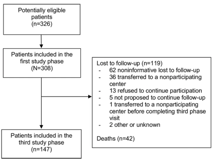
Of the 308 patients included in the first study phase, 142 (46.1%) were no longer followed at the participating center due to death (35/142, 24.6%), lost to follow-up (69/142, 48.5%), followed by a nonparticipating center (36/142, 25.3%), incarcerated (1/142, 0.7%), or unknown reasons (1/142, 0.7%). Of the 166 patients still in care at the participating center, 5 (3.5%) were not proposed to participate in the third phase cross-sectional visit and 13 (7.8%) refused participation. The remaining 148 patients provided informed consent to participate in the third phase cross-sectional study visit. One patient continued care in a nonparticipating center after signing consent and did not complete their visit, so 147 patients were considered to have completed the third phase cross-sectional visit. Patient flow from the beginning of the cohort inclusion until the third phase cross-sectional visit is summarized in Figure 1.
Figure 1. Patient flow. Patient numbers are given between the first and third phases of the French HIV–hepatitis B virus cohort. Reasons for study
discontinuation between these phases are also provided. Seven individuals initially considered as lost to follow-up were in fact deceased based on date linked to the CépiDS database.
Participants of the Third Phase Cross-sectional Study Visit and Their Characteristics
Of the 147 patients participating, most were male (119/147, 80.9%) with a median age of 55 years (IQR 49-59) at the time of the third phase cross-sectional visit. The median time since the first positive HIV and HBsAg tests was 24 years (IQR 18-28) and 22 years (IQR 17-26), respectively. Immunosuppression was for the most part mild with median CD4+ cell count at 548/mm3 (IQR 426-719), and HIV RNA was predominately undetectable (136/139, 97.8% with available data). Most patients
were HBeAg negative (111/134, 82.8% with available data) and many had undetectable HBV DNA (124/132, 93.9% with available data) with a median HBV-DNA VL of 2.53 log10
IU/mL (IQR 2.53-2.95) for those with detectable HBV DNA. Most patients (114/147, 77.6%) underwent either TFV or TFV alafenamide–containing ART.
When comparing patients who did and did not participate in the third phase cross-sectional visit (Table 2), the only difference in inclusion characteristics observed was a lower proportion with an AIDS-defining illness (30/147, 20.4% vs 49/161, 30.4%, respectively; P=.04).
Table 2. Characteristic of the study population at cohort inclusion, stratified by participation in the third phase cross-sectional study visit. P valuea Phase 3 Phase 1 (N=308) Characteristics Nonparticipating (n=161) Participatingb (n=147) .15 140 (87.0) 119 (81.0) 259 (84.1) Male, n (%) .93 39 (35-45) 40 (35-44) 40 (35-45)
Age (years), median (IQR)
.59 89 (58.2)
87 (61.2) 176 (59.7)
Alcohol consumption >1 glass/day (n=295), n (%)
.94 BMI (kg/m2; n=291), n (%) 8 (5.3) 8 (5.8) 16 (5.5) Underweight (16.5-18.5) 116 (76.3) 105 (75.5) 221 (75.9) Normal (18.5-25.0) 23 (15.1) 23 (16.6) 46 (15.8) Overweight (25-30) 5 (3.3) 3 (2.2) 8 (2.8) Moderate or severe obesity (>30)
.29 10.2 (3.6-14.6)
9.4 (3.8-13.1) 9.9 (3.6-14.0)
Estimated HIV infection duration (years), median (IQR)
.04 49 (30.4) 30 (20.4) 79 (25.6) AIDS-defining illness, n (%) .83 399 (264-554) 404 (283-557) 400 (269-555)
CD4+ T cell count (per mm3), median (IQR)
.81 215 (90-344)
212 (107-309) 212 (103-325)
Nadir CD4+ T cell count (per mm3; n=271), median (IQR)
.59 129 (80.6)
122 (83.0) 251 (81.8)
ARTcexperienced at inclusion, n (%)
.21 81 (50.6)
64 (43.5) 145 (47.2)
Detectable HIV RNA (>50 copies/mL), n (%)
.68 3.89 (2.87-4.50)
3.91 (2.48-4.41) 3.90 (2.59-4.44)
HIV-RNA viral load (log10copies/mL)d, median (IQR)
.16 39 (24.2)
47 (32.0) 86 (27.9)
From country of high endemicity, n (%)
.14 5.3 (2.2-10.4)
7.3 (2.8-11.2) 6.1 (2.2-10.8)
Estimated HBVeinfection duration (years), median (IQR)
.90 124 (77.5) 113 (76.9) 238 (77.2) HBV DNA >60 IU/mL, n (%) .12 4.68 (2.66-6.69) 3.39 (2.36-6.58) 4.26 (2.55-6.58)
HBV DNA viral load (log10IU/mL)d, median (IQR)
.44 87 (54.0) 73 (49.7) 160 (52.0) HBeAgfpositive, n (%) .18 HBV genotype ( n =170), n (%) 54 (56.8) 51 (68.0) 105 (61.8) A 1 (1.1) 0 (0.0) 1 (0.6) B 10 (10.5) 7 (9.3) 17 (10.0) D 11 (11.6) 8 (10.7) 19 (11.2) E 14 (14.7) 5 (6.7) 19 (11.2) G 5 (5.3) 4 (5.3) 9 (5.3) Mixed A/D or A/G
.31 29 (31.9)
18 (24.7) 47 (28.7)
Precore W28stop mutation (n=164), n (%)
.67 55 (67.9) 42 (64.6) 97 (66.4) Lamivudine-resistance mutations (n=146), n (%) .48
Other viral hepatitis, n (%)
10 (6.2) 9 (6.1)
19 (6.2) Anti-HCVgpositive serology
6 (3.7) 6 (4.1)
12 (3.9) Anti-HDVhpositive serology
9 (5.6) 3 (2.0)
12 (3.9) Anti-HCV and anti-HDV positive serology
aSignificance was determined using the Kruskal-Wallis test for continuous variables and Pearson χ² test or Fisher exact test for categorical variables. bParticipating patients were defined as those signing written informed consent and completing the third phase cross-sectional study visit. All characteristics reported in the table are from data collected at the inclusion visit of the French HIV-HBV cohort (2002-2003).
cART: antiretroviral therapy. d
Among patients with detectable viral loads. eHBV: hepatitis B virus.
fHBeAg: hepatitis B e virus antigen. gHCV: hepatitis C virus.
hHDV: hepatitis D virus.
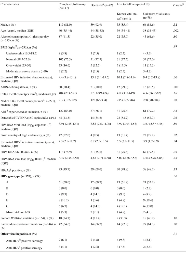
Lost to Follow-up and Deceased Participants of the Cohort Study and Their Characteristics
Of the 161 patients not followed up at the third phase cross-sectional visit, we were able to establish vital status for the 55 patients who were already known to have been deceased (n=35), incarcerated (n=1), not proposed or refused to participate (n=5 and 13, respectively), or did not complete the third phase study visit (n=1). The remaining 106 patients not followed at the third phase cross-sectional visit did not have known vital status because of an unknown reason for lost to follow-up (n=69), being followed at a nonparticipating center (n=36), or unknown reason for noninclusion (n=1). Of these, 28 patients were able to be linked to the CépiDC database: 21 were still known to be alive and 7 were identified as deceased from the death certificate or notification registry. These deaths occurred between 2007 and 2015.
From a total of 308 participants, 147 (47.7%) participants completed the third phase cross-sectional visit, 42 (13.6%) discontinued follow-up due to death, 41 (13.3%) were lost to follow-up with known alive vital status, and 78 (25.3%) were
lost to follow-up with unknown vital status. When comparing characteristics at the inclusion visit of the cohort, there were several significant differences across these patient groups (Table 3). Most differences were observed in deceased individuals who, respectively compared with all others, were significantly more likely to be older (median age 44 vs 40 years; P<.001), were not from a country of high HBV endemicity (4/42, 10% vs 82/266, 30.8%; P=.003), have a longer duration since first HIV-positive serology (median 13.1 vs 9.4 years; P<.001), have a higher proportion with AIDS-defining illness (21/42, 50% vs 58/266, 21.8%; P<.001), and have a lower nadir CD4+ cell count (median 128 vs 221 per mm3; P=.03). No significant differences in characteristics at cohort inclusion were observed between individuals completing the third phase cross-sectional visit versus lost to follow-up with known vital status. There were also no significant differences in characteristics at cohort inclusion between individuals completing the third phase cross-sectional visit versus lost to follow-up with unknown vital status, with the exception of longer duration since first HBsAg-positive serology (median 7.3 vs 3.9 years, respectively;
P=.009) and higher proportion with detectable HIV RNA (45/78,
Table 3. Characteristics of the study population at cohort inclusion, stratified by those completing the third phase cross-sectional visit, deceased, and
lost to follow-up with known or unknown vital status.
P valueb Lost to follow-up (n=119) Deceaseda(n=42) Completed follow-up (n=147) Characteristics
Unknown vital status (n=78)
Known vital sta-tusc(n=41) .32 66 (84.6) 35 (85.4) 39 (92.9) 119 (81.0) Male, n (%) .002 38 (34-43) 39 (34-41) 44 (38-53) 40 (35-44) Age (years), median (IQR)
.80 45 (61.6)
22 (55.0) 22 (55.0)
87 (61.3) Alcohol consumption >1 glass per day
(n=295), n (%) .99 BMI (kg/m2; n=291), n (%) 4 (5.6) 1 (2.5) 3 (7.5) 8 (5.8) Underweight (16.5-18.5) 54 (75.0) 31 (77.5) 31 (77.5) 105 (75.5) Normal (18.5-25.0) 11 (15.3) 7 (17.5) 5 (12.5) 23 (16.6) Overweight (25-30) 3 (4.2) 1 (2.5) 1 (2.5) 3 (2.2)
Moderate or severe obesity (>30) .06 9.4 (3.2-13.8) 10.1 (2.8-14.6) 13.1 (7.1-15.6) 9.4 (3.8-13.1) Estimated HIV infection duration (years),
median (IQR) .001 16 (20.5) 12 (29.3) 21 (50.0) 30 (20.4) AIDS-defining illness, n (%) .65 406 (268-562) 411 (338-619) 370 (249-474) 404 (283-557) CD4+ T cell count (per mm3), median (IQR)
.08 238 (70-384)
235 (172-346) 128 (65-304)
212 (107-309) Nadir CD4+ T cell count (per mm3; n=271),
median (IQR) .45 61 (79.2) 31 (75.6) 37 (88.1) 122 (83.0) ARTdexperienced at inclusion, n (%)
.05 45 (57.7)
22 (53.7) 14 (34.2)
64 (43.5) Detectable HIV RNA (>50 copies/mL), n (%)
.89 3.67 (2.87-4.46)
3.99 (3.04-4.53) 3.83 (2.59-4.05)
3.91 (2.48-4.41) HIV-RNA viral load (log10copies/mL)e,
median (IQR) .02 22 (28.2) 13 (31.7) 4 (9.5) 47 (32.0) From country of high endemicity, n (%)
.04 3.9 (1.7-8.9)
5.5 (2.8-11.5) 6.7 (2.3-13.5)
7.3 (2.8-11.2) Estimated HBVfinfection duration (years),
median (IQR) .95 62 (79.5) 31 (75.6) 31 (75.6) 113 (76.9) HBV DNA >60 IU/mL, n (%) .45 4.54 (2.76-6.88) 5.02 (2.28-6.58) 4.63 (2.71-6.88) 3.39 (2.36-6.58) HBV-DNA viral load (log10IU/mL)e, median
(IQR) .13 38 (48.7) 20 (48.8) 29 (69.0) 73 (49.7) HBeAggpositive, n (%) .36 HBV genotype (n=170), n (%) 24 (52.2) 13 (61.9) 17 (60.7) 51 (68.0) A 1 (2.2) 0 (0.0) 0 (0.0) 0 (0.0) B 4 (8.7) 2 (9.5) 4 (14.3) 7 (9.3) D 9 (19.6) 1 (4.8) 1 (3.6) 8 (10.7) E 6 (13.0) 4 (19.1) 4 (14.3) 5 (6.7) G 2 (4.3) 1 (4.8) 2 (7.1) 4 (5.3)
Mixed A/D or A/G .10 18 (40.9) 7 (33.3) 4 (15.4) 18 (24.7) Precore W28stop mutation (n=164), n (%)
.80 27 (64.3) 14 (77.8) 14 (66.7) 42 (64.6) Lamivudine-resistance mutations (n=146), n (%) .31
Other viral hepatitis, n (%)
4 (5.1) 4 (9.8)
2 (4.8) 9 (6.1)
Anti-HCVhpositive serology
2 (2.6) 3 (7.3)
1 (2.4) 6 (4.1)
P valueb Lost to follow-up (n=119) Deceaseda(n=42) Completed follow-up (n=147) Characteristics
Unknown vital status (n=78)
Known vital sta-tusc(n=41)
3 (3.9) 1 (2.4)
5 (11.9) 3 (2.0)
Anti-HCV and anti-HDV positive serol-ogy
aIncludes deaths observed during the study and from vital records.
bOverall significance between groups determined using the Kruskal-Wallis test for continuous variables and Pearson χ² test or Fisher exact test for categorical variables.
cIncludes patients whose vital status is known: 13 refusing to continue participation, 5 not proposed to continue follow-up, 1 transferred to a nonparticipating center before completing the third phase visit, 2 other or unknown reasons, and 22 with vital status determined by CépiDC.
dART: antiretroviral therapy. e
Among patients with detectable viral loads. fHBV: hepatitis B virus.
g
HBeAg: hepatitis B e virus antigen. hHCV: hepatitis C virus.
i
HDV: hepatitis D virus.
Discussion
Principal FindingsA handful of prospective cohort studies have examined the effect of HIV-HBV coinfection on mortality outcomes [10], but most of these studies have incomplete data on HBV status, making it difficult to determine how HBV infection contributes to HBV-related disease. We present the design of a recent extension of a large observational cohort of patients coinfected with HIV-HBV in France to address the shortcomings of these previous cohorts.
Strengths and Weaknesses
One of the major strengths of our cohort is the consistent collection of HBV-DNA VLs, complete HBV serological battery, HBV genetic characterization (specifically regarding antiviral resistance mutations in the pol gene, vaccine escape mutations in the S gene, and W28* precore mutations [25]), and liver fibrosis measurements. To our knowledge, few cohorts have regularly collected these data and those that have this information are no longer in active follow-up [26,27]. The regular collection of markers of HBV replication and HBV serological data will help provide more precise estimates of when certain events occur (eg, time to undetectable HBV-DNA or HBeAg or HBsAg seroclearance) as well as their durability (eg, persistent HBV-DNA viremia or sustained HBeAg or HBsAg seroclearance) or variability (as was observed for noninvasive measures of liver fibrosis [28]). At the same time, we aim to associate these primary outcomes with all-cause mortality.
In addition, by extending our previous cohort, we collected data for up to 15 years of follow-up. There are certain facets of HIV-HBV coinfection that make data sets containing longer follow-up important. HBsAg seroclearance is a slow process [29] and is achieved in approximately 1% per year in treated coinfected patients [17]. TFV-containing ART regimens have shown effective antiviral potency [4]. However, long-term toxicity issues related to treatment and/or comorbidities among
clinical effectiveness. It is also unknown how HIV-HBV coinfection evolves in an aging population, especially with respect to severe non-AIDS and non-HBV-related clinical events. The longer follow-up provided in the French HIV-HBV cohort could help establish the frequency of these end points and their determinants, provided that there are a sufficient number of events.
Another advantage of our cohort is the availability of frozen samples dating back to the beginning of the cohort. As these samples have been stored at a centralized location, they have facilitated their use in several collaborations, some of which have involved the genetic variability of HBV [25,30], compared with patients undergoing intensification with pegylated-interferon [31], HDV-RNA replication in tri-infected patients [32], kinetics of HBsAg or HBeAg quantification [33], hepatitis B core-related antigen quantification [34], and covalently closed circular DNA [35] during TFV treatment and ongoing projects related to markers of replication during treatment. Nevertheless, it should be mentioned that stored samples are only available in the first and second phases and at the third phase cross-sectional visit. As a result, any future analysis using available samples will have a gap in follow-up time.
There are other limitations in the French HIV-HBV cohort. First, most patients had years of ART experience before inclusion in the first phase, were HBeAg positive, came from predominately Western Europe, and sexually acquired HBV. The vast majority of individuals with HIV currently diagnosed with an HBV coinfection in Europe come from regions of high HBV endemicity, while new HBV infections have declined dramatically in other Western European settings [36,37]. Therefore, our study population may not be fully representative of modern coinfected patients. Second, despite the large number of patients and extensive follow-up, we might not have enough end points to ensure sufficiently powered comparisons. Third, certain data were not consistently collected during follow-up (eg, TE measurements only began at the start of the second phase) or across the entire study population (eg, HBsAg levels
patients). Fourth, metabolic data (insulin, adiponectin, controlled attenuation parameter of the FibroScan, etc) were not collected at any point during follow-up, making it difficult to understand the increasing role of metabolic disorders in this patient population [38].
Assessment of Participation in the Third Phase Study Visit and Loss to Follow-up
Similar to other longitudinal studies, one major concern is loss to follow-up and the possible biases induced thereto. At the third phase of follow-up, occurring roughly 15 years after the start of the cohort, almost half of the patients did not continue follow-up. When compared with rates of loss to follow-up in HIV-positive (5.4% of patients lost to follow-up from 1999 to 2009 in the French Hospital Database on HIV study [39]) or HIV-HCV coinfected (10% of patients lost to follow-up from 2006 to 2010 in the Agence Nationale de Recherche sur le Sida et les Hépatites (ANRS) CO13 HEPAVIH study [40]) cohorts from France, loss to follow-up would seem to occur at a much higher rate in our cohort.
Individuals lost to follow-up could have a higher risk of more advanced disease (opportunistic infections, advanced liver fibrosis or cirrhosis, cancer, advanced cardiovascular diseases, etc), which might make them less willing to participate or continue follow-up. No particular population characteristic at cohort inclusion was significantly different between participating and nonparticipating patients across phases, except for a higher proportion of AIDS-defining illnesses in those not participating in the third phase cross-sectional visit. There could also be other characteristics during follow-up, either measured or unmeasured (eg, nonreported comorbidities) in our cohort that were associated with loss to follow-up. This might induce differential loss to follow-up bias, the effect of which depends on the
research question addressed. Therefore, the assessment of this bias is imperative in future studies from our cohort.
Previous studies linking data from the French Hospital Database on HIV study and other mortality outcome registries or cohorts demonstrated that 29.8% of patients with HIV who were lost to follow-up had in fact died [41]. In our cohort, this proportion was similar at 32% (7/22), and notwithstanding the fact that 61.9% (78/126) of individuals with lost to follow-up were unable to have their vital status obtained is comparable with the proportion deceased of those remaining in follow-up (35/182, 19.2% either died or participated in the third phase cross-sectional visit).
Conclusions
Long-term and more comprehensive cohorts of coinfected individuals with HIV-HBV are pivotal in understanding how liver-related and nonliver-related morbidity and mortality can develop in this patient population. We were able to extend our cohort data on HBV replication, HBV serology, and liver fibrosis to up to 15 years; however, given the fact that approximately half of the patients were unable to participate in the third phase cross-sectional study, either due to death or lost to follow-up, the biases associated with this high rate of loss to follow-up need to be considered in future studies from this cohort. Nevertheless, these data will be helpful in enhancing our knowledge of the clinical trajectory during coinfection. More specifically, the clinical implications of persistent viremia, lack of HBeAg or HBsAg seroclearance, and progressive development of liver fibrosis will be evaluated along with their association with all-cause mortality. The French HIV-HBV cohort may also contribute to future collaborations aimed at assessing rarer outcomes, such as hepatic decompensation, hepatocellular carcinoma, and cause-specific death.
Acknowledgments
The authors are grateful to the patients and clinical teams for their commitment to the French HIV-HBV cohort study. The authors acknowledge L Roguet for managing the logistics of the French HIV-HBV cohort; L Melila, R Kassime, and R Loisel for their help with data entry; G Pannetier and Prof F Carrat for their help in data management; and Prof P Cales for kindly providing Fibrometre scores. Finally, the authors would like to thank Prof J-F Flejou and E Roux of the Tumorothèque HUEP at Hôpital Saint-Antoine for storing samples.
This work was supported by a grant from the ANRS. Gilead Sciences, Inc, provided an unrestricted grant for the French HIV-HBV cohort in the first phase and was not involved in any part of the data collection, analysis, and writing of the paper. A doctoral fellowship was awarded to KL from the ANRS (2003-2006) and postdoctoral fellowships from the ANRS and SIDACTION were awarded to AB (2012-2014 and 2014-2017, respectively), and a postdoctoral fellowship from the ANRS was awarded to LD (2018-2020).
The funder of the study had no role in study design, data collection, data analysis, data interpretation, or writing of the report. The corresponding author had full access to all the data in the study and had final responsibility for the decision to submit for publication.
Authors' Contributions
AB, LD, and RK managed data for the French HIV-HBV cohort, performed statistical analysis, and drafted the paper; PM, CL-C, and JC helped conceptualize and design the French HIV-HBV cohort, participated in patient recruitment, and provided critical input to the paper; JG and CD validated virological and serological measures and provided critical input to the paper; FZ coordinated data collection on genetic variability and gave valuable comments on the paper; HR coordinated and participated in data collection and helped draft parts of the paper; P-MG and KL initiated the French HIV-HBV cohort study, coordinated data collection, and drafted parts of the paper. All authors approved the final version of the paper.
Conflicts of Interest
KL has served on advisory boards and received travel grants from Gilead, Abbvie, MSD, ViiVHealthcare, and Janssen. All other authors have conflicts to declare.
Multimedia Appendix 1
Classes of concomitant treatment collected during the third phase cross-sectional study visit. [DOCX File , 26 KB-Multimedia Appendix 1]
Multimedia Appendix 2
Types of clinical events collected during the third phase cross-sectional study visit. [DOCX File , 26 KB-Multimedia Appendix 2]
References
1. Data Collection on Adverse Events of Anti-HIV drugs (D:A:D) Study Group, Smith C, Sabin CA, Lundgren JD, Thiebaut R, Weber R, et al. Factors associated with specific causes of death amongst HIV-positive individuals in the D:A:D Study. AIDS 2010 Jun 19;24(10):1537-1548. [doi: 10.1097/QAD.0b013e32833a0918] [Medline: 20453631]
2. Smith CJ, Ryom L, Weber R, Morlat P, Pradier C, Reiss P, D:A:D Study Group. Trends in underlying causes of death in people with HIV from 1999 to 2011 (D:A:D): a multicohort collaboration. Lancet 2014 Jul 19;384(9939):241-248. [doi: 10.1016/S0140-6736(14)60604-8] [Medline: 25042234]
3. Chen CJ, Yang HI, Iloeje UH, REVEAL-HBV Study Group. Hepatitis B virus DNA levels and outcomes in chronic hepatitis B. Hepatology 2009 May 27;49(5 Suppl):72-84. [doi: 10.1002/hep.22884] [Medline: 19399801]
4. Boyd A, Gozlan J, Maylin S, Delaugerre C, Peytavin G, Girard PM, et al. Persistent viremia in human immunodeficiency virus/hepatitis B coinfected patients undergoing long-term tenofovir: virological and clinical implications. Hepatology 2014 Aug 20;60(2):497-507. [doi: 10.1002/hep.27182] [Medline: 24752996]
5. Falade-Nwulia O, Seaberg EC, Rinaldo CR, Badri S, Witt M, Thio CL. Comparative risk of liver-related mortality from chronic hepatitis B versus chronic hepatitis C virus infection. Clin Infect Dis 2012 Aug 20;55(4):507-513 [FREE Full text] [doi: 10.1093/cid/cis432] [Medline: 22523269]
6. Piroth L, Pol S, Miailhes P, Lacombe K, Lopes A, Fillion A, GERMIVIC Study Group. Therapeutic management and evolution of chronic hepatitis B: does HIV still have an impact? The EPIB 2012 study. Liver Int 2015 Aug
22;35(8):1950-1958. [doi: 10.1111/liv.12777] [Medline: 25559645]
7. Ryom L, Lundgren JD, De Wit S, Kovari H, Reiss P, Law M, D:A:D Study Group. Use of antiretroviral therapy and risk of end-stage liver disease and hepatocellular carcinoma in HIV-positive persons. AIDS 2016 Jul 17;30(11):1731-1743. [doi: 10.1097/QAD.0000000000001018] [Medline: 26752282]
8. Kouamé GM, Boyd A, Moh R, Badje A, Gabillard D, Ouattara E, French National Agency for Research on AIDSViral Hepatitis (ANRS) 12136 TempranoANRS 12240 VarBVA Study Groups. Higher mortality despite early antiretroviral therapy in Human Immunodeficiency Virus and Hepatitis B Virus (HBV)-coinfected patients with high HBV replication. Clin Infect Dis 2018 Jan 06;66(1):112-120. [doi: 10.1093/cid/cix747] [Medline: 29020361]
9. Thornton AC, Jose S, Bhagani S, Chadwick D, Dunn D, Gilson R, UK Collaborative HIV cohort (UK CHIC) steering committee. Hepatitis B, hepatitis C, and mortality among HIV-positive individuals. AIDS 2017 Nov 28;31(18):2525-2532 [FREE Full text] [doi: 10.1097/QAD.0000000000001646] [Medline: 28926400]
10. Singh KP, Crane M, Audsley J, Avihingsanon A, Sasadeusz J, Lewin SR. HIV-hepatitis B virus coinfection: epidemiology, pathogenesis, and treatment. AIDS 2017 Sep 24;31(15):2035-2052 [FREE Full text] [doi: 10.1097/QAD.0000000000001574] [Medline: 28692539]
11. Price H, Bansi L, Sabin CA, Bhagani S, Burroughs A, Chadwick D, UK Collaborative HIV Cohort Hepatitis Group‚ Steering Committee. Hepatitis B virus infection in HIV-positive individuals in the UK collaborative HIV cohort (UK CHIC) study. PLoS One 2012 Nov 7;7(11):e49314 [FREE Full text] [doi: 10.1371/journal.pone.0049314] [Medline: 23145150] 12. Boyd A, Lacombe K, Girard P. An improved understanding of severe liver morbidity in HIV-infected individuals. AIDS
2016 Jul 17;30(11):1843-1845. [doi: 10.1097/QAD.0000000000001170] [Medline: 27351928]
13. Soriano V, Mocroft A, Peters L, Rockstroh J, Antunes F, Kirkby N, EuroSIDA. Predictors of hepatitis B virus genotype and viraemia in HIV-infected patients with chronic hepatitis B in Europe. J Antimicrob Chemother 2010 Mar
05;65(3):548-555. [doi: 10.1093/jac/dkp479] [Medline: 20051475]
14. Kosi L, Reiberger T, Payer B, Grabmeier-Pfistershammer K, Strassl R, Rieger A, et al. Five-year on-treatment efficacy of lamivudine-, tenofovir- and tenofovir + emtricitabine-based HAART in HBV-HIV-coinfected patients. J Viral Hepat 2012 Nov;19(11):801-810. [doi: 10.1111/j.1365-2893.2012.01601.x] [Medline: 23043387]
15. Strassl R, Reiberger T, Honsig C, Payer BA, Mandorfer M, Grabmeier-Pfistershammer K, et al. Viral determinants predicting hepatitis B surface antigen (HBsAg) seroclearance in HIV-/HBV-coinfected patients. J Viral Hepat 2014 Jul 24;21(7):508-516. [doi: 10.1111/jvh.12175] [Medline: 24112778]
16. Martín-Carbonero L, Teixeira T, Poveda E, Plaza Z, Vispo E, González-Lahoz J, et al. Clinical and virological outcomes in HIV-infected patients with chronic hepatitis B on long-term nucleos(t)ide analogues. AIDS 2011 Jan 02;25(1):73-79. [doi: 10.1097/QAD.0b013e328340fde2] [Medline: 21076274]
17. Boyd A, Gozlan J, Miailhes P, Lascoux-Combe C, Cam MS, Rougier H, et al. Rates and determinants of hepatitis B 'e' antigen and hepatitis B surface antigen seroclearance during long-term follow-up of patients coinfected with HIV and hepatitis B virus. AIDS 2015 Sep 24;29(15):1963-1973. [doi: 10.1097/QAD.0000000000000795] [Medline: 26153669] 18. Rockstroh JK, Bhagani S, Benhamou Y, Bruno R, Mauss S, Peters L, EACS Executive Committee. European AIDS Clinical
Society (EACS) guidelines for the clinical management and treatment of chronic hepatitis B and C coinfection in HIV-infected adults. HIV Med 2008 Feb;9(2):82-88 [FREE Full text] [doi: 10.1111/j.1468-1293.2007.00535.x] [Medline: 18257771] 19. Bottero J, Lacombe K, Guéchot J, Serfaty L, Miailhes P, Bonnard P, et al. Performance of 11 biomarkers for liver fibrosis
assessment in HIV/HBV co-infected patients. J Hepatol 2009 Jun;50(6):1074-1083. [doi: 10.1016/j.jhep.2009.01.022] [Medline: 19398234]
20. Boursier J, Zarski JP, de Ledinghen V, Rousselet M, Sturm N, Lebail B, Multicentric Group from ANRS/HC/EP23 FIBROSTAR Studies. Determination of reliability criteria for liver stiffness evaluation by transient elastography. Hepatology 2013 Mar;57(3):1182-1191. [doi: 10.1002/hep.25993] [Medline: 22899556]
21. Gal FL, Gordien E, Affolabi D, Hanslik T, Alloui C, Deny P, et al. Quantification of hepatitis delta virus RNA in serum by consensus real-time PCR indicates different patterns of virological response to interferon therapy in chronically infected patients. J Clin Microbiol 2005 May 04;43(5):2363-2369. [doi: 10.1128/jcm.43.5.2363-2369.2005]
22. Scholtes C, Icard V, Amiri M, Chevallier-Queyron P, Trabaud MA, Ramière C, et al. Standardized One-Step Real-Time Reverse Transcription-PCR Assay for Universal Detection and Quantification of Hepatitis Delta Virus from Clinical Samples in the Presence of a Heterologous Internal-Control RNA. J Clin Microbiol 2012 Mar 14;50(6):2126-2128. [doi:
10.1128/jcm.06829-11]
23. ATC classification index with DDDs. WHO Collaborating Centre for Drug Statistics Methodology. Geneva: World Health Organization; 2020. URL: https://www.whocc.no/[accessed 2021-03-15]
24. World Health Organization. ICD-10 : international statistical classification of diseases and related health problems : tenth revision, 2nd ed. Geneva: World Health Organization; 2004. URL: https://apps.who.int/iris/handle/10665/42980[accessed 2021-03-15]
25. Lacombe K, Boyd A, Lavocat F, Pichoud C, Gozlan J, Miailhes P, et al. High incidence of treatment-induced and
vaccine-escape hepatitis B virus mutants among human immunodeficiency virus/hepatitis B-infected patients. Hepatology 2013 Sep 25;58(3):912-922. [doi: 10.1002/hep.26374] [Medline: 23468093]
26. Matthews GV, Seaberg EC, Avihingsanon A, Bowden S, Dore GJ, Lewin SR, et al. Patterns and causes of suboptimal response to tenofovir-based therapy in individuals coinfected with HIV and hepatitis B virus. Clin Infect Dis 2013 May;56(9):e87-e94 [FREE Full text] [doi: 10.1093/cid/cit002] [Medline: 23315316]
27. Matthews GV, Ali RJ, Avihingsanon A, Amin J, Hammond R, Bowden S, et al. Quantitative HBsAg and HBeAg predict hepatitis B seroconversion after initiation of HAART in HIV-HBV coinfected individuals. PLoS One 2013 Apr 9;8(4):e61297 [FREE Full text] [doi: 10.1371/journal.pone.0061297] [Medline: 23593455]
28. Boyd A, Bottero J, Miailhes P, Lascoux-Combe C, Rougier H, Girard P, et al. Liver fibrosis regression and progression during controlled hepatitis B virus infection among HIV-HBV patients treated with tenofovir disoproxil fumarate in France: a prospective cohort study. J Int AIDS Soc 2017 Jan 01;20(1):21426. [doi: 10.7448/ias.20.1.21426]
29. Chevaliez S, Hézode C, Bahrami S, Grare M, Pawlotsky J. Long-term hepatitis B surface antigen (HBsAg) kinetics during nucleoside/nucleotide analogue therapy: finite treatment duration unlikely. J Hepatol 2013 Apr;58(4):676-683. [doi: 10.1016/j.jhep.2012.11.039] [Medline: 23219442]
30. Boyd A, Lacombe K, Lavocat F, Miailhes P, Lascoux-Combe C, Girard P, et al. Low incidence of precore W28* mutant variants in treated hepatitis B virus and human immunodeficiency virus co-infected patients. Antiviral Res 2018 Jan;149:174-178. [doi: 10.1016/j.antiviral.2017.11.014] [Medline: 29169914]
31. Boyd A, Piroth L, Maylin S, Maynard-Muet M, Lebossé F, Bouix C, et al. Intensification with pegylated interferon during treatment with tenofovir in HIV-hepatitis B virus co-infected patients. J Viral Hepat 2016 Dec 03;23(12):1017-1026. [doi: 10.1111/jvh.12581] [Medline: 27486094]
32. Boyd A, Miailhes P, Brichler S, Scholtès C, Maylin S, Delaugerre C, et al. Effect of tenofovir with and without interferon on Hepatitis D Virus replication in HIV–Hepatitis B Virus–Hepatitis D Virus-infected patients. AIDS Res Hum Retroviruses 2013 Dec;29(12):1535-1540. [doi: 10.1089/aid.2013.0008]
33. Boyd A, Maylin S, Gozlan J, Delaugerre C, Simon F, Girard P, et al. Use of hepatitis B surface and "e" antigen quantification during extensive treatment with tenofovir in patients co-infected with HIV-HBV. Liver Int 2015 Mar 31;35(3):795-804. [doi: 10.1111/liv.12521] [Medline: 24606220]
34. Dezanet LNC, Maylin S, Gabassi A, Rougier H, Miailhes P, Lascoux-Combe C, et al. Kinetics of Hepatitis B core-related antigen and Anti-Hepatitis B Core Antibody and their association with serological response in Human Immunodeficiency Virus-Hepatitis B coinfection. J Infect Dis 2020 May 11;221(11):1826-1837. [doi: 10.1093/infdis/jiaa013] [Medline: 31960918]
35. Boyd A, Lacombe K, Lavocat F, Maylin S, Miailhes P, Lascoux-Combe C, et al. Decay of ccc-DNA marks persistence of intrahepatic viral DNA synthesis under tenofovir in HIV-HBV co-infected patients. J Hepatol 2016 Oct;65(4):683-691. [doi: 10.1016/j.jhep.2016.05.014] [Medline: 27210429]
36. Ireland G, Simmons R, Balogun K, Kirwan P, Sabin C, Ramsay M, et al. HIV coinfection among persons diagnosed with hepatitis B in England in 2008-2014. HIV Med 2019 Apr 28;20(4):255-263. [doi: 10.1111/hiv.12707] [Medline: 30693643] 37. van Welzen BJ, Smit C, Boyd A, Lieveld F, Mudrikova T, Reiss P, et al. Decreased all-cause and liver-related mortality
risk in HIV/Hepatitis B Virus coinfection coinciding with the introduction of tenofovir-containing combination antiretroviral therapy. Open Forum Infect Dis 2020 Jul;7(7):ofaa226 [FREE Full text] [doi: 10.1093/ofid/ofaa226] [Medline: 32665961] 38. Morse CG, McLaughlin M, Matthews L, Proschan M, Thomas F, Gharib AM, et al. Nonalcoholic steatohepatitis and hepatic fibrosis in HIV-1-monoinfected adults with elevated aminotransferase levels on antiretroviral therapy. Clin Infect Dis 2015 May 15;60(10):1569-1578 [FREE Full text] [doi: 10.1093/cid/civ101] [Medline: 25681381]
39. Mary-Krause M, Grabar S, Lièvre L, Abgrall S, Billaud E, Boué F, et al. Cohort profile: French hospital database on HIV (FHDH-ANRS CO4). Int J Epidemiol 2014 Oct 17;43(5):1425-1436. [doi: 10.1093/ije/dyu002] [Medline: 24550249] 40. Loko MA, Salmon D, Carrieri P, Winnock M, Mora M, Merchadou L, ANRS CO 13 HEPAVIH Study Group. The French
national prospective cohort of patients co-infected with HIV and HCV (ANRS CO13 HEPAVIH): early findings, 2006-2010. BMC Infect Dis 2010 Oct 22;10(1):303 [FREE Full text] [doi: 10.1186/1471-2334-10-303] [Medline: 20969743] 41. Lanoy E, Lewden C, Lièvre L, Tattevin P, Boileau J, Aouba A, Clinical Epidemiologic Group of the French Hospital
Database on HIV (ANRS CO4 FHDH), Groupe d'Etude Mortalité 2000. How does loss to follow-up influence cohort findings on HIV infection? A joint analysis of the French hospital database on HIV, Mortalité 2000 survey and death certificates. HIV Med 2009 Apr;10(4):236-245 [FREE Full text] [doi: 10.1111/j.1468-1293.2008.00678.x] [Medline: 19178591]
Abbreviations
ANRS: Agence Nationale de Recherche sur le Sida et les Hépatites ART: antiretroviral therapy
ATC: Anatomical Therapeutic Chemical HBeAg: hepatitis B e antigen
HBsAb: hepatitis B surface antibody HBsAg: hepatitis B surface antigen HBV: hepatitis B virus
HCV: hepatitis C virus HDV: hepatitis D virus
ICD: International Classification of Diseases IU: international unit
PCR: polymerase chain reaction TFV: tenofovir
TE: transient elastography VL: viral load
Edited by G Eysenbach; submitted 02.10.20; peer-reviewed by R Staerkle, K Fitzner; comments to author 31.12.20; revised version received 17.01.21; accepted 19.01.21; published 06.04.21
Please cite as:
Boyd A, Dezanet LNC, Kassime R, Miailhes P, Lascoux-Combe C, Chas J, Girard PM, Gozlan J, Zoulim F, Delaugerre C, Rougier H, Lacombe K
Subclinical and Clinical Outcomes in Patients Coinfected With HIV and Chronic Hepatitis B Virus From Clinical Outpatient Centers in France: Protocol for an Ambispective, Longitudinal Cohort Study
JMIR Res Protoc 2021;10(4):e24731
URL: https://www.researchprotocols.org/2021/4/e24731
doi: 10.2196/24731
PMID:
©Anders Boyd, Lorenza N C Dezanet, Raisha Kassime, Patrick Miailhes, Caroline Lascoux-Combe, Julie Chas, Pierre-Marie Girard, Joël Gozlan, Fabien Zoulim, Constance Delaugerre, Hayette Rougier, Karine Lacombe. Originally published in JMIR Research Protocols (http://www.researchprotocols.org), 06.04.2021. This is an open-access article distributed under the terms of the Creative Commons Attribution License (https://creativecommons.org/licenses/by/4.0/), which permits unrestricted use,
distribution, and reproduction in any medium, provided the original work, first published in JMIR Research Protocols, is properly cited. The complete bibliographic information, a link to the original publication on http://www.researchprotocols.org, as well as this copyright and license information must be included.