backbone-backbone interactions
between closely packed RNA double
helices
By
Fatou TAO
Submitted to:
The Faculty of Health Sciences in partial fulfillment of the requirements for the
Master degree of Bioinformatics
Department of Biochemistry University of Montreal
Avril, 2013
1
To my sisters, brothers and nephew
To my professors and colleagues
To all my friends
2
Acknowledgements
I definitely need to start by thanking God for being by my side throughout this journey. Thanks to Him who kept me on the right path, gave me faith, wisdom and patience, to make the good decisions, to stubbornly rise and pursue my dreams no matter the obstacles I met.
I am deeply indebted to my supervisor, Pr. Sergey Steinberg, for his unfailing support and guidance through the completion of this thesis. My thanks go to him for giving me the chance to experience both part of the Bioinformatics science in wet and dry lab. However, his greatest contribution to my academic education is his teaching of how to look at science beyond the evidence and how to pay attention to apparently meaningless details where the truth might be discovered. This richness did not only serve me as a research student, but also as person.
Thanks to Pr. François Major and Pr. Stephen Michnick for accepting to be the judges this thesis. I am grateful to you and appraise the time you will spend on my thesis. I must also thank the members of my laboratory. Each of them taught me in his own way. Thanks to Natalia Kotlova, Tetsu Ishii, Yury Butorin and Kostantine Bokov for sharing with me their immense knowledge, for their assistance and support.
I have a special thought for two ladies who lighted up those years I spent in this university. Elaine Meunier, I don’t have enough space to thank you up to everything you did for me. Marie Pageau, you brought me more support and recognition than I could expect. Both of you have been like an ice cream cone under the hot sun of July. Many thanks for your listening to my academic and personal issues.
Finally, I thank my family and my best friend Judicaël Zounmevo for supporting me through this journey. They have been my cheerleaders I could rely on for better and for worst. I deeply thank M. and Mrs TAO, my beloved parents, for their unerring overseas’ support, advice and for all those hours spent on videoconference to lift my spirits when I needed it the most. I dedicate this thesis to my mother and father, for sharing their tremendous experience with me, for caring about my present, my future and for their unfailing belief in me.
3
Abstract
Although backbone-backbone interactions play an important role in stabilization of the tertiary structure of large RNA molecules, the particular rules that govern the formation of these interactions remain basically unknown. One RNA structural element for which the backbone-backbone interactions are essential is the along-groove packing motif. This motif is found in numerous locations in the ribosome structure; it consists of two double helices arranged such that the backbone of one helix is packed in the minor groove of the other helix and vice versa. The contact area between the two helices is mostly formed by riboses and totally involves twelve nucleotides. Here we analyze the internal structure of the along-groove packing motif and the dependence of stability of the association of the helices on their nucleotide sequences. We show that the proper positioning of the riboses that allows them to form inter-helix contacts is achieved through the particular choice of the identities of the base pairs involved. For different base pairs participating in the inter-helix contacts the optimal identities can be Watson-Crick, GC/CG, or certain non-Watson-Crick base pairs. The proper choice of the base pairs provides for the stable inter-helix interaction. In some cases of the motif, the identities of certain base pairs do not correspond to the most stable structure, which may reflect the fact that these motifs should break and form during the ribosome function.
Keywords: RNA structure; ribosomal RNA; along-groove packing motif; ribosome
4
Résumé
Les interactions entre les squelettes sucre-phosphate de nucléotides jouent un rôle important dans la stabilisation des structures tertiaires de larges molécules d’ARN. Elles sont régies par des règles particulières qui gouverne leur formation mais qui jusque là demeure quasiment inconnues. Un élément structural d’ARN pour lequel les interactions sucre-phosphate sont importantes est le motif d’empaquetage de deux doubles hélices d’ARN le long du sillon mineur. Ce motif se trouve à divers endroits dans la structure du ribosome. Il consiste en deux doubles hélices interagissant de manière à ce que le squelette sucre-phosphate de l’une se niche dans le sillon mineur de l’autre et vice versa. La surface de contact entre les deux hélices est majoritairement formée par les riboses et implique au total douze nucléotides. La présente thèse a pour but d’analyser la structure interne de ce motif et sa dépendance de stabilité résultant de l’association optimale ou non des hélices, selon leurs séquences nucléotidiques. Il est démontré dans cette thèse qu’un positionnement approprié des riboses leur permet de former des contacts inter-hélices, par l’entremise d’un choix particulier de l’identité des pairs de bases impliquées. Pour différentes pairs de bases participant à ce contact inter-hélices, l’identité optimale peut être du type Watson-Crick, GC/CG, or certaines pairs de bases non Watson-Crick. Le choix adéquat de paires de bases fournit une interaction inter-hélice stable. Dans quelques cas du motif, l’identité de certaines paires de bases ne correspond pas à la structure la plus stable, ce qui pourrait refléter le fait que ces motifs devraient avoir une liberté de formation et de déformation lors du fonctionnement du ribosome.
Mots clés: Structure d’ARN; ARN ribosomique; motif d’empaquetage le long du sillon
5
Table of Contents
ABSTRACT ... 3 RÉSUMÉ ... 4 TABLE OF CONTENTS ... 5 LIST OF FIGURES ... 7 LIST OF TABLES ... 8 1. INTRODUCTION... 10 2. METHODS ... 13 2.1. Insight II Software ... 13 2.1.1. Overview ... 132.1.2. Biopolymers module for molecular modeling ... 15
2.1.3. Discover module ... 23
2.1.4. Minimization ... 24
2.1.5. Steepest descents method ... 25
2.1.6. Conjugate gradient method ... 25
2.1.7. Molecular Dynamic ... 26
2.1.8. Quantitative evaluation of AGPM stability ... 26
2.1.9. Analysis module... 27
3. RESULTS ... 29
3.1. Background: the general description of AGPM... 29
3.2. Nomenclature of different elements of AGPM ... 31
3.3. Collection of the set of AGPMs ... 32
3.4. Principles of helix packing within AGPM: triangle-over-triangle model ... 33
3.5. Molecular dynamics of specially modeled AGPM constructs ... 38
3.6. The inter-helix interactions in contact zones QR and PS ... 45
3.6.1. The central role of the -1-base pairs ... 45
6
3.7. The inter-helix interactions in contact zone QS ... 56
3.7.1. The 0-base pairs ... 56
3.7.2. The +1-base pairs ... 62
3.8. The asymmetric requirement for the GC/CG identity of base pairs [-1P; -1Q]: additional H-bond with the ribose of nucleotide 0S ... 68
3.9. The optimal secondary structure of AGPM ... 72
4. DISCUSSION AND CONCLUSION ... 74
7
List of Figures
Figure 1: Description of the along-groove packing motif (AGPM) in a schematic
representation. ... 12
Figure 2: Diagram of the main steps followed in InsightII software. ... 14
Figure 3: Nucleotide sequences of all known AGPMs identified within ribosomal RNA. ... 16
Figure 4: Schematic representation and stereo-view of the AGPM structure. ... 18
Figure 5: First attempt of modelisation of an AGPM bearing AG base pair at the +1-level of thw Watson-Crick helix. ... 21
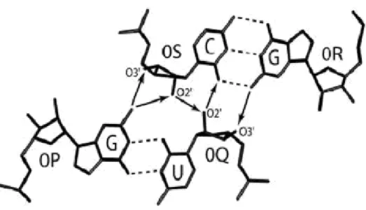
Figure 6: Hydrogen bonds network at the central part of the AGPM arrangement. ... 30
Figure 7: The arrangement of the three contact zones within AGPM. ... 35
Figure 8: Example of profiles from different MD simulations. ... 40
Figure 9: Stereo view of the superposition of the available AGPMs ... 47
Figure 10: Ribose-ribose interactions within contact zones PS and QR. ... 50
Figure 11: Stereo view of the interaction between the riboses of the external -2-nucleotide (-2e) and of the internal -1-nucleotide (-1i) for different -2-base pairs. ... 53
Figure 12: The potential collision of the internal nucleotides at the 0- and +1-levels and its consequences for the structure and position of base pair [0P; 0Q]. ... 58
Figure 13: The over-twist observed at the +1-level when both base pairs are either WC or WC-nonWC. ... 67
Figure 14: Stereo view of the asymmetry between base pairs [-1P;-1Q] and [-1R;-1S] caused by the displacement of nucleotide 0Q. ... 70
8
List of Tables
Table I : Structure of the contact zones ... 37 Table II: Summary of the molecular dynamics simulations at -1-, -2-, 0- and +1-level of modeled AGPM used in our analysis... 43 Table III: Occurrence of different combinations of the -1-base pairs in AGPMs existing in prokaryotic rRNA. ... 46 Table IV: Occurrence of different identities of the -2-base pairs in AGPMs existing in prokaryotic rRNA. ... 55 Table V: Occurrence of different combinations of the 0-base pairs in AGPMs existing in prokaryotic rRNA. ... 60 Table VI: Occurrence of different combinations of the +1-base pairs in AGPMs existing in prokaryotic rRNA. ... 64
9
List of Abbreviations and Symbols
Ǻ : Angstrom
AGPM : Along Groove Packing Motif
MD : Molecular Dynamic
ns : Nanoseconds
PDB : Protein Data Bank
ps : Picoseconds
R.M.S.D : Root Mean Square Deviation
RNA : Ribonucleic Acid
rRNA : Ribosomal RNA
tRNA : Transfer RNA
10
1. Introduction
An essential part of our knowledge on RNA structure is accumulated in the form of recurrent structural motifs, which appear in the same or different molecules and have identical or very similar conformation1-11, 28, 35, 36. The fact that such motifs can form in different structural contexts demonstrates a certain level of autonomy of their folding. Therefore, analysis of the aspects that govern formation of RNA recurrent motifs is important for understanding how larger RNA molecules fold and function. In most cases, analysis of the requirements for formation of RNA motifs has been focused on specific interactions that involve nitrogen bases, while the role of the sugar-phosphate backbone has been largely ignored. However, when the backbone participates in the formation of the core of the arrangement, the role of the interactions formed by the backbone can no longer be ignored.
A case of this kind represents the so-called along-groove packing motif (AGPM)12, which consists of two double helices closely packed via minor grooves in the way that a sugar-phosphate backbone of one helix interacts with the minor groove of the other helix and vice versa (Figure 1). AGPM has been found in more than a dozen places in ribosomal RNA (rRNA), which makes this motif an important element of the ribosome architecture. Two more cases of AGPM are involved in the association of the P- and E-site tRNAs with the 50S subunit. The recurrence of AGPM and the active involvement of the sugar-phosphate backbone in its formation make this motif an excellent model for studying the general role of the backbone in RNA structure formation. Although since the discovery of AGPM, several studies concerning different aspects of the AGPM formation have been reported12-15, 37, in none of them has the role of the backbone-backbone interactions been specifically analyzed.
In this paper, we undertake a systematic analysis of the aspects governing the interaction between the two double helices within AGPM. This analysis is based on the available X-ray conformations of the motif, on the collected data for more than sixty-five thousand available nucleotide sequences of AGPM, and on molecular dynamics (MD) simulations of specially modeled AGPM constructs. The analysis demonstrates a very
11 active role of backbone-backbone interactions in the shaping of the motif. We show that in different parts of the motif, the nucleotide identities are specifically tuned to provide for a stable collision-free interaction between the backbones of both helices. Because backbone-backbone interactions play essential role in the formation of different RNA complexes, including the ribosome, the rules that we discuss here are expected to be of general importance for RNA structure formation.
12
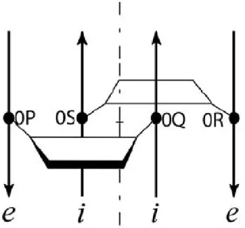
Figure 1: Description of the along-groove packing motif (AGPM) in a schematic representation.
Trapezoids represent the so-called central base pairs found the 0-level of AGPM and opened toward the minor grooves. The four nucleotides of this layer are named 0P, 0Q, 0R and 0S due to the strand of the helices to which they belong. Thus strands P and Q are respectively the external and internal of one helix while strands R and S are their homologs in the other helix. Arrows represent backbones directed 5’→3’. The internal and external strands of each helix are marked by italic letters i and e, respectively. The internal strand of each helix interacts with the minor groove of the other helix. Rotation of one helix for 180° around the axis of symmetry (dash-dotted line) leads to the superposition of both helices. (b): Juxtaposition of the central base pairs within AGPM. Arrows designate inter-helix hydrogen bonds directed from the donor to acceptor atom. The characteristic geometry of the GU base pair allows one helix to closely pack against the other helix in a collision free manner.
13
2. Methods
2.1. Insight II Software
2.1.1. Overview
Insight II is a graphic molecular modeling program used in conjunction with the molecular mechanics/dynamics program such as Discover or CHARMm to build and manipulate tridimensional virtual biomolecules, but also to study their molecular properties 42. For the purpose of our study, we worked with Silicon Graphics Fuel computer in a UNIX environment. Insight II provide us with an adequate environment to model in silico AGPMs, undergo minimization in order to define the optimal structure, meaning the one with the lowest energy and ultimately, to perform dynamics simulations and analysis structural rearrangements within each AGPM mutant providing for a stability inherent to a specific base pair replacement. Figure 2 resumes the main steps of the current work with Insight II.
14
Figure 2: Diagram of the main steps followed in InsightII software.
Each modeled construct has been submitted to specifics forcefield and potential before minimization and dynamics. The details of those steps are presented in the sections below.
AGPM molecular
15
2.1.2. Biopolymers module for molecular modeling
The Biopolymer module facilitates the building and modification of peptides, proteins, polynucleic acids and carbohydrates. In the case of the current study, it allows to perform modifications through base pairs replacements, from the initial AGPM molecule construct to its clones. The Biopolymer module has been useful to perform the AGPMs molecular modeling. The mutants AGPM models are based on the conformation of the motif L657 (Figure 3) in the crystal structure of E. coli ribosome (pdb entry code 2aw4) with few base pair modifications. For instance, the [0R; 0S] central base pair of L657 was replaced by GC, resulting in the GU-GC base pair juxtaposition at the 0-level, which is the most favorable and stable arrangement for the central base pairs within AGPM (Appendix 1). Moreover, AU base pair [+1P; +1Q] was replaced by GC, which resulted in the GC-GU base pair juxtaposition at the +1-level (Appendix 2). The latter modification increased the stability of the helix formed by strands P and Q, and at the same time, the presence of the GU base pair [+1R; +1S] provided for a relaxed ribose-ribose contact between the internal nucleotides of the Q and S strands (as explained in the sections above). The 1nucleotides remain unchanged such that [1P; 1Q] and [1R; -1S] are represented by a CG-CG base pairs combination; which respects the strong conservation of WC base pairs observed at the -1-level (see Figure 3, Appendix 4) while the -2-nucleotides of the motif L657, -2P and -2Q, as well as, -2R and -2S, do not form base pairs (Figure 3b, g). For the purpose of this analysis, we arranged the latter nucleotides such that they formed two WC base pairs [-2P; -2Q] and [-2R; -2S] (Appendix 3). Finally, we add additional WC-WC base pairs to form a -3-level.
From all those modifications of motif L657, the resulting AGPM construct we used as the initial model and as consensus structure is shown in Figure 4a.
16
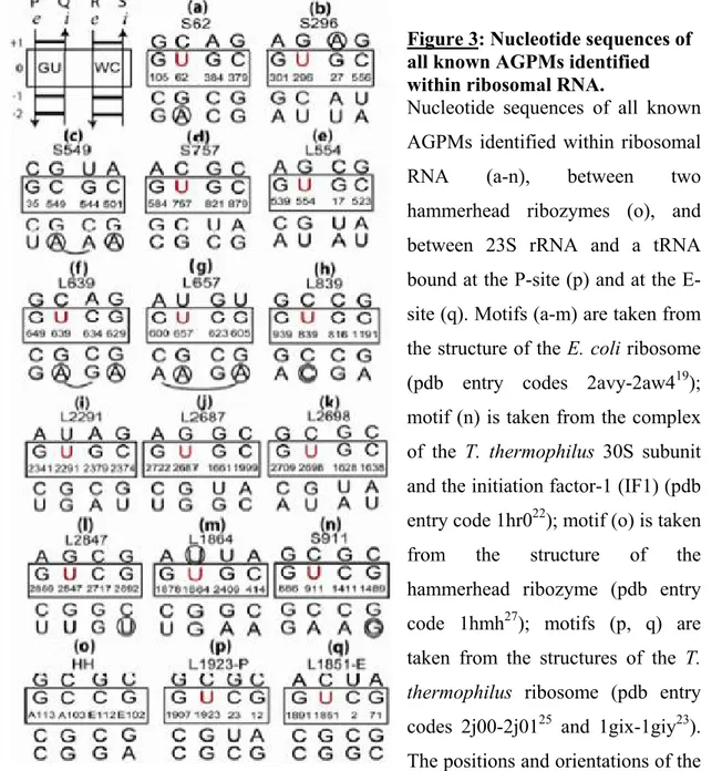
Figure 3: Nucleotide sequences of all known AGPMs identified within ribosomal RNA.
Nucleotide sequences of all known AGPMs identified within ribosomal RNA (a-n), between two hammerhead ribozymes (o), and between 23S rRNA and a tRNA bound at the P-site (p) and at the E-site (q). Motifs (a-m) are taken from the structure of the E. coli ribosome (pdb entry codes 2avy-2aw419); motif (n) is taken from the complex of the T. thermophilus 30S subunit and the initiation factor-1 (IF1) (pdb entry code 1hr022); motif (o) is taken from the structure of the hammerhead ribozyme (pdb entry code 1hmh27); motifs (p, q) are taken from the structures of the T.
thermophilus ribosome (pdb entry
codes 2j00-2j0125 and 1gix-1giy23).
The positions and orientations of the GU- and WC-containing helices correspond to those shown at the upper left corner. Central base pairs are boxed. U in position 0Q is red. The E. coli nucleotide numbering is used for all cases found within the ribosome. The name of each motif starts with letter ‘S’ or ‘L’, which reflects the small or large subunit in which it is found, followed by the number in the standard E. coli nomenclature of the nucleotide occupying position 0Q in 16S or 23S rRNA.
17 Each modeled construct has identical nucleotide sequences except for one base pairs located at the level of interest. Moreover, all the constructs are composed of 18 nucleotide-helices forming AGPM and cap on both ends of both helices by GAGA tetraloops, in order to provide for additional stability to the structure during the simulations (Figure 4a, c). In the following discussion, we explain how we limit the effect on this supplemental feature. The simulations were performed on thirty-one different constructs.
18
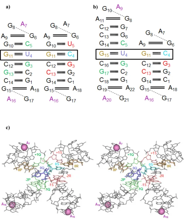
Figure 4: Schematic representation and stereo-view of the AGPM structure.
(a, b) shows the URS and the AGPM construct bearing a AG base pair at +1-level of the Watson-Crick helix respectively. The central base pairs are boxed. Each helix of the URS consists of 18 nucleotides and is capped by a GAGA tetraloop on both ends. For the AGPM in b), the helix containing GU at 0-level (GU-helix) bears two additional layers which increase the number of its nucleotides to twenty-two, while the opposite helix
a) b)
19 (WC-helix) has one missing layer. Thus, the latter carries 16 nucleotides. Within base pairs, each solid line represents a distance constraint corresponding to an H-bond
between two bases, while a dash line stands for a distance constraint corresponding to an H-bond between a base and a ribose. Details of the constraints used are given in the sections below. The nucleotides involved in the formation of the contact zones are green in the GU-helix and red in the WC-helix. Also, 0Q is dark blue, 0S is light blue, while 0P and 0R are orange. All other nucleotides not involved in inter-helix contacts are black. (c): Stereo-drawing of the tertiary structure of the modeled AGPM construct shown in panel a). The C1’ atoms of the pink nucleotides, located within the GAGA tetraloops, were fixed during the molecular dynamic simulations. Their fixation helps to avoid uncontrolled deterioration of the construct at the regions outside the inter-helix contact. Based on such fixation, the WC-helix had enough freedom to dissociate or not from the GU-helix when the requirements for the stability of the overall structure where optimal or absent. Consequently, it becomes possible to estimate the stability of the whole
20 From Figure 3, one can observe the predominance of AGPMs having +1AG in one the double helices and as previously mentioned, we capped both ends of the double helices forming AGPM with GAGA tetraloops. For instance, having a +1-AG base pair would have forced the superimposition of two AG base pairs; one at +1-level and the other one standing for the base pair of the tetraloop. Such arrangement is not observed in real structure because it is not structurally possible thus, it cannot did not fit our previous model (URS). Therefore, we modeled a construct which were structurally different from the URS. To overcome the problem of AGs superimposition and perform dynamic simulations on an AGPM harboring AG base pair at +1-level, several measures have been taken and are explained below. Ultimately, their lead to the AGPM shown in Figure 4b.
Measure 1: AG base pair of the tetraloop as +1-base pair
In our first attempt, we removed the +1-base pair in the helix where we wanted to insert the AG base pair. This modification brings the AG base pair of the GAGA tetraloop at the +1-level as shown in Figure 5.
21
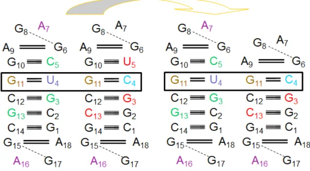
Figure 5: First attempt of modelisation of an AGPM bearing AG base pair at the +1-level of thw Watson-Crick helix.
The URS (left side) is reduced from its +1-level in the WC-helix which is replaced by the closing base pair of the tetraloop (right side).
22 We thus perform our +1-level analysis using this altered structure. However, we soon notice a parasite phenomenon that affects all performed simulations. Right after the beginning of the simulation the two unpaired nucleotides G and A, from the tetraloop of the shorter double helix (Figure 5), were interacting with the opposite tetraloop. Both A7 nucleotides (Figure 5) of opposite tetraloops come together and stack one on top of the other. This secondary interaction favors the interaction at the 0-level by bringing the two internal nucleotides close enough to interact without collision and prevent the interactions at -1- and -2-levels to occur by pulling upward the GU-helix. This phenomenon resulting of the solution 1 constitutes the problem 2. As our purpose is to study the interactions within the AGPM in response to base pair replacement, we needed to find a way out to prevent any interaction from tetraloop to influence the atom-atom contacts of our mutants. We thus introduce additional constraints between the two tetraloops of interest.
Measure 2: Introduction of distance constraints
We stated that the distance between the two A7 nucleotides should not be less than 15Å, which was the distance observe between both nucleotides before performing the molecular dynamic simulation. In case those nucleotides get close to each other at less than 15Å, we imposed a penalty of 100 to the energy of the overall arrangement. On top of the fact that this restriction didn’t solve the initial problem, we decided to lower the distance constraint from 15Å to 5Å with the same penalty on the energy. This new constraint was more realistic and more than enough to enable the molecule to ‘breathe’ during simulations and still preventing the secondary interaction described above to occur.
For this second attempt, we realise that during the simulation, as the AGPM construct breathes, if for whatever reason the two restrained A7 become close to each other for less than 5Å, the molecule ‘explode’ because of too high penalty.
A third try was therefore to associate an energetic penalty of 10 to the energy of the overall structure if the distance between the A7 was less than 5Å. Unfortunately, this ultimate attempt concerning the introduction of distance constraints did not solve the
23 problem of secondary interaction in the tetraloops that favor 0-level atom-atom contact within the AGPM.
Measure 3: Introduction of two additional layers on top of the +1-base pair which is not AG
To prevent the parasite interaction of the A7 nucleotides from the two tetraloop capping the positive side of the mutant AGPM containing a +1AG-base pair, we model a new AGPM having two additional layers on top of the +1-base pair which was not AG (Figure 4b).
At this point, the constraints imposed on this motif were the same than the ones on the initial constructs and they are discussed in the following argumentation. After each AGPM modification, the use of the Discover program helped us to create a proper environment for the AGPM constructs molecular dynamic simulations.
Another issue we had to deal with was the cyclisation of AGPM construct. Building a new constructs necessitates creating and/or breaking of covalent bonds for the introduction of base pair replacement. The biopolymer module enables to do so through its ‘Modify’ menu. Moreover, one can also create covalent bonds from the ‘Ligate’ option of the ‘Nucleic Acid’ menu. The latter was used for each AGPM construct modeling while only the last covalent bonds from each double helices were created, after all modifications and before setting of potentials, through the ‘Modify’ menu. Besides, the two double helices forming the APGM were unmerged through the ‘Unmerge’ option of the ‘Modify’ menu before base pairs manipulations, which allows working on two separated molecules. This strategy enables us to solve the problem of cyclic molecule which led us for a while, to false positive molecular dynamic results.
2.1.3. Discover module
Discover module provides an interface to the Discover program which allows performing minimization, template forcing, derivatives, means square displacements, vibrational frequencies 39. It also provides tools for performing simulations under various conditions including constant temperature, pressure and stress, periodic boundaries, fixed
24 and restrained atoms. In the case of the current study where we want to determine the local and global rearrangements on the surface of both interacting helices caused by specific base pairs replacement and how those structural reorganisations influence the ability of one helix to niche optimally in the minor groove the other and vice-versa, particular methods and strategies have been adopted. As we are dealing with dynamic calculations, our results are directly related to the forcefield we use, which is the calculation of the potential energy for a given configuration of atoms 39. The forcefield represents the single largest approximation in molecular modeling and its quality, applicability to the system at hand and its ability to predict the particular properties measured in the simulation directly determine the validity of the results 39. Among the available four families of forcefields supported by the Discover program – CVFF, CFF91, ESFF, AMBER, the latter has been used in our study.
2.1.4. Minimization
When an AGPM is built through a process of adding fragments which can generates serious atom clashes, it usually needs to be refined. Thus, the molecule is brought to a stable, sterically acceptable, conformation by the process of minimization. Minimisation is an iterative procedure in which the tridimensional structure is brought to a minimum through adjustments of atoms coordinates and where the obtained molecule with the lowest energy is considered to have the most stable arrangement and the closest resemblance with the real physical AGPM structure. The calculation of the energy follows a classical approach in which the molecule is considered as a set of charged point masses (the atoms), coupled together with springs and the total energy of the system is calculated through the summation of a number of individual energy terms. This calculation encompasses bond stretching, valence angle bending, torsion and nonbond interaction terms which associate an energetic penalty to the structure based upon deviations from an idealised equilibrium geometry. The nonbond interaction energy sums the van der waals attraction and repulsion as well as electrostatic forces for all atom pairs in the structure that are 1-4 nonbonded and above. Moreover, the value of bonds and angles cannot be based upon element type alone. As an example, a carbon-carbon single
25 bond is longer than a carbon-carbon double bond. For instance, before running energy calculation, the structure needs to have a potential type assigned. In the case of our study, all potentials are fixed.
2.1.5. Steepest descents method
In this low but robust method, each line search produces a new direction that is perpendicular to the previous gradient and the directions oscillate along the way to minimum. Even though the convergence is slow near the minimum because the gradient approaches zero, the steepest descent method is extremely robust. It helps generating a lower-energy structure regardless of the function. For this reason, the steepest descent method is used when the gradient is large and the conformations are far from the minimum, which was our case. We performed 300 iterations per steepest descent minimization. No constraints have been imposed on our mutant AGPMs during the simulations.
2.1.6. Conjugate gradient method
The second move in term of minimization was to use the conjugate gradient method. It allowed us to refine our search of the lowest-energy structure by refining step by step the direction toward the minimum. Moreover, to maintain the integrity of both helices forming AGPM, minor distances constraints were imposed on the lengths of the hydrogen bonds in all base pairs, except the central ones. These constraints were introduced as penalty K x (R - 3.3)2 added to the energy function when the distance R between the two electro-negative atoms involved in the formation of a corresponding hydrogen bond exceeded 3.3Å. Besides, all nucleotides of all GAGA tetraloops were fixed. Each simulation necessitates 2000 iterations, which enabled the structure to reach convergence.
26
2.1.7. Molecular Dynamic
Even though the minimization step is of an important use to reach the lowest energy structure, molecules still static and do not represent the reality where molecules are flexible structure subjects to thermal motion. This is the reason why, from the energetically stable structure obtained from previous steps, we used molecular dynamic to simulate the thermal motion of AGPM as a function of time, using the forces acting on the atoms to drive the motion. The forces acting on the atoms can be evaluated with Newton’s second law of motion which is F = m * a, where F is the force, m the masse of an atom and a is the acceleration. The acceleration and velocity are then used to calculate new positions for the atoms over a short time step, thus moving each atom to a new position in space. The process iterates, generating series of conformations of the structure which we call trajectory. The velocity of the atoms is directly related to the temperature at which the simulation is run. In our study, the simulations were run at 300K, which provides information on the structural fluctuations that occur around the starting conformation, and the pathways of conformational transitions. The C1’ atoms of A7 nucleotides from both GAGA tetraloops in the GU-helix have been fixed during all molecular dynamic simulation, as well as the one from the lower tetraloop of GC-helix. This restriction was necessary to maintain some stability and avoid parasite movements during the simulation. The fourth tetraloop remained unrestrained to give AGPMs some level of freedom and allow them to break or form according to the affinity of interaction of both double helices describing this motif. All mutants AGPM were simulated several times (3 - 10) in a probabilistic process where the number of repeat was determined by the tendency of stability for each simulation. For each repeat, we follow the thermal motion of our mutants throughout one nanosecond (1 000 000 iterations).
2.1.8. Quantitative evaluation of AGPM stability
As mentioned above, AGPM is a motif whose formation involves non-specific interactions of base pairs spreading over four levels. To study the structural rules governing the formation of this motif, we undertake a systematic analysis of inter-helix
27 interactions at each level, one at a time. This analysis consists in the replacements of base pairs followed by testing of the stability of the modified AGPM. The stability was tested through dynamic simulations of one nanosecond. For each simulation, we monitored the four inter-ribose interactions, which involved the areas of contact described in Figure 7. For backbone-backbone interactions at the +1- and 0-levels where only internal nucleotides are involved (QS zone), the atom-atom contact is made between both C4’ of +1Q and +1S and both O2’ of 0Q and 0S. For interactions spreading over -1- and -2-levels, the C4’-atom of the -1Q internal nucleotide contacts the -2R external nucleotide via its O2’atom. The similar situation occurs for the symmetric SP zone where 1S and -2P interact together via their C4’ and O2’ respectively (Figure 7, Table I).
Each simulation (monitoring 4 contacts at the same time) gave us the overall stability of the studied construct. After several simulations of the same construct, we calculate the average stability, which becomes a characteristic of the given construct. We discuss this stability in the following text. For the current study, we did not use molecular dynamic simulation to generate a space of different conformations AGPM was able to tolerate. We used it as a tool to understand the structural rules governing the AGPM formation through specific base pairs replacements at each level where inter-helix interactions occur. Based on the assumption that each base pair replacement affects the complementarities of shape between the two double helices and thus affects the stability of the modified AGPM, each dynamic provides for a quantitative evaluation of the stability corresponding to a particular base pair replacement. For each construct, 3 to 10 simulations have been made as a probabilistic process which told us how beneficial or detrimental a base pair replacement was for the AGPM structure. As each simulation is defined by four atom-atom contacts, we defined the stability of a specific construct as the sum of all four partial stabilities.
2.1.9. Analysis module
This module helps to trace the history of the simulated AGPMs motion during dynamics through the trajectory function. Data collected from the appropriated file enable to plot profiles of the stability of the AGPMs over one nanosecond. From those profiles
28 and the different frames witness of the internal rearrangement during dynamic simulations, one can approximate the period of time a modeled AGPM is stable for a quantitative evaluation of the stability, give more insight into the structural constraints guiding the formation of such modified AGPM and how a specific base pair replacement at one level can affect its overall stability.
29
3. Results
3.1. Background: the general description of AGPM
As mentioned above, AGPM consists of two double helices arranged such that a sugar-phosphate backbone of one helix is packed along the minor groove of the other helix and vice versa (Figure 1)12. In each helix, the strand that interacts with the minor groove of the opposite helix is positioned closely to the center of the arrangement and is thus called internal. The other strand in each helix stays at the periphery of the arrangement and is called external. The internal strands of both helices go in the same direction, opposite to that of the external strands. The conventional representation of the motif given in Figure 1 demonstrates the existence of symmetry between the two helices12. Indeed, within AGPM, the position of each helix can be roughly determined
based on the position of the other helix through the rotation for 180º around the common symmetry axis.
At the center of the contact area, there are two base pairs, called central, that pack with each other most closely. Despite the general symmetry of AGPM, the packing of the central base pairs is essentially asymmetric. In most identified cases, one of these base pairs is Watson-Crick (WC), while the other one is GU. In the GU base pair, G and U stay, respectively, in the external and internal strand12. The arrangement of these base pairs shown in Figure 6 provides for a close contact between the helices and allows the formation of a network of several inter-helix hydrogen bonds. Such GU-WC arrangement at the center of the contact area is found in most known cases of AGPM. Moreover, as pointed out by Mokdad et al.15, the GU base pairs involved in AGPM are among the most conserved GU base pairs in rRNA. Based on these findings, the coexistence of a WC and GU base pair in the middle of a helix-helix contact has been considered as a signature of AGPM that could facilitate the identification of new cases of the motif. In particular, this pattern enabled us to identify the two AGPMs formed by the P- and E-site tRNAs and 23S rRNA12.
30
Figure 6: Hydrogen bonds network at the central part of the AGPM arrangement.
This network is possible because of the presence of the GU base pair which allows the displacement of G and U toward the minor and major groove respectively.
31 Despite the almost universal presence of the GU-WC combination, some cases of AGPM do not follow this pattern, which indicates the existence of other aspects within the structure of the motif that are important for its integrity. The presence of such aspects can also be deduced from the fact that in AGPM, the area in each helix that interacts with the other helix is not limited to one base pair but instead, spreads over several consecutive base pairs. These additional contacts largely represent interactions between the backbones of both helices. Given that the interacting surfaces of the two helices are not flat, each next base pair participates in inter-helix contacts differently from its neighbors within the helix. The latter makes analysis of the aspects responsible for the integrity and stability of AGPM a rather complex exercise and has determined our choice of the approach to be used for this purpose. In this paper, we perform a systematic analysis of the inter-helix interactions in order to understand the role of each pixel of these interactions in the integrity and stability of AGPM. This analysis is based on the MD simulations of thirty-one specially modeled constructs of AGPM harboring a variety of identities for each base pair involved in inter-helix interactions. The results of the simulations are discussed in the context of the statistical data on the occurrence of particular nucleotides in different positions of AGPM. This analysis allowed us to rationalize the identity preferences for all base pairs involved in interaction with the opposite helix. These preferences can now be used for evaluation of stability of particular AGPMs and for further identification of AGPM in yet unresolved RNA structures.
3.2. Nomenclature of different elements of AGPM
To facilitate the discussion of the inter-helix interactions within AGPM, we will use the following nomenclature. For the four strands of these helices, capital letters P, Q, R and S are assigned as shown in Figure 3 (upper left corner). One helix is formed by strands P and Q, while the other one is formed by strands R and S. Strands P and R are external, while strands Q and S are internal. The helix containing GU as the central base pair in the Escherichia coli rRNA is composed of strands P and Q and is called the GU-helix (Figure 3). The opposite GU-helix is called the WC-GU-helix. For each base pair of each helix, a number is assigned, so that the central base pairs carry number zero, and the
32 positive propagation of the numbering corresponds to the 5’→3’ direction of the internal chains. The two base pairs of the opposite helices carrying the same number form a layer. In the identity of a base pair, the first and last letter will correspond to the external and internal nucleotide position.
3.3. Collection of the set of AGPMs
The nucleotide sequences of all identified motifs are shown in Figure 3. The original set of 12 motifs12 presented in Figure 3(a-l) exists in all ribosome structures. In addition, Mokdad et al.15 showed that motif L1864 (Figure 3m) exists in the bacterial 50S subunits of Deinococcus radiodurans (pdb entry codes 1kpj-1lnr18) and E. coli (pdb entry codes 2aw4-2awb19) but not in the archaeal 50S subunit of Haloarcula marismortui (pdb entry codes 1jj2-1s7220), where it was replaced by an A-minor interaction21. For this work, we undertook an additional analysis of all ribosome-related crystal structures and found one more motif, S911 (Figure 3n), which is formed between helices h27 and h44 of the 16S rRNA as a result of a conformational rearrangement in the 30S subunit caused by its association with the initiation factor-1 (IF1) (pdb entry code 1hr022).
Two more motifs, named L1923-P and L1851-E, are formed between the D-stem of the P-site tRNA and helix 69 of the 23S rRNA (Figure 3p) as well as between the acceptor stem of the E-site tRNA and helix 68 of the 23S rRNA (Figure 3q)12. These motifs exist in two low-resolution structures of the Thermus thermophilus 70S ribosome (pdb entry codes 1gix-1giy23 and 2ow8-1vsa24, respectively). The high resolution ribosome structure from the same organism (pdb entry codes 2j00-2j01 and 2j02-2j0325) confirmed the presence of motif L1923-P, while the E-site tRNA in this structure was positioned differently. It is also observable in the X-ray structure of the 70S ribosome determined at 3.3 Å resolution of the same organism (pdb entry code 3uzn 38). The structures of the
yeast ribosome 39 and of both ribosomal subunits of Tetrahymena thermophila 40, 41 confirmed the presence of all AGPMs existing in the bacterial ribosomes except L1864 and S911. Finally, a systematic analysis of all RNA-containing structures in the PDB database26 revealed a case of AGPM, named HH, which was formed by two hammerhead ribozyme molecules within the same asymmetric unit of the crystal (Figure 3o) (pdb
33 entry code 1hmh27). So far, this case of AGPM has been the only one identified outside the ribosome.
The data on nucleotide sequences of AGPMs were collected from the set of available nucleotide sequences of prokaryotic rRNA, which included 12 107 bacterial and 590 archaeal sequences of 16S rRNA as well as 399 bacterial and 37 archaeal sequences of 23S rRNA16. For the tRNA sequence analysis, the database containing 819 bacterial and 220 archaeal tRNA sequences was used17.
3.4. Principles of helix packing within AGPM: triangle-over-triangle
model
Prior to this paper, several studies of AGPM were reported and focused exclusively on the role of the central base pairs and their variations on the stability of the motif12-15, 37. Based on the analysis of all cases of AGPM in the available ribosome structures, it was already known that two cases did not follow the standard GU-WC pattern. In particular, motifs S549 in all available 30S subunit structures19,25,29,30 and L2291 in the 50S subunit of H. marismortui20 have two GC base pairs at the 0-level12,15. A similar situation also occurs in motif HH, which forms between two hammerhead ribozyme molecules27 and where two WC base pairs pack together at the 0-level (Figure 3o). Moreover, further in vivo studies of other cases of AGPM showed that the central base pairs can display an array of nucleotide combinations, while still being able to provide for functional ribosomes14, 37. These observations downplayed the importance of the central base pairs for the integrity of AGPM and prompted us to consider the role of other inter-helix contacts in the formation of AGPM. Regardless of how effective the interaction between the central base pairs is, it can occur only once per motif. Indeed, because of the spiral character of both helices, the juxtaposition of the base pairs at each level is different. Only at the 0-level, the arrangement of the base pairs is such as it is shown in Figure 6, while even at the neighboring +1 and -1 layers, it is so different that it can no longer be described as a packing of a backbone of one helix in a groove of the other helix. It does not mean, however, that outside the 0-level the two helices do not interact. On the contrary, analysis of the available AGPM conformations shows that the
34 inter-helix contacts spread over four layers in each helix between -2 and +1. While at the 0-level, these contacts include three out of four bases (bases of nucleotides 0P, 0R and 0S, see Figure 6), at the other levels contacts are mainly formed by elements of the backbones. Most of the backbone contacts are formed by riboses and are thus mainly hydrophobic.
Analysis of the AGPM structure shows that the whole contact area outside the 0-base pairs can be divided in three zones, depending on the particular strands involved in the inter-helix contacts. The first zone, named QS, corresponds to the interaction between chains Q and S. The Q- and S-moieties of this zone are mainly formed by the riboses of nucleotides +1Q and +1S, but also include some atoms of the neighboring nucleotides 0Q and 0S (Figure 7 and Table I. The second zone QR is formed by chains Q and R. It mainly consists of the contact between riboses -1Q and -2R, but also includes some atoms of 0Q and -1R. The third zone PS is symmetrical to zone QR with strands P and S being equivalent to R and Q, respectively. A complete list of the atoms participating in the formation of the three contact zones is given in Table I. On the surface of each helix, these zones form a triangle with the vertices positioned at the riboses of the internal +1- and -1-nucleotides as well as of the external -2-nucleotide (Figure 7b). The interaction of the two helices can thus be seen as superposition of the triangle in one helix on the equivalent triangle in the other helix (shown by orange arrows in Figure 7b). Analysis of the known AGPMs shows that the three contact zones are preserved in all cases regardless of the presence of other features and are thus considered important for the integrity of the motif. The formation of the contacts within the three contact zones depends on the particular positions of the riboses involved and is thus expected to be sensitive to the structures of the corresponding base pairs. In the following sections, we will show how the system of backbone-backbone contacts shapes AGPM and how it restricts the identities of the essential base pairs.
35
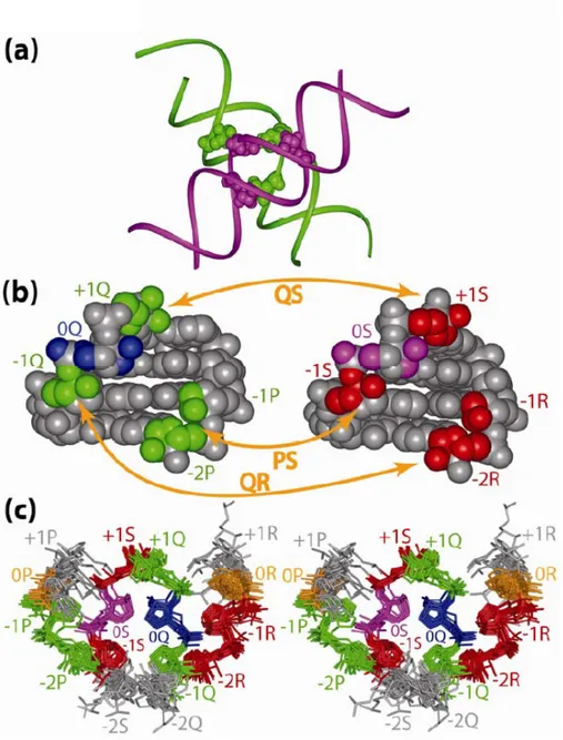
Figure 7: The arrangement of the three contact zones within AGPM.
(a): Schematic representation of the three contact zones when both helices interact together (b): Location of the atoms forming the three contact zones in the GU-helix (left) and in the WC-helix (right). The contact zones in the GU-helix are green; they which encompass nucleotides +1Q, -1Q, -1P and -2P. The symmetrical contact zones in the WC-helix are red; they encompass nucleotides +1S, -1S, -1R and -2R. Also, 0Q is blue; 0S is magenta. The external +1-nucleotides, +1P and +1Q as well as internal -2-nucleotides, -2Q and -2S are grey. Grey nucleotides are not involved in inter-helix contacts, which can explain the freedom allowed to the positions of their nucleotides
36 (Figure 7c). Orange two-headed arrows indicate the nucleotides forming the three contact zones when both helices interact together. The complete list of the atoms involved in the contact zones is given in Table I.
(c): Stereo view of all known AGPM structures (Figure 3a-p) superposed based on the positions of the C4΄ atoms (r.m.s.d. = 0.87 Å). The high resolution structure of the T.
thermophilus ribosome (pdb entry codes 2j00-2j01 and 2j02-2j0324) allowed us to include motif L1923-P (Figure 3p) in this superposition, while the E-site tRNA was positioned differently. For clarity, the bases are not shown. For the atoms of the contact zones, the same colors are used as in panel (b). The 0P and 0R are colored in orange.
37
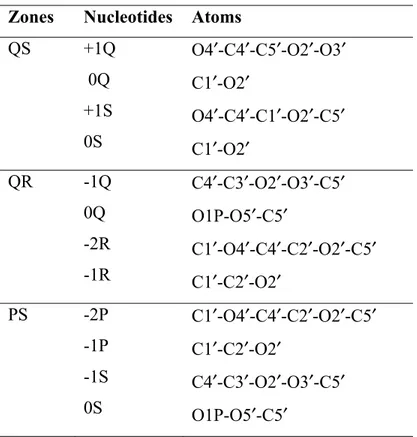
Table I : Structure of the contact zones
A non-hydrogen atom was considered a part of a contact zone if its distance from the closest non-hydrogen atom of the opposite helix was within 4.2 Å. See also Figure 7.
Zones Nucleotides Atoms
QS +1Q 0Q +1S 0S O4′-C4′-C5′-O2′-O3′ C1′-O2′ O4′-C4′-C1′-O2′-C5′ C1′-O2′ QR -1Q 0Q -2R -1R C4′-C3′-O2′-O3′-C5′ O1P-O5′-C5′ C1′-O4′-C4′-C2′-O2′-C5′ C1′-C2′-O2′ PS -2P -1P -1S 0S C1′-O4′-C4′-C2′-O2′-C5′ C1′-C2′-O2′ C4′-C3′-O2′-O3′-C5′ O1P-O5′-C5′
38
3.5. Molecular dynamics of specially modeled AGPM constructs
To check how different modifications of each element of the AGPM structure affect the integrity and stability of the whole arrangement, we made different in silico constructs and submitted them to MD simulations. Most constructs were made based on the design shown in Figure 4a, which is henceforth named the Universal Reference Structure (URS). It consisted of two closely packed double-helices. Each helix was composed of five base pairs corresponding to layers from -3 to +1 and was capped at both ends by tetraloops GAGA. For URS, the identities of the base pairs were chosen based on the nucleotide sequence of motif L657 in the E. coli ribosome (pdb entry code 2aw419). The presence of the two tetraloops made each helix a cyclic structure. The modeling procedure and the particular conditions of the MD simulations are described in the Methods. Figure 4c provides the stereo view of a construct based on the design shown in Figure 4a. For those constructs containing an A-G shared base pair at the +1 level, we used another design shown in Figure 4b.
The effect of base pair replacements on the stability of AGPM was assessed through monitoring the distances within four pairs of atoms. The integrity of the QS contact zone was followed by measuring the distance between the C4΄ atoms of nucleotides +1Q and +1S and between the O2΄ atoms of nucleotides 0Q and 0S (in the standard AGPM structure these O2΄ atoms are connected by a hydrogen bond). In the second zone QR, the contact between riboses of nucleotides -1Q and -2R was monitored following the distance between atoms C4΄ of -1Q and O2΄ of -2R. Finally, the integrity of the third zone PS was monitored following the distance between the symmetrically positioned atoms C4΄ and O2΄ of nucleotides -1S and -2P, respectively. At each moment of a simulation, each of the four contacts was considered as existing if the distance between the two assigned atoms did not exceed 4.0 Å (only for the contact between the O2΄ atoms of nucleotides 0Q and 0S) or 4.5 Å (for the other three contacts). During a particular MD simulation, each of the four measured contacts could follow one of three patterns of either being stable or undergoing reversible or irreversible breakages. Figure 8 an example of such profiles in
39 different constructs and when monitoring different distances within the AGPM contact zones.
40
Figure 8: Example of profiles from different MD simulations.
(a, b) represent the profiles of a base pair replacement at the -1-level leading to -1-GC-GU complex. The profile showed in a) measures the period of time the contact between the O2’ atoms of both nucleotides 0Q and 0S last throughout the 1ns the simulation lasts.
a)
b)
41 This profile is considered as stable while the profile (b), measuring the period of time the contact between the C4’ atoms of both +1Q and +1S breaks irreversibly after 207 ps. The profile in c) shows that the contact between the C4’ atom of -1Q and O2’ atom of -2R breaks instantly after the beginning of the simulation. However, this breakage is reversible and the complex demonstrates stability between 168 ps and 840 ps. In this construct, the base pair GC-AU has been introduced at the -1-level.
42 The stability of each of the four contacts was evaluated as the fraction of the whole simulation time during which the assigned contact kept its integrity. The stability of the whole construct was calculated as the sum of the stabilities of all four contacts, which thus varied between 0 and 4 for each simulation. For each construct, the final stability was averaged over several simulations. Table II provides the list of all constructs for which MD simulations were performed, the final stability of each construct, as well as the details of all simulations. For URS the stability was 2.4, which was higher than for almost all other constructs. To make the analysis more systematic, the overall energy of most constructs, including URS, was calculated using a specifically modified formula. As discussed below, when base pair [-1P, -1Q] is GC or CG, the amino group of the guanosine forms an H-bond with atom O4’ of the 0S nucleotide. In most calculations, the energy of this hydrogen bond was excluded from the formula of the overall energy and was taken into account only when the role of this H-bond was specifically analyzed.
43
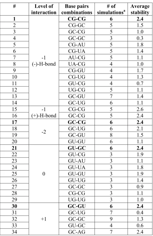
Table II: Summary of the molecular dynamics simulations at -1-, -2-, 0- and +1-level of modeled AGPM used in our analysis
# Level of interaction Base pairs combinations # of simulationsa Average stability 1 -1 (-)-H-bond CG-CG 6 2.4 2 CG-GC 5 1.5 3 GC-CG 5 1.0 4 GC-GC 3 0.3 5 CG-AU 5 1.8 6 CG-UA 5 1.4 7 AU-CG 5 1.1 8 UA-CG 4 1.0 9 CG-GU 4 1.7 10 CG-UG 4 1.3 11 GU-CG 4 0.7 12 UG-CG 5 1.1 13 GC-GU 7 1.4 14 GC-UG 6 1.1 15 -1 (+)-H-bond CG-CG 5 2.6 16 GC-CG 5 2.4 17 -2 GC-CG 6 2.4 18 GC-UG 6 2.1 19 GC-GU 8 1.5 20 GU-GU 6 1.1 21 0 GU-GC 6 2.4 22 GU-CG 3 1.9 23 GU-AU 3 1.1 24 GU-UA 3 1.8 25 GU-GU 3 1.9 26 GU-UG 3 1.4 27 GC-GC 3 0.9 28 CG-CG 3 1.1 29 UG-UG 3 1.0 30 +1 GC-GU 6 2.4 31 GC-UG 7 0.4 32 GC-GC 9 1.3 33 GU-GC 4 0.6 34 GC-AG 7 2.4
This table shows the results of MD simulations for all AGPM constructs studied in this paper. Each construct is characterized by the particular identity of the base pairs (the third column) located at the particular level (shown in the second column). The identities of all
44 other base pairs were as in URS, except construct 34, which was made based on the alternative design shown in Figure 4.
45
3.6. The inter-helix interactions in contact zones QR and PS
3.6.1. The central role of the -1-base pairs
As one can judge from Figure 7 and Table I, among all base pairs participating in the formation of AGPM, only in the -1-base pairs both nucleotides are involved in inter-helix backbone-backbone contacts. In particular, the internal nucleotide -1Q forms a major part of the Q-moiety of zone QR, while its external base pair partner -1P participates in the formation of the P-moiety of zone PS. The same is true for nucleotides -1S and -1R with respect to zones PS and QR. The necessity of the proper fitting for all four -1-nucleotides to the inter-helix contacts within zones QR and PS will impose strong restrictions on the structure of the -1-base pairs. As shown in Figure 3, in all presented examples of AGPM, both -1-base pairs are always WC. Moreover, analysis of the available nucleotide sequences of AGPM shows that in different organisms, the WC identities of both -1-base pairs are maintained in all motifs at the average level of 98% (Table III)16. The superposition (Figure 9) of the available AGPM conformations shows that in all of them the two -1-base pairs occupy the same positions, which are symmetrical with respect to the common symmetry axis. Given that a replacement of any of the two -1-base pairs by a non-WC dinucleotide combination will unavoidably affect both contact zones QR and PS, we can suggest that the maintenance of the WC identity of both -1-base pairs is important for the AGPM stability.
46
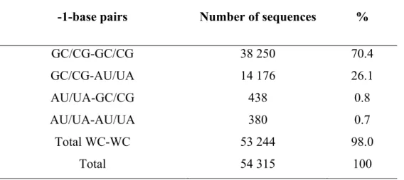
Table III: Occurrence of different combinations of the -1-base pairs in AGPMs existing in prokaryotic rRNA.
-1-base pairs Number of sequences %
GC/CG-GC/CG GC/CG-AU/UA AU/UA-GC/CG AU/UA-AU/UA Total WC-WC Total 38 250 14 176 438 380 53 244 54 315 70.4 26.1 0.8 0.7 98.0 100
For Table III-Table VI, the data were obtained from the available rRNA alignments16. In all these tables, “Total” refers to the total number of nucleotide sequences for which the identities of all nucleotides in question are known.
47
Figure 9: Stereo view of the superposition of the available AGPMs
a) The superposition excludes the motif (2o) from the structure of the hammerhead ribozyme (pdb entry code 1hmh27) and motifs (2p, q) from the structures of the T.
thermophilus ribosome (pdb entry codes 2j00-2j0125 and 1gix-1giy23). The -1-base pairs from each helix are presented in color. The superposition at levels -1 and 0 is optimal while at -2 and +1-levels it is notably poor. The set of four arrows in the middle represents the strands P, Q, R and S (shown in red) and the direction 5’ 3’. The trapeziums in the superposition represent the base pairs as shown in b). The base pairs are linked via their N1/N9 atoms (dotted line).
48 To test whether indeed both -1-base pairs must be WC, we made six constructs in which, starting from URS, we introduced the GU or UG base pair in either position [-1P;-1Q] or [-1R;-1S]. Compared to other non-WC base pairs, combinations GU and UG provided relatively minor deviations from the WC geometry and thus were expected to fit to these positions better than any other non-WC combination. As one can see in Table II, a replacement of any of the two -1-base pairs by a GU or UG combination resulted in a notable drop of the AGPM stability. Interestingly, the damaging effect of the presence of a GU/UG base pair at the -1-level was notably stronger when it was located in the GU-helix than in the WC-GU-helix. We attribute this effect to the steric intolerance between the two neighboring non-WC base pairs occupying positions [-1P; -1Q] and [0P; 0Q]. Below we will see that on top of being WC, -1-base pairs have additional asymmetric restrictions of their identities caused by the interaction with the 0-base pairs.
3.6.2. The -2-base pairs
Unlike the -1-base pairs, in which all internal and external nucleotides participate in the inter-helix contacts, at the -2-level only the external nucleotides -2P and -2R are involved in the interaction with the opposite helix (Figure 7). It is not surprising, therefore, that contrary to the -1-base pairs, the -2-base pairs harbor a variety of different structural forms. In particular, among the 17 cases of AGPM shown in Figure 3, there are 34 potential -2-dinucleotide combinations, which in the real structures correspond to 16 WC base pairs, 8 non-WC base pairs, while in 10 remaining cases the base pair does not exist at all.
The 16 WC -2-base pairs are distributed between 10 AGPMs: in four motifs S296, S757, L554 and L2698 both base pairs are WC, and in six more motifs only one base pair is WC, while the other base pair is either non-WC or inexistent. When both -2-base pairs are WC, the two -1- and two -2--2-base pairs form a four--2-base-pair arrangement seen in Figure 10a, b. All cases of this arrangement are superposable with r.m.s.d. 0.56 Å. Also, each arrangement is symmetrical with respect to the symmetry axis of the whole AGPM. At the center of contact zone QR, the fivemember rings of two riboses 1Q and -2R are closely packed with each other. In total, between the Q and R strands there are
49 about 15 atom-atom van der Waals contacts in which two non-hydrogen atoms are positioned within 4.2 Å of each other. The same interactions occur in the PS contact zone between nucleotides -2P and -1S.
50
Figure 10: Ribose-ribose interactions within contact zones PS and QR.
(a-b): Two different stereo views of the same superposition of motifs S296, S757, L554 and L2698, in which both 2base pairs are WC (r.m.s.d. = 0.56 Å). Nucleotides 2P and -1S form a major part of contact zone PS, while nucleotides -1Q and -2R form a major part of zone QR. Nucleotide -1P and -1Q are respectively in yellow and blue. Nucleotides -1R and -1S are respectively in pink and green.
51 The superposition of the motifs containing only one -2-base pair WC with the motifs having two such base pairs showed that in all cases, a WC -2-base pair stays at the same position with respect to the neighboring -1-base pair regardless of the structure or even of the existence of the -2-base pair in the opposite helix. This fact is taken as an indication that at the -1- and -2-levels the conformations of both helices are optimal and do not change upon the formation of the AGPM.
For the other helix, harboring at the -2-level a non-WC combination, there are two possibilities of having a non-WC base pair or having no base pair. In both cases, the external nucleotide (-2P or -2R) maintains the same position, which, however, is different from that within a WC base pair. Compared to the latter, this external nucleotide is over-twisted by 10˚-15˚, so that its WC edge becomes more open to the major groove (Figure 11). Such over-twist allows the formation of an additional hydrogen bond between the O2΄ atoms of the two nucleotides (-2P and -1S) or (-2R and -1Q). Although this hydrogen bond does not exist when the -2-base pair is WC, it is found in most cases when this base pair is either non-WC or non-existent.
52
a)
b)
53
Figure 11: Stereo view of the interaction between the riboses of the external -2-nucleotide (-2e) and of the internal -1--2-nucleotide (-1i) for different -2-base pairs.
For the -2-base pair, AU is blue (panel a), UG is pink (panel b), and GA is orange (panel c). The -1-internal and external nucleotides are presented in green. In all -2-base pairs, the ribose of the external nucleotide -2e is positioned closely enough to the ribose of the internal nucleotide -1i of the -1-base pair to form a ribose-ribose contact. Compared to the -2-base pair AU, in both base pairs UG and GA, the external nucleotide -2e is slightly over-twisted, which allows the formation of a hydrogen bond between the O2′ atoms of the two riboses (dashed line). The internal nucleotide -2i of the -2-base pair shows a strong variability in its position, which is not surprising given that this nucleotide is not involved in inter-helix interactions.
54 When the external -2-nucleotide (-2P or -2R) is a part of a non-WC base pair, the structure of the pair cooperates with the arrangement described above: in all such cases, the external and internal nucleotides are displaced towards the major and minor grooves, respectively (Figure 11). This displacement allows the over-twist of the external nucleotide and leads to the formation of the above-mentioned inter-helix hydrogen bond. The necessity for such displacement limits the set of the acceptable non-WC dinucleotide combinations to UG (3 times), GA (3), AA (1) and UU (1), all of which are able to support the opening of the base of the external -2-nucleotide towards the major groove. These dinucleotide combinations are also predominant non-WC ones observed at the -2 level in the available sequences of prokaryotic rRNA of AGPM (Table IV)16. In total, for those -2-dinucleotide combinations that correspond to a base pair in the ribosome tertiary structure, WC, GA, UG, AA and UU combinations amount to 99% of all cases (Table IV). More specifically, combination GA is almost 600 times more frequent than AG, while combination UG is almost six times more frequent than GU. Such assumption is corroborated with the known AGPMs presented in Figure 3.
55
Table IV: Occurrence of different identities of the -2-base pairs in AGPMs existing in prokaryotic rRNA.
-2-base pairs Number of
base pairs %
b
WC and closely related conformations a
WC UG GU UU GA AA AG 54 815 1669 293 286 42 19 14 82.1 2.5 0.4 0.4 0.06 0.03 0.02 Sheared conformations a GA AA UA UG AG 8 507 856 320 27 1 12.7 1.3 0.5 0.04 0.001 Sub-total Others c Total d 66 849 42 031 108 880 100
a: The base pair conformations were deduced from the available crystal structures of AGPM.
b: Percentages were calculated with respect to the total number of the -2-dinucleotide combinations that form a base pair.
c: This number relates to those -2-dinucleotide combinations in motifs S62, S549, S911, L639, L657, L839 and L2847 that do not form a base pair.
d:The total number of -2-base pairs is based on 54440 sequences for which the identities of all four nucleotides are known. See also the footnote to Table III.
56 To check the importance of the correct ribose-ribose interactions between the -1 and -2 base pairs for stability of the whole AGPM, we performed molecular dynamic simulation of several constructs having alternative nucleotide combinations at the -2-level. Starting from URS (Figure 4a, c), we replaced base pair [-2R;-2S] by either the UG or GU base pairs. The resulting constructs were submitted to several MD simulations. For the construct containing combination UG, the stability was 2.1, i.e. slightly lower than of URS. Most of the simulation time, nucleotides -2R and -2S formed the normal UG base pair. In the case of the GU combination, the two nucleotides did not form a base pair, and the stability of the arrangement was notably lower: S=1.5. When both helices contained the GU combinations at the -2-level, the stability of the construct was even lower: S=1.1. These results corroborated our suggestion that base pair UG, which displaces the external uridine towards the major groove, provides for a more stable AGPM structure than GU, which displaces the external guanosine towards the minor groove, thus causing its collision with the opposite helix.
3.7. The inter-helix interactions in contact zone QS
3.7.1. The 0-base pairs
Although the presence at the 0-level of the asymmetric base pair combination GU-WC is considered as a signature of AGPM, the necessity of such asymmetry in view of the rather symmetric arrangement of two base pairs at the -1-level has never been properly understood. To make a step towards this understanding, we performed a simple
in silico experiment consisted in the extension of both double helices from layer -1 to
layers 0 and +1 in the standard A-RNA conformation. As one can see in Figure 12, after such extension, the two helices become colliding with each other at both levels 0 and +1 with the degree of mutual penetration of the two helices of about 1.0 Å (level 0) and 1.5 Å (level +1) (Figure 12). At both levels, the collision mainly affects the internal nucleotides. However, while at the 0-level the inter-helix contacts include not only the riboses but also the bases of the internal nucleotides 0Q and 0S, at the +1-level the
inter-57 helix contacts are made only by the riboses of +1Q and +1S. This difference makes the strategies for resolving the collisions at levels 0 and +1 different as well.
58
Figure 12: The potential collision of the internal nucleotides at the 0- and +1-levels and its consequences for the structure and position of base pair [0P; 0Q].
(a,b): The extension of both double helices from the -1-level (black) to -1-levels 0 (a) and +1 (b) leads to the collision of the internal nucleotides (green and magenta in (a), blue and red in (b)). (c): the displacement of the WC base pair [0P;0Q] to the major groove allows nucleotide 0Q to avoid the collision with 0S. Grey: the juxtaposition of the central base pairs in the same theoretically obtained structure as in (a) and (b). Black: the adjustment of the position of a WC base pair [0P; 0Q] that allows it to avoid the collision with base pair [0R;0S]. (d): the subsequent adjustment of the position of nucleotide 0P when a WC base pair [0P; 0Q] (brown) is replaced by GU (blue).
59 At the 0-level, the most convenient way to resolve the collision between the two internal nucleotides consists in introduction of a GU base pair at position [0P;0Q] or [0R;0S]. In this case, the uridine of the GU base pair becomes displaced in the direction of the major groove, i.e. farther from the opposite base pair. Such displacement would allow the uridine to avoid the collision with the opposite internal nucleotide without creation of conformational tensions within the GU-helix. The network of five inter-helix hydrogen bonds (Figure 6) will additionally stabilize this collision-free arrangement. We thus can conclude that the asymmetry between the two base pairs at the 0-level is initially caused by the potential collision between the two WC base pairs and is resolved through replacement of one of them by GU. Correspondingly, the GU base pairs involved in AGPMs are among the most conserved GU pairs existing in rRNA15 (Table V).