• Aucun résultat trouvé

Arabidopsis Response Regulator 6 (ARR6) Modulates Plant Cell-Wall Composition and Disease Resistance

N/A
N/A
Protected

Academic year: 2021

Partager "Arabidopsis Response Regulator 6 (ARR6) Modulates Plant Cell-Wall Composition and Disease Resistance"

Copied!
120
0
0

Texte intégral

(1)

HAL Id: hal-03023355

https://hal.archives-ouvertes.fr/hal-03023355

Submitted on 27 Nov 2020

HAL is a multi-disciplinary open access

archive for the deposit and dissemination of

sci-entific research documents, whether they are

pub-lished or not. The documents may come from

teaching and research institutions in France or

abroad, or from public or private research centers.

L’archive ouverte pluridisciplinaire HAL, est

destinée au dépôt et à la diffusion de documents

scientifiques de niveau recherche, publiés ou non,

émanant des établissements d’enseignement et de

recherche français ou étrangers, des laboratoires

publics ou privés.

Laura Bacete, Hugo Mélida, Gemma López, Patrick Dabos, Dominique

Tremousaygue, Nicolas Denancé, Eva Miedes, Vincent Bulone, Deborah

Goffner, Antonio Molina

To cite this version:

Laura Bacete, Hugo Mélida, Gemma López, Patrick Dabos, Dominique Tremousaygue, et al..

Ara-bidopsis Response Regulator 6 (ARR6) Modulates Plant Cell-Wall Composition and Disease

Re-sistance. Molecular Plant-Microbe Interactions, American Phytopathological Society, 2020, 33 (5),

pp.767-780. �10.1094/MPMI-12-19-0341-R�. �hal-03023355�

(2)

1

Arabidopsis Response Regulator 6 (ARR6) modulates plant

2

cell wall composition and disease resistance

3

Laura Bacete

1,2,#

, Hugo Mélida

1

, Gemma López

1

, Patrick Dabos

3

, Dominique

4

Tremousaygue

3

, Nicolas Denancé

3,4,†

, Eva Miedes

1,2

, Vincent Bulone

5,6

, Deborah

5

Goffner

4

, and Antonio Molina

1,2*

6

1

Centro de Biotecnología y Genómica de Plantas, Universidad Politécnica de Madrid

7

(UPM)-Instituto Nacional de Investigación y Tecnología Agraria y Alimentaria (INIA),

8

Campus Montegancedo-UPM, 28223-Pozuelo de Alarcón (Madrid), Spain.

9

2

Departamento de Biotecnología-Biología Vegetal, Escuela Técnica Superior de

10

Ingeniería Agronómica, Alimentaria y de Biosistemas, Universidad Politécnica de

11

Madrid, 28040-Madrid, Spain.

12

3

LIPM, Université de Toulouse, INRA, CNRS, UPS, Castanet‐Tolosan Cedex, France

13

4

Laboratoire de Recherche en Sciences Végétales, CNRS, Université Paul Sabatier, UMR

14

5546, Chemin de Borde Rouge, F-31326 Castanet-Tolosan, France

15

5

Royal Institute of Technology (KTH), School of Engineering Sciences in Chemistry,

16

Biotechnology and Health, Division of Glycoscience, AlbaNova University Center,

SE-17

106 91 Stockholm, Sweden.

18

6

ARC Centre of Excellence in Plant Cell Walls and School of Agriculture, Food and

19

Wine, The University of Adelaide, Waite Campus, Urrbrae, SA 5064, Australia.

20

*Corresponding author: Antonio Molina, antonio.molina@upm.es

21

#

Current address: Institute for Biology, Faculty of Natural Sciences, Norwegian

22

University of Science and Technology, 7491 Trondheim, Norway.

23

Current address: GEVES, Station Nationale des Essais de Semences, Laboratoire de

24

Pathologie, Beaucouzé, France.

(3)

25

ORCID#: L. Bacete (0000-0003-3171-8181), H. Mélida (0000-0003-1792-0113), G.

26

López (0000-0001-8390-0466), P. Dabos (0000-0002-9486-5720), D. Tremousaygue

27

3992-4924), N. Denancé 0173-3970), E. Miedes

(0000-0003-28

2899-1494), V. Bulone (0000-0002-9742-4701), D. Goffner (0000-0002-1232-7911), A.

29

Molina (0000-0003-3137-7938).

30

31

Author Contributions: A.M. initiated, conceived and coordinated all the experiments

32

except those related to R. solanacearum, which were conceived and initiated by D.T and

33

D.G.. L.B. performed the experiments described in Fig. 1, 2, 4, 5, Fig. S1-S4, S6-S7, and

34

Tables S1-S5. H.M. conceived and initiated the experiments to determine cell wall

35

composition and generate the pectin subfractions, and carried out the experiments

36

described in Fig. 3, Fig. S9-S10, and Table S6. P.D., D.T. and N.D. performed the disease

37

resistance experiment with R. solanacearum. D.G. initially selected the arr6-3 allele. E.M.

38

performed the cell wall analyses included in Fig. S9b and contributed to microarray

39

analysis. V.B. contributed in the design of the cell wall analyses included in Fig. S9-S10.

40

G.L. isolated RNA for microarray analysis, prepared PcBMM spores and contributed to

41

the generation of transgenic plants (Fig.1 and S2) and the experiments of Fig. S1B and

42

Fig. S8. L.B and H.M prepared the tables and figures. A.M., L.B. and H.M. wrote the

43

paper. N.D., D.T. and V.B. edited the paper.

(4)

44

Abstract

45

The cytokinin signaling pathway, which is mediated by Arabidopsis Response Regulators

46

(ARRs) proteins, has been involved in the modulation of some disease resistance

47

responses. Here, we describe novel functions of ARR6 in the control of plant disease

48

resistance and cell wall composition. Plants impaired in ARR6 function (arr6) were more

49

resistant and susceptible, respectively, to the necrotrophic fungus Plectosphaerella

50

cucumerina and to the vascular bacterium Ralstonia solanacearum, whereas Arabidopsis

51

plants that overexpress ARR6 showed the opposite phenotypes, which further support a

52

role of ARR6 in the modulation of disease resistance responses against these pathogens.

53

Transcriptomics and metabolomics analyses revealed that, in arr6 plants, canonical

54

disease resistance pathways, like those activated by defensive phytohormones, were not

55

altered, whereas immune responses triggered by Microbe-Associated Molecular Patterns

56

were slightly enhanced. Cell wall composition of arr6 plants was found to be severely

57

altered compared to that of wild-type plants. Remarkably, pectin-enriched cell wall

58

fractions extracted from arr6 walls triggered more intense immune responses than those

59

activated by similar wall fractions from wild-type plants, suggesting that arr6 pectin

60

fraction is enriched in wall-related Damage-Associated Molecular Patterns, which trigger

61

immune responses. This work supports a novel function of ARR6 in the control of

cell-62

wall composition and disease resistance and reinforces the role of the plant cell wall in

63

the modulation of specific immune responses.

(5)

64

Short title: ARR6 effects on wall composition and resistance.

65

66

Keywords: Arabidopsis response regulators (ARR), cell wall, cytokinin,

Damage-67

Associated Molecular Pattern (DAMPs), disease resistance, immunity, Plectosphaerella

68

cucumerina, Ralstonia solanacearum.

(6)

69

Plants are sessile organisms that need to develop robust disease resistance

70

mechanisms to efficiently defend themselves from pathogens and pests. Plant defense is

71

tightly regulated by a complex network of phytohormones, which precisely integrates

72

external and internal cues to maintain homeostasis and coordinate the defense responses

73

at the spatial and temporal levels (Couto and Zipfel 2016; Pieterse et al. 2012). In

74

Arabidopsis, salicylic acid (SA), jasmonic acid (JA), and ethylene (ET) are the most

75

important hormones regulating plant disease resistance responses (Denancé et al. 2013a;

76

Robert-Seilaniantz et al. 2011; Shigenaga et al. 2017). The SA pathway is usually

77

effective in mediating resistance against biotrophic pathogens, whereas JA and ET

78

pathways are commonly required for immune responses against necrotrophic pathogens

79

and insects (Bari and Jones 2008; Denancé et al. 2013a; Glazebrook 2005). In addition to

80

SA, JA and ET, other phytohormones may also function as plant immunity modulators

81

and play specific roles in resistance to different types of pathogens (Robert-Seilaniantz et

82

al. 2011; Shigenaga et al. 2017).

83

Cytokinins have emerged as an important hub integrating defense responses

84

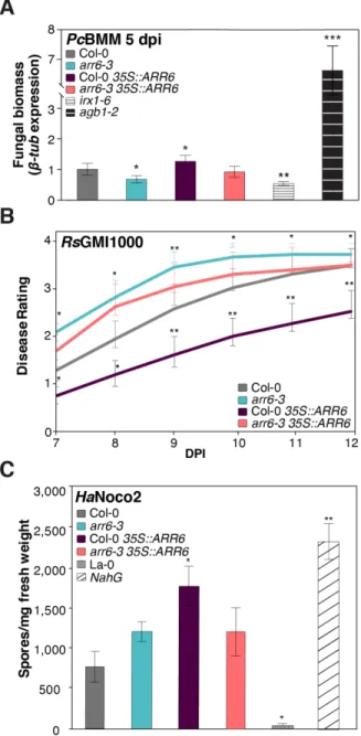
mediated by other hormones (Choi et al. 2011), and have been shown to regulate the

85

expression of defense genes and the activation of immune responses as determined in

86

plants treated with cytokinins or in cytokinin-overaccumulating lines (Choi et al., 2010).

87

Cytokinins are a family of N

6

-substituted adenine derivatives and chemically unrelated

88

phenylurea type hormones primarily involved in cell growth and differentiation. In

89

Arabidopsis, cytokinins are perceived by Arabidopsis Histidine Kinase 2–4 (AHK2–4)

90

receptors, that are two-component system proteins which initiate a downstream

91

phosphotransfer cascade that leads to the phosphorylation of Arabidopsis Response

92

Regulator (ARR) proteins through Arabidopsis Histidine Phosphotransfer (AHP) proteins

93

(Hwang et al. 2012; Naseem et al. 2014; To et al. 2004, 2007). The activation of cytokinin

(7)

94

pathway results in the transcriptional expression of a set of specific cytokinin-regulated

95

genes that have been identified by meta-analyses (Bhargava et al. 2013).

96

ARRs are encoded by a multigenic family comprising 21 members that have been

97

classified in three types (A-C) depending on their structural domains and functions: (i)

98

type A, comprising ARR3–ARR9 and ARR15–ARR17, negatively regulate cytokinin

99

responses and are transcriptionally regulated by type B ARRs; (ii) type B, namely

ARR1-100

ARR2, ARR10–ARR14, and ARR18–ARR21, are transcription factors that positively

101

regulate cytokinin signaling (Hwang et al. 2012). A type C group of ARRs has also been

102

described, but it its role in cytokinin signaling is unclear (Horák et al. 2008; Kiba et al.

103

2004; Pils and Heyl 2009).

104

The activity of ARR members is, in general, partially redundant in the regulation

105

of biological processes, and thus even mutants impaired in six ARRs genes may still retain

106

partial functions (To et al. 2004, 2007). Moreover, the existing regulatory loop between

107

types A and B ARRs could explain, at least in part, the observed contradictory functions

108

of cytokinins in the regulation of some biological processes, like plant immune responses.

109

For example, it has been suggested that type B ARRs might enhance immunity by

110

promoting SA responses, whereas type A ARRs have been proposed to attenuate immune

111

responses by inhibiting the SA pathway (Argueso et al. 2012; Naseem et al. 2015).

112

Additionally, some ARRs, such as ARR2, ARR5, ARR7 and ARR15, can interact with

113

ET signaling and modulate ET biosynthesis, which provides additional levels of

114

regulation of immunity due to hormonal crosstalk (Hass et al. 2004; Shi et al. 2012).

115

Cytokinins have also been suggested to modulate plant cell wall structure by

116

regulating the expression of genes encoding cell wall remodeling proteins such as

pectin-117

modifying enzymes, expansins and laccases (Brenner et al. 2012). Furthermore,

118

degradation of cytokinins has been linked to alterations in cell wall integrity (CWI) that

(8)

119

might function as a monitoring system to regulate developmental processes such as cell

120

cycle progression (Gigli-Bisceglia et al. 2018). Cytokinins also play important roles in

121

vascular development and, for instance, ARR5 and ARR6 genes have been shown to

122

regulate the formation of protoxylem vessels of the vascular system (Kondo et al. 2011).

123

All these structural and biochemical cell wall modifications regulated by cytokinins and

124

ARRs might affect disease resistance responses and the in planta spread of some

125

pathogens, like vascular ones.

126

Activation of plant defensive responses requires the perception of signals which

127

trigger specific resistance mechanisms through diverse molecular monitoring systems

128

(Atkinson and Urwin 2012). Among these monitoring mechanisms are Pattern-Triggered

129

Immunity (PTI) and Effector-Triggered Immunity (Dodds and Rathjen 2010). PTI is

130

based on the perception through pattern recognition receptors (PRRs) of “non-self”

131

microbe/pathogen-associated molecular patterns (MAMPs/PAMPs) or of “modified-self”

132

damage-associated molecular patterns (DAMPs) derived from the plant (Boller and Felix

133

2009; Boutrot and Zipfel 2017). MAMPs and DAMPs from different biochemical nature,

134

i.e., proteins, carbohydrates, lipids and nucleic acids, have been identified, thus reflecting

135

the diversity of immunogenic structures recognized by plants (Boutrot and Zipfel 2017).

136

Compared to MAMPs far fewer DAMPs derived from plants have been identified to date

137

(Bacete et al. 2018; Choi and Klessig 2016; Claverie et al. 2018; De Lorenzo et al. 2018).

138

An important source of DAMPs which currently is attracting research interest is

139

the plant cell wall, that is a dynamic and highly regulated structure essential for plant

140

growth and development. The functional integrity of cell walls is controlled by CWI

141

monitoring systems (Engelsdorf and Hamann 2014; Engelsdorf et al. 2018). These

142

systems trigger countervailing responses to cell wall damage (CWD) which occurs upon

143

pathogen infection, abiotic stress and cell expansion during growth and development

(9)

144

(Vaahtera et al. 2019). The plant CWI pathway is strongly involved in the regulation of

145

growth, immune responses and resource allocation between development and immunity

146

(Engelsdorf et al. 2018; Hamann et al. 2009; Wolf et al. 2012). Modification of cell wall

147

composition or integrity by genetic or chemical means might affect the capacity of some

148

pathogens to degrade the wall during plant colonization, and might lead to the activation

149

of defensive signaling pathways, including those regulated by hormones (Bacete et al.

150

2018; Houston et al. 2016; Miedes et al. 2014; Nafisi et al. 2015). For example, enhanced

151

resistance to pathogens has been observed in Arabidopsis mutants defective in cellulose

152

synthases (CESAs) required for the biosynthesis of primary (prc1/ixr1/cev1,

153

procuste1/isoxaben resistant 1/constitutive expression of VSP1) or secondary cell walls

154

(irx1/cesa8, irx3/cesa7 and irx5/cesa4, from irregular xylem) (Ellis et al. 2002;

Escudero

155

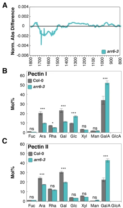
et al. 2017; Hernández-Blanco et al. 2007)

.

The disease resistance phenotypes of

156

irx1/irx3/irx5 mutants are in part explained by the constitutive activation of abscisic acid

157

(ABA) pathway and the biosynthesis of antimicrobial compounds (Escudero et al. 2017;

158

Hernández-Blanco et al. 2007). In contrast, prc1/ixr1/cev1 resistance phenotypes are

159

associated to the activation of ET/JA signaling (Ellis et al. 2002)

.

Similarly, the alteration

160

of O-acetylation patterns from cell wall xylans caused by mutations in ESKIMO1 (ESK1)

161

is associated with stress resistance phenotypes of esk1 plants and an enhanced

162

accumulation of ABA and strigolactones (Escudero et al. 2017; Lugan et al. 2009;

163

Ramírez et al. 2018; Xin et al. 2007; Xu et al. 2014). In another example, the Arabidopsis

164

walls are thin 1 (wat1) mutant exhibits increased resistance to vascular pathogens, e.g. R.

165

solanacearum, which is accompanied by higher SA levels and a general repression of

166

indole metabolism (Denancé et al. 2013b). The alteration of the composition of wall

167

pectins (e.g. degree of methylesterification) also affect disease resistance responses, some

168

of which can be reverted by auxins (Ferrari et al. 2008; Raiola et al. 2011). Also, cell wall

(10)

169

mutants with differential resistance to pathogens have been described whose immune

170

responses are not associated with the activation of hormonal pathways (Delgado-Cerezo

171

et al. 2012; Klopffleisch et al. 2014; Llorente et al. 2005; Raiola et al. 2011;

Sánchez-172

Rodríguez et al. 2009).

173

Modifications of cell wall structure or composition may also alter the presence

174

and/or release of cell wall-derived signaling molecules (e.g. DAMPs), that regulate

175

immune responses. It has been shown that biochemical modification of plant wall by cell

176

wall degrading enzymes secreted by pathogens during the colonization process can result

177

in the release of DAMPs, such as pectic oligogalacturonides (OGs) derived from

178

homogalacturonan (Benedetti et al. 2015; Lionetti et al. 2017; Ridley et al. 2001; Voxeur

179

et al., 2019). Moreover, the overexpression or inactivation of plant genes encoding

180

enzymes involved in the control of pectin structure (e.g. pectin methyl esterases (PME)

181

and PMEs inhibitors) results in the modification of the degree of OGs release upon

182

infection, and in disease resistance phenotypes alterations (De Lorenzo et al. 2018; Ferrari

183

et al. 2008; Lionetti et al. 2017; Raiola et al. 2011). However, it is not well understood

184

the relationship between composition/structure of the cell wall, the dynamic of the release

185

of wall DAMPs, the activation of canonical defensive responses and the resistance to

186

pathogens.

187

Here, we show that arr6-3 plants defective in ARR6 gene and ARR6

188

overexpression lines have altered and opposing disease resistance outcome phenotypes to

189

different pathogens. We also demonstrate that arr6-3 plants display alterations in the

190

biochemical composition of their cell walls and that pectin-enriched fractions extracted

191

from arr6-3 walls contain wall-related DAMPs triggering enhanced immune responses

192

in comparison to pectin-enriched fractions extracted from wild-type plant walls. Our data

(11)

193

demonstrate, a novel link between the activity of regulatory components of the cytokinin

194

pathway and Arabidopsis cell wall composition and disease resistance responses.

195

196

RESULTS

197

ARR6 modulates differential disease resistance responses to pathogens.

198

Arabidopsis mutants impaired in genes putatively involved in cell wall

199

biosynthesis or remodeling represent useful tools to study and characterize plant

200

immunity mechanisms mediated by CWI (Bacete et al. 2018; Miedes et al. 2014). We

201

selected ARR6 gene to investigate the function of CWI on immunity as this is the

202

Arabidopsis orthologue of DV017520 from Zinnia elegans, a gene that is upregulated

203

during late xylogenesis, which is a developmental process involved in secondary cell wall

204

formation (Pesquet et al. 2005). ARR6 has also been described to regulate the formation

205

of protoxylem vessels of the Arabidopsis vascular system and accordingly arr6 mutants

206

shows alterations in the xylem (Kondo et al. 2011). Moreover, data retrieved from

Bio-207

Analytic Resource for Plant Biology (BAR, http://bar.utoronto.ca/eplant) also indicated

208

that ARR6 expression was repressed in wild-type Col-0 plants upon infection with

209

different types of pathogens, like the vascular bacterium R. solanacearum (Hanemian et

210

al. 2016; Hu et al. 2008) or the biotrophic oomycete Hyaloperonospora parasitica (Hpa),

211

after treatment with MAMPs (e.g. bacterial flg22 peptide) or under some abiotic stress

212

conditions (Supplementary Fig. S1). Also, mutants impaired in several ARR members,

213

including ARR6 (e.g. arr5,6,8,9 and arr3,4,5,6,8,9 mutants), but not the single arr6-1

214

mutant, have been described to show slightly enhanced resistance to Hpa (Argueso et al.

215

2012). Altogether, these data suggested that ARR6 could be associated with the

216

modulation of cell wall composition and activation of defense responses.

(12)

217

To further probe these putative ARR6 functions in the modulation of disease

218

resistance and cell wall composition, we selected for disease resistance studies two

T-219

DNA insertional alleles of ARR6 (arr6-2 and arr6-3; Supplementary Fig. S2A).

qRT-220

PCR analysis of ARR6 expression in the mutant alleles and Col-0 wild-type plants

221

(Supplementary Fig. S2B) indicated that arr6-3 allele was a knock-out (null) whereas

222

arr6-2, was a knock-down (hypomorphic) mutant. arr6-2 allele harbors the T-DNA

223

insertion very close to that present in arr6-1 allele, that was also previously described as

224

an hypomorphic allele (To et al. 2004). The developmental phenotype of arr6 plants does

225

not differ from that of wild-type plants, as previously described (To et al. 2004). We

226

tested the disease resistance phenotypes of arr6-2 and arr6-3 to pathogens with different

227

life styles: the necrotrophic fungus P. cucumerina BMM (PcBMM), the vascular

228

bacterium R. solanacearum GMI1000 (RsGMI100), and the biotrophic oomycete Hpa

229

Noco2. Notably, the arr6-3 mutant was slightly more resistant to the necrotrophic fungus

230

PcBMM than Col-0 wild-type plants, as determined by fungal biomass quantification by

231

qPCR at 5 days post-inoculation (dpi) and by disease rating (DR) determination at 7 dpi

232

(Fig. 1A and Supplementary Fig. 3A). The accumulation of PcBMM biomass and DR

233

were slightly higher in arr6-3 than that observed in irx1-6 plants included as resistant

234

control, and in both mutants these values were significantly lower than that of Col-0

wild-235

type plants (Figure 1A and Supplementary Fig. 3A). This contrasted with the enhanced

236

fungal biomass and DR observed in the agb1-2 mutant, which was included as

237

hypersusceptible control (Fig. 1A and Supplementary Fig. 3A; Hernández-Blanco et al.

238

2007; Llorente et al. 2005). Remarkably, the arr6-3 mutant was highly susceptible to

239

RsGMI1000 and plants decayed at 10-12 dpi, as determined by visual evaluation of

240

disease rating (Fig. 1B). As previously described for arr single mutants (Argueso et al.

241

2012) the susceptibility to the oomycete Hpa of the arr6-3 allele was found to be similar

(13)

242

to that of Col-0 plants, as determined by quantification of conidiosphores/mg of fresh

243

weight at 7 dpi (Fig. 1C). Similarly, the hypomorphic arr6-2 allele showed similar disease

244

symptoms as Col-0 wild-type plants to the three pathogens tested (Supplementary Fig.

245

S3B-D), that is in line with the previously described lack of differential phenotypes of

246

arr6-1 hypomorphic allele (To et al. 2004; Argueso et al. 2012).

247

In view of the disease resistance responses of arr6-3 mutant and to corroborate

248

the role of ARR6 in the modulation of these responses, we generated different Col-0 lines

249

that overexpress ARR6 (35S::ARR6) or ARR6 fused to the green fluorescence protein

250

(GFP) or the human influenza hemagglutinin (HA) tags (35S::ARR6-GFP and

251

35S::ARR6-3HA), as well as plant lines overexpressing ARR6 in the arr6-3 background

252

(arr6-3 35S::ARR6; Supplementary Fig. S2C). ARR6 expression in these lines was

253

evaluated by qRT-PCR and we selected for further characterization different

254

overexpression lines (in Col-0 background) with expression levels of ARR6 transgenes

4-255

10 fold higher than in Col-0, and arr6-3 lines (arr6-3 35S::ARR6) with ARR6 expression

256

levels similar to that of wild-type plants (Supplementary Fig. S2B). Notably, in the arr6-3

257

35S::ARR6 lines tested the enhanced resistance to PcBMM and susceptibility to

258

RsGMI100 was almost restored to wild-type levels, further confirming the role of ARR6

259

in regulating these disease resistance responses (Fig. 1A and B, Supplementary Fig. S3A).

260

In line with these results, we found that PcBMM fungal growth (5 dpi) and DR (7 dpi) in

261

the 35S::ARR6 overexpression line were significantly increased compared to Col-0

262

plants, whereas 35S::ARR6 plants were more resistant to the bacterium RsGMI1000 than

263

Col-0 plants, as revealed by their delayed disease symptoms (Fig. 1A and B,

264

Supplementary Fig. S3A). To further confirm these phenotypes associated to ARR6

265

overexpression, independent overexpression lines (3HA and

35S::ARR6-266

GFP) were tested for disease resistance and we found that they also showed enhanced

(14)

267

susceptibility and resistance to PcBMM and RsGMI1000, respectively, as compared to

268

Col-0 wild-type plant plants (Supplementary Fig. S3B and C). In agreement with previous

269

results (Argueso et al. 2012), the 35S::ARR6 lines tested were slightly more susceptible

270

than Col-0 plants to the oomycete Hpa (Fig. 1C; Supplementary Fig. S3D). Altogether,

271

these observations support a novel function of ARR6 in the differential modulation of

272

Arabidopsis resistance responses to PcBMM and RsGMI1000 and corroborate the

273

redundant role of type A ARRs (including ARR6) in controlling the resistance to Hpa

274

(Argueso et al. 2012).

275

276

arr6-3 plants show differential expression of defense and cell wall-associated genes.

277

A comparative transcriptomic analysis of 18-day-old arr6-3 and Col-0 plants was

278

performed. RNA was extracted from the plants one day after PcBMM-inoculation or

279

mock-treatment. Microarray data analysis of mock-treated plants revealed 205

280

differentially expressed genes (DEGs) in arr6-3 compared to Col-0 plants, of which 153

281

and 53 were up- and down-regulated, respectively (Supplementary Table S1). Functional

282

classification and Gene Ontology (GO) terms enrichment analyses performed using the

283

hypergeometric test and Benjamini & Hochberg False Discovery Rate (FDR, corrected

284

with a p value cut-off of 0.05) indicated that 126 GOs were overrepresented among the

285

DEGs (Fig. 2A; Supplementary Table S2). These GO included “Response to stimulus”

286

(GO:0050896, p value = 2.82×10

-5

) and “Regulation of transcription, DNA-templated”

287

(GO:0006355, p value 1.18×10

-5

), each with 23 unique genes (11.2%), and “Response to

288

chitin” (GO:001020018, p value of 3.09×10

-16

, 18 genes representing 8.7%) (Fig. 2A;

289

Table S2). A closer view of “Response to stimulus” category revealed that most of the

290

genes were related to defense responses (10 out of 23) and response to stress (14 out of

291

23), specifically biotic stress (9 out of 23) (Table S2). In the case of “Regulation of

(15)

292

transcription, DNA-templated” category, we observed that a significant number of DEGs

293

were related to the ET pathway (7 out of 23). In addition, analysis of the 205 DGEs using

294

the Thalemine web tool (Krishnakumar et al. 2017) indicated that the

295

APETALA2/ethylene responsive factor (AP2/ERF) domain (IPR001471) was

296

overrepresented (p value = 5.71×10

-4

) with 11 ERFs (5.34% of DEGs) up-regulated in

297

arr6-3 (Supplementary Table S3). Among these ERFs, four belonged to the

dehydration-298

responsive element binding (DREB) subfamily (Group A), known to be involved in the

299

regulation of dehydration- and cold-inducible genes (Sakuma et al. 2002), whereas seven

300

were ERFs from Group B (Sakuma et al. 2002) or IX (according to Nakano 2006). Of

301

note, six out of these seven ERFs have been implicated in pathogen resistance (Nakano,

302

2006), and the majority of these ERFs were found to be up-regulated in microarray data

303

of Col-0 plants infected with either PcBMM or RsGMI100 (Supplementary Fig. S4).

304

These microarray values were validated in arr6-3 and Col-0 plants by qRT-PCR analysis

305

of ARR6 and a set of genes selected based on their putative functional relevance in disease

306

resistance or abiotic stress responses (Fig. 2B).

307

To determine if alteration of cytokinin perception contributes to explain arr6-3

308

disease resistance phenotypes, we compared the arr6-3 DEGs with those of ahk1 and

309

ahk2 ahk3 mutants (E-MEXP-1155; Tran et al. 2007). Notably, only five DEGs were

310

common between ahk2 ahk3 and arr6-3 (including the downregulated ARR6) and one

311

between ahk1 and arr6-3 (Supplementary Fig. S5A). To strength these results, we also

312

compared arr6-3 DEGs with the dataset of 75 cytokinin-specific regulated genes

313

proposed by Bhargava et al. (2013). In line with our previous comparison with ahk1 and

314

ahk2 ahk3 datasets, we did not find any of these 75 cytokinin responsive genes among

315

the arr6-3 DEG, further indicating that arr6-3 DEGs are not cytokinin-associated genes

316

(Supplementary Fig. S5B).

(16)

317

We next studied the disease resistance response of arr6-3 plants upon inoculation

318

with PcBMM, and we found 2816 DEGs in arr6-3 inoculated plants, of which 905 were

319

specifically expressed in arr6-3, but not in Col-0 (Fig. 2C; Supplementary Tables S5 and

320

S6). According to GO term enrichment analysis, among these arr6-3 DEGs there were

321

GO related to defense responses, to diverse biotic and abiotic stresses as well as to other

322

physiological processes (Supplementary Fig. S6). Notably, among these 905 DEG we

323

found 75 out of the 205 genes (36.59%) constitutively up-regulated in non-inoculated

324

arr6-3 plants (Supplementary Fig. S5B and Supplementary Table S5), further supporting

325

the function of this set of genes in disease resistance responses. Strikingly, only 9 of these

326

arr6-3 DEGs upon PcBMM infection were also among cytokinin-regulated marker genes

327

described by Bhargava et al. (2013) (Supplementary Fig. S5). Together, these results

328

indicate that the arr6-3 disease resistance phenotypes are unlikely to be associated with

329

differential regulation of the cytokinin pathway.

330

Of note, canonical defense pathways (i.e. SA, ET, JA and biosynthesis of

331

secondary metabolites) and PTI signaling were up-regulated in response to PcBMM

332

infection both in Col-0 and arr6-3 plants, with a slightly higher up-regulation in arr6-3

333

plants as corroborated by qRT-PCR expression analysis of marker genes (Fig. 2D). These

334

data suggested that the arr6-3 disease resistance phenotypes are not associated to the

335

misregulation of canonical defensive pathways. To further validate this hypothesis and to

336

identify potential defense-associated metabolites in arr6-3, we performed a comparative

337

and global metabolite profiling of four-week-old, non-inoculated arr6-3 and Col-0 plants.

338

Within the 373 analyzed metabolites (including all the defensive hormones or some of

339

their precursors), only 11 were more abundant (n-fold higher than 1.5 and p < 0.1) in

340

arr6-3 compared to Col-0 (Supplementary Fig. S7A and Supplementary Table S7).

(17)

342

camalexin and sulforaphane-cysteine-glycine glucosinolate, two antimicrobial

343

compounds (Zhou et al. 1998; Buxdorf et al. 2013), were found to be more abundant in

344

the mutant than in Col-0 plants (Supplementary Fig. S7A and Supplementary Table S7).

345

Glucosinolates, but not camalexin, have been previously shown to be required for

346

Arabidopsis resistance to PcBMM (Sánchez-Vallet et al. 2010). With the exception of

347

cyano-alanine, a product of ET metabolism, that showed a slightly higher abundance in

348

arr6-3, we did not find significant alterations in the levels of hormones or their precursors

349

(e.g. SA, JA and brassinosteroids) that regulate canonical immune pathways in arr6-3

350

plants (Supplementary Figure S7B and Supplementary Table S7). The slight increased

351

abundance of a small subset of antimicrobial compounds is, most likely, not sufficient to

352

fully explain arr6-3 disease resistance phenotypes to two different pathogens with distinct

353

colonization styles.

354

Since some PTI marker genes (e.g. PHI1) were up-regulated in non-inoculated

355

arr6-3 plants (Fig. 2B) and showed a slightly enhanced expression upon PcBMM

356

infection, we determined PTI responses in arr6-3 plants upon treatment with two MAMPs

357

(flg22 and chitin hexamer (Mélida et al. 2018)), and compared these responses with that

358

of 35S:ARR6 and Col-0 plants. The phosphorylation of Mitogen Activated Protein

359

Kinases (MAPKs, MAPK3/MAPK6/MAPK4/MAPK11) and expression of PHI1 gene

360

were slightly higher in arr6-3 than in Col-0 plants (Supplementary Fig. S8), which is in

361

line with the transcriptomic data (Fig 2B). Notably, MAPK phosphorylation was weaker

362

in 35S:ARR6 plants than in Col-0, suggesting a defective activation of PTI in the

363

overexpressor line (Supplementary Fig. S8). We then tested the expression of ARR6 in

364

these genotypes and we found that ARR6 expression was downregulated upon MAMPs

365

treatment in Col-0, as described previously (Supplementary Fig. 1), but also in 35S:ARR6

(18)

366

plants. The results point to a complex mechanism of regulation of ARR6 function during

367

immune activation.

368

369

arr6 mutants show alterations in their cell wall composition.

370

Among the arr6-3 DEGs, we found 16 genes that were associated with cell wall

371

biosynthesis and/or remodeling, or with responses to CWI impairments (Supplementary

372

Fig. S9), which suggested some cell wall alterations in arr6-3 plants. Since ARR6 has

373

been involved in regulation of formation of protoxylem vessels of the vascular system

374

(Kondo et al. 2011), we determined the cell wall composition of arr6-3 plants and

375

compared it with that of wild-type plants. First, we performed a cell wall characterization

376

using Fourier-transform infrared (FTIR) spectroscopy to obtain “cell wall fingerprints”

377

that could reveal general differences between Col-0 and arr6-3 cell walls. FTIR is able

378

to recognize polymers and functional groups and is used as a powerful and rapid

379

technique for analyzing cell wall components, since they can be assigned to different

380

wavenumbers of the FTIR-spectra (Alonso-Simón et al. 2011). The analysis of the

381

differential FTIR-spectra obtained after digital subtraction of the wild-type values,

382

revealed differences mainly in the region between 1500 and 1750 cm

-1

(Fig. 3A). This

383

region is characterized by the absorption of uronic acids in wavenumbers 1600-1630 and

384

1740 cm

-1

(McCann et al. 1992; Mouille et al. 2003) and lignin at 1515, 1630 and 1720

385

cm

-1

(Séné et al. 1994; Carpita et al. 2001). With FTIR data pointing to differences in cell

386

wall components, such as uronic acids and/or lignin, and bearing in mind that overlap in

387

absorption and vibrational coupling between chemical bonds corresponding to different

388

cell wall polymers may occur, a more detailed biochemical characterization of the cell

389

wall composition was carried out.

(19)

390

Lignin and cellulose quantifications showed no differences in the total amount of

391

these cell wall components in arr6-3 and Col-0 plants (Supplementary Fig. S9). Once

392

discarded the contribution of these two load-bearing cell wall components to the

393

differences observed by FTIR, crude highly complex cell walls were chemically

394

fractionated. Four fractions enriched in different cell wall components (two pectic and

395

two hemicellulosic ones) were analyzed for their monosaccharide composition and their

396

glycosidic linkages types. arr6-3 pectin fractions displayed some striking differences in

397

monosaccharides composition compared to their wild-type counterparts, whereas no

398

significant differences in composition were found between their hemicellulose fractions

399

(Fig. 3B and C; Supplementary Fig. S10). In comparison to Col-0 pectin I fraction the

400

arr6-3 fraction was highly enriched in galacturonic acid (57 mol% versus 34 mol% in

401

Col-0) and glucose (17 mol% versus 9 mol% in Col-0), while they contained less

402

arabinose, rhamnose and galactose amounts than the fractions from wild-type plants (Fig.

403

3B). Similarly, the pectin II fraction from the mutant contained more galacturonic acid

404

(45 mol% versus 22 mol%) and lower arabinose and galactose content than that of Col-0

405

plants (Fig. 3C). Glycosidic linkage analyses of the pectin fractions confirmed that the

406

enrichment in 1,4-linked galacturonic acid observed in the arr6 pectin fractions was from

407

homogalacturonan-type polymers (Supplementary Fig. S11). Other 1,4-galacturonic acid

408

containing polymers such as rhamnogalacturonan did not show any increased levels of

409

other residues from these structures, such as 1,2-linked rhamnose, ruling out the

410

possibility that the observed increased levels in galacturonic were due to modifications in

411

rhamnogalacturonan content (Supplementary Fig. S11). These analyses confirmed that

412

arr6-3 cell walls are altered and that these modifications are mainly associated to the

413

pectic fractions.

414

(20)

415

The pectin-enriched fractions from arr6 trigger immune responses.

416

From the results presented above, we hypothesized that an enhanced and

417

differential presence of carbohydrate-based DAMPs in the cell walls of arr6-3 in

418

comparison to Col-0 might explain, at least partially, the differential disease resistance

419

responses of arr6-3 plants. These DAMPs, when released, would activate immune

420

responses, thus triggering disease resistance. arr6-3 cell wall fractions were tested for

421

their capacity to trigger intracellular Ca

2+

influxes, an early immune response, by using

422

Col-0

AEQ

sensor lines, that express the Apoaequorin gene from Aequorea Victoria and

423

can be used as an in vivo bioluminescent Ca

2+

sensor (Knight et al. 1991; Mélida et al.

424

2018). Interestingly, we found that Ca

2+

entry was activated by Col-0 and arr6-3 pectin

425

fractions, but not by hemicellulose ones. Moreover, arr6-3 pectin fractions induced

426

higher Ca

2+

influxes than those from Col-0 (Fig. 4A). These calcium signatures were

427

compatible with a ligand-receptor binding interaction, like the observed for flg22 MAMP

428

and OG

10-15

(degree of polymerization (DP) of 10-15) DAMP, which were included in

429

the experiment as controls (Fig. 4A). Of note, the DAMPs contained in pectin I and pectin

430

II fractions from Col-0 and arr6-3 remained active (e. g. triggered [Ca

2+

]

cyt

entry) after

431

different treatments affecting proteins stability, i.e. denaturation by heat treatment and

432

proteolysis with proteinase K combined with heat denaturation (Fig. 4B). Slight, but not

433

significant, increases in the activity of these fractions were observed in some cases upon

434

these treatments (Fig. 4B), that are consistent with solubilization effects of the fractions,

435

which would facilitate the release of their DAMPs, as it has been previously reported for

436

insoluble polysaccharides subjected to heat solubilization (Mélida et al. 2018). Such

437

observation contrasted with the immune activity of flg22 after heat + proteinase K

438

treatment that was significantly reduced (Fig. 4B). The resistance to proteolysis of the

(21)

439

pectin I fraction from arr6-3 and Col-0 suggested that this fraction contains DAMPs that

440

are not peptides and might be of glycosidic nature.

441

The pectin-enriched fractions extracted from arr6-3 and Col-0 cell walls triggered

442

phosphorylation of MAPK3 and MAPK6, but not MAPK4 or MAPK11 in Col-0

wild-443

type plants, being the activities of pectin I fractions higher than those of pectin II ones

444

(Fig. 5A and B). The pectin I-induced MAPK phosphorylation was similar to that

445

triggered by OG

10-15

, since no phosphorylation of MAPK4/MAPK11 occurred, in contrast

446

to the phosphorylation triggered by flg22 (Fig. 5C). We also evaluated the expression of

447

PTI marker genes in wild-type plants 30 minutes after treatments with pectin fractions

448

from Col-0 and arr6-3 plants, or water. The selected genes for the qRT-PCR analyses

449

were either specifically activated by the calcium-dependent protein kinase (CDPK)

450

pathway [i.e. PHI-1, which is constitutively up-regulated in arr6-3], the CDPKs +

451

MAPKs pathways (i.e. CYP81F2), or by pathogens (e.g. WRKY33; Boudsocq et al. 2010;

452

Boudsocq and Sheen 2013). Remarkably, up-regulation of these three genes was observed

453

in Col-0 plants treated with the arr6-3 pectin fractions (Fig. 5D and E), and such induction

454

was comparable to that observed upon treatments with highly purified MAMPs/DAMPs,

455

such as flg22 or OG

10-15

(Fig. 5F). Of note, Col-0 pectin I fractions did not trigger the

456

expression of any of these marker genes up-regulated by the arr6-3 pectin I fraction (Fig.

457

5D and E), whereas the pectin II fractions from arr6-3 and Col-0 plants induced similar

458

expression of the tested genes (Fig. 5D and E). Together these data suggested that

arr6-459

3 pectin I fraction contained either enhanced levels of DAMPs than Col-0 fraction or

460

specific DAMPs, of glycosidic nature, which triggered differential immune responses.

461

462

463

(22)

464

DISCUSSION

465

The plant cell wall is currently viewed as a dynamic structure regulating different

466

processes like immunity and development (Bacete et al. 2018; Bethke et al. 2016; de

467

Lorenzo et al. 2018 Engelsdorf et al. 2018; Voxeur and Höfte, 2016). To better understand

468

the relationship between plant cell walls and immunity we have characterized the function

469

of the Arabidopsis ARR6 gene in the regulation of CWI and disease resistance responses.

470

This gene was selected based on: (i) its expression profile in Arabidopsis plants subjected

471

to different stresses, including MAMPs treatment (Supplementary Fig. S1 and S8); (ii) its

472

described role on protoxylem vessel formation (Kondo et al. 2011); (iii) the up-regulation

473

of its Z. elegans orthologue during xylogenesis, a secondary cell wall biosynthetic process

474

triggered by auxin/cytokinin (Pesquet et al. 2005); and (iv) its expression during R.

475

solanacearum infections, since ARR6 expression is repressed in leaves from susceptible

476

Arabidopsis genotypes, but it is up-regulated in clavata1 mutants that are more tolerant

477

to this bacterium than wild-type plants (Hanemian et al. 2016; Hu et al. 2008). These data

478

suggest that ARR6 could be involved in the regulation of stress responses and/or

479

modulation of plant cell wall composition. In line with this initial hypothesis we have

480

demonstrated that, compared to wild-type plants, arr6-3 loss of function mutant allele

481

shows differential disease resistance phenotypes to different pathogens (Fig. 1 and

482

Supplementary Fig S3) and alterations in its cell wall composition (Fig. 3).

483

ARR6 belongs to type A ARRs, which have been previously described to be

484

involved in disease resistance. For example, an arr3,4,5,6,8,9 sextuple mutant exhibited

485

decreased susceptibility to the biotrophic pathogen Hpa, whereas transgenic lines

486

overexpressing either ARR6, ARR5, or ARR9 were more susceptible to this pathogen

487

(Argueso et al. 2012). The unaltered resistance phenotype of the arr6-3 single mutant to

488

Hpa (Fig. 1) compared to the described enhanced resistance of the arr3,4,5,6,8,9 mutant

(23)

489

indicates that ARR6 ha soverlapping functions with other ARRs in the regulation of

490

disease resistance responses to Hpa, as previously reported (Argueso et al. 2012).

491

Notably, we have shown that arr6-3 is more resistant and susceptible than Col-0 plants

492

to the necrotrophic fungus P. cucumerina and to the vascular bacterium R. solanacearum,

493

respectively (Fig. 1 and Supplementary Fig S3), further supporting a relevant and specific

494

function of ARR6 in the regulation of disease resistance responses to these two pathogens

495

with different life styles. As previously described for arr6-1 (To et al. 2004), the

496

additional hypomorphic allele (arr6-2) tested here did not show any alteration on the

497

disease resistance phenotypes tested (Supplementary Fig. S3). Interestingly, ARR6

498

expression is downregulated by MAMPs (e.g. flg22) and different biotroph and

499

necrotroph pathogens, in both compatible and incompatible interactions (Supplementary

500

Fig. S1 and S8). This highlights the relevance of ARR6 in the immune response and

501

suggests that ARR6 may act as a negative regulator of disease resistance activation against

502

certain pathogens (e.g. PcBMM) similarly to what it has been described for other genes,

503

such as PMR6, that mediate cell wall-based resistance to pathogens (Vogel et al. 2002).

504

The specific function of ARR6 in immunity was further supported by the disease

505

phenotypes of lines overexpressing ARR6, which showed opposite disease resistance

506

phenotypes to arr6-3 (e.g. in 35S::ARR6, 35S::ARR6-GFP, and 35S::ARR6-HA) or

507

complemented them (e.g. arr6-3 35S::ARR6 lines; Fig. 1; Supplementary Fig. S3).

508

Notably, ARR6 expression was downregulated in 35S::ARR6 plants upon MAMPs

509

treatment, suggesting a complex regulation of ARR6 function during PTI activation

510

(Supplementary Fig. S8C). Together, these results corroborate a role of type A ARRs in

511

the regulation of disease resistance responses and support a relevant function of ARR6 in

512

this process.

(24)

513

Cytokinins have been suggested to up-regulate plant immunity via an elevation of

514

SA–dependent defense responses that, in turn, inhibit cytokinin signaling in a feedback

515

loop (Argueso et al. 2012). Type-A ARRs are considered central components of this

516

process and of other hormonal cross-talks (e.g. cytokinin-ET) that are activated in

517

response to different stresses (O’Brien and Benková 2013; Shi et al. 2012). These

518

regulatory pathways are mediated by the cytokinin receptors AHK2 and AHK3 that

519

function upstream of ARRs (including ARR6) sensing cytokinin concentrations (Argueso

520

et al. 2012; Hwang and Sheen 2001). However, the transcriptome pattern of ahk2 ahk3

521

double mutants did not overlap with that of arr6-3, and neither did that of ahk1 mutant

522

(Supplementary Fig. S5A), which is impaired in the CWI sensor AHK1 (Hamann et al.

523

2009; Wormit et al. 2012). Therefore, AHK1, AHK2 and AHK3 do not seem to be linked,

524

at least at the transcriptional level, with the alterations in gene expression observed in the

525

arr6-3 plants, suggesting that the immune responses regulated in arr6-3 are not related

526

with the cytokinin signaling pathway, or at least with AHK2 and AHK3 function. This is

527

further supported by the fact that none of the cytokinin-regulated genes identified in

528

previous works (Bhargava et al. 2013) are among the DEGs in arr6-3 mock-treated

529

plants, and only 9 of the arr6-3 DEGs upon PcBMM infection were among the set of

530

cytokinin-regulated marker genes (Bhargava et al. 2013) (Supplementary Fig. 5B).

531

Similarly, metabolomic and transcriptomic analyses of arr6-3 plants demonstrated that

532

canonical immune pathways, like those regulated by hormones, are not misregulated in

533

the arr6-3 in comparison to wild-type plants, since hormone homeostasis and expression

534

of marker genes of the pathways mediated by these hormones are not altered in the mutant

535

(Fig. 2; Supplementary Tables S4 and S7).

536

Transcriptomic analyses of arr6-3 plants identified defense-associated genes

537

which are specifically up-regulated in non-inoculated and PcBMM infected plants (Fig.

(25)

538

2; Supplementary Fig. 6; Supplementary Tables S4 and S5), and that may contribute to

539

explain arr6-3 resistance to this fungus. For example, arr6-3 plants are characterized by

540

a clear overrepresentation of up-regulated genes encoding ERFs transcriptional factors

541

belonging to group IX-b, whose members are known to be involved in responses to

542

pathogens (Licausi et al. 2013). Transgenic lines overexpressing ERF5 or ERF6 are

543

resistant to the necrotrophic fungus Botrytis cinerea but show enhanced susceptibility to

544

Pseudomonas syringae pv. tomato DC3000 (Huang et al. 2015; Moffat et al. 2012; Wang

545

et al. 2013), ERF104 overexpression lines are resistant to P. syringae pv. phaseolicola

546

(Bethke et al. 2009) and overexpression of ERF1 confers enhanced resistance to the

547

necrotrophic fungi P. cucumerina and B. cinerea (Berrocal-Lobo et al. 2002). Thus, the

548

observed enhanced resistance of arr6-3 to PcBMM could be explained, at least in part,

549

by the expression pattern of some ERFs genes. Additionally, the enhanced accumulation

550

of some antimicrobial secondary metabolites in arr6-3 (Supplementary Figure S7) may

551

also contribute to arr6-3 disease resistance (Sánchez-Vallet et al. 2010). Moreover, some

552

PTI responses (e.g. MAPKs phosphorylation) are enhanced in arr6-3 upon MAMP

553

treatment, in comparison to wild-type plants (Supplementary Fig. S8A-B), suggesting a

554

negative function of ARR6 in immune regulation. In line with this function, ARR6

555

expression was down regulated upon pathogen infection and MAMPs treatment

556

(Supplementary Fig. S1 and S8).

557

Arabidopsis resistance to R. solanacearum has been shown to be enhanced in ET

558

signaling mutants (Hirsch et al. 2002) indicating that ET has a negative regulatory role

559

on Arabidopsis resistance response to this bacterium, which is in line with the finding

560

that ET is one of the effectors produced by R. solanacearum to colonize Arabidopsis

561

(Weingart et al. 1999). The constitutive up-regulation in arr6-3 plants of some ERFs,

562

regulated by ET pathway, as well as other genes that are DEGs in susceptible

(26)

563

Arabidopsis/R. solanacearum interactions might explain the enhanced susceptibility of

564

arr6-3 to the bacterium (Hanemian et al. 2016; Hu et al. 2008). Notably, ARR6 expression

565

is repressed in leaves from susceptible compared to resistant plants during R.

566

solanacearum infections and is activated in clavata1 mutants that are more tolerant to

567

this bacterium than wild-type plants (Hanemian et al. 2016; Hu et al. 2008). Also, the

568

alteration of protoxylem vessel structure in arr6-3 plants might contribute to explain the

569

enhanced susceptibility of arr6-3 to the vascular bacterium, that colonize systemically

570

the plant through the xylem (Hirsch et al. 2002). All these R. solanacearum-associated

571

susceptibility factors of arr6-3 plants might favor bacterial colonization and are not

572

counterbalanced by the slight enhanced activation of PTI observed in the arr6-3 or the

573

higher PTI activity of the DAMPs found in mutant wall fractions in comparison to Col-0

574

ones.

575

Here, using biochemical analyses we show that leaves of arr6-3 plants display

576

significant alterations in their cell wall composition, particularly in their pectic fractions

577

(Fig 3), which is in line with the described alteration of protoxylem vessel formation in a

578

different arr6 allele (Kondo et al. 2011). These arr6 cell wall features may contribute to

579

the differential disease resistance responses of arr6 plants as the cell wall structure

580

influences Arabidopsis resistance to R. solanacearum and P. cucumerina, as observed in

581

irx1, esk1 and wat1 mutants (Denancé et al. 2013b; Digonnet et al. 2012; Escudero et al.

582

2017; Hernández-Blanco et al. 2007), and pectins have been described to be involved in

583

Arabidopsis resistance to necrotrophic fungi, like B. cinerea (Bethke et al. 2016; Lionetti

584

et al. 2017). Beyond structural reasons, pectins, in particular homogalacturonan, are a

585

source of different types of OGs, the best characterized plant cell wall-derived DAMPs,

586

which regulate DAMP-Triggered Immunity (Davidsson et al. 2017; Ferrari et al. 2013;

587

Galletti et al. 2008; Voxeur et al., 2019).

(27)

588

The increased abundance of homogalacturonan in the arr6-3 pectic fractions

589

suggested that some OGs, or OGs concentration or OGs availability might be higher in

590

arr6-3 walls than in wild-type ones, and accordingly we found that the pectin I and pectin

591

II fractions from arr6-3 mutant triggered enhanced immune responses in Arabidopsis

592

Col-0 plants, in comparison to the responses activated by the corresponding fractions

593

from wild-type plants (Figs. 4 and 5). These data suggest that additional or higher

594

concentrations of DAMPs are present in arr6 wall fractions compared to Col-0 ones.

595

Also, we can hypothesize that differential release of specific DAMPs might occurs upon

596

infection of arr6-3 and Col-0 plants by distinct pathogens expressing different sets of cell

597

wall degrading enzymes. Interestingly, the arr6-3 pectin I fraction, but not that of Col-0

598

plants, triggers the up-regulation of some marker genes (e.g. PHI-1) that are also

599

constitutively up-regulated in arr6-3 plants, suggesting that some of these genes would

600

be markers of the immune responses activated by the arr6 pectin I fraction. We also

601

show here that MAPK phosphorylation cascades, that have a crucial role in signal

602

transduction in response to pathogens (Meng and Zhang 2013) and regulate the immune

603

responses, are triggered by the pectin I and pectin II fractions of arr6-3 and Col-0 plants.

604

Phosphorylation patterns upon treatment with the pectin I, pectin II fractions or OG

10-15

,

605

support that the pathways activated by these fractions are not identical to those activated

606

by flg22 (Fig. 5). Based on the enhanced PTI-triggering capacities of arr6-3 pectic

607

fractions, their direct application to plant leaves should confer disease resistance.

608

However, future research will be needed in this direction in order to get suited

609

formulations of these complex carbohydrate mixtures that will favor DAMPs penetration

610

through the plant cuticle and will avoid their degradation by leave epiphytic

611

microorganisms or pathogens, as it has been described previously (Trouvelot et al. 2014).

(28)

612

In summary, we show here that impairment of ARR6 gene affects cell wall

613

composition, which may impact plant-pathogen interactions, and lead to the accumulation

614

of precursors of the differential DAMPs (e.g. OGs) that would favor a “defense-ready”

615

state instead of resting stage. Under the attack of a necrotrophic pathogen harboring a

616

repertoire of cell wall degrading enzymes, these DAMPs would be released and perceived

617

through unidentified PRR complexes, contributing to the observed increased resistance

618

of arr6-3 plants. However, this response would not take place in arr6-3 plants being

619

attacked by pathogens that colonize the plant through the vascular system, but that do not

620

massively degrade plant cell walls, such as RsGMI1000. Further characterization of

621

ARR6-mediated immunity will pave the way to understand its role in CWI perception

622

and maintenance and PTI regulation.

623

624

MATERIALS AND METHODS

625

Plant material and growth conditions.

626

The Arabidopsis thaliana mutants used in this work, i.e. arr6-2 (SALK_008866)

627

and arr6-3 (SALK_133123) were in Columbia-0 (Col-0) genetic background. Details of

628

all mutant lines are provided in Supplementary Table S8. Arabidopsis plants were grown

629

as previously described (Sánchez-Vallet et al. 2010; Mélida et al. 2018). For generation

630

of ARR6 transgenic lines, constructs presented in Figure S1 were brought into the

631

Agrobacterium tumefaciens strain C58 using the Gateway technology (Earley et al. 2006).

632

ARR6 (At5g62920) cDNA was cloned in Gateway plasmids pGWB2, pGWB5 and

633

pGWB14. The constructs were used to transform wild-type Col-0 and arr6-3 plants by

634

the standard floral dip method (Clough and Bent 1998). Segregation in 50 µg/L

635

hygromycin ½ MS 1% sucrose agar plates was analyzed until T4 generation. For each

(29)

636

combination of background and construct, expression levels of 10 independent transgenic

637

lines were analyzed by qRT-PCR using oligonucleotide primers (Supplementary Table

638

S9). Two lines from each construct, with different expression levels of the transgene,

639

were selected for further analyses and one of them tested for resistance (Fig. 1;

640

Supplementary Figs. S2 and S3; data not shown).

641

Pathogenicity assays.

642

For P. cucumerina BMM (PcBMM) pathogenicity assays, 18-day-old plants (n

643

>12) growing on soil were sprayed with a spore suspension (4x10

6

spores/ml) of the

644

fungus as previously described (Sánchez-Vallet et al. 2010; Delgado-Cerezo et al. 2012).

645

Fungal biomass in planta was quantified at 5 days post-inoculation (dpi) by qPCR

646

determining the β-tubulin gene from the fungus and normalizing these values to those of

647

the UBC21 (At5g25760) gene from Arabidopsis (Delgado-Cerezo et al. 2012). The

648

progress of PcBMM infection was estimated at 7 dpi as average disease rating (DR) from

649

0 to 5, where 0 = no symptoms; 1 = plant with some necrotic spots; 2 = one or two necrotic

650

leaves; 3 = three or more leaves showing necrosis; 4 = more than half of the plant showing

651

profuse necrosis; and 5 = decayed/dead plant (Delgado-Cerezo et al. 2012). For H.

652

arabidopsidis Noco2 assays, 12-day-old plants (n > 20) were sprayed with a conidiospore

653

suspension (2x10

4

spores/ml). Plants were incubated under short day conditions for 7

654

days, and the aerial parts of all plants were harvested and released conidiospores were

655

counted (Mélida et al. 2018). Bacterial suspension of R. solanacearum GMI1000, cells

656

grown as described in Deslandes et al. (1998), were used for root-inoculation of

28-day-657

old plants and then bacterial growth curves were calculated by determining disease index

658

(from 0 (no symptoms) to 4 (100% wilted leaves or dead plant)) of inoculated plants, as

659

described previously (Deslandes et al. 1998).

Figure

Fig. 1. Mutants and overexpression ARR6 lines display altered resistance to pathogens
Fig. 2. Genes associated with response to stimuli are overrepresented among arr6-3 differentially expressed  genes
Fig. 3. arr6-3 plants display alterations in their cell wall compositions. A, Fourier-transform infrared  spectroscopy (FTIR) difference spectra obtained after digital subtraction of Col-0 cell wall
Fig. 4. Pectin-enriched cell wall fractions trigger calcium influxes.
+2

Références

Documents relatifs

We conclude that plant crude extracts containing H5 oligomers are more immunogenic than the trim- eric H5-S·Tag containing extracts as measured by an Figure 7 Strong and

Results showed a well defined localization of cell wall epitopes with highly esterified homogalacturonan and arabinogalactan-protein mainly in the tip region, weakly

Accordingly, after 10 days of drought, significant changes in a number of secondary metabolites for the mutant lines trxo1-1 and trxo1-2 were observed, including

[r]

Values are expression levels relative to the water control (defined value of 1). Experiments were repeated twice with similar results... further tested whether BABA-IR is functional

triple mutant exhibited a higher callose deposition than sid2 plants... We also studied callose accumulation in leaf tissues in response to mechanical

(C) Root lengths of Col-0 and azi1 seedlings grown with Azospiril- lum brasilense (Sp245) on +Zn or –Zn medium for five days. Data are represented as mean ± s.d.. Letters

By contrast to the idd4 mutant, the overexpression of IDD4 (IDD4ox) reduces defense- related gene expression depicted by the GO analysis of significantly down-regulated