[CANCER RESEARCH 52, 5788-5793, October 15, 1992]
Genomic Rearrangements
in Mouse C3H/10T1/2 Cells Transformed
by X-Rays,
UV-C, and 3-Methylcholanthrene,
Detected by a DNA Fingerprint
Assay1
Benoit Paquette and John B. Little2
Harvard School of Public Health, Laboratory of Radiobiology, Boston, Massachusetts 02115
ABSTRACT
Genomic rearrangements occurring in C3H/10T'/2 cells transformed by X-rays were examined with a DNA fingerprint assay. Four multilo-cus and multiallele probes were employed (M, X, H10, and H16) that detect different families of minisatellite sequences dispersed throughout the genome. Genomic rearrangements were detectable only with probe M. This specificity may be explained by a genomic instability owing to a specific sequence or structure of DNA recognized by probe M. Genomic rearrangements were detected in 5 of 12 type III foci trans formed by 600 cGy of X-rays and in all clones isolated from a previously transformed clone exposed to a second dose of 600 cGy and recloned. The latter data suggest that the stage of transformation and the occur rence of genomic rearrangement induced by X-rays may be related. An intensity shift or a complete deletion of band 2 was common to these X-ray-induced clones, as well as to clones transformed by UV-C (1 of 5) or 3-methylcholanthrene (4 of 6). This band did not hybridize to probes for the ri>tim»biasimilagene Ufi or forpSS. We hypothesize that the loss of band 2 may reflect a significant genetic change in the transformation of 101 ' 2 cells, perhaps representing the inactivation of a tumor sup pressor gene other than RB orp53. Additional rearrangements occurred in X-ray-transformed clones; these rearrangements were not observed with the other carcinogens. Aside from the changes in band 2, however, no specific pattern of genomic rearrangement was associated with X-ray transformation, and the presence or absence of rearrangements did not correlate with tumorigenicity in syngeneic nonimmunosuppressed C3H mice.
INTRODUCTION
Although ionizing radiation is a well established carcinogen, our current knowledge concerning the cellular oncogenes in volved in radiation carcinogenesis is still fragmentary. Specific activation of K-ras (1-3) or N-ras (2, 4) has been observed in a variable but usually small fraction of certain radiation-induced rodent tumors and seems to be related to species and strain genetic factors. Using the NIH 3T3 transfection assay, two teams have reported the activation of unidentified, non-ras on cogenes in C3H/10T'/2 cells transformed by X-rays (5, 6), whereas amplification of abl andaos oncogenes was sometimes found in epithelial tumors (7). Amplification of c-myc was re ported to be a frequent late event in radiation-induced rat skin carcinogenesis, suggesting that this oncogene was not directly affected by ionizing radiation (7).
Recently, the DNA fingerprint assay has been used to detect genomic rearrangements arising in spontaneous and chemically induced liver tumors in CD-I mice (8). This tool might allow rapid screening for genomic rearrangement in transformed cells, instead of analyzing for specific oncogenes, suppressor genes, and senescence genes that may be involved. As a second step, a frequent or common modification in the DNA
finger-Received 5/19/92; accepted 8/8/92.
The costs of publication of this article were defrayed in part by the payment of page charges. This article must therefore be hereby marked advertisement in accord ance with 18 U.S.C. Section 1734 solely to indicate this fact.
1This research was supported by Grants CA-47542 and ES-00002 from the NIH and a fellowship to B. P. from les Fonds de la Recherche en Santédu Québec.
2 To whom requests for reprints should be addressed, at Laboratory of Radio-biology, Harvard School of Public Health, 665 Huntington Ave., Boston, MA 02115.
print pattern could be studied in more detail to identify the molecular event it represents.
The DNA fingerprint assay is based on detection of mini-satellite sequences dispersed throughout the genome and char acterized by a short consensus sequence repeated in tandem arrays (9). Multilocus and multiallele probes have been devel oped that detect simultaneously up to 40 related minisatellite sequences to produce individual-specific DNA fingerprints (10, 11). Allelic variation at each locus is due to differences in the number of consensus sequence repeats, arising presum ably by unequal mitotic or meiotic exchanges between tandem arrays (12). It has been postulated that some of these minisat ellite sequences may serve as recombination signals to promote crossing over (13-15), such as in the mouse major histocom-patibility locus where hypervariable minisatellites may serve as meiotic recombinational hot-spots (14, 16). To support this hypothesis, Wahls et al. (12) reported that the consensus se quence d(AGAGGTGGGCAGGTGG)6.5 can stimulate homol ogous recombination up to 13.5-fold between two nonreplicat-ing plasmids introduced into human cells.
In order to study the mechanisms of radiation transformation
in vitro, patterns of genomic rearrangements in transformed
mouse C3H/10T'/2 cells were examined with a DNA fingerprint assay. The probes M, X, H10, and H16 were hybridized to different families of minisatellite sequences. Specific genomic rearrangements induced by one or two doses of X-rays were identified and compared to those induced by UV-C and 3-methylcholanthrene. Finally, the tumorigenicity of each clone was evaluated.
MATERIALS AND METHODS
Cells and Culture Conditions. The mouse C3H embryo-derived fibroblast cell line (lOT'/i, clone 8) characterized by Reznikoff et al.
(17) was grown in Eagle's basal medium supplemented with 10%
heat-inactivated fetal bovine serum (Hazleton, Lenexa, KS), penicillin (50 units/ml), and slreptomycin (50 jig/ml). These cells were aneuploid wilh a stable mode of 81 chromosomes and were used between passages 8 and 12.
Assay for Transformation by X-Rays. Cells in exponential growth were irradiated (600 cGy) at a dose rale of 71 cGy/min wilh a Philips MG 102 conslani-polential X-ray source. This radialion exposure yielded a surviving fraclion of about 10%. Following irradiation, the cells were trypsinized and seeded (irradiated cells, 10,000; control,
1,000) in replicate 100-mm Petri dishes. Medium was changed twice each week for the firsl 3 weeks and then once each week until morpho logically iransformed foci developed (total of 6-7 weeks). Transformed foci were characterized according to the criteria outlined by Reznikoff
et al. (18). Type III foci were harvested with a small piece of Whatman
1-mm paper soaked in trypsin and were cultured in Eagle's basal medium.
Transformation by UV-C and 3-Methylcholanthrene. The protocol for the transformation by UV-C of lOT'/z cells in exponential growth has been described previously ( 19). P-100 dishes containing 10,000 cells were exposed to a dose of 7.5 J/m2 from a bank of five GE G8T5 germicida! lamps emitting 254-nm UV light at a dose rate of 0.45 J/m2 per s. The culture medium was removed prior to exposure and then 5788
DNA FINGERPRINT OF CELLS TRANSFORMED BY X-RAY, UV-C. AND MCA
Table 1 Probes for minisatellite sequences ProbeM XH10 H16Oligonucleotide sequence5'-AGGC 5-GCTGGTGG 5-GGGCAGGAAG 5 -GGAGGTGGGCAGGAGGRepeats52 x 4 bp" 34 x 8 bp 38 x 10 bp 36 x 16 bp " bp, base pairs.
replaced as described previously (19). For transformation by 3-methyl-cholanthrene (Sigma), 2000 cells were seeded per 100-mm Petri dish and were incubated with 15 ¿ig/mlchemical at 37°Cfor 24 h. After incubation, medium was removed and cells were washed twice with Earle's balanced salt solution and overlayed with fresh medium. For both UV-C and 3-methylcholanthrene, treated cultures were returned to the incubator for 6-7 weeks and type III foci were isolated as de scribed above.
Tumorigenicity. Tumorigenicity testing of cells isolated from type III foci and of nontransformed C3H/10T'/2 cells was carried out by inoculating IO6 cells s.c. in the dorsal region of syngeneic nonimmun-osuppressed C3H mice (C3H/HeNCrlBR; Charles River). Four to nine animals were given injections for each transformed cell clone. The number of progressively growing nonregressing tumors that developed by 5 months after injection was scored.
Isolation of Genomic DNA. Genomic DNA was isolated according to the method previously reported (20), with some modifications. Cells were washed with ice-cold phosphate-buffered saline and lysed in the 100-mm Petri dishes for 2 h at 37°Cwith 1 ml of 20 miviTris-HCl (pH 7.6), 100 miviNaCl, 10 mivi EDTA, 0.5% SDS,1 with 0.1 mg of proteinase K. The samples were then extracted twice with phenol: CHClj:isoamyl alcohol (25:24:1), followed by two extractions with CHCI.visoamyl alcohol (24:1). DNA was precipitated by addition of 40 ill of a solution of 5 M NaCl and 2-3 volumes of 95% ethanol and was stored at —¿20°Covernight. The DNA was spooled out, air dried briefly, and dissolved in TE buffer (10 min Tris-HCl, I miviEDTA, pH 8.0). RNase A (0.05 ml of a 10 mg/ml solution; Sigma) was added and incubated for l h at 37°C.The DNA samples were precipitated with ethanol as described above and redissolved in TE buffer.
DNA Fingerprinting Analysis. Ten Mgof each sample of DNA were digested with either //«ellI, Hinfl, or Pstl according to the recommen dations of the manufacturer (New England Biolabs, Inc., Beverly, MA). Digested DNA was separated in 0.8% agarose gels (25-cm long). Elec-trophoresis was performed at 3.3 V/cm for 32 h, and then the gel was soaked in 0.25 M HC1 for 20 min to induce chemical cleavage of the DNA. Transfer of DNA onto Duralose-UV nitrocellulose membranes (Stratagene, La Jolla, CA) was performed according to the procedure previouly described (8). The membranes were baked for 2 h at 80°C, prehybridized for 3 h at 65°Cin 6x SSC, 5x Denhardt's, (0.2% ficoll, 0.2% polyvinylpyrrolidone, 0.2% BSA) 1% SDS, and hybridized for 16 h at 65°C(probe M) or 42°C(probes HI0, HI6, and X) in 6x SSC, 5x Denhardt's, 1% SDS, 10% dextran sulfate, with 5 x 10s cpm/ml 32P-labeled probe. The membranes were washed 3 times for 15 min at room temperature in lx SSC/1% SDS and 1-3 times at 65°C(probe M) or 42°C(probes H10, HI6, and X) in 0.1% SSC/0.5% SDS. The washed filters were exposed at —¿70°Cto Fuji RX film, with intensifying screens, for 2-7 days.
Plasmids carrying the multilocus multiallele probes used for this study were provided by Dr. Brian J. Ledwith (Merck Sharp and Dohme Research Laboratories, West Point, PA). Synthetically generated, the probes represented a tandem-repetitive DNA fragment based on a mini-satellite sequence (Table 1). Probes H10 and H16 are based on a human consensus core sequence present in minisatellites known to be effective
as DNA fingerprinting probes (10, 11) (H10, 38 x
5'-GGGCAGG-AAG; HI6, 36 x 5 -GGAGGTGGGCAGGAGG). Probe M is based on
a minisatellite sequence found in the mouse major histocompatibility complex ( 16), and probe X on the Escherichia coli x sequence (M, 52 x 5'-AGGC; X, 34 x S'-GCTGGTGG).
These plasmids (>10 Mg/ml) were transfected in DH5a E. coli by means of an electroporation device (Gene Pulser; Bio-Rad). The appa ratus was set up at 200 Q, 25 nFd, and 2.5 kV, according to the man ufacturer's recommendations. After electroporation, transfected E. coli were plated and colonies were picked up and grown in Luria-Bertani medium. Plasmids were isolated by means of a Plasmid Midi kit (Qiagen Inc.) and were co-digested with BamH\ and £c0RIto release the probe, which was then purified by low-melting temperature agarose (FMC Bioproducts) gel electrophoresis.
Detection of RB and p53 Suppressor Genes. A damp Duralose-UV nitrcellulose membrane, previously used for the fingerprinting assay, was washed twice in distilled water at 65°Cfor 5 min. This procedure washed out the multilocus multiallele probes previously hybridized while preserving the DNA samples fixed on the membrane. The pre-hybridization, pre-hybridization, and washing were carried out at 42°Cwith the same solutions as described for the fingerprinting assay.
Plasmids carrying the probes for RB and p53 genes were provided by Dr. Thaddeus P. Dryja (Harvard Medical School, Boston, MA) and by the American Type Culture Collection (Rockville, MD), respectively. The plasmids were cultured as described above for multilocus probes. Probe for RB (3.8 kilobases) was released following enzymatic digestion of the plasmid with EcoRl, while BamHl was required for the probe hybridizing with p53 (2 kilobases).
RESULTS
Three restriction enzymes were tested to optimize the condi tions for DNA fingerprinting of C3H/10T'/2 mouse DNA. Pstl was not informative (data not shown), whereas both Haelll and
Hinfl gave the same general pattern of bands. Because the best
resolution was obtained with Hinfl (Fig. 1), this enzyme was chosen for use in this study.
Under these conditions, the fingerprinting assay was highly polymorphic. The four probes hybridized to many alÃ-elessimul taneously, and the pattern of bands was specific to each probe (Fig. 1) as well as to any single mouse tested (21). We focused our analysis on the bands representing DNA fragments varying approximately from 5 to 20 kilobases; resolution was poor be low 5 kilobases, because too many bands of smaller size were detected. Approximately 15-20 different bands were detected
PROBES MMX Ilio Hit 8-==
—¿?
.¿51
•¿-
•¿
1234 limi I 5678 9 IO 11 12 Mue III lineiliRESTRICTION KN/.YMKS
17 1* 19 20 II.,. Ill
3 The abbreviations used are: SDS, sodium dodecyl sulfate: SSC, standard saline citrate (150 mm sodium chloride/15 ITIMsodium citrate, pH 7.4): RB, retinoblas-toma; MCA, 3-methylcholanthrene.
Fig. 1. DNA polymorphisms detected by probes M, X, H10, and HI6. Ten ¿ig of DNA digested with Hinfl or Hae\\\ were loaded onto a 0.8% agarose gel and allowed to migrate for 26 h. DNA samples loaded are: lane I, F-l 7; lanes 2, 5, 9,
13. and 17. F-17-2; lanes 6, 10. 14. and IS. F-17-3: lanes 3, 7. 11. 15, and 19, F-l7-4; lanes 4, 8, 12, 16. and 20. F-l7-5.
DNA FINGERPRINT OF CELLS TRANSFORMED BY X-RAY, UV-C, AND MCA
on the resolvable part of the gel for each probe, allowing anal ysis of about 70 minisatellite alÃ-elesspread throughout the ge nome. Obviously, point mutations or small deletions of only a few base pairs in a minisatellite sequence were not detectable with our assay. Therefore, we have scored only a band shift or loss, as well as an obvious intensity shift, as a positive genomic rearrangement.
According to these criteria, genomic rearrangements induced by X-rays were detected only with probe M, which is based on a consensus repeat found in the mouse major histocompatibility complex and reported as a possible hot-spot of recombination (14, 16). Migration of longer restriction fragments of genomic DNA has been detected with probes X, H10, and H16, but, although the patterns of bands were different for all of these probes, no genomic rearrangements were apparent in trans formed cell clones on the resolvable part of the gel (Fig. 1). Thus, within the limits of our assay, genomic rearrangements induced by X-rays were specific to minisatellite sequences hy bridized by probe M.
DNA Fingerprinting of C3H/10T>/2 Cells Transformed by a Single Dose of X-Rays. In the first part of this study, genomic rearrangements occurring in cells transformed by X-rays were investigated with the DNA fingerprinting assay. C3H/10T'/2 cells were irradiated with a single dose of 600 cGy, and the type III foci were harvested 6-7 weeks later. As a control, subclones isolated from nonirradiated, wild-type, CSH/lOT'/z clone 8 cells were also analyzed. These controls showed no genomic rearrangements, whereas about 40% (5 of 12) of the clones derived from type III transformed foci showed at least one genomic rearrangement when hybridized with probe M (Fig. 2). Loss or an obvious intensity shift of band 2 occurred in five
23kb
9.4kb
6.6kb
%•••
•¿**
2 3 4 5 6 7 g 9 10 11 12 13 14li
17 18 19Fig. 3. Genomic rearrangements detected with probe M in clones isolated from a previously transformed clone (F-17) exposed to a second dose of 600 cGy. DNA isolated from nontransformed C3H/10T'/i cells is shown as a control (lane on
left).
23 kh
9.4kb 6.6kb•¿Sfffi
Xi 1 52 a ss2
* ? 2 22
X X 23
—¿ K»W oevö g j-9 10 11 12 x »*i33
Fig. 2. Genomic rearrangements induced by 600 cGy of X-rays, detected with probe M. DNA digested with //in/I was allowed to migrate for 32 h. DNA isolated from C3H/10T'/2 cells was used as a control (lane on left) .
transformed clones (loss, clones X-ray-3, X-ray-6, and F-17; intensity shift, clones X-ray-2 and X-ray-7). For clones X-ray-3 and X-ray-6, loss of band 2 appeared to be associated with a large deletion, because no smaller band was detected on the gel. With clone F-17, on the other hand, the presence of new bands identified as bands 5 and 8 could have originated from a rear rangement that had altered the size of band 2 or from the translocation of a small minisatellite fragment previously un resolved on the assay. Since our DNA fingerprinting probes are not locus specific, we cannot determine whether one of these new bands represents a single rearrangement in the same mini-satellite as band 2 or an independent rearrangement in different minisatellites. These two new bands could not result from the creation of a new restriction site within the minisatellite se quence of band 2, however, because the same pattern of bands was detected with another restriction enzyme, Haelll (data not shown).
In order to identify the nature of band 2, probes hybridizing to the suppressor genes RB and p53 were incubated on the nitrocellulose membrane previously used for the fingerprinting assay. Fuji RX films exposed for 14 days with the nitrocellulose membrane showed no correlation between band 2 and either of these suppressor genes (data not shown).
Transformed C3H/10T>/2 Cells Exposed to a Second Dose of X-Rays. To study the cumulative effect of X-rays on the fre quency of genomic rearrangement, cells from transformed clone F-17 were irradiated with 600 cGy, seeded at 10,000 cells/dish, and maintained for 6-7 weeks as in the original transformation assay, and new type III foci were harvested. Unlike clones transformed by a single dose of 600 cGy (Fig. 2), new genomic rearrangements occurred in 100% of clones
DNA FINGERPRINT OF CELLS TRANSFORMED BY X-RAY, UV-C, AND MCA
irradiated twice (5 of 5) and more modifications were detected (Fig. 3). The new band 8 that occurred in clone F-17 trans formed by a single dose of 600 cGy was completely deleted in 3 of the 5 transformed clones selected after the second dose (clones F-17-1, F-17-3, and F-17-5). Clone F-17-5 showed the most changes in banding pattern, because new bands 6 and 7 also appeared, while clones F-17-2, F-17-3, and F-17-4 lost band 17 (Fig. 3).
C3H/10T/2 Cells Transformed by UV-C or MCA. In order to determine whether the pattern of rearrangements observed in Figs. 2 and 3 are specific for X-ray, or simply characteristic of transformed lOT'A cells in general, fingerprinting was carried out in clones transformed by UV-C light, as well as the chem ical carcinogen MCA. These results are shown in Fig. 4. For the five clones transformed by UV-C, a single genomic rearrange ment was observed in only one clone (UV-2), whereas 4 of 6 clones transformed by MCA showed rearrangements (Fig. 4). Surprisingly, all these clones showed the same loss of band 2, a genomic rearrangement also common to clones transformed by X-ray. Only band 3.5 of clone MCA-6 represented a genomic rearrangement that appears specific to transformation by MCA; alternatively, the loss of bands 9, 11, and 17 and the migration of the new bands 5, 6, and 7 (Fig. 3) were induced only by X-ray.
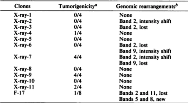
Tumorigenicity and Genomic Rearrangement. One million cells from each clone isolated from type III foci were injected s.c. into syngeneic nonimmunosuppressed C3H mice to evalu ate the tumorigenicity of the clones. Tumors appeared from 6 to 20 weeks after injection. As can be seen in Table 2, only 5 of the 11 clones transformed by X-rays gave rise to tumors, at a fre quency ranging from 1 of 8 to 4 of 4. The appearance of specific
genomic rearrangements as detected by the fingerprinting assay did not correlate with the tumorigenic potential of the cells (Table 2). Similar results were found with clones derived from F-17 that were exposed to a second dose of X-rays (Table 3); although multiple genomic rearrangements were observed, these did not correlate with tumorigenicity. No tumors arose in mice given injections of cells from UV-C-transformed clones (Table 4). On the other hand, we found a perfect correlation for clones transformed by MCA; loss of band 2 was always associ ated with tumorigenicity.
DISCUSSION
In the present study, the occurrence of genomic rearrange ments in cells transformed by specific carcinogens was analyzed by a DNA fingerprinting assay. Multilocus and multiallele probes identified as M, X, H10, and H16 were selected to detect genomic rearrangements in clones of C3H/10T'/2 fibroblast cells transformed by X-rays, UV-C, or 3-methylcholanthrene. We found genomic rearrangements in DNA only with probe M; these occurred in about 40% (5 of 12) of clones isolated from type III foci induced by X-rays. By use of the same probe, a similar frequency of genomic rearrangement has been reported in CD-I mouse liver tumors induced by 7,12-dimethylbenz[a]-anthracene (8). On the other hand, we were unable to detect any modification in the resolvable part of the gel with the others probes (X, H10, and H16) in clones irradiated with either one or two doses of 600 cGy.
These latter results could appear surprising in that each probe allowed detection of many minisatellite sequences dispersed
B
23 kh 23 kh
Fig. 4. Genomic rearrangements induced by 3-methylcholanthrene or UV-C. A, DNA di gested with //in/I was allowed to migrate for 32 h. and bands were detected with probe M.
B, DNA from clone MCA-6 was allowed to
migrate for 26 h.
•¿mi
^p<*•>
*»
9.4 kh 6.6 khe
2 3 3.5 4 9.4 kh •¿Â»Â«ft ** ~* ** 9 IO II 12 * 8•¿ÃŽI
•¿Â» 12 6.6 kh^22222^ccq
gjQG<"*or5Ã-tÃ-?r
N- I I I I I ~ O •¿"f^ '•*>*» 'J\ Kt •¿J»G
O ¿N 5791DNA FINGERPRINT OF CELLS TRANSFORMED BY X-RAY. UV-C. AND MCA
Table 2 Relationship between tumorigenicity and genomic rearrangements
induced by a single dose of X-rays (600 cGy)
ClonesX-ray- 1X-ray-2X-ray-3X-ray-4X-ray-5X-ray-6X-ray-7X-ray-8X-ray-9X-ray-10X-rav-11F-17Tumorigenicity"0/40/40/41/40/40/44/40/44/40/42/41/8Genomic rearrangements*NoneBand 2, intensity shiftBand 2, lostNoneNoneBand 2, lostBand 9. intensity shiftBand 2. intensity shiftBand 9, lostNoneNoneNoneNoneBands 2 and 11, lostBands 5 and 8. new " Number of animals with tumors/number of animals given injections.
h Genomic rearrangement was observed only with probe M (frequency, 5 of 12).
Table 3 Tumorigenicity and genomic rearrangements of transformed
C3H/IOT'/! cells exposed to a second dose of 600 cGy of X-rays
Clones"F-17-1F-17-2F-17-3F-17-4F-17-5F-17Tumorigenicity*0/41/30/41/44/91/8Genomic
rearrangements'2"LostLostLostLostLostLost5 6NewNewNewNewNew
NewNew789New-NewNewLostNew11LostLostLostLostLostLost17LostLostLost "Clones F-17-1 to F-17-5 were isolated from a previously transformed clone
(F-17) exposed to a second dose of 600 cGy and recloned.
* Number of animals with tumors/number of animals given injections. ' Genomic rearrangement frequency. 5 of 5.
''Band number.
throughout the genome. Such specificity suggests that the dam age produced by X-rays in the genome may not be a random process. It seems unlikely that a nonuniform distribution of radiation energy within the DNA could explain this phenome non. Wolfe et al. (22) have reported variation in mutation rate among regions in the genome. They showed a correlation with the base composition of genes and their flanking DNA for the rate of silent substitution. It is tempting to speculate that a particular structure or sequence of DNA confers genetic insta bility or a mutational hot-spot not only to X-rays but also to other carcinogens such as UV-C and 3-methylcholanthrene, because the loss of band 2 detected only with probe M was common to all three of these carcinogens.
This complete or partial deletion of band 2 could reflect the inactivation of the suppressor gene. However, we found that band 2 was not apparently related to either the RB orp53 genes. For clones which showed no loss of band 2, inactivation of a suppressor gene could be mediated either by a point mutation or a short deletion not resolvable with our assay or by mutations in other tumor suppressor genes for which the fragments gener ated by enzymatic digestion with //in/I were too small to be analyzed in our assay. This interpretation could explain the lack of correlation between the tumorigenicity in syngeneic nonim-munosuppressed mice of clones transformed by X-rays and a systematic loss of band 2.
Our tumorigenicity test appears to result in a lower incidence of tumors than previously reported (19, 23, 24); however, these earlier experiments were carried out with nude, newborn, or immunosuppressed mice, facilitating the growth of cancer cells. Our assay is probably more stringent and may thus make the link between a modification in the fingerprint pattern and the tumorigenicity of a clone more informative. Although some
modifications in the fingerprint could be irrelevant, the involve ment of the loss of band 2 in the mechanism of transformation by X-rays deserves further investigation.
Carcinogenesis is recognized as a multistep process involving different combinations of oncogenes (25) and including both early and late events. For clones X-ray-3, X-ray-6, and F-17 loss of band 2 would appear to represent an early event in the mechanism of carcinogenesis, because a complete deletion was detected. On the other hand, with clones X-ray-2 and X-ray-7 a genomic rearrangement affecting band 2 may be a later event, because an intensity shift was observed in the fingerprint assay, suggesting that the change occurred in only a fraction of the cell population. As a result, clones X-ray-2 and X-ray-7 would rep resent a mixed population in which some cells retained and others lost band 2.
Our DNA fingerprinting assay has also detected some ge nomic rearrangements more specific to the carcinogens. The new band 3.5 of clone MCA-6 represents a specific modifica tion induced by MCA, while the loss of bands 9, 11, and 17 and the migration of the new bands 5, 6, and 7 were induced only by X-ray. With appropriate probes, therefore, it seems possible that the DNA fingerprinting technique might be useful in a predictive assay to identify the carcinogenic agent involved in the induction of malignant transformation or a clinical cancer. This hypothesis is supported by the observation that different fingerprint patterns occurred in spontaneous, compared with 7,12-dimethylbenz[a]anthracene-induced, mouse liver tumors (8).
The results of the second irradiation of clone F-17 emphasize the apparent genomic instability of transformed cells. All clones isolated from a previously transformed clone exposed to a sec ond dose of X-rays showed genomic rearrangements, compared to a frequency of about 40% for clones arising after one dose of X-rays. Although preliminary, this result suggests that the ac cumulation of genomic rearrangements is related to the state of transformation of the cell. This phenomenon could be due to the creation of genomic instability during the process of trans formation. Thus, we might expect that the rate of mutation would increase in the same manner, accelerating the process of transformation or inducing a cascade of events once the cell has reached a certain stage of transformation. To support a rela tionship between genomic instability and tumorigenicity, mu tation rates of normal and transformed cells have already been compared. Embryonic Chinese hamster cells transformed by simian virus 40 and polyoma virus showed a higher mutation
Table 4 Relationship between tumorigenicity and genomic rearrangements
induced by VV-C and 3-methylcholanthrene
Clones Tumorigenicity" Genomic rearrangements*
UV-1UV-2UV-3UV-4UV-5MCA-1MCA-2MCA-3MCA-4MCA-5MCA-60/40/40/40/40/44/44/40/44/40/4ND<NoneBand 2. lostNoneNoneNoneBand 2. lostBand 2, lostNoneBand 2. lostNoneBand 2, lostBand 3.5, newBand 8, new
" Number of animals with tumors/number of animals given injections.
* Genomic rearrangement frequency detected with probe M: UV-C, 1 MCA, 4 of 6.
' ND, not determined.
of 5;
DNA FINGERPRINT OF CELLS TRANSFORMED BY X-RAY, UV-C. AND MCA
rate at loci responsible for 6-thioguanine and ouabain resistance than did nontransformed cells (26). Cifone and Fidler (27) re ported that murine fibrosarcoma UV-2237 with high metastatic potential had a 3-7-fold increase in the rate of mutation, com pared with a subclone with low metastatic potential. Mutation at the hypoxanthine-guanine phosphoribosyltransferase locus was also reported to be higher for neoplastic lymphocytes than normal lymphocytes (28).
A number of mechanisms could lead to genomic instability. A genome-wide process could be involved; for example, mutant enzymes in DNA replication or repair could play a role in neoplastic progression by causing extensive deletions in DNA (29). On the other hand, genomic instability might involve only a specific class of genes. For example, mutation rates in dere-pressed genes were significantly higher than in genes in the repressed state for yeast Saccharomyces (30), suggesting that genes which are more actively transcribed are also more vul nerable to mutation. Our results, however, tend to support a more specific mechanism perhaps related to DNA structure or sequence, in that the increase in genomic rearrangements was detected with only one of the four probes used. Clearly, more experiments are necessary to support and clarify this hypothe sis.
In conclusion, the DNA fingerprinting assay has allowed de tection of specific genomic rearrangements in a number of (but not all) clones transformed with the three carcinogenic agents; however, X-rays induced a greater variety of rearrange ments than those observed with UV-C or 3-methylcholan-threne. This result suggests that the molecular mechanisms of transformation by various carcinogens may differ. Because the genomic rearrangements were detected with only one of the four probes used, it is tempting to speculate that a particular structure or sequence of DNA confers a genomic instability or a mutational hot-spot to these carcinogens. Our results suggest also that the stage of transformation and the occurrence of genomic rearrangement induced by X-rays may be related.
ACKNOWLEDGMENTS
We thank Dr. Brian T. Ledwith for providing the probes. REFERENCES
1. Guerrero, I., Calzada, P., Mayer, A., and Pellicer, A. A molecular approach to leukemogenesis: mouse lymphomas contain an activated c-ras oncogene. Proc. Nati. Acad. Sci. USA, 81: 202-205, 1984.
2. Newcomb, E. W., Steinberg, J. J., and Pellicer, A. ras oncogenes and phe-notypic staging in jV-methylnitrosourea- and 7-irradiation-induced thymic lymphomas in C57BL/6J mice. Cancer Res., 48: 5514-5521, 1988. 3. Sawey, M. J.. Hood, A. T., Burns, F. J., and Carte, S. J. Activation of c-myc
and c-K-raj oncogenes in primary rat tumors induced by ionizing radiation. Mol. Cell. Biol., 7: 932-935, 1987.
4. Gumerlock, P. H., Meyers, F. J., Foster, B. A., Kawakami, T. G., and deVere White, R. W. Activated c-N-ras in radiation-induced acute nonlymphocytic leukemia: twelfth codon aspartic acid. Radiât.Res.. /17: 198-206. 1989. 5. Borek, C., Ong, A., and Mason. H. Distinctive transforming genes in
X-ray-transformed mammalian cells. Proc. Nati. Acad. Sci. USA, 84: 794-798, 1987.
6. Krolewski, B., and Little, J. B. Molecular analysis of DNA isolated from the different stages of X-ray-induced transformation in vitro. Mol. Carcinog., 2: 27-33, 1989.
7. Garte. S. J., and Burns, F. J. Oncogenes and radiation carcinogenesis. En viron. Health Perspect., 93: 45-49, 1991.
8. Ledwith, B. J., Storer, R. D., Prahalada, S., Manam, S., Leander, K. R., van Zwicten, M. J., Nichols, W. W., and Bradley, M. O. DNA fingerprinting of 7,12-dimethylbenz[o]anthracene-induced and spontaneous CD-I mouse liver tumors. Cancer Res., 50: 5245-5249, 1990.
9. Wong, Z., Wilson, V., Patel, I., Povey, S., and Jeffreys, A. J. Characteriza tion of a panel of highly variable minisatellites cloned from human DNA. Ann. Hum. Genet., 51: 269-288, 1987.
10. Jeffreys, A. J., Wilson. V., and Thein, S. L. Hypervariable minisatellite regions in human DNA. Nature (Lond.), 314: 67-73, 1985.
11. Jeffreys, A. J., Wilson, V., and Thein, S. L. Individual-specific 'fingerprints' of human DNA. Nature (Lond.). 316: 76-79, 1985.
12. Wahls, W. P., Wallace, L. J., and Moore, P. D. Hypervariable minisatellite DNA is a hot-spot for homologous recombination in human cells. Cell, 60: 95-103, 1990.
13. Jeffreys, A. J., Bookfield, J. F. Y., and Semeonoff, R. Positive identification of an immigrant test-case using human DNA fingerprints. Nature (Lond.),
317: 818-819, 1985.
14. Steinmetz, M., Stephen, D., and Lindahl, K. F. Gene organization and re-combinational hot-spots in the murine major histocompatibility complex. Cell, 44: 895-904, 1986.
15. Jeffreys, A. J. Highly variable minisatellites and DNA fingerprints. Biochem. Soc. Trans., 15: 309-317. 1987.
16. Kobari, J. A.. Strauss, E., Minard, K., and Hood, L. Molecular analysis of the hot-spot of recombination in the murine major histocompatibility complex. Science (Washington DC), 234: 173-179, 1986.
17. Reznikoff, C. A., Bertram, J. S., Brankow, D. W., and Heidelberger, C. Establishment and characterization of a cloned line of C3H mouse embryo cells sensitive to postconfluence inhibition of division. Cancer Res., 33: 3231-3238, 1973.
18. Reznikoff, C. A., Bertram. J. S., Brankow, D. W., and Heidelberger, C. Quantitative and qualitiative studies of chemical transformation of cloned C3H mouse embryo cells sensitive to postconfluence inhibition of cell divi sion. Cancer Res., 33: 3239-3249. 1973.
19. Chan. G. L., and Little, J. B. Correlation of in vitro transformation with in
vivo tumorigenicity in 10T'/2 mouse cells exposed to UV light. Br. J. Cancer,
59:590-593, 1979.
20. Perbal, B. Purification of high molecular weight cellular DNA. In: B. Perbal (éd.),A Practical Guide to Molecular Cloning, Ed. 2, pp. 324-325. New York: John Wiley & Sons, 1988.
21. Ledwith, B. J., Manam, S., Nichols, W. W., and Bradley, M. O. Preparation of synthetic tandem-repetitive probes for DNA fingerprinting. Biotechniques.
9: 149-152, 1990.
22. Wolfe, K. H., Sharp, P. M., and Li, W-H. Mutation rates differ among regions of mammalian genome. Nature (Lond.), 337: 283-285, 1989. 23. Terzaghi, M., and Little, J. B. X-radiation-induced transformation in a C3H
mouse embryo-derived cell line. Cancer Res., 36: 1367-1374, 1976. 24. Hahn, G. M. Comparison of the malignant potential of lOT'/z cells and
transformants with their survival responses to hyperthermia and to ampho-tericin B. Cancer Res., 40: 3763-3767, 1980.
25. Boyd, J. A., and Barrett, J. C. Genetic and cellular basis of multistep car cinogenesis. Pharmacol. Ther.. 46: 469-486, 1990.
26. Goldberg, S., and Defendi, V. Increased mutation rates in doubly viral trans formed Chinese hamster cells. Somat. Cell Genet., 5: 887-895, 1979. 27. Cifone, M. A., and Fidler, I. J. Increasing metastatic potential is associated
with increasing genetic instabiity of clones isolated from murine neoplasms. Proc. Nati. Acad. Sci. USA, 78: 6949-6952, 1981.
28. Seshadri, R.. Kutlaca. R. J., Trainor. K.. Matthews, C., and Morley, A. A. Mutation rate of normal and malignant human lymphocytes. Cancer Res.. 49:407-409, 1987.
29. Kaden, D. A., Bardwell, L., Newmark, P., Anisowicz, A., Skopek, T. R., and Sager, R. High frequency of large spontaneous deletions of DNA in tumor derived CHEF cells. Proc. Nati. Acad. Sci. USA, 86: 2306-2310, 1989. 30. Korogodin, V. I., Korogdina, V. L., Fajszi, Cs. Chepurnoy. A. I.,
Mikhova-Tsenova, N., and Simonyan, N. V. On the dependence of spontaneous mu tation rates on the functional state of genes. Yeast, 7: 105-117. 1991.