DOCTORAT DE L'UNIVERSITÉ DE TOULOUSE
Délivré par :
Institut National Polytechnique de Toulouse (INP Toulouse)
Discipline ou spécialité :
Pathologie, Toxicologie, Génétique et Nutrition
Présentée et soutenue par :
Mme THAIS HAUTBERGUE
le mardi 14 novembre 2017
Titre :
Unité de recherche :
Ecole doctorale :
Caractérisation chimique des métabolomes secondaires de Penicillium et
Fusarium par marquage isotopique couplé à la spectrométrie de masse
haute résolution
Sciences Ecologiques, Vétérinaires, Agronomiques et Bioingénieries (SEVAB)
Toxicologie Alimentaire (ToxAlim)
Directeur(s) de Thèse :
MME ISABELLE OSWALDM. EMILIEN JAMIN
Rapporteurs :
M. DAVID TOUBOUL, CNRS GIF SUR YVETTE M. JOSE DIANA DI MAVUNGU, UNIVERSITE DE GENT
Membre(s) du jury :
Mme FLORENCE MATHIEU, INP TOULOUSE, Président M. CEDRIC BERTRAND, UNIVERSITE DE PERPIGNAN, Membre
M. FABIEN JOURDAN, INRA TOULOUSE, Membre M. OLIVIER PUEL, INRA TOULOUSE, Membre
« Le rôle des infiniment petits dans la nature
est infiniment grand »
Louis Pasteur
Je remercie le Ministère de l’Éducation nationale, de l’Enseignement Supérieur et de la Recherche, les porteurs du projet MetaboHUB-ANR-11-INBS-0010, l’ensemble des représentants de l’INRA et en particulier de l’unité Toxalim pour leur soutien matériel et financier sans lesquels je n’aurais pas pu mener cette thèse à son terme. Je remercie également les représentants de la plateforme MetaboHub-MetaToul de m’avoir intégrée dans leur structure ainsi que l’INP et l’école doctorale SEVAB. Parmi eux, je remercie tout particulièrement Claude Maranges pour l’attention et la bienveillance dont il fait preuve envers les doctorants de l’école doctorale SEVAB.
Je tiens à remercier Isabelle Oswald et Emilien Jamin qui ont accepté de diriger ma thèse, et bien sûr Olivier Puel pour avoir été mon troisième encadrant majeur. Merci Isabelle pour m’avoir accueillie au sein de votre équipe et pour m’avoir fait confiance au cours de cette thèse. Je vous remercie d’avoir été le chef d’orchestre nécessaire au bon fonctionnement de cette thèse ainsi que pour votre importante réactivité malgré les nombreuses responsabilités qui vous incombent. Emilien, merci également de m’avoir formée sur les techniques analytiques, pour ta réactivité et pour ton implication particulière dans ce projet. Merci de m’avoir fait confiance, de m’avoir accordé une grande autonomie sur ce projet, tout en étant bien sûr présent à chacune des étapes. De nombreux paramètres (aspect spectrométrie de masse de ma thèse, le fait que tu sois mon directeur de thèse, que je sois ta première thésarde, cohabitation au sein du bâtiment A) ont fait que tu as été l’encadrant avec lequel j’ai le plus échangé au cours de ces trois années. Tu as notamment veillé à ce que j’apprenne à canaliser mon énergie et à ce que je découvre tous les aspects de la vie en entreprise. Olivier, merci pour ton expertise, mais aussi pour ton optimisme qui m’a plus d’une fois réconfortée dans mes moments de doute. Merci beaucoup pour ton implication, pour avoir supervisé l’aspect fongique de ma thèse et pour m’avoir accordé le temps dont j’avais besoin malgré tes nombreux projets.
Je voudrais également remercier les membres de mon comité de thèse. Laurent Debrauwer, la force tranquille qui veille de loin sur chacun des membres de son équipe. Merci pour votre humanité et votre gentillesse qui m’ont permis de garder la tête hors de l’eau dans les moments difficiles. Cette énergie pour continuer malgré les difficultés, je l’ai également puisée chez Jean-Claude Tabet. Les semaines intensives d’interprétation de nos spectres et de réflexion scientifique m’ont permis de vivre pleinement ma passion pour ces casse-têtes et de m’épanouir professionnellement. J’ai rencontré au début de ma thèse un véritable mentor, un exemple autant sur le plan technique qu’humain. Merci pour tout Jean-Claude, de la part de tous tes étudiants, sincèrement. Enfin, merci Yann Guitton pour ton expertise en bioinformatique. Tu as cru dans ce projet et dans ses perspectives. Ton regard extérieur toujours très positif n’a fait que renforcer ma motivation.
Je souhaite remercier les rapporteurs de ma thèse, David Touboul et José Diana di Mavungu d’avoir accepté d’accorder de leur précieux temps à la lecture de mon manuscrit, ainsi que les membres du jury pour avoir accepté d’être présents le jour de ma soutenance. Merci à vous tous de le faire part de votre expertise.
Je tiens à remercier également tous les membres d’AXIOM pour leur aide tout au long de ce projet. Merci à l’équipe 5 de m’avoir accueillie chaleureusement. Merci Soraya pour tes précieux conseils, merci Sylviane et Jean-Denis pour votre bienveillance et votre aide. Je dédie une pensée affective pour tous les « jeunes » du labo grâce à qui la thèse représente également de super souvenirs amicaux. Ceux du bureau, tout d’abord, avec qui j’ai partagé notre « placard ». Aurélien (désolée encore de t’avoir rendu la vie aussi difficile !), Christelle, Djawed, Adéline, Salim, Robin et tout particulièrement Claire notre petite sœur si présente. Alyssa, on est arrivée en même temps mais toi, tu as pris perpet’ comme on dit. Selma, Maria, Clément, Cyndel, Jade, Elodie, Morgane, Laura, Perrine, mais aussi tous les geeks, les jeunes de MeX et les filles de l’équipe 5, merci. Je reste fière d’avoir supervisé la première et sûrement dernière maison de campagne construite à l’intérieur des bureaux de l’INRA. La glycine n’a jamais aussi bien poussé que dans notre cagibi. Enfin, merci à tous ceux qui m’ont entourée de près ou de loin dans les moments difficiles et qui ont stimulé mes désirs de réussite.
TABLE DES MATIÈRES
COMMUNICATIONS SCIENTIFIQUES ...i
TABLE DES ILLUSTRATIONS ... iii
LISTE DES ABBRÉVIATIONS ... v
INTRODUCTION ... 1
1. CONTEXTE DE L’ÉTUDE ... 3
2. OBJECTIFS ET DÉROULEMENT DE LA THÈSE ... 5
3. ARTICLE 1 : CARACTÉRISATION DES MÉTABOLITES SECONDAIRES FONGIQUES... 7
4. DES ESPÈCES FONGIQUES TOXINOGÈNES ... 33
4.1. P. verrucosum et P. nordicum ... 33
4.1.1. Le genre Penicillium ... 33
4.1.2. Propriétés de P. verrucosum et P. nordicum ... 34
4.1.3. Critères de classification de P. verrucosum et P. nordicum ... 35
4.1.4. Métabolomes secondaires de P. verrucosum et P. nordicum ... 37
4.2. F. graminearum, un champignon phytopathogène prédominant en Europe ... 39
4.2.1. Présentation de F. graminearum ... 39
4.2.2. Le métabolome secondaire de F. graminearum ... 41
5.LES FUNGISPORINES ... 42
5.1. Une découverte ancienne, une caractérisation récente ... 42
5.2. Origines génétiques des fungisporines ... 44
5.3. Des métabolites d'intérêt ... 45
6.RÉFÉRENCES DE L'INTRODUCTION ... 47
MATÉRIELS & MÉTHODES ... 49
1. LA GÉNÉRATION DES DONNÉES ... 51
1.1. La méthode analytique ... 51
1.1.1. Le principe ... 51
1.1.2. La métabolomique non ciblée ... 52
1.1.3. Marquage isotopique du métabolome secondaire fongique ... 52
1.1.4. Analyse des métabolomes par HPLC-HRMS ... 53
1.2. Analyse des données ... 54
1.2.1. Extraction des pics... 54
1.2.2. Suppression de la redondance d’information ... 57
1.2.3. Détermination des formules chimiques ... 58
1.3. Analyse des métabolites secondaires par MS/MS ... 61
1.3.1. Génération des spectres MS/MS par acquisition data dépendante ... 61
1.3.2. Analyse ciblée des métabolites secondaires fongiques en mode CID ... 61
1.3.3. Analyse ciblée des métabolites secondaires fongiques en mode HCD ... 62
2. INTERPRÉTATION DES RÉSULTATS ... 62
2.1. Les bases de données ... 62
2.2. Validation des annotations : identification ... 62
2.3. Les réseaux moléculaires : Le “GNPS Molecular Networking System” ... 63
3. LE SÉQUENÇAGE PEPTIDIQUE DE NOVO ... 64
3.1. Les outils de séquençage peptidique automatique ... 64
3.2. Le séquençage peptidique de novo ... 65
3.2.1. Le modèle du proton mobile ... 65
3.2.2. Étude théorique des mécanismes de fragmentation des peptides linéaires... 65
3.2.3. Les mécanismes de fragmentation des adduits sodium ... 70
TRAVAIL EXPÉRIMENTAL ... 77
PARTIE 1. CARACTÉRISATION DU MÉTABOLOME SECONDAIRE DE P. verrucosum ... 79
1.ARTICLE 2. CARACTÉRISATION DU MÉTABOLOME SECONDAIRE DE P. verrucosum ... 81
2.OPTIMISATION DES RÉSEAUX MOLÉCULAIRES ... 102
2.1. Optimisation des données ... 102
2.2. Optimisation desparamètres du réseau moléculaire ... 102
2.3.Visualisation du réseau moléculaire ... 105
3.INTERPRÉTATION DES RÉSEAUX MOLÉCULAIRES ... 105
4.RÉFÉRENCES QUANT À L’ÉTUDE DE P. verrucosum ... 107
PARTIE 2. LA PRODUCTION DE FUNGISPORINES PAR P. nordicum ... 109
1. ARTICLE 3. RECHERCHE DE NOUVEAUX PEPTIDES CHEZ P. nordicum ... 111
2. ARTICLE 4. UN MÉCANISME DE FRAGMENTATION PEPTIDIQUE NON RÉFÉRENCÉ ... 137
PARTIE 3. ÉTUDE DU MÉTABOLOME SECONDAIRE DE F. graminearum ... 147
1. ARTICLE 5. LE MÉTABOLOME D'UN PUISSANT PHYTOPATHOGÈNE ... 149
DISCUSSION ... 173
1. ANALYSE CRITIQUE DE LA MÉTHODE... 177
1.1. Marquage isotopique du substrat fongique ... 177
1.1.1. Une méthode adaptable à de multiples sujets d’étude ... 177
1.1.2. Limites de la méthode ... 177
1.2. Les outils bioinformatiques ... 178
1.2.1. CFM-ID ... 178
1.2.2. Les réseaux moléculaires ... 179
1.2.3. Développement d’un nouvel outil bioinformatique ... 180
1.3. Analyse critique de l’étude métabolomique des champignons filamenteux ... 181
1.4. Discussion autour de la fragmentation peptidique ... 183
2. ANALYSE DES RÉSULTATS DE L'ÉTUDE ... 184
2.1. Environ 80% de métabolites secondaires inconnus ... 184
2.2. Le métabolome secondaire de F. graminearum ... 185
2.2.1. Des métabolomes secondaires différents selon la souche et l’environnement ... 185
2.2.2. Découvertes de nouvelles fusaristatines chez F. graminearum ... 186
2.3. Les métabolomes secondaires des espèces de Penicillium productrices d’ochratoxine A ... 189
2.3.1. Caractérisation de métabolites secondaires connus ... 189
2.3.2. Découverte d’un nouveau peptide cyclique ... 190
2.3.3. Caractérisation de nouveaux tétrapeptides linéaires ... 192
2.3.4. Production de nouveaux pentapeptides par P. nordicum ... 194
2.3.5. Hypothèses quant aux propriétés des tétrapeptides produits les Penicilllium ... 194
2.3.6. Perspectives pour l’analyse de ces composés ... 195
3. CLASSIFICATION MÉTABOLIQUE ET NOTION D’ESPÈCE FONGIQUE ... 197
3.1. Proximité phylogénique et similarité métabolique ... 197
3.2. Une souche de P. verrucosum chémotype anacine ... 197
3.3. Réflexion autour de la notion de caractérisation métabolique ... 198
3.3.1. Mise au point du système ... 198
3.3.2. Identification d’une souche grâce à ce système ... 200
3.3.3. Analyse critique d’un tel système de classification métabolique ... 200
4. RÉFÉRENCES DE LA DISCUSSION ... 202
COMMUNICATIONS SCIENTIFIQUES
ARTICLES
T. Hautbergue, O. Puel, S. Tadrist, L. Meneghetti, M. Péan, M. Delaforge, L. Debrauwer, I. P. Oswald and E. L. Jamin « Evidencing 98 secondary metabolites of Penicillium verrucosum using substrate isotopic labeling and high resolution mass spectrometry », Journal of Chromatography B. Sous presse. doi: 10.1016/j.jchromb.2017.03.011
T. Hautbergue, O. Puel, L. Debrauwer, E. L. Jamin and I. Oswald « From genomic to metabolomic, moving toward an integrated strategy for the discovery of fungal secondary metabolites », soumis à Natural Product Reports
T. Hautbergue et al. « Ochratoxin A producing Penicillium species: new tetrapeptides on Penicillium nordicum’s secondary metabolome », en preparation
T. Hautbergue et al. « Evidencing a new peptidic fragmentation mechanism », en préparation
P. Cano, E. Jamin, S. Tadrist, T. Hautbergue, P. Bourdaudhui, M. Péan, L. Debrauwer, I. P. Oswald, M. Delaforge, O. Puel « Identification of new secondary metabolites produced in vitro and in planta by F. graminearum on wheat using isotopic labeling and HPLC-HRMS », en préparation
CONFÉRENCES
T. Hautbergue, O. Puel, S. Tadrist, M. Péan, M. Delaforge, L. Debrauwer, I. P. Oswald, E. L. Jamin « Identification of six new fungisporins in Penicillium nordicum by stable isotope labeling and HPLC-HRMS » 33e Journées de Spectrométrie de Masse (JFSM), 2016, Bordeaux, France.
T. Hautbergue, O. Puel, S. Tadrist, M. Péan, M. Delaforge, L. Debrauwer, I. P. Oswald, E. L. Jamin « Molecular networks: optimization of parameters to study the secondary metabolome of Penicillium verrucosum and Penicillium nordicum » Rencontre du Club des Jeunes en Spectrométrie de Masse, 2016, Moustier-Sainte-Marie, France.
Prix de la meilleure présentation orale
T. Hautbergue, O. Puel, S. Tadrist, M. Péan, M. Delaforge, L. Debrauwer, I. P. Oswald, E. L. Jamin « Evidencing 161 new secondary metabolites in Penicillium by stable isotope labeling and HPLC-HRMS » Journée Nationale des Mycotoxines, 2016, Toulouse, France.
T. Hautbergue, O. Puel, S. Tadrist, M. Péan, M. Delaforge, L. Debrauwer, I. P. Oswald, E. L. Jamin « Double stable isotope labeling and high-resolution mass spectrometry for the characterization of fungal secondary metabolomes » Journée de Spectrométrie de Masse en Midi-Pyrénées (SMMP), 2015, Toulouse, France.
S. Chevolleau, I. Jouanin, T. Hautbergue, O. Martin, F. Guéraud, F. Pierre, L. Debrauwer
« Development of a LC-HRMS method for the non-targeted analysis of aldehydes: application for the detection of peroxidation products in sewage waters » 10th Congress of the Francophone Association Francophone des Sciences Séparatives (AFSEP), 2013, Paris, France.
POSTERS
T. Hautbergue, E. L. Jamin, O. Puel, L. Debrauwer and I. P. Oswald « Characterization of the secondary metabolome of Penicillium by stable isotope labeling and HPLC HRMS » 65e Congrès de l’American Society for Mass Spectrometry (ASMS), 2016, Indianapolis, USA.
T. Hautbergue, E. L. Jamin, C. Canlet, S. Tadrist, P. Bourdaudhui, O. Puel, L. Debrauwer and I. P. Oswald « Characterization of new secondary metabolites of Penicillium using isotopic labeling and HPLC-HRMS » 21e International Mass Spectrometry Conference (IMSC), 2016, Toronto, Canada.
T. Hautbergue, O. Puel, S. Tadrist, M. Péan, M. Delaforge, L. Debrauwer, I. P. Oswald, E. L. Jamin « Characterization of the secondary metabolome of Penicillium by stable isotope labeling and HPLC HRMS » Congrès de Spectrométrie de Masse et d’Analyse Protéomique (SMAP), 2015, Ajaccio, France.
T. Hautbergue, E. L. Jamin, C. Canlet, S. Tadrist, P. Bourdaudhui, O. Puel, L. Debrauwer and I. P. Oswald « Double stable isotope labeling and high-resolution mass spectrometry for the characterization of fungal secondary metabolomes » 11e Annual International Conference of the Metabolomics Society, 2015, San Francisco, USA.
MÉDIATION SCIENTIFIQUE
• Article Web pour l’Institut National de Recherche Agronomique (INRA)
Titre : « Un champignon passé au crible des chercheurs : 86 nouvelles molécules chez Penicillium verrucosum »
Lien : http://www.sa.inra.fr/Toutes-les-actualites/Un-champignon-passe-au-crible-des-chercheurs-86-nouvelles-molecules-chez-Penicillium-verrucosum
• Concours de médiation scientifique « Ma thèse en 180 secondes », France
Lien vers la vidéo de la prestation en finale nationale : https://www.youtube.com/watch?v=JxQY-0N1Sgk Règles : Les participants ont 180 secondes pour expliquer le sujet de leur thèse en français et en termes simples, à un auditoire profane et diversifié. Ils sont évalués par des représentants du monde socio-économique et du monde médiatique ainsi que des acteurs de la recherche sur le contenu et la clarté de leur présentation mais également sur leur charisme.
Etapes de sélection : Mars 2016 Premier prix du jury et premier prix du public en finale régionale Mai 2016 Première place en demie finale nationale
Conférences invitées dans le cadre de MT180 :
Juin 2016 Présentation pour BioMedical Alliance et Bioméridies (Narbonne, France)
Janvier 2017 Présentation lors des vœux de la Ministre de l’Education nationale, de l’Enseignement supérieur et de la Recherche Najat Vallaud-Belkacem (Musée de l’Homme, Paris, France)
TABLE DES ILLUSTRATIONS
Figure 1 Présentation des travaux expérimentaux réalisés aux cours de la thèse Figure 2 Classification des Penicillium selon le degré de ramification de leur mycélium. Figure 3 P. verrucosum : conidiophores et conidies.
Figure 4 Classification phylogénique de P. verrucosum et P. nordicum au sein du genre Penicillium.
Figure 5 Culture sur milieu YES agar de P. verrucosum et P. nordicum après 7 jours à 25°C. Figure 6 Métabolites secondaires de P. verrucosum et P. nordicum recensés dans la littérature. Figure 7 Observation macroscopique et microscopique de F. graminearum.
Figure 8 Épi de blé infesté par F. graminearum.
Figure 9 Principaux métabolites secondaires de F. graminearum.
Figure 10 Lien hypothétique entre les tétrapeptides linéaires et les tétrapeptides fongiques. Figure 11 Schématisation des gènes hcpA d’A. niger et de P. chrysogenum responsables
notamment de la synthèse de fungisporines chez ces champignons. Figure 12 Culture de P. chrysogenum. Souche sauvage et mutant ΔhcpA.
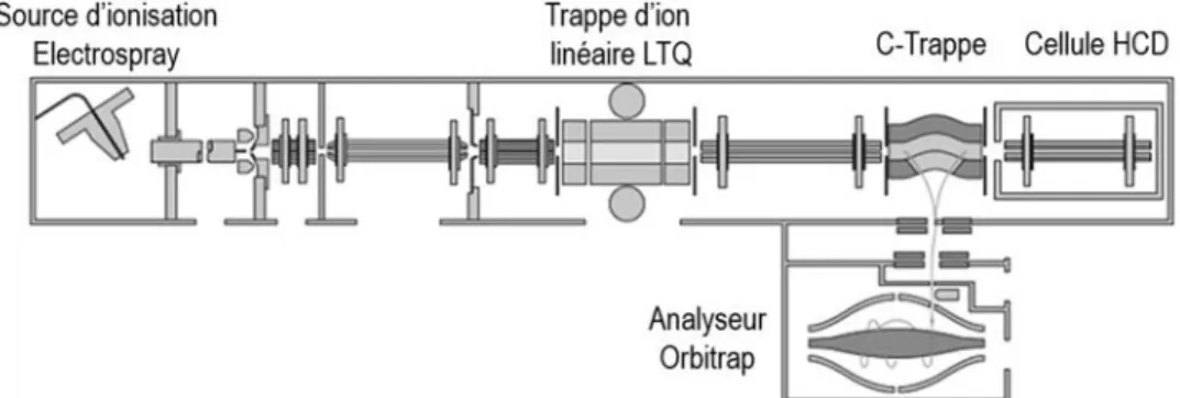
Figure 13 Stratégie analytique mise en œuvre pour l’analyse des métabolomes secondaires fongiques par double marquage isotopique et spectrométrie de masse haute résolution. Figure 14 Cibles d’un marquage isotopique selon son degré de spécificité du précurseur marqué. Figure 15 Schéma d’un spectromètre de masse LTQ-Orbitrap.
Figure 16 Protocole d’extraction des pics.
Figure 17 Extrait du spectre de masse issu de l’analyse du métabolome secondaire de P. verrucosum cultivé sur blé 13C15N.
Figure 18 Superposition des massifs isotopiques observés sur les spectres de masse d’un même métabolite marqué 53% 13C 97% 15N (A), 97% 13C (B) et 99% 12C (C).
Figure 19 Comparaison des spectres MS/MS obtenus par acquisition en mode data-dépendant des trois ions moléculaires détectés dans les spectres de masse 12C, 13C et 13C15N, d’un métabolite équivalent.
Figure 20 Utilisation du logiciel MassToFormulaCompare. Figure 21 Lexique relatif aux réseaux.
Figure 22 Nomenclature de la fragmentation peptidique en spectrométrie de masse.
Figure 23 Fragmentation peptidique par formation d’une oxazolone et dissociations consécutives. Figure 24 Fragmentation peptidique générée par formation d’une dicétopipérazine.
Figure 25 Formation des ions y par rupture hétérolytique ou par formation d’une aziridinone. Figure 26 Perte d’eau à partir de la fonction acide carboxylique terminale.
Figure 27 Formation d’ions immonium internes à partir d’ions y.
Figure 28 Hypothèses d’attaque nucléophile expliquant la perte de NH3 à partir de la fonction amine terminale d’un tryptophane.
Figure 29 Mécanisme de fragmentation peptidique des adduits sodium d’après Renner et al. Figure 30 Interaction d’un atome de sodium avec les diverses fonctions d’un tétrapeptide. Figure 31 Mécanismes de fragmentation des peptides cycliques protonés en mode CID de basse
énergie.
Figure 32 Mécanismes de fragmentation des peptides cycliques adduits au sodium.
Figure 33 Complexation du sodium sous forme de zwitterion et fragmentation des peptidiques cycliques adduits au sodium.
Figure 34 Optimisation des paramètres de GNPS en fonction de la diversité des spectres MS/MS à analyser.
Figure 35 Comparaison des spectres MS/MS de l’ochratoxine A, de l’ochratoxine B et du métabolite C17H14O5 détecté à 11,5 min.
Figure 36 Protocole d’extraction et de traitement des données de l’outil en développement. Figure 37 Utilisation de l’échange hrydrogène/deutérium pour la validation d’hypothèses structurales.
Figure 39 Hypothèses quant à l’origine génétique de la synthèse de fusaristatine C.
Figure 40 Schématisation d’un système de classification potentiel permettant la classification des espèces fongiques selon leur métabolome secondaire.
LISTE DES TABLEAUX
Tableau 1 Critères de classification de P. verrucosum et P. nordicum.
Tableau 2 Tétrapeptides cycliques, tétrapeptides linéaires non modifiés et autres composés associés référencés dans la littérature.
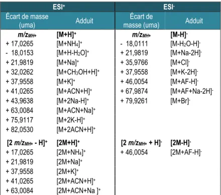
Tableau 3 Écarts de masse théoriques entre l’ion moléculaire ou le dimère et les adduits correspondants.
Tableau 4 Exemple du calcul de la composition en carbone et en azote de l’ion détecté à m/z12C=553.2814 et détermination de sa formule brute.
Tableau 5 Informations relatives aux 20 acides aminés protéinogènes standards. Tableau 6 Paramètres de génération de réseaux moléculaires par l’outil du GNPS.
Tableau 7 Analyse des similarités entre les sommets annotés et les sommets adjacents inconnus du réseau moléculaire de P. verrucosum.
LISTE DES ABRÉVIATIONS
Abréviations en anglais
CID Collision-induced dissociation
DDA Data dependent acquisition
ECD Electron-capture dissociation
ESI Electrospray ionization
ETD Electron-transfert dissociation
HCD Higher-energy collisional dissociation
HPLC High performance liquid chromatography
HRMS High resolution mass spectrometry
IC50 Half-maximal inhibitory concentration
ITS Internal transcribed spacer
LC/MS Liquid chromatography coupled to mass spectrometry
MS Mass spectrometry
MS/MS Tandem mass spectrometry
NRPS Non-ribosomal peptide synthetase
OSMAC One strain many compounds
PDA Potato dextose agar
PKS Polyketides Synthase
TIC Total ion current
USA United states of america
YES Yeast extract sucrose
Abréviations en français
ADN Acide désoxyribonucléique
CEA Commissariat à l’énergie atomique et aux énergies alternatives
IR infrarouge
m/z Rapport masse sur charge
OTA Ochratoxine A
ppm Partie par million (e.g. mg/kg)
RMN Résonance magnétique nucléaire
TR Temps de rétention
UE/EU Union européenne/European union
Introduction
« Which is better: to have fun with fungi or to have
tomorrow’s misdeeds out of yesterday’s miscreeds? »
Aldous Huxley
1. CONTEXTE DE L’ÉTUDE
Les moisissures sont des microorganismes eucaryotes appartenant au règne fongique et dotés d’un développement filamenteux. Selon leur pouvoir pathogène et le milieu qu’ils colonisent, ces champignons microscopiques peuvent être impliqués dans des problématiques très diverses. Certaines espèces sont notamment responsables d’altérations directes de la santé humaine appelées mycoses. Alors que les dermatophytoses localisées sont pour la plupart bénignes, les mycoses systémiques affectant les individus immunodéprimés entraînent 1,6 à 2 millions de décès chaque année dans le monde1. Les genres Candida et Aspergillus comptent parmi les genres les plus impliqués dans ces problématiques. Contrairement aux mycoses, les mycotoxicoses consistent en des intoxications de l’Homme et des animaux via l’ingestion ou l’inhalation de toxines produites par ces moisissures. Historiquement, l'ergotisme a été la première maladie fongique épidémique reconnue en 1676 comme résultant de la consommation d'aliments. Dans les années 1960, la maladie « turkey X disease » qui a décimé des élevages de dindes est l’un des épisodes de mycotoxicoses les plus référencés et signe le point de départ d’une nouvelle discipline « la mycotoxicologie »1. D’autres espèces de moisissures sont quant à elles phytopathogènes, entraînant des menaces pour la santé alimentaire, une destruction partielle ou totale des cultures et, par extension, d'importantes pertes économiques. En effet, la quantité de nourriture détruite chaque année en raison d’une infestation fongique est équivalente à la quantité de nourriture suffisante pour 600 millions d'individus, soit 8,5% de la population mondiale2.
Le pouvoir pathogène des moisissures résulte en partie de la production par ces microorganismes de métabolites secondaires toxiques appelés mycotoxines. Les métabolites secondaires sont des molécules de structures très variables qui ne sont pas nécessaires à la croissance des moisissures mais qui leur confèrent, en général, un avantage sélectif dans leur environnement. Ces composés ne sont pas nécessairement toxiques et certains sont, au contraire, utilisés en médecine comme antimicrobiens (pénicilline), hypolipidémiants (lovastatine), immunosuppresseurs (cyclosporine, acide mycophénolique) ou vasoconstricteurs (ergométrine). En revanche, on recense aujourd’hui plus de 400 mycotoxines. Alors que 30 d'entre elles ont des effets toxiques relativement bien caractérisés pour l'Homme et/ou les animaux, seules six familles de mycotoxines sont réglementées au niveau mondial. C’est le cas notamment de l’aflatoxine qui est l’un des plus puissants agents cancérigènes d’origine naturelle. Les genres Aspergillus, Penicillium et Fusarium sont ceux qui produisent le plus de mycotoxines.
Afin de lutter contre les champignons filamenteux, 21 000 tonnes de fongicides ont été utilisées sur les surfaces agricoles françaises en 2014. Face aux inquiétudes environnementales et sanitaires qui impliquent une diminution de l’utilisation de pesticides d’une part, et au besoin croissant de nourriture nécessaire pour satisfaire une population mondiale estimée à neuf milliards d’individus en 2050 d’autre part, la lutte contre la toxicité de ces agents pathogènes apparaît comme cruciale3. La caractérisation des molécules toxiques impliquées dans ces problématiques est une étape préalable à leur contrôle dans les denrées alimentaires. Cependant, on estime que les 11 250 métabolites secondaires fongiques connus aujourd’hui4 ne représentent que 20% de la totalité des métabolites secondaires sécrétés par les champignons filamenteux.
En Europe et dans les régions tempérées du globe, P. verrucosum et F. graminearum constituent un risque important pour les productions céréalières. Alors que P. verrucosum est un
champignon saprophyte infectant les grains lors de leur stockage, F. graminearum affecte les épis de blé lors de leur croissance dans les champs. P. nordicum infecte quant à lui principalement les denrées alimentaires protéinées telles que les viandes séchées ou cuites. La caractérisation des métabolites secondaires de ces champignons est essentielle à la diminution des pertes économiques et à la protection des populations. D’autant plus qu’il est suggéré que ces espèces fongiques sont dotées d’importantes capacités métaboliques. En effet, F. graminearum est considéré comme l’un des champignons les plus producteurs de métabolites secondaires, et une étude récente a permis de souligner l’importance du potentiel métabolique des champignons du genre Penicillium grâce à l’analyse des génomes de 24 de ses espèces5.
Au sein du laboratoire, une méthode d’analyse des métabolomes secondaires fongiques a été précédemment développée6. Celle-ci repose sur le marquage de l’ensemble des composés produits par les champignons. Dans ce but, les champignons sont cultivés sur des grains de blés marqués afin qu’ils puisent le marquage de ce substrat et l’intègrent dans tous les composés qu’ils génèrent. Cette méthode permet de détecter spécifiquement les molécules d’origine fongique au sein des extraits analysés par spectrométrie de masse et de fournir des informations quant à la composition chimique des molécules analysées.
2. OBJECTIFS ET DÉROULEMENT DE LA THÈSE
De nombreuses méthodes existent pour la recherche de nouveaux métabolites secondaires fongiques. Le choix de la méthode d’analyse mise en œuvre au sein d’un tel projet doit être effectué en connaissance du contexte analytique global. Pour cela, une revue des différentes stratégies disponibles pour la recherche de composés fongiques est présentée en introduction.
Ce projet de thèse a été élaboré afin de répondre aux objectifs suivants :
1. Caractériser les métabolomes secondaires de P. verrucosum, P. nordicum et F. graminearum. 2. Appréhender différents outils informatiques disponibles pour assister l’identification de nouveaux produits naturels.
3. Élucider la structure de métabolites secondaires inconnus.
4. Comparer les métabolomes secondaires d’espèces fongiques voisines afin d’apprécier la relation entre proximité phylogénique et similarité métabolique.
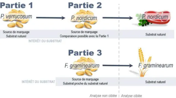
Afin de répondre à ces problématiques, ce projet se présente en trois grandes parties distinctes correspondant à l’analyse de chacune des trois espèces fongiques (Figure 1). Dans un premier temps, le métabolome secondaire de P. verrucosum est investigué. Grâce à une culture sur grains de blé marqués, il est possible de caractériser les composés produits par ce champignon phytopathogène saprophyte lorsque celui-ci se développe dans des conditions représentatives de son environnement naturel. En effet, puisque le métabolome secondaire des moisissures est finement régulé selon le milieu dans lequel elles se développent, il est important de prendre en considération ce facteur lors de la recherche de composés fongiques. Des outils bioinformatiques permettent de faciliter l’élucidation structurale de composés inconnus parmi les métabolites secondaires détectés. Afin d’apprécier la distribution de la production des nouveaux métabolites secondaires au sein du règne fongique, les composés identifiés peuvent être recherchés au sein des métabolomes secondaires d’espèces voisines et éloignées.
P. nordicum est une espèce phylogéniquement très proche de P. verrucosum. Afin de comparer les métabolomes secondaires de ces deux moisissures, les conditions de cultures et d’analyses doivent être identiques. P. nordicum est donc cultivé dans un premier temps sur grains de blé marqués. Premièrement, cela permet de rechercher la production par P. nordicum des nouveaux composés identifiés chez P. verrucosum. Deuxièmement, l’étude des similarités entre les deux métabolomes permet d’apprécier la relation entre proximité phylogénique et similarité métabolique. Enfin, ce substrat modèle constitue une source de marquage nécessaire pour la caractérisation chimique des métabolites secondaires de ce champignon toxinogène. Afin de considérer P. nordicum dans son contexte environnemental et de prendre en compte l’influence du milieu de culture sur la production de métabolites secondaires, les composés caractérisés grâce au marquage sont ensuite recherchés de manière ciblée lorsque P. nordicum se développe sur son substrat naturel.
Dans la troisième partie du travail expérimental mené au sein de cette thèse, le métabolome secondaire de F. graminearum est investigué. Puisque ce champignon est phytopathogène, la problématique environnementale dans lequel il s’inscrit est similaire à celle concernant P. verrucosum. Dans un premier temps, le champignon est donc cultivé sur grains de blé marqués afin de détecter spécifiquement et de caractériser les molécules qu’il produit sur un substrat végétal. En revanche, contrairement à P. verrucosum, F. graminearum n’est pas saprophyte et de nouveaux paramètres tels que la réponse du végétal à l’infection fongique sont susceptibles d’intervenir dans sa production de métabolites secondaires. Pour cette raison, les composés caractérisés grâce au marquage sont dans un deuxième temps recherchés dans des échantillons de blé naturellement infestés dans les champs.
3. ARTICLE 1 : CARACTÉRISATION DES MÉTABOLITES SECONDAIRES FONGIQUES
Cette partie présente les différentes méthodes ayant conduit à la découverte de nouveaux métabolites secondaires fongiques. Elle fait l’objet d’une revue soumise pour publication dans Natural Product Reports.
Deux types de stratégies sont particulièrement mis en œuvre pour la recherche de nouveaux métabolites secondaires fongiques. D’une part, les méthodes orientées vers l’analyse directe des métabolites secondaires permettent la détection des molécules sécrétées par le champignon dans les conditions expérimentales. D’autre part, l’analyse des génomes fongiques est une stratégie largement utilisée. Des approches transcriptomiques et protéomiques complémentaires peuvent également être mises en œuvre pour appuyer les résultats des analyses métabolomiques et génomiques. Aujourd’hui, le développement rapide de méthodes d’analyses omiques non ciblées permet d’atteindre des niveaux de résolution et de sensibilité qui révolutionnent l’analyse des extraits naturels. Des outils informatiques utilisés en particulier dans l’étude des métabolomes fongiques pour le traitement des importants jeux de données qui en résultent sont présentés dans cette partie. Enfin, l'influence de l'environnement de culture sur la synthèse des métabolites secondaires par les champignons est mise en évidence comme un facteur majeur à prendre en compte pour la recherche de nouveaux métabolites secondaires fongiques. Par la présentation critique de ces outils, cette revue préconise une orientation de la recherche vers une approche intégrée et souligne l’importance du développement des outils bioinformatiques associés.
Natural Product Reports
REVIEW
From Genomics to Metabolomics, Moving toward an Integrated
Strategy for the Discovery of Fungal Secondary Metabolites
T. Hautberguea,b E. L. Jamina,b, L. Debrauwera,b, O. Puela and I. P. Oswalda
Fungal secondary metabolites are defined by bioactive properties that ensure adaptation of the fungus to its environment. Although some of these natural products are promising sources of new lead compounds especially for the pharmaceutical industry, others pose risks to human and animal health. The identification of secondary metabolites is critical to assessing both the utility and risks of these compounds. Since fungi present biological specificities different from other microorganisms, this review covers the different strategies specifically used in fungal studies to perform this critical identification. Strategies focused on the direct detection of the secondary metabolites are reported since they specifically highlight the expressed part of the fungal genome. Particularly, advances in high-throughput untargeted metabolomics have led to the generation of large datasets whose exploitation and interpretation generally require bioinformatics tools. Then, the genome-based methods used to study the entire fungal metabolic potential are reported. Transcriptomic and proteomic tools used in the discovery of fungal secondary metabolites are presented as links between genomic methods and metabolomic experiments. Finally, the influence of the culture environment on the synthesis of secondary metabolites by fungi is highlighted as a major factor to consider in research on fungal secondary metabolites. Through this review, we seek to emphasize that the discovery of natural products should integrate all of these valuable tools. Attention is also drawn to emerging technologies that will certainly revolutionize fungal research and to the use of computational tools that are necessary but whose results should be interpreted carefully.
1. Introduction
Fungi are well known for their capacity to produce a broad diversity of secondary metabolites that provide them with beneficial properties for adequate growth in a fluctuating environment. On the one hand, these proprieties are of interest to various industries (particularly pharmaceutical, cosmetic and alimentary industries) that either can commercialize the natural compounds directly or develop derived products from the fungal molecules. Newman and Cragg showed that between 1981 and 2006, 28% of the newly developed industrial chemicals were of natural origin and 24% were inspired by
natural products1. Over 40% of filamentous fungi are presumed
to produce antibiotics under natural growth conditions2. Since
the discovery of penicillin by Alexander Fleming in 19283,
medicine has exploited natural fungal defenses against bacteria
to protect humans and animals from pathogenic
microorganisms. Likewise, many fungal compounds are used as antimicrobials as well as lipid-lowering medications (lovastatin),
immunosuppressants (cyclosporine and mycophenolic acid) and vasoconstrictors (ergometrine).
On the other hand, some fungal secondary metabolites named mycotoxins are the subject of major concern because of their toxicity. Each year, systemic mycoses affecting immunocompromised individuals lead to 1.6-2 million deaths
globally4. In contrast to mycoses, mycotoxicoses involve
intoxication from the exposure to mycotoxins. Ergotism was an epidemic fungal disease recognized in 1676 to result from foods
consumption5. In the 1960s, the identification of turkey X
disease, due to the presence of Aflatoxins represented a turning
point in the use of the term mycotoxin6, with far more than 400
secondary metabolites being considered as such today7. Of
these, 30 mycotoxins have been demonstrated to be toxic to humans and/or animals, and only six mycotoxin families are
regulated worldwide8. Some mycotoxins are also involved in
plant diseases as pathogenic or aggressiveness factors, leading to partial or complete destruction of crops and by extension to huge economic losses. The destruction of infected crops was estimated to be equivalent each year to a quantity of food that could feed 600 million individuals, i.e., 8.5% of the world
population9. Given the environmental concerns seeking to
decrease the use of fungicides and the increasing demand for food to feed nine billion people in 2050, the irrepressible
development of fungal resistance to pesticides must be faced4.
Presubmission accepted Submitted May 2017
a. Toxalim, Université de Toulouse, INRA, INP-ENVT, INP-EI-Purpan, Univ.
Toulouse 3 Paul Sabatier, 31027 Toulouse, France.
b. Axiom Platform, MetaToul-MetaboHUB, National Infrastructure for
REVIEW Natural Product Reports
The inherent properties, both beneficial and harmful, of fungal secondary metabolites make the study of these natural products of great importance. Today, 99,000 fungal species are identified and it is estimated that as many as 5 million fungal
species exist10. Nevertheless, fungal genome investigations of
identified species suggest that 80% of their secondary metabolome remains unknown, highlighting the large
proportion of compounds waiting to be discovered11. Structural
elucidation of unknown fungal secondary metabolites is difficult since many of these natural compounds are synthesized in low amounts in very complex matrices. Each secondary metabolite is secreted under particular environmental conditions to adapt the colonization process, resulting in the production of only a small proportion of the total compounds under standard laboratory growth conditions. Moreover, the toxicity of mycotoxins may be observed after long-term exposure or exposure to mixtures with other fungal compounds. For these reasons, mycotoxin characterization should not be restricted to the predominant metabolites and to the observation of acute biological effects.
This review presents an overview of the large spectrum of methods that have been developed to discover fungal natural products (Figure 1) and presents the advantages of each strategy in addressing the abovementioned difficulties. Strategies focused on the direct detection of the secondary metabolites are reported since they specifically highlight the expressed part of the fungal genome. Particularly, advances in high-throughput untargeted metabolomics have led to the generation of large datasets whose exploitation and interpretation generally require bioinformatics tools. Some of the promising algorithms for natural product research are presented herein. Then, the genome-based methods used to study the entire fungal metabolic potential are reported. Transcriptomic and proteomic tools used in the discovery of fungal secondary metabolites are presented as links between genomic methods and metabolomic experiments. Finally, the significant influence of the culture environment on the synthesis of secondary metabolites by the fungus is highlighted. Through this review, we seek to emphasize that the discovery of natural products should integrate all of these valuable tools.
Fig. 1 From genomics to metabolomics, the multiple strategies for the discovery of unknown fungal secondary metabolites.
2. Analysis of Expressed Secondary Metabolome
2.1 Targeted Purification
Purification of Crude Extracts. Many natural products were discovered after observation of an uncharacterized signal in the analytical profile of a fungal extract. Chromatographic purification of the corresponding unknown secondary
metabolite and structural analysis by mass spectrometry (MS) and nuclear magnetic resonance (NMR) spectroscopy lead to the characterization of fungal compounds. Among the very numerous examples, two unknown secondary metabolites, arugosins H and G, were isolated from Emericella nidulans using normal-phase vacuum liquid chromatography (VLC) and
REVIEW Natural Product Reports
modified peptides were discovered by Garo et al.13. One of
these trichodermamides bears a chlorinated group and displays antimicrobial activities and cytotoxicity against human colon carcinoma. Using this method, Wangun et al. identified three unknown antibacterial tetramic acid pyridones from the fruit
pathogen fungus Epicoccum sp.14. Study of the soil fungus
Trichoderma harzianum revealed the isoharzianic acid, which is responsible for the inhibition of fungal mycelium radial growth, the stimulation of tomato seed germination and the induction
of disease resistance15. After purification of unknown
compounds from an extract of Aspergillus variecolor, Wang et
al. detected 12 variecolorins (A to L)16.
The secondary metabolites characterized in these later works displayed biological activities while there was no guarantee about this hypothesis. This strategy is likely unsuccessful in this respect since research on natural compounds is not guided by precise criteria. To guide research on unknown natural compounds, a recent study presented the MeHaloCoA algorithm developed to specifically identify halogenated compounds in untargeted MS data from natural
extracts17. Many halogenated natural compounds, such as
ochratoxin A, a well-known mycotoxin, and the antifungal
griseofulvin, display biological effects18. A successful application
of MeHaloCoA led to the discovery of griseophenone I and
chlorogriseofulvin, which have antiproliferative effects17.
Although this targeting of halogenated products increases the probability of discovering active compounds, it does not guarantee it. Alternatively, the historical strategy for secondary metabolite characterization consists of extractions guided by the observation of biological effects.
Bioguided Purification. Several toxic fungal secondary metabolites have been identified during sanitary crises, and their characterization was guided by tracking their toxic effects. Until today, bioguided research on natural compounds has been performed to trace back to the origin of the deleterious effect or of the pharmaceutical activity. Bioguided purification consists of the consecutive fractionation of a fungal extract that retains the studied effect to ultimately obtain the smallest fraction containing only the active substance (Figure 2). In 1988, two fumonisins, B1 and B2, which are the cause of leukoencephalomalacia in horses and hepatocarcinogenic in rats, were discovered from Fusarium verticillioides cultures
using this approach19.
Many examples of fungal natural product identification based on the bioguided strategy have been reported in the literature, such as the discovery of mevinolin, an HMG-CoA
reductase from A. terreus20, and a tyrosine-derived alkaloid
characterized from F. incarnatum21. Another study on the fruit
pathogen fungus Mollisia benesuada revealed the synthesis of an antimicrobial, cytotoxic and phytotoxic compound named
besudon22. In the same way, bioassay-guided fractionation was
performed on an extract of Neosetophoma samarorum and led
to the isolation of four antimicrobial compounds23. In an effort
to identify anticancer products, Liu et al. also used this strategy with Spicaria elegans and characterized trichodermamide A,
espergillazine A and six cytochalasins24,25. The crude extract of a
using liquid chromatography (LC) and led to the discovery of
three compounds, variecolortides A to C26. Communesins were
also characterized in extracts of Penicillium expansum27.
Recently, this strategy was used to test the antioxidant activity
of Bipolaris sorokiniana, leading to the discovery of sorokiniol28.
Fig. 2 Principle of the bioguided fractionation strategy to discover secondary metabolites with biological properties: the fungal extract demonstrating bioactivity (here, a phytotoxic effect) is fractionated, and each resulting fraction is tested for the specific activity. The bioactive fraction is then also fractioned, and each resulting subfraction is tested in turn. This protocol is carried forward until the last subfraction contains the pure bioactive compound that can then be characterized (here, by MS and NMR, for example).
This approach can be extended to the high-throughput screening of thousands of secondary metabolites from many different fungal extracts to identify compounds with a targeted biological effect. For example, Vansteelandt et al. selected a Penicillium strain after screening for cytotoxicity against cancer cell lines versus non-tumor cell lines. These activities were found to originate from the synthesis of a chlorinated
sesquiterpenoid named ligerin29. In searching for molecules
capable of inhibiting the SecA protein (involved in bacterial resistance to toxins), Parish et al. screened extracts of
Geomyces pannorum and characterized pannomycin30.
Bioguided purification is suitable for secondary metabolites that have strong activities or are produced in considerable quantities but could be limited by the “cocktail effect” and not be effective in cases where an effect observed in mixed extract is lost in the
purified fraction11. Nevertheless, this method is commonly
integrated in studies of the fungal secondary metabolome as a
complement to other strategies reported in this review31,32.
2.2 Untargeted Metabolomics
Metabolomics consists of the determination of whole metabolites in a biological system. In particular, untargeted metabolomics seeks to obtain a fingerprint of the metabolome
of a studied organism using MS or NMR data33. Technological
advances in MS and NMR methods in recent decades have revolutionized the research on natural compounds by providing high-resolution datasets. At present, this research needs to be directed toward the development of computational algorithms for processing such complex results. Data mining and spectral
REVIEW Natural Product Reports
on secondary metabolites34,35, particularly when analyzing
complex matrices such as fungal extracts36. Analytical
strategies, dereplication tools and software developed to assist untargeted studies of new natural products are described hereafter.
Stable Isotope Labeling. One strategy to guide the detection of fungal secondary metabolites in complex metabolomic fingerprints is to specifically label fungal products using stable isotope labeling (SIL). When growing on a labeled substrate, fungi produce labeled secondary metabolites that can be distinguished from contaminants and background noise. Depending on the level of specificity of the labeled substrate, the labeling of fungal secondary metabolites can be restricted to a specific family or spread to the entire secondary metabolome. In their experiment, Klitgaard et al. studied the secondary metabolites synthesized from phenylalanine by feeding A. nidulans with labeled phenylalanine. The resulting extract was compared with the extract of a non-labeled culture. The compounds of interest were specifically detected using MS by exploiting the given mass difference between the secondary
metabolites produced via the incorporation of the 13C
915
N-labeled phenylalanine and the secondary metabolites produced from non-labeled amino acids. Several unknown compounds were identified as several analogues of nidulanin A and
fungisporin37.
In addition to the precursor-focused SIL strategies, labeling
the global source of carbon atoms (13C) and/or nitrogen atoms
(15N) leads to the untargeted labeling of all fungal secondary
metabolites38. As mentioned above for the precursor-focused
strategies, the observation of particular isotopic patterns allows the specific detection of fungal secondary metabolites. Moreover, the labeling of fungal metabolites at the atomic scale has the additional advantage of helping to determine the chemical formula of each secondary metabolite. Considering the mass difference between the labeled and unlabeled signals, the number of carbon and/or nitrogen atoms contained in each secondary metabolite can be determined. Bueschl et al. cultivated F. graminearum on two distinct media, one
containing native glucose with almost 100% 12C and another
containing 13C-glucose as a unique carbon source. Based on the
mass difference between the symmetric isotopic patterns of the labeled and non-labeled co-analyzed metabolites, the carbon atom composition of all the fungal compounds was elucidated
and used to determine the chemical formulas39. Another
approach to the 13C-labeling of fungal secondary metabolites
called isotopic ratio outlier analysis (IROA) is based on the preparation of two labeled metabolomes with specific
proportions of labeled nutrients from 5% and 95% 13C-labeled
cultures40. Some overviews of IROA methods are available40,41.
Automatic detection of the isotopic patterns can be performed
using ClusterFinder for IROA42,43 and additional data mining is
possible with FragExtract44. This latter tool was applied in the
study of F. graminearum, and 9 unknown compounds were
identified44.
Although calculation of the number of carbon atoms could be sufficient to unambiguously determine the chemical formula
high molecular weights could have several possible formulas, even with measurements of high mass accuracy. To overcome this limitation, Cano et al. developed a protocol using double isotope labeling of the fungal substrate with both labeled carbon atoms and labeled nitrogen atoms. This method allows the specific detection of all the fungal compounds as well as the
unambiguous determination of their chemical formulas45. Three
differently labeled wheats were generated after culturing in
hermetic chambers alimented with native or 13C-labeled CO
2
and with native or 15N-labeled nutrients: (i) a native wheat
(containing 99% 12C), (ii) a 13C-labeled wheat (containing 97%
13C) and (iii) a 13C15N-labeled wheat (containing 97% 15N and
50% 13C). The harvested wheat grains were then uses as only
source of carbon and nitrogen atoms for the fungus. After distinct extractions and MS analyses of the three cultures, a list of all secondary metabolites produced by the fungus grown on the wheat grains was generated, and their chemical formulas were unambiguously determined, even for high molecular mass compounds. The second advantage of this method lies in the
labeling of the plant with 50% of 13C and 97% of 15N, which
enables specific and fast detection of each fungal secondary metabolite in the complex mass spectra based on the specific isotopic patterns. This method was successfully validated for the well-documented secondary metabolism of A. fumigatus,
and a new member of the fumigaclavine family was identified45.
Similarly, the application of double SIL of a fungal substrate led to the detection of 98 secondary metabolites in P. verrucosum and the determination of their chemical formulas. Among them,
82 compounds were unknown46.
Dereplication. Untargeted metabolomics often leads to complex fingerprints of fungal secondary metabolomes. In this context, dereplication, i.e., the annotation of known
compounds in a mixture, is a key step47,48,49. Particularly in the
field of secondary metabolite research, Bills et al. stated that “dereplication is a vital step in natural product discovery
processes”11. To avoid wasting time rediscovering known
metabolites11, this early data-mining strategy seeks to highlight
known compounds and guide the analysis toward unknowns by submitting the MS or NMR results to databases. Dereplication must be based on the appropriate databases that correspond to the studied organism and are adapted to the type of data obtained (UV, NMR or MS spectra, for example). For fungal
studies, databases such as Antibase50 or the Dictionary of
Natural Products51 list known compounds with the associated
metadata such as the chemical formula and exact mass. Since many isomeric products may have the same exact mass, i.e., chemical formula, restricting the databases from global natural products to fungal secondary metabolites could limit the number of matching compounds.
Tandem mass spectrometry (MS/MS or MSn) coupled with
LC provides both chromatographic retention times and fragmentation pattern specific to each detected secondary metabolite. Hence, some databases reference fungal secondary metabolites with their chromatographic and MS or MS/MS profiles. El-Elimat et al. recorded ultra-high performance liquid chromatography (UHPLC) retention times and MS, MS/MS, and
REVIEW Natural Product Reports
Similarly, Nielsen et al. listed the LC-UV-MS properties of 474
fungal secondary metabolites53, and Kildgaard et al. presented
a database of 1300 compounds for the dereplication of
marine-derived fungal secondary metabolomes54. In addition to these
homemade databases, the Global Natural Products Social Molecular Networking (GNPS) system is a general tool for the dereplication of natural products. This open-access library shares approximately 220,000 MS/MS spectra representing
more than 18,000 natural products from the MassBank55,
ReSpect56 and NIST57 databases as well as reference compounds
from approved worldwide contributors58,59. Recently, the
dereplication algorithm DEREPLICATOR was associated with the
GNPS system60. This tool, which specializes in the dereplication
of peptidic natural products from metabolomic experiments, was created to complete the spectral alignment algorithms for the non-ribosomal peptide (NRP) dereplication of cyclic
peptides61 and the platform for the classical dereplication of
NRPs, iSNAP62. While such databases increase the dereplication
efficiency, full identification is only possible from comparison (of the chromatographic pattern and MS/MS, NMR or UV spectrum, for example) with a standard compound.
In addition to the tens of thousands of MS/MS spectra of known compounds, the GNPS system, as the largest library of publicly shared datasets, containing more than 80 million public, uncharacterized, MS/MS spectra, facilitates unknown compound dereplication. Presented as a data-mediated social network, public datasets deposited by laboratories across the globe can be compared with each other. Thus, unknown secondary metabolites identified in different studies can be linked and knowledge can be shared in the interest of natural
product characterization59.
Computational Approaches to Assist Data-Mining in Metabolomics. Further advances in metabolomic research on natural products has produced large datasets with hundreds of MS/MS spectra per experiment, leading to the development of computational tools that can help in data interpretation. GNPS enables the organization of hundreds of MS/MS spectra according to their similarity, assuming that their similar MS/MS
spectra may originate from similar structures63. This
organization highlights metabolites with potentially similar structures, including knowns and unknowns, and therefore assists in their structural elucidation. Mohimani and Pevzner focused on the application of molecular networks for peptidic natural products. Since these secondary metabolites are primarily produced as group of analogs, GNPS simplifies their
detection by clustering their similar MS/MS spectra64. Based on
latent Dirichlet allocation (LDA), a generative statistical model, the software MS2LDA was also developed to group molecules
sharing specific substructures according to their MS2 profiles65.
Contrary to the GNPS system, MS2LDA users can specify the neutral losses or mass fragments of interest to avoid grouping secondary metabolites by non-specific fragmentations (i.e.,
losses of CO and H2O).
In addition to these data mining tools, software has been developed to support the interpretation of fragmentation mass spectra and the structural characterization of metabolites.
particular computational tools to interpret or compare MS/MS
spectra66,67. Each node in an FT represents an ion (precursor or
fragment), and the edges illustrate the fragmentation reactions. Such an algorithm was first designed by Rasche et al. based on
known fragmentation rules68 but was limited by the
tremendous number of rules and the fact that totally unknown compounds could fragment according to unknown rules. To address this limitation, Rasche et al. created an FT alignment algorithm that compares standard FTs with uncharacterized FTs to highlight the structural similarities between known and
unknown natural products69. Pairwise alignment similarity
matrices of standard FTs showed good correlation with the structural similarities, validating this method. Therefore, this completely automated and “rule-free” analysis is considered a guide to highlight the structural similarities between standards and unknown secondary metabolites. The main limitations of
FTs lie in the generation of MSn experiments from low
abundances of parent ions, particularly in the context of natural product analyses. Caution must also be used because the fragmentation pattern of a molecule depends on the mass spectrometer and fragmentation parameters used. While the GNPS software compares the fragmentation patterns generated from a single analysis or from analyses performed using the same mass spectrometer, FT alignment compares unknown FTs with standard FTs from databases that were generated using different equipment. Users should ensure to not compare the incomparable.
In terms of the structural elucidation of peptidic natural products, classical proteomic tools for peptide sequencing can fail due to the incorporation of non-proteinogenic amino acids and the recurring important structural modifications. To overcome these limitations, NRPquest was created to compare the MS/MS spectra of multiple peptidic natural products and
was successfully used for the de novo sequencing of peptides70.
In addition, the freely available open-source software mMass includes a specific algorithm for the in silico fragmentation of peptidic natural products to assist the interpretation of mass
spectra of linear and cyclic peptides71.
Some other bioinformatics tools, such as MassTRIX72,
CSI:FingerID73, CFM-ID74,75, MAGMa76, MetFrag77 and
MassFrontier, have been developed to support the structural elucidation of unknown compounds, but not specifically natural products. Although these tools may provide valuable assistance, predictions from all these computational tools need to be carefully considered.
3. Proteomics
In the quest of fungal product discovery, proteomic approaches can be implemented for direct analysis of the enzymes involved in the synthesis of the secondary metabolites. The specificity of such analyses results from the fact that fungal secondary metabolites are mainly synthesized by core enzymes belonging to well-defined families, such as non-ribosomal peptide synthases (NRPSs), polyketide synthases (PKS) and
dimethylallyltryptophan synthases (DMATSs)78–80. Although
REVIEW Natural Product Reports
a way to support one or several of the methods mentioned in this review, rather than fully fledged strategies for the discovery of fungal secondary metabolites.
3.1 Gel-Based Proteomics
Using comparative 2D polyacrylamide gel electrophoresis (PAGE)-LC-MS/MS analyses, Owens et al. studied the proteome variations of A. fumigatus under different conditions of oxidative stress. The proteins in the crude extract were separated by 2D sodium dodecyl sulfate (SDS)-PAGE. Proteins with quantitative differences between the experiments were excised, digested by trypsin and analyzed by LC-MS/MS. The results demonstrated the presence of approximately 20 enzymes that were involved in the secondary metabolome and
specifically increased during oxidative stress81. A similar
approach, called the Proteomic Investigation of Secondary Metabolism (PrISM), targets the core enzymes of the secondary metabolome by selecting the high-molecular weight proteins after 2D SDS-PAGE separation. After a tryptic digest, NRPSs and PKSs are specifically detected in MS/MS data by tracking the neutral losses corresponding to the labile posttranslational modifications of the specific domains of these enzymes. After characterization of the enzymes, their PCR (polymerase chain reaction) primers are designed, the corresponding gene clusters are be identified, and the corresponding secondary metabolites
are characterized82,83. Of note, gel-based proteomics methods
are often restricted to the analysis of abundant proteins84.
3.2 Non-Gel-Based Proteomics
To study proteins of low abundance, a strategy involving the use of isobaric tags for relative and absolute quantification
(iTRAQ) was developed85. Concerning fungi, this technique has
the advantage of enabling the analysis of proteins secreted in low amounts (for example in the early stage of plant infection) and the quantitative comparison of 4 samples in the same
analysis86 (to compare different culture conditions or different
growth stages, for example). This strategy was used by Taylor et al. to detect the biosynthetic pathways specifically upregulated by F. graminearum when growing in conditions favorable to
mycotoxin production86. Labeling digested proteins followed by
LC-MS/MS analysis led to the observation of 18 proteins strongly upregulated during the mycotoxin production stage. This method to discover numerous candidate pathogenicity-related proteins is an interesting starting point for the characterization of pathogenic fungal products. Other methods have been developed to improve the interpretation of complex mass spectra generated from protein extracts but have not been applied in fungal studies. Among others, the orthogonal active site identification system (OASIS) and the activity based protein profiling (ABPP) consist of the derivatization of the active site of NRPSs and PKSs with a specific chemical probe, followed by an affinity purification to eliminate the MS signals
of all proteins not involved in secondary metabolism87,88.
4. Genome Mining for Fungal Product Discovery
Thanks to technological advances in molecular biology, the sequencing of microbial genomes has substantially increased in recent decades. Observation of these fungal genomes has led to the establishment of two general principles: (i) genes involved in the synthesis of secondary metabolites are mostly organized in clusters that are principally located in the non-syntenic
regions of subtelomeric extremities of the genomes89 and (ii)
these genes code for core enzymes belonging to well-defined
families such as NRPS, PKS and DMATS78–80. The observation of
orphan clusters, defined by Gross et al. as gene clusters whose
corresponding secondary metabolites are still undiscovered90,
indicates that more than 80% of the fungal compounds remain to be identified. This unexplored portion is partly explained by
the fact that many gene clusters, called silent clusters90, are not
expressed under standard laboratory culture conditions. The misemployment of the term “cryptic clusters” to characterize silent clusters was exposed by Harald Gross who states that “gene clusters are more of cryptic nature due to their
undiscovered characters, but not in a sense of silence”90. To
avoid confusion and ensure the accuracy of the information, only the terms orphan and silent will be used in this review to specify the status of unknown clusters. Based on genetic observations, a strategy for fungal secondary metabolite discovery consists of the targeted analysis of orphan clusters and more specifically of the awakening of silent clusters. Whereas the abovementioned studies directly targeted the secondary metabolites by specifically focusing on the expressed part of the genome, genetic engineering allows the characterization of every unknown fungal gene cluster regardless of whether it is expressed in the natural environment. Several reviews report the different genome
mining tools employed in natural products research91–95. In this
section, all methods employed in fungal genome studies are reported and discussed. First, several algorithms developed to detect the gene clusters involved in the secondary metabolome are described. Then, knock-out experiments to characterize the natural products from orphan clusters are detailed. Finally, we focus on strategies elaborated to activate the transcription of silent gene clusters.
4.1 Gene Cluster Analysis
In silico Cluster Detection. The first step of genome mining strategies involves the detection of gene clusters involved in the synthesis of secondary metabolites. The mostly rule-based bioinformatics algorithms developed for the untargeted detection of these clusters from sequenced fungal genomes are
described in some specific reviews96,97,98. Owing to the
identification of genes coding for well-conserved protein domains related to the synthesis of secondary metabolites, these approaches are very effective at detecting gene clusters that code for known biosynthetic pathways with high
precision96. SMURF identifies putative clusters by recognizing
the conserved sequences of the three major core enzymes, NRPSs, PKSs and DMATSs and then evaluates their adjacent