HAL Id: hal-02696179
https://hal.inrae.fr/hal-02696179
Submitted on 1 Jun 2020HAL is a multi-disciplinary open access archive for the deposit and dissemination of sci-entific research documents, whether they are pub-lished or not. The documents may come from teaching and research institutions in France or abroad, or from public or private research centers.
L’archive ouverte pluridisciplinaire HAL, est destinée au dépôt et à la diffusion de documents scientifiques de niveau recherche, publiés ou non, émanant des établissements d’enseignement et de recherche français ou étrangers, des laboratoires publics ou privés.
RLIP76, an effector of the GTPase Ral, interacts with
the AP2 complex: involvement of the Ral pathway in
receptor endocytosis
Viviana Jullien-Flores, Yannick Mahe, Gladys Mirey, Corinne Leprince,
Brigitte Meunier-Bisceuil, Alexander Sorkin, Jacques H Camonis
To cite this version:
Viviana Jullien-Flores, Yannick Mahe, Gladys Mirey, Corinne Leprince, Brigitte Meunier-Bisceuil, et al.. RLIP76, an effector of the GTPase Ral, interacts with the AP2 complex: involvement of the Ral pathway in receptor endocytosis. Journal of Cell Science, Company of Biologists, 2000, 113 (16), pp.2837-2844. �hal-02696179�
INTRODUCTION
In the Ras superfamily of GTPases, within the Ras family, Ral proteins constitute a special case, presenting in their effector region three amino acid differences with the other Ras. Two of them are not conservative. These differences have functional consequences (Bauer et al., 1999): (i) the only protein known to interact with Ral effector domain, RLIP76 (also called RalBP1 and RIP1; herein referred also as RLIP), does not bind to any other member of the Ras family (Jullien-Flores et al., 1995; Cantor et al., 1995; Park and Weinberg, 1996); (ii) reciprocally, domains binding to the effector region of Ras, Rap, R-Ras and TC21 proteins do not bind to Ral, even those that display some molecular promiscuity (they bind to several GTPases of the Ras family); (iii) finally, RalGEF proteins function as effectors of Ras to which they bind by their Ras-binding domain, and activate Ral by their GEF (guanine nucleotide exchange factor) domain (Albright et al., 1993; Hofer et al., 1994; Wolthuis et al., 1996; Kikuchi et al., 1994; Lopez-Barahona et al., 1996; Spaargaren and Bischoff, 1994). Biochemical and biological data are converging toward a model where, after receptor tyrosine kinase activation, activated Ras interacts with RalGEFs that triggers activation of Ral (Wolthuis et al., 1998b), although in platelets, RalGEF
activation might be dependent on Rap activation (Wolthuis et al., 1998a).
In order to understand Ral contribution to signal transduction, Ral effectors were searched and RLIP76 was identified by a two-hybrid screen (Jullien-Flores et al., 1995). RLIP76 is a 655 amino acid modular protein made of a N-terminal region of unknown function (amino acids 1-209), a Rac/CDC42-GAP domain (amino acids 210-353), a Ral binding domain (RalBD; amino acids 403-499) and a C-terminal region predicted to fold in part as a ‘coiled-coil’ (amino acids 440-610) and containing a leucine zipper, that dimerizes in yeast (J. Camonis, unpublished data). To investigate further the role of RLIP76, we have addressed the question of the role of its N-terminal ‘orphan’ domain. We present data in yeast, in vitro and in vivo that support a role for Ral and RLIP76 in linking signal transduction and receptor endocytosis. In this role, RLIP76 sorts out the plasma membrane clathrin adaptor AP2 complex from the trans-Golgi network clathrin adaptor AP1 complex. The recent discovery that the C-terminal domain of RLIP is involved in EGF-receptor endocytosis via an interaction with POB1 (Nakashima et al., 1999) is discussed in a unified model for the contribution of both the N-terminal and the C-terminal domain of RLIP76 in receptor endocytosis.
Printed in Great Britain © The Company of Biologists Limited 2000 JCS1613
RLIP76 is a modular protein that was identified as a putative effector of Ral, a GTPase activated during Ras signaling. To explore further the contribution of the Ral-RLIP76 pathway to Ras signaling, we have looked for partners of RLIP76. µ2, the medium chain of the AP2 complex is shown to interact with RLIP76. We show also that in vivo endogenous AP2 and RLIP76 form a complex and that this in vivo interaction is independent of cells being stimulated by a growth factor. Furthermore, RLIP76 differentiates AP2 from AP1 in vivo as RLIP76 differentiates µ2 from µ1 in vitro and in two hybrid assays.
We show that activated Ral interferes with both tranferrin receptor endocytosis and epidermal growth factor (EGF) receptor endocytosis in HeLa cells. We propose a model where the Ral-RLIP76 pathway connects signal transduction and endocytosis through interaction on one hand between the Ras-Ral pathway and RLIP, on the other hand between RLIP and proteins belonging to the endocytotic machinery.
Key words: Ras signalling, Ral, RLIP76, Adaptin, Endocytosis
SUMMARY
RLIP76, an effector of the GTPase Ral, interacts with the AP2 complex:
involvement of the Ral pathway in receptor endocytosis
Viviana Jullien-Flores1,*, Yannick Mahé1,*, Gladys Mirey1, Corinne Leprince1, Brigitte Meunier-Bisceuil1,
Alexander Sorkin2 and Jacques H. Camonis1,‡
1Institut Curie, INSERM (Institut National de la Santé et de la Recherche Scientifique) U-528, 26 rue d’Ulm, 75248 Paris Cedex
05, France
2Department of Pharmacology, University of Colorado Health Sciences Center, 4200 E. Ninth Ave., Denver, CO 80262, USA *Both authors contributed equally to this work
‡Author for correspondence (e-mail: [email protected])
2838
MATERIALS AND METHODS
Bacterial strains, plasmid constructions and Ral and RLIP76 alleles generation
Cloning, plasmid amplification, deletions and other molecular biology procedures were performed according to standard procedures (Sambrook et al., 1989), using bacterial strains DH5αor XL1-Blue. RLIP76 deletion alleles and Ral alleles have been generated by PCR, and all PCR products have been sequenced.
Two-hybrid screening and in vitro binding
Two-hybrid screening was performed in the L40 yeast strain, using a fusion of LexA with the first 209 amino acids of RLIP76 as a bait and a Jurkat cell line cDNA library (Jullien-Flores et al., 1995; Vojtek et al., 1993). Diploids expressing proteins fused to LexA and proteins fused to GAL4 activation domain (GAL4 AD) were tested for LacZ expression by a color assay on paper, using X-Gal. Results of histidine prototrophy tests were consistent with X-Gal data. Full-length mouse µ1, µ2 and µ3 cDNAs as well as µ2 deletion mutants were provided by Dr J. Bonifacino.
For in vitro binding assay, µ2 was in vitro transcribed/translated (using a µ2 template in pcDNA3, a gift of Dr J. Bonifacino) and incubated with glutathione-Sepharose beads bound to equal amounts of glutathione S-transferase (GST) or GST-RLIP76. After overnight binding at 4°C, beads were centrifuged and washed three times. Unbound and bound fractions were analyzed by SDS-PAGE followed by autoradiography.
Cell lines
Plasmid pUHD10-3.B3FLAG, a gift from Pr. D. Wallach, is derived from pUHD10-3, a tTA/rtTA responsive cloning vector, and drives expression of proteins tagged with a FLAGepitope at their N terminus in a doxycycline-dependent manner (Gossen et al., 1995). RLIP76, including its 3′ untranslated region (UTR), was cloned in plasmid pUHD10-3.B3FLAG, resulting in plasmid pTet-F-RLIP. HR5 cells expressing a rtTA protein were transfected with pTet-F-RLIP and pX343, a plasmid conferring hygromycin resistance (ratio 20:1; a gift from Dr H. Bujard). Cells were submitted to geneticin (Life Technology; 20 µg/ml) and hygromycin (Sigma; 200 µg/ml) selection, and isolated clones were tested by western blot for doxycycline-dependent expression of FLAG-RLIP, using the monoclonal antibody anti-FLAGM2 (Kodak).
Antibodies
To generate polyclonal antibodies, a DNA fragment encoding the C-terminal region of RLIP76, starting after the RhoGAP domain, at amino acid 403, was cloned in frame with the maltose binding protein (MBP) coding region in plasmid pMAL-C2 (NewEngland Biolabs). The resulting fusion protein (MBP-CterRLIP) was produced in XL1-Blue cells, purified on an amylose resin according to the supplier’s instructions, and injected into rabbits. The complete open reading frame of RLIP76 was cloned in plasmid pGEX-4T1 (Pharmacia), allowing an IPTG-dependent expression of a GST-RLIP76 fusion protein. GST-RLIP76 fusion protein produced in DH5αcells was purified by affinity chromatography on a glutathione Sepharose column. Sera from rabbits injected with MBP-CterRLIP were used to purify anti-RLIP76 antibodies by affinity chromatography on immobilized GST-RLIP76 protein. Affinity purified anti-RLIP antibodies were used at 2.5 µg per immunoprecipitation/per 100 mm plate, and at 1:1000 dilution for western blotting. Antibodies against α-adaptin (AC1-M11) were purchased from Alexis Corp. Anti-myc (9E10) and anti-phosphotyrosine (PY20) were both purchased from Santa Cruz. Antibodies against γ-adaptin and RalB were purchased from Transduction Laboratories.
Immunoprecipitation
For immunoprecipitation and coimmunoprecipitation, cells at 70%
confluence were washed twice with cold PBS and incubated on ice with 400 µl of cold lysis solution (0.05 M Tris-HCl pH 7.5, 0.15 M NaCl, 1% Triton, 0.001 M EDTA, 0.1 M Na3VO4, with a cocktail of protease inhibitors (Boehringer) and pepstatin 1 µg/ml) per 100 mm plate for 30 minutes. Cells were scraped, the suspension was centrifuged at 13000 rpm for 15 minutes at 4°C, the supernatant was incubated with antibodies for 3 hours at 4°C, and immuno-precipitation was carried out further according to standard procedures, using Protein A- or Protein G-Sepharose, depending on the antibodies. For depletion experiments, affinity purified anti-RLIP antibodies were incubated overnight prior to immuno-precipitation either on immobilized GST or on immobilized GST-RLIP.
Endocytosis assays and immunofluorescence
Transiently transfected HeLa cells were grown on coverslips in Dulbecco’s modified Eagle medium (DMEM) supplemented with 10% fetal calf serum, 2 mM glutamine and antibiotics.
For endocytosis of transferrin, cells were preincubated for 45 minutes in DMEM 20 mM HEPES, pH 7.5, at 37°C. Endocytosis of Texas Red-conjugated transferrin (Molecular Probes) was performed at 37°C for 15 minutes in endocytosis medium (DMEM, 20 mM HEPES pH 7.5, 1 mg/ml BSA) containing 100 nM Texas Red-conjugated transferrin. Cells were rapidly cooled to 4°C, washed twice in cold PBS, and then fixed with 4% paraformaldehyde for 3 hours at 4°C. Cells were washed with PBS, incubated for 15 minutes in 50 mM NH4Cl (in PBS), washed with PBS again and permeabilized for 10 minutes with 0.05% saponin (in PBS). Incubation with primary antibodies diluted in IF-buffer (1% BSA, 0.025% saponin in PBS) was performed for 30 minutes. After 3 washes with IF-buffer, cells were labeled with secondary antibodies diluted in IF-buffer for 30 minutes. Coverslips were rinsed in PBS and mounted in Mowiol. For endocytosis of the EGFR-GFP (Carter and Sorkin, 1998), cells were harvested overnight in serum-free medium. Cells were stimulated or not with 200 ng/ml human EGF diluted in endocytosis medium for 30 minutes at 37°C, rapidly cooled to 4°C, washed twice in cold PBS, and fixed with 4% paraformaldehyde for 15 minutes at 4°C. After washing cells in PBS, they were incubated for 15 minutes in 50 mM NH4Cl (in PBS), washed with PBS and permeabilized for 10 minutes with 0.1% Triton X-100 (in PBS). Incubation with primary antibodies diluted in IF-buffer X (0.2% BSA in PBS) was performed for 1 hour. After 3 washes with IF-bufferX, cells were incubated with secondary antibodies (diluted in IF-bufferX) for 1 hour. Coverslips were rinsed in PBS and mounted in Mowiol.
RESULTS
A two-hybrid screen identifies µ2 as a partner of RLIP76
The 209 N-terminal amino acids of RLIP76 (N-RLIP) upstream of the RLIP76 GAP domain (Jullien-Flores et al., 1995) were used to screen a Jurkat cell cDNA library by the two-hybrid method. Out of 2 million clones, we have isolated 3 partial cDNAs that express polypeptides that interact specifically with N-RLIP. All 3 clones correspond to µ2, the medium chain of the AP2 complex, and start at codon 127 of
µ2 (Fig. 1A). Full-length µ2 interacts also with N-RLIP (Fig. 1A).
The AP2 complex is a tetramer made of two heavy chains
αand β2, a medium chain µ2 and a light chain σ2. In endocytic clathrin coated pits, AP2 complexes function to anchor clathrin triskelions to the membrane and to initiate the formation of the clathrin lattice (Schmid et al., 1998). AP-2 also concentrates various cargo proteins in coated pits by means of selective recognition of the internalization signals. The binding between V. Jullien-Flores and others
the cytoplasmic tail of transmembrane receptors and AP2 involves on one hand a so-called tyrosine-based signal, on the other hand µ2 (Ohno et al., 1995).
Deletion analysis of the interaction between µ2 and N-RLIP
Various deletions of µ2 were tested to delineate regions involved in the interaction with N-RLIP. Fig. 1A shows that µ2 truncated of its 140 N-terminal amino acids (µ2141−435) still binds to N-RLIP whereas µ2 truncated of its 163 N-terminal amino acids (µ2164−435) does not. This latter mutant has been shown to interact with tyrosine-based signals in two-hybrid assays (Aguilar et al., 1997). In summary, µ2 140 N-terminal amino acids are not required for its interaction with N-RLIP and amino acids 141 to 164 that are dispensable for the interaction between µ2 and tyrosine-based signals are indispensable for µ2/RLIP76 interaction. Thus within µ2, regions of interaction with tyrosine-based signals and with N-RLIP are not identical.
Reciprocally, we have shortened N-RLIP and we tested the capacity of regions 1-165 and 1-203 of RLIP to bind to µ2141−435: both do (data not shown).
In vitro binding between length RLIP76 and full-length µ2
To confirm that RLIP and µ2 interact and do it directly, a fusion protein made of full-length RLIP76 and GST was used for affinity chromatography in the presence of in vitro transcribed-translated µ2. GST was used as a control. Fig. 1B shows that full-length µ2 is able to bind specifically and directly to full-length RLIP76.
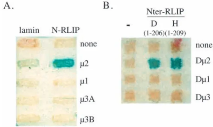
RLIP76 distinguishes µ2 from µ1 and µ3
At the trans-Golgi network (TGN), clathrin coated vesicle formation requires a complex equivalent to AP2 called AP1, made of γand β1 heavy chains, µ1 medium chain and σ1 light chain (Bonifacino and Dell’Angelica, 1999). AP3 is another AP complex of unclear function in mammalian cells (Odorizzi et al., 1998). In AP1 and AP3, µ2 homologues are µ1 and µ3, respectively. We tested the ability of N-RLIP to differentiate between µ1, µ2 and µ3. Fig. 2A shows that N-RLIP does not interact with µ1 nor with µ3A and µ3B, two isoforms of µ3 (Bonifacino and Dell’Angelica, 1999). N-RLIP does not interact either with any other tested chain of the AP1 or AP2 complexes (α, β2, γ, σ1, σ2) nor with σ3 (data not shown; A. Rocca, personal communication). Thus the N-RLIP-µ2 interaction is specific.
The interaction between RLIP and µ2 is conserved through evolution
If the interaction of the N terminus of RLIP76 with µ2 is biologically significant, it might be expected to be conserved through evolution. We have cloned the Drosophila homologue
of human RLIP, D-RLIP as a partner of activated Drosophila Ral (this work will be described elsewhere. D-RLIP GenBank accession number: AF037470). We tested whether the N-terminal domain of D-RLIP (amino acids 1-206) interacts specifically with the fly µ2 protein (Zhang and Broadie, 1999). Two-hybrid tests were performed using the N-terminal regions of human RLIP76 and of Drosophila RLIP, and the human and
Drosophila µ1, µ2 and µ3 proteins. Fig. 2B shows that fly RLIP interacts with fly µ2 and not with fly µ1 and µ3 proteins: here again the interaction between RLIP and µ2 is specific. Fig. 1. RLIP76 and µ2 interact specifically in the two-hybrid system
and in vitro. (A) The N-terminal domain of RLIP76, N-RLIP (amino acids 1-209), fused to LexA was used in a two-hybrid assay. Various truncations of µ2 (numbers in-between brackets represent the first and the last amino acid of µ2 expressed in the fusion protein) were tested for their capacity to interact with N-RLIP, and with lamin as a negative control. On the right side is listed the capacity of the µ2 mutants to interact with a tyrosine-based YQRL-motif (Aguilar et al., 1997, and our data not shown). (B) Equal amounts of in vitro synthesized 35S-µ2 were incubated with glutathione-Sepharose beads bound to equal amounts of GST or GST-RLIP76. Half of the beads (the bound fraction, b) and all the supernatant (the unbound fraction, u) were analyzed by SDS-PAGE followed by autoradiography.
Fig. 2. The interaction between RLIP and µ2 is specific and conserved through evolution. (A) N-RLIP was tested in a two-hybrid assay for its specificity towards µ1, µ2, µ3A and µ3B proteins. (B) The equivalent N-terminal domain of the Drosophila RLIP, D(1-206), and the human N-RLIP, H(1-209), were analyzed for their interaction with fly µproteins, Dµ2, Dµ1 and Dµ3, in a two-hybrid test.
2840
This interaction crosses species: Drosophilaµ2 interacts with human and fly RLIP. Reciprocally human µ2 but not human µ1 and µ3 interacts with fly RLIP (data not shown). Expression of all µproteins was tested by western-blotting and turned out to be equivalent (data not shown).
RLIP76 interacts specifically with AP2 in vivo
Since we have shown that RLIP76 binds µ2 in two-hybrid and in vitro, we addressed the question of the biological relevance of this interaction by testing whether RLIP76 binds AP2 in mammalian cells.
To this aim we have established a HeLa cell line where RLIP76 is expressed in a doxycycline-dependent manner (Gossen et al., 1995), fused to a FLAGepitope at its N terminus.
HR5 cells were transfected and selected in presence of G418 and hygromycin, and tested for RLIP76 expression. One geneticin resistant clone (FRD1.1) was found to express FLAG -RLIP76 only in the presence of doxycycline (Fig. 3, upper panel).
When cells are treated with doxycycline, FLAG-RLIP76 can be immunoprecipitated with affinity purified polyclonal anti-RLIP76 antibodies as tested with an anti-FLAG-antibody by western blot (data not shown). The signal is specific to RLIP as it can be depleted when anti-RLIP antibodies are incubated with GST-RLIP prior immunoprecipitation (Fig. 3, upper panel). When the antibodies are incubated with GST, FLAG-RLIP can still be immunoprecipitated.
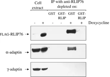
We tested the presence of AP2 proteins in complexes immunoprecipitated with anti-RLIP76 antibodies by western blotting the immunoprecipitates with antibodies against AP2 α -subunit (anti-αadaptin antibodies). We found that when FRD1.1 cells are treated with doxycycline, AP2 complexes are detected in anti-RLIP76 immunoprecipitates. We questioned the
specificity of this result by incubating the anti-RLIP antibodies with GST or GST-RLIP proteins prior to immunoprecipitation. Fig. 3 (middle panel) shows that α chain is detected in the presence of doxycycline but only when the antibodies have not been depleted on GST-RLIP. When the same experiment is done with the parental cell line HR5, no αadaptin can be detected in anti-RLIP immunoprecipitates independent of cells being treated with doxycycline or not (data not shown).
Since N-RLIP binds µ2 but not µ1 in two-hybrid tests, we tested whether this reflects the ability of RLIP76 to bind AP2 but not AP1 in vivo. Anti-RLIP76 immunocomplexes were tested for the presence of AP1 by western blot with anti-γadaptin antibodies. Fig. 3 (lower panel) shows that no AP1 can be co-immunoprecipitated with RLIP76. We tested whether treatment of cells with doxycyclin changes the level of expression of α -and of γ-adaptins: it does not (Fig. 3, two left lanes). We conclude that RLIP76 and AP2 can be found complexed to each other in vivo while RLIP76 and AP1 cannot.
EGF independence
We have shown here that RLIP76 is found complexed to AP2. We tested whether this interaction is dependent on EGF stimulation, a growth factor that triggers the Ras-Ral signaling pathway. Two cell lines overexpressing the EGF receptor, A431 and ER22 (L’Allemain et al., 1989), where found to allow co-immunoprecipitation of α adaptin with endogenous RLIP76. A431 cells were starved overnight in 0.5% fetal calf serum and stimulated or not with 0.1 µg/ml of epidermal growth factor (EGF) for 10 minutes. Cell extracts were prepared and tested for EGF receptor stimulation with anti-phosphotyrosine antibodies. Fig. 4 shows that EGF stimulation was efficient: much more phosphotyrosines were detected in EGF stimulated cells than in starved cells. Cell extracts were prepared using the same method that allowed detection of RLIP76/AP2 interaction in doxycycline-induced FRD1.1 cells, and were submitted to immunoprecipitation with anti-RLIP76 antibodies followed by western blot with anti-α antibodies. Fig. 4 shows that RLIP76/AP2 complexes exist in starved A431 cells before any EGF stimulation and this interaction does not seem to vary upon EGF addition at 10 minutes of stimulation. As in the previous experiment, this signal is specific since neither RLIP nor α adaptin are detected in the immunoprecipitate when anti-RLIP antibodies are incubated with GST-anti-RLIP prior immunoprecipitation - as opposed to a pre-incubation with GST (data not shown). When control rabbit IgG were used instead of anti-RLIP76 antibodies for immunoprecipitation, no AP2 protein was detected. Similar results were obtained with ER22 cells (data not shown).
Ral inhibits endocytosis of transferrin
It has been suggested a role for RLIP76/RalBP1 in ligand-induced endocytosis through the interaction of RLIP76 with POB1 (Nakashima et al., 1999). Since RLIP behaves as a Ral effector and since we found that RLIP interacts also with AP2, the complex linking the clathrin coat and transmembrane receptors, we have examined Ral involvement in both ligand-independent and ligand-dependent endocytosis. HeLa cells were transfected with plasmids allowing expression of different RalB mutants and endocytosis of fluorescent-labeled transferrin was analyzed. As shown in Fig. 5, the expression of constitutively active mutant of RalB (RalB G23V) inhibits
V. Jullien-Flores and others
Fig. 3. AP2 and RLIP76 form a complex in vivo. FRD1.1 cells were stimulated (+) or not (−) with doxycycline, and cell extracts were prepared (see Materials and Methods) in conditions that maintain integrity of the AP complexes. Cell extracts were incubated with affinity purified anti-RLIP76 antibodies that have been depleted prior immunoprecipitation on either GST or GST-RLIP. The
immunocomplexes were precipitated using Protein A-Sepharose, washed, boiled in SDS-PAGE sample buffer, migrated in SDS-PAGE gels and transferred to nitrocellulose. Membranes were then probed with anti-FLAG, anti-α-adaptin and anti-γ-adaptin antibodies.
endocytosis of transferrin receptors (upper panel). In contrast, the dominant-negative mutant (RalB S28N) does not affect the uptake of transferrin (middle panel). The inhibitory action of active RalB requires its localization to the membrane as an active but cytosolic RalB mutant (RalB G23V ∆CAAX) is unable to inhibit endocytosis of transferrin (lower panel). Moreover, the recruitment of RLIP76 to the membrane is supported by RalB G23V, but neither by RalB S25N nor by cytoplasmic RalB G23V ∆CAAX (data not shown; Matsubara et al., 1998). These data are consistent with the hypothesis that the increase of the membrane-associated RLIP-RalB complexes results in the inhibition of endocytosis.
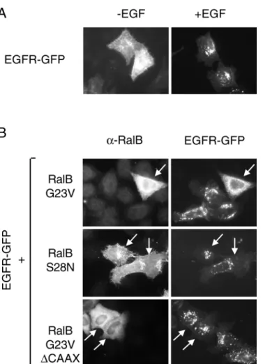
Ral interferes with endocytosis of EGFR-GFP
We have also analyzed the effect of Ral in ligand-induced endocytosis of the EGF receptor - having in mind that stimulation of cells by EGF triggers Ral activation. To this aim we expressed a functional EGF receptor-GFP chimera (EGFR-GFP) in HeLa cells cotransfected with a control plasmid or plasmids allowing expression of different RalB mutants. Stimulation by EGF leads to a rapid internalization of EGF receptors (Fig. 6A) with constitution of large vesicles where the EGF receptor is concentrated. Co-expression of the constitutively active mutant of RalB (RalB G23V) dramatically inhibits these phenomena (Fig. 6B). This inhibition is not observed either by co-expression of the dominant-negative RalB S28N or the cytoplasmic activated mutant RalB G23V ∆CAAX suggesting that RalB-mediated inhibition of EGF receptor internalization requires activation of Ral as well as its anchoring to membranes.
Ral has previously been demonstrated to be involved in ARF1-mediated phospholipase D (PLD) activation
(Kim et al., 1998) and this function requires the integrity of the N terminus of RalB. In order to define if the inhibitory effect of RalB G23V upon endocytosis is due to the action of PLD, we used a RalB G23V mutant, RalB ∆11 G23V, missing the first eleven N-terminal amino acids. This mutant has a decreased but no null capacity to interfere with EGFR endocytosis and roughly 50% of transfected cells still show an inhibition of EGF receptor endocytosis (Fig. 7).
We further addressed the question whether Ral inhibits endocytosis through the action of its effector RLIP76. Therefore we analyzed the effect of a previously described Ral effector domain mutant (RalB G23V D49N) which has a weakened but not null interaction with RLIP76 (Bauer et al.,
Fig. 4. Interaction between AP2 and RLIP76 is not modulated by stimulation of cells with EGF. (A) A431 cells were starved overnight and then stimulated or not with EGF. Cell extracts were run on a SDS-PAGE gel, transferred to a nitrocellulose membrane and probed with an antiphosphotyrosine antibody. (B) The same cell extracts were similarly probed with an anti-α-adaptin antibody; or submitted to immunoprecipitation using anti-RLIP76 antibodies or rabbit IgG antibodies as a control. The immunoprecipitates were probed by western blot with anti-α-adaptin antibody. Aliquots of whole cell extract used directly in western blots correspond to 1/50 of what was used for immunoprecipitation. Molecular masses are indicated.
Fig. 5. Active RalB (G23V) inhibits endocytosis of transferrin. HeLa cells expressing different RalB mutants (RalB G23V, RalB S28N or RalB G23V ∆CAAX) were subjected to endocytosis of TexasRed-labelled transferrin for 15 minutes at 37°C. Expression of the RalB constructs was visualized by a subsequent indirect immunofluorescence using anti-RalB and anti-rabbit-FITC conjugated antibodies.
2842
1999). As shown in Fig. 7, cells expressing this Ral mutant show a ‘mixed’ phenotype with 50% of transfected cells displaying no longer an inhibition of EGFR endocytosis (Fig. 7). This ‘mixed’ result might be due to the level of expression of Ral, since the interaction between RalB G23V D49N is only weakened but not completely abolished (Bauer et al., 1999).
The same kind of results was obtained with another effector domain mutant, RalB G23V A48E, which also displays a weakened but not null interaction with RLIP. Again this mutant yields a ‘mixed’ phenotype with a reproducible decreased capacity to inhibit EGFR endocytosis, as the RalB G23V D49N phenotype. By contrast, a RalB G23V D49E mutant, which is not affected in its binding properties to RLIP, is able to inhibit EGF receptor endocytosis as well as the RalB G23V allele (Fig. 7). Thus, there is a correlation between the capacity of Ral to inhibit EGFR endocytosis and its ability to interact with RLIP: RLIP76 is a potential candidate to mediate Ral function in endocytosis.
DISCUSSION
For a brief time, it was believed that Ras-mediated signalling downstream of transmembrane receptors was a simple linear cascade of kinases, from Raf to MAP kinases. It is now clear, however, that Ras action is mediated by a combination of effectors - including RalGEFs, which are activators of the GTPase Ral and indeed activation of Ras leads to the activation of Ral (Urano et al., 1996; White et al., 1996; Wolthuis et al., 1997). Activated Ral associates with the Ral effector RLIP76 (also called RalBP1, RIP1), a cytosolic protein that is recruited to membranes after Ral activation (J. Camonis, unpublished data; Matsubara et al., 1998). RLIP76 can be divided into four regions: two central domains, one carrying a Rac1/CDC42 GAP activity, the other which binds to activated Ral, and two flanking domains of unknown function.
We have performed two-hybrid screens to identify partners of these latter two ‘orphan’ regions of RLIP76. Using the first 209 amino acids of RLIP76 (N-RLIP) as a bait, we identified
V. Jullien-Flores and others
Fig. 6. Active RalB (G23V) inhibits endocytosis of the EGF receptor. (A) HeLa cells were transfected with a plasmid allowing expressing EGF receptor fused to GFP (EGFR-GFP), were serum-starved overnight and stimulated (+EGF) or not (−EGF) with epidermal growth factor for 30 minutes. (B) HeLa cells were co-transfected with a EGFR-GFP expression plasmid and different RalB mutants (RalB G23V, RalB S28N or RalB G23V ∆CAAX), serum-starved overnight and stimulated with EGF (200 ng/ml) for 30 minutes at 37°C. Expression of RalB (cells expressing RalB are indicated by arrows) was visualized by a subsequent indirect immunofluorescence.
Fig. 7. Deficiency in RLIP interaction affects RalB-mediated inhibition of endocytosis. HeLa cells were co-transfected with a EGFR-GFP expression plasmid and different RalB mutants affected or not in their interaction with effectors. Cells were serum-starved overnight and endocytosis of the EGFR-GFP was induced by stimulation with EGF (200 ng/ml) for 30 minutes at 37°C. GFP allowed for detection of EGFR. RalB G23V D49N is an activated mutant deficient in its interaction with RLIP76, RalB ∆11 G23V is deficient in PLD-activation, RalB G23V D49E is an effector domain mutant still interacting with RLIP76, and RalB G23V A48E is an effector domain mutant deficient in its interaction with RLIP76.
µ2, the medium chain of the AP2 complex, as a partner of RLIP76. Our in vitro binding assay shows that this interaction is direct. This interaction is specific since N-RLIP binds µ2 but not µ1 and µ3, two close homologues of µ2 that are subunits of AP1 and AP3 complexes, respectively. The specific interaction between RLIP76 and µ2 has been conserved throughout evolution as it also exists in flies and such a conservation supports the relevance of this partnership.
µ2 plays a central role in the interaction of AP2 with integral membrane proteins and their recruitment into coated pits. It has been proposed that receptors endocytosed in clathrin-coated pits display a cytoplasmic ‘endocytic’ tyrosine-based signal Y-X-X-Φ (X is any amino acid and Φ is a hydrophobic amino acid) and that this motif is recognized by µ2 (Ohno et al., 1995). This model was shown to be true for transferrin receptors (Nesterov et al., 1999). However, the affinity between µ2 and tyrosine-based signals is rather weak (Boll et al., 1996), and most existing Y-based signals are unable to differentiate between µ1, µ2 and µ3 (Ohno et al., 1995). In contrast to this promiscuity, AP2 works exclusively at the plasma membrane and AP1 works only at the trans Golgi network. This suggests that one or more additional factors along with µ2 may be responsible for the specificity of the interaction of AP2 with receptors. RLIP76 may be one of these factors.
We show that the region encompassing amino acids 141-163 of µ2 is essential for its binding to N-RLIP. This region of µ2 is not directly involved in binding of µ2 to Y-X-X-Φsignals and is within a region of divergence between µ2 and µ1 (Aguilar et al., 1997). Therefore this region of interaction might be the molecular avatar for RLIP capacity to differentiate µ2 from the other µsubunits.
The involvement of a Ral-monitored pathway in endocytosis has been recently documented neatly by Nakashima et al. (1999), These authors suggest that the molecular basis of such involvement is the interaction between RLIP76 and POB1, a protein of yet unknown function containing an Eps15 homology domain (EH) domain. Our data with Ral mutants are consistent with Ral playing a role in endocytosis. We show that a constitutively active Ral mutant interferes with the endocytosis of EGF receptors as well as the endocytosis of transferrin receptors. Ral stimulates phospholipase D, but the effect of Ral upon endocytosis cannot be explained only by this activity since Ral mutants unable to activate PLD can still inhibit endocytosis. Finally results with Ral alleles mutated in the effector domain suggest that the effect of Ral occurs at least in part via its interaction with RLIP76.
Our results differ marginally from those of Nakashima et al. (1999). While they observe that both a dominant negative Ral allele as well as an activated Ral allele show inhibitory effects on endocytosis, we see an effect only with activated alleles. Since dominant negative GTPases work by titrating their exchange factor(s), it could be that in HeLa cells (our experiments) RalGEFs are abundant proteins and cannot be titrated by dominant negative Ral - as opposed to A-431 cells. We also see an effect of activated Ral on transferrin endocytosis whereas Nakashima et al. do not. We have no obvious explanation for this discrepancy except that experiments were not performed in the same cell types, and that we might achieve higher levels of transient expression in HeLa cells compared to A-431 cells, which are typically very difficult to transfect.
The requirement for GTPases at various stages of a coated pit cycle has been demonstrated in endocytosis assays in semi-intact cells (Carter et al., 1993). Whereas the role of one GTPase, dynamin, is clearly established, the requirement of GTP-binding proteins at the stage of membrane docking of AP2 and formation of coated pits remains to be elucidated. Membrane targeting and docking of AP2 rely on the interaction of AP2 with phospholipids and integral membrane proteins. The µ2 subunit of AP2 is responsible for interactions with various cargo proteins containing Y-X-X-Φmotifs. Therefore, µ2/Y-X-X-Φ interactions could serve as part of a docking mechanism for the membrane recruitment of AP2. We hypothesize that µ2/RLIP76 interactions modulate the ability of µ2 to bind Y-X-X-Φ-containing proteins, thus regulating membrane association of AP2 and formation of coated pits.
According to our model, GTP-Ral dependent recruitment of RLIP76 to the membrane may facilitate receptor interaction with the µ2 subunit of AP2. Under steady-state conditions, the equilibrium between GTP- and GDP-bound Ral could regulate the association of AP2 with the membrane. Trapping Ral in its GTP-bound form, as is the case of Ral G23V, would stabilise the association of µ2/RLIP with membranes and thus disrupt the dynamic cycle of AP-membrane interactions, i.e. association and dissociation, that are required for coated pits assembly and disassembly. Therefore, overexpression of GTP-Ral leads to inhibition of clathrin-dependent endocytosis of transferrin and EGF receptors.
When signaling receptors such as EGFR are stimulated, the activation of Ral would result in additional local recruitment of AP2 and formation of new coated pits. In fact, formation of specialized coated pits in response to receptor stimulation has been recently demonstrated (Cao et al., 1998; Wilde et al.,
Ras-GTP Ral-GTP RalGEF RLIP76 AP2 POB1
1
2
3
4
Clathrin?
Vesicle s formati o nOUT
IN
Fig. 8. Local signalosome formation pinpointsreceptors that qualify for endocytosis. Arrows refer either to interaction leading to activation (black-blue arrows) or to interactions leading to recruitment of cytoplasmic complexes (white-blue arrows). Numbers refer to the order of events: activated receptor activates - indirectly - Ras (1) that recruits RalGEFs (2) that activates Ral (3) that recruits RLIP76 and its companion proteins (4). This signalosome formation allows vesicles formation and subsequent endocytosis of the activated receptors. Both activated receptors submitted to endocytosis and quiescent receptors that stay put at the membrane display Y-based motifs but only the former ones participate in this cascade of events, leading to their endocytosis.
2844
1999). Fig. 8 depicts such a ‘local activation model’ where sequential cycles of activation and recruitment of GTPases and their partners lead to activated receptors engulfed in coated pits. Obviously, other Ral and RLIP interacting proteins may participate in the regulation of AP2 dynamics, such as POB1 (Ikeda et al., 1998) and Reps1 (Yamaguchi et al., 1997), another EH-containing protein that we have identified in a two-hybrid screen (data not shown). We propose that RLIP76 must bind to the partners of its N-terminal domain (i.e. to AP2 proteins) and its C-terminal domain (i.e. to POB1 and/or REPS1) to behave properly in endocytosis. Further studies using functional approaches will elucidate the precise mechanisms by which RLIP76 and other Ral effectors control clathrin-mediated endocytosis.
This work was supported by grants from Ligue Nationale contre le Cancer, Association de Recherche sur le Cancer (ARC) and EC grants # BIO4-CT96-1110 and QLK3-CT-1999-00875. V.J.F. was supported by a fellowship from Ministère de l’Education Nationale, de la Recherche et de la Technologie (MENRT) and by an ARC fellowship, G.M. is supported by a MENRT fellowship and Y.M. is a recipient of a post-doctoral fellowship from the EC (# BIO4-CT96-1110). We thank Drs J. Bonifacino, A. Rocca, A. Benmerah, H. Bujard, D. Ricol, D. Wallach, A.Dautry-Varsat, A. Subtil, N. Cerf-Bensussan, J. Flanders, S. Whiteside and J. Pouysségur for reagents and/or wise advices and stimulating discussions.
REFERENCES
Aguilar, R. C., Ohno, H., Roche, K. W. and Bonifacino, J. S. (1997).
Functional domain mapping of the clathrin-associated adaptor medium chains mu1 and mu2. J. Biol. Chem. 272, 27160-27166.
Albright, C. F., Giddings, B. W., Liu, J., Vito, M. and Weinberg, R. A.
(1993). Characterization of a guanine nucleotide dissociation stimulator for a ras-related GTPase. EMBO J. 12, 339-347.
Bauer, B., Mirey, G., Vetter, I. R., Garcia-Ranea, J. A., Valencia, A., Wittinghofer, A., Camonis, J. H. and Cool, R. H. (1999). Effector
recognition by the small GTP-binding proteins Ras and Ral. J. Biol Chem.
274, 17763-17770.
Boll, W., Ohno, H., Songyang, Z., Rapoport, I., Cantley, L. C., Bonifacino, J. S. and Kirchhausen, T. (1996). Sequence requirements for the
recognition of tyrosine-based endocytic signals by clathrin AP-2 complexes. EMBO J. 15, 5789-5795.
Bonifacino, J. S. and Dell’Angelica, E. C. (1999). Molecular bases for the
recognition of tyrosine-based sorting signals. J. Cell Biol. 145, 923-926.
Cantor, S. B., Urano, T. and Feig, L. A. (1995). Identification and
characterization of Ral-binding protein 1, a potential downstream target of Ral GTPases. Mol. Cell. Biol. 15, 4578-4584.
Cao, T. T., Mays, R. W. and von Zastrow, M. (1998). Regulated endocytosis
of G-protein-coupled receptors by a biochemically and functionally distinct subpopulation of clathrin-coated pits. J. Biol. Chem. 273, 24592-24602.
Carter, L. L., Redelmeier, T. E., Woollenweber, L. A. and Schmid, S. L.
(1993). Multiple GTP-binding proteins participate in clathrin-coated vesicle-mediated endocytosis. J. Cell Biol. 120, 37-45.
Carter, R. E. and Sorkin, A. (1998). Endocytosis of functional epidermal
growth factor receptor-green fluorescent protein chimera. J. Biol. Chem.
273, 35000-35007.
Gossen, M., Freundlieb, S., Bender, G., Muller, G., Hillen, W. and Bujard, H. (1995). Transcriptional activation by tetracyclines in mammalian cells.
Science 268, 1766-1769.
Hofer, F., Fields, S., Schneider, C. and Martin, G. S. (1994). Activated Ras
interacts with the Ral guanine nucleotide dissociation stimulator. Proc. Nat. Acad. Sci. USA 91, 11089-11093.
Ikeda, M., Ishida, O., Hinoi, T., Kishida, S. and Kikuchi, A. (1998).
Identification and characterization of a novel protein interacting with Ral-binding protein 1, a putative effector protein of Ral. J. Biol. Chem. 273, 814-821.
Jullien-Flores, V., Dorseuil, O., Romero, F., Letourneur, F., Saragosti, S., Berger, R., Tavitian, A., Gacon, G. and Camonis, J. (1995). Bridging the
Ral GTPase to Rho pathways: RLIP76, a Ral effector with CDC42/Rac GTPase activating protein (GAP) activity. J. Biol. Chem. 270, 22473-22477.
Kikuchi, A., Demo, S. D., Ye, Z.-H., Chen, Y.-W. and Williams, L. T.
(1994). RalGDS family members interact with the effector loop of ras p21. Mol. Cell. Biol. 14, 7483-7491.
Kim, J. H., Lee, S. D., Han, J. M., Lee, T. G., Kim, Y., Park, J. B., Lambeth, J. D., Suh, P. G. and Ryu, S. H. (1998). Activation of phospholipase D1
by direct interaction with ADP-ribosylation factor 1 and RalA. FEBS Lett.
430, 231-235.
L’Allemain, G., Seuwen, K., Velu, T. and Pouyssegur, J. (1989). Signal
transduction in hamster fibroblasts overexpressing the human EGF receptor. Growth Factors 1, 311-321.
Lopez-Barahona, M., Bustelo, X. R. and Barbacid, M. (1996). The TC21
oncoprotein interacts with the Ral guanosine nucleotide dissociation factor. Oncogene 12, 463-470.
Matsubara, K., Hinoi, T., Koyama, S. and Kikuchi, A. (1998).
Post-translational modifications of Ral and Rac1 are important for the action of Ral-binding protein 1, a putative effector protein of Ral. FEBS Lett. 410, 169-174.
Nakashima, S., Morinaka, K., Koyama, S., Ikeda, M., Kishida, M., Okawa, K., Iwamatsu, A., Kishida, S. and Kikuchi, A. (1999). Small G
protein Ral and its downstream molecules regulate endocytosis of EGF and insulin receptors. EMBO J. 18, 3629-3642.
Nesterov, A., Carter, R. E., Sorkina, T., Gill, G. N. and Sorkin, A. (1999).
Inhibition of the receptor-binding function of clathrin adaptor protein AP-2 by dominant-negative mutant mu2 subunit and its effects on endocytosis. EMBO J. 18, 2489-2499.
Odorizzi, G., Cowles, C. R. and Emr, S. D. (1998). The AP-3 complex: a
coat of many colours. Trends Cell Biol. 8, 282-288.
Ohno, H., Stewart, J., Fournier, M. C., Bosshart, H., Rhee, I., Miyatake, S., Saito, T., Gallusser, A., Kirchhausen, T. and Bonifacino, J. S. (1995).
Interaction of tyrosine-based sorting signals with clathrin-associated proteins. Science 269, 1872-1875.
Park, S.-H. and Weinberg, R. A. (1996). A putative effector of Ral has
homology to Rho/Rac GTPase activating proteins. Oncogene 11, 2349-2355.
Sambrook, J., Fritsch, E. F. and Maniatis, T. (1989). Molecular Cloning. A
Laboratory Manual. Cold Spring Harbor, NY: Cold Spring Harbor Laboratory Press.
Schmid, S. L., McNiven, M. A. and De Camilli, P. (1998). Dynamin and its
partners: a progress report. Curr. Opin. Cell. Biol. 10, 504-512.
Spaargaren, M. and Bischoff, J. R. (1994). Identification of the guanine
nucleotide dissociation stimulator for Ral as a putative effector molecule of R-ras, H-ras, K-ras and Rap. Proc. Nat. Acad. Sci. USA 91, 12609-12613.
Urano, T., Emkey, R. and Feig, L. A. (1996). Ral-GTPases mediate a distinct
downstream signaling pathway from Ras that facilitates cellular transformation. EMBO J. 15, 810-816.
Vojtek, A. B., Hollenberg, S. M. and Cooper, J. A. (1993). Mammalian Ras
interacts directly with the serine/threonine kinase Raf. Cell 74, 205-214.
White, M. A., Vale, T., Camonis, J. H., Schaefer, E. and Wigler, M. H.
(1996). A role for Ral guanine nucleotide dissociation stimulator in mediating Ras-induced transformation. J. Biol. Chem. 271, 16439-16442.
Wilde, A., Beattie, E. C., Lem, L., Riethof, D. A., Liu, S. H., Mobley, W. C., Soriano, P. and Brodsky, F. M. (1999). EGF receptor signaling
stimulates SRC kinase phosphorylation of clathrin, influencing clathrin redistribution and EGF uptake. Cell 96, 677-687.
Wolthuis, R. M., Bauer, B., van’t Veer, L. J., de Vries-Smits, A. M., Cool, R. H., Spaargaren, M., Wittinghofer, A., Burgering, B. M. and Bos, J. L. (1996). RalGDS-like factor (Rlf) is a novel Ras and Rap 1A-associating
protein. Oncogene 13, 353-362.
Wolthuis, R. M., de Ruiter, N. D., Cool, R. H. and Bos, J. L. (1997).
Stimulation of gene induction and cell growth by the Ras effector Rlf. EMBO J. 16, 6748-6761.
Wolthuis, R. M., Franke, B., van Triest, M., Bauer, B., Cool, R. H., Camonis, J. H., Akkerman, J. W. and Bos, J. L. (1998a). Activation of
the small GTPase Ral in platelets. Mol. Cell. Biol. 18, 2486-2491.
Wolthuis, R. M., Zwartkruis, F., Moen, T. C. and Bos, J. L. (1998b).
Ras-dependent activation of the small GTPase Ral. Curr. Biol. 8, 471-474.
Yamaguchi, A., Urano, T., Goi, T. and Feig, L. A. (1997). An Eps homology
(EH) domain protein that binds to the Ral-GTPase target, RalBP1. J. Biol. Chem. 272, 31230-31234.
Zhang, Y. Q. and Broadie, K. (1999). Cloning, mapping and tissue-specific
expression of Drosophila clathrin-associated protein AP50 gene. Gene 233, 171-179.